Embed Size (px)
Citation preview

Cell suspension cultures

Defini&on of cell culture A cell suspension culture can be defined in a prac2cal way has an homogenous suspension of dividing cells easily to take up
some aliquot only by a glass-‐pipe=e
A cell suspension culture consists of cell aggregates dispersed and growing in moving liquid media

Cell suspension culture uses ! Understanding of biosynthe2c pathway
! Mutant selec2on
! Secondary metabolite produc2on
! Use of suspension cultures in plant propaga2on.

Establishment steps for cell suspension culture
1. Choice of the explants and induc2on to cell division
2. Inoculum in a liquid culture medium.
3. Subculture of cell suspension culture

! it usually started by placing an inoculum of
friable callus in a liquid
medium or,
! by placing an explant in
liquid culture
Starting a cell suspension culture

Leaf sec&ons floated in liquid culture medium
Leaf sec2ons of Chenopodium rubrum floated on Murashige and
Skoog (1962) medium in the light, show rapid growth and cell
division in the mesophyll, and aNer 4 days on a rotary shaker they
can be disintegrated completely to release a great number of cells
into suspension (Geile and Wagner, 1980).
Disadvantages to use explants floated in liquid culture medium
1. High probability for contamina2on
2. No forma2on of callus due to a low gaseous exchange
between explants and liquid medium
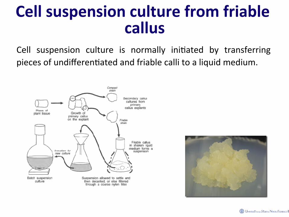
Cell suspension culture from friable callus
Cell suspension culture is normally ini2ated by transferring pieces of undifferen2ated and friable calli to a liquid medium.

Obtaining friable callus. 1) Choice of explant 2) Iden2fica2on of a suitable growth medium

Establishment steps for cell suspension culture
1. Choice of the explant and induc2on to cell division
2. Inocolum in a liquid culture medium.
3. Subculturing

Callus as inoculum in liquid culture medium
! Callus:
! is separated from the parent explant and transferred to a
fresh medium to build up reasonable amount of callus 2ssue.
! is transferred to fresh medium every 4-‐6 weeks.
! is an essen2al step to avoid cell aging that is visible as
reduc2on of growing and dark spot.
Causes of explant aging
I. Nutri2ve element declining
II. Water loss by evapora2on, therefore modifica2on of
nutri2ve compound concentra2on
III. Build up of compounds products from cell that can have
an inhibi2on of cell growth or induce apoptosis

Callus produc&on from zygote embryos of Croton bonplandianum.
1. 10 days old embryo cultyre.; 2. One month culture, see callus from root (cr) and form endopserm (ce); 3. Healthy callus
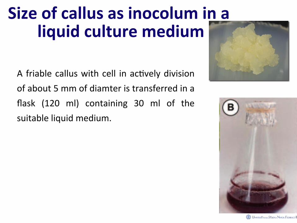
Size of callus as inocolum in a liquid culture medium
A friable callus with cell in ac2vely division of about 5 mm of diamter is transferred in a flask (120 ml) containing 30 ml of the suitable liquid medium.
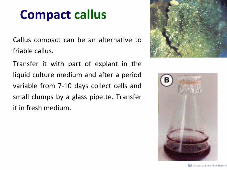
Compact callus
Callus compact can be an alterna2ve to friable callus.
Transfer it with part of explant in the liquid culture medium and aNer a period variable from 7-‐10 days collect cells and small clumps by a glass pipe=e. Transfer it in fresh medium.

Media composi&on
• A wide variety of explant and media composi2on has been
used : Heller, B5 Gamborg and MS
• To these media are added, vitamins, inositol, sucrose, and
auxin (2,4D at ∼ 1-‐5 µM concentra2ons for cell to divide

Culture vessels • Wide-‐mounthed Erlemeyer flasks
are widely used as culture vessels.
• The flasks are normally sealed with aluminium foil.
• Co=on wool plugs may be used for sealing flasks during autoclaving but not for culturing cells. They are a common source of contamina2on on flasks that are sibng on a shaker for several weeks.
• Flasks closure must maintain sterility, allow gas exchange and reduce evaporation.
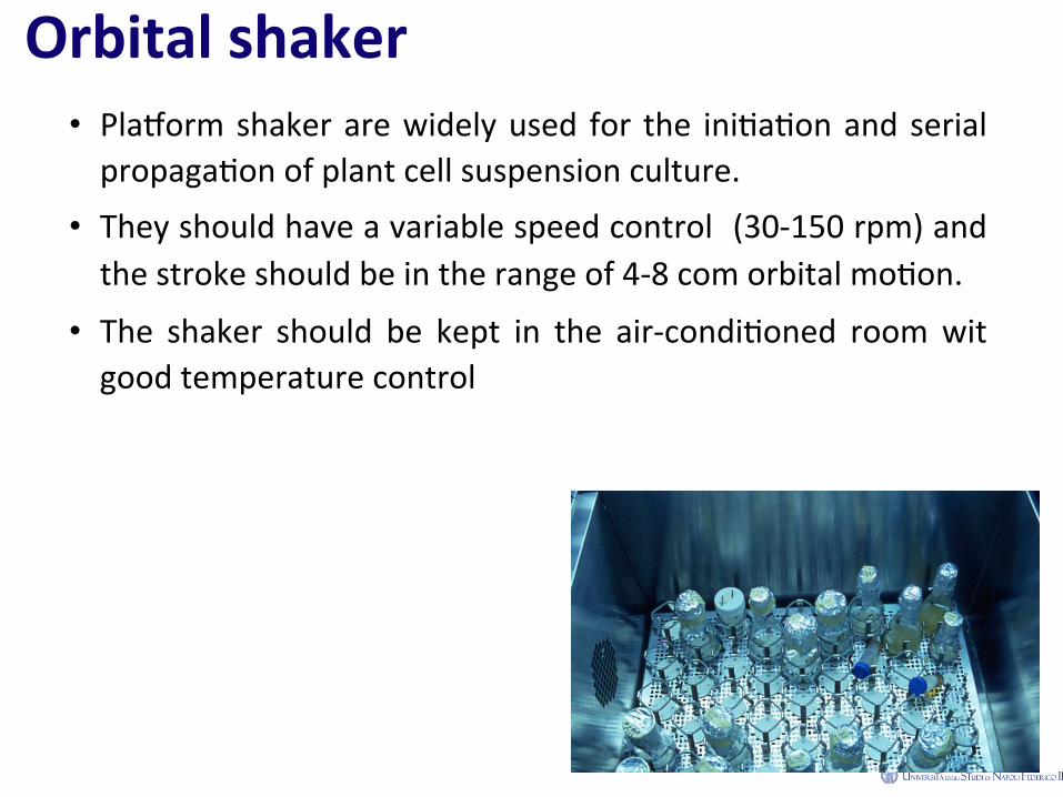
Orbital shaker • Pladorm shaker are widely used for the ini2a2on and serial propaga2on of plant cell suspension culture.
• They should have a variable speed control (30-‐150 rpm) and the stroke should be in the range of 4-‐8 com orbital mo2on.
• The shaker should be kept in the air-‐condi2oned room wit good temperature control

Agita&on of medium serves two purposes.
1. It exerts a mild pressure on cell aggregates, breaking them
into smaller clumps and single cells.
2. It maintains uniform distribu2on of cell and cell clumps in
the medium. Movement of the medium also provides good
gaseous exchange between the culture medium and air.

Fine cell suspension culture
! In an ideal cell suspension culture there are single isodiametric cells and few clumps of 20-‐100 cells.
! Mostly of cell suspension culture is made up by an heterogeneous cell popula2on either by size than by specific density.

Cell suspension culture completely isolated have yet be obtained
! Because the walls of plant cells have a natural tendency to adhere, it is not possible to obtain suspensions that consist only of dispersed single cells.
! Some progress has been made in selec2ng cell lines with increased cell separa2on, but cultures of completely isolated cells have yet to be obtained.
Cell propor&on and size
• The propor2on and size of small cell aggregates varies
according to plant variety and the medium in which the
culture is grown.
• As cells tend to divide more frequently in aggregates than in
isola2on, the size of cell clusters increases during the phase of
rapid cell division.

Methods for obtaining a well-‐dispersed suspension culture
By : De-‐Long flasks Sieving Siringe Adding in the
medium cellualse and pec2nase

Cell size
Mesh 150 x 150 µm

Use of cell density for obtaining a fine cell suspension culture.
Cell can contain: vacuole, starch and other
Therefore cell can be separated on own density by a
centrifuga2on.

Discon&nuous gradient in an appropriate solu&on
Ficoll is largely used as solute due to this features :
! can be sterilized by autoclave
! has got low osmomolarity
! at high concentra2on (10-‐20%) has low viscosity
Type of hormone influence degree of cell dispersion
The degree of cell dispersion in suspension cultures is par2cularly influenced by the concentra2on of growth regulators in the culture medium.
Auxinic growth regulators: increase the specific ac2vity of enzymes, which bring about the dissolu2on of the middle lamella of plant cell walls (Torrey and Reinert, 1961).
Effects of PGRs on cell suspension aggregates
• Auxin at rela2vely high concentra2on and a low concentra2on of a cytokin in a liquid culture medium usually increase cell
dispersion (Narayanaswamy, 1977).
• Use of high auxin levels to obtain maximum cell dispersion will
ensure that the cultured cells remain undifferen2ated.
Establishment steps for cell suspension culture
1. Choice of the explant and induc2on to cell division
2. Inocolum in a liquid culture medium.
3. Subculturing of cell suspension culture
Subculturing
Cell suspension culture must be frequently and on regular bases transfer in a fresh medium.
The lag period is usually between one or two weeks.
The ra2o of dilu2on (cell vs medium) is experimentally determined-‐
As general rule
1: 4 aNer one week
1:10 aNer two weeks
Is a cell suspension culture not sterile ?
• The sterility of plant cell suspension can be monitored by
several parameters:
– change in colour of solu2on
– change in pH
– smell
– interface analysis
– use of microscope
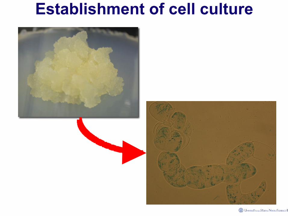
Establishment of cell culture
The accurate, fast, and reliable determination of cell growth is of
critical importance in plant cell and tissue culture
However, the measurement of growth parameters in the different types of cultures, and concomitantly the use of various containers along with the heterogenity in cell morphology, introduce diverse problems that must be
addressed by using a specific methodology for each case callus and cell suspension cultures represent two of the most
common in vitro systems
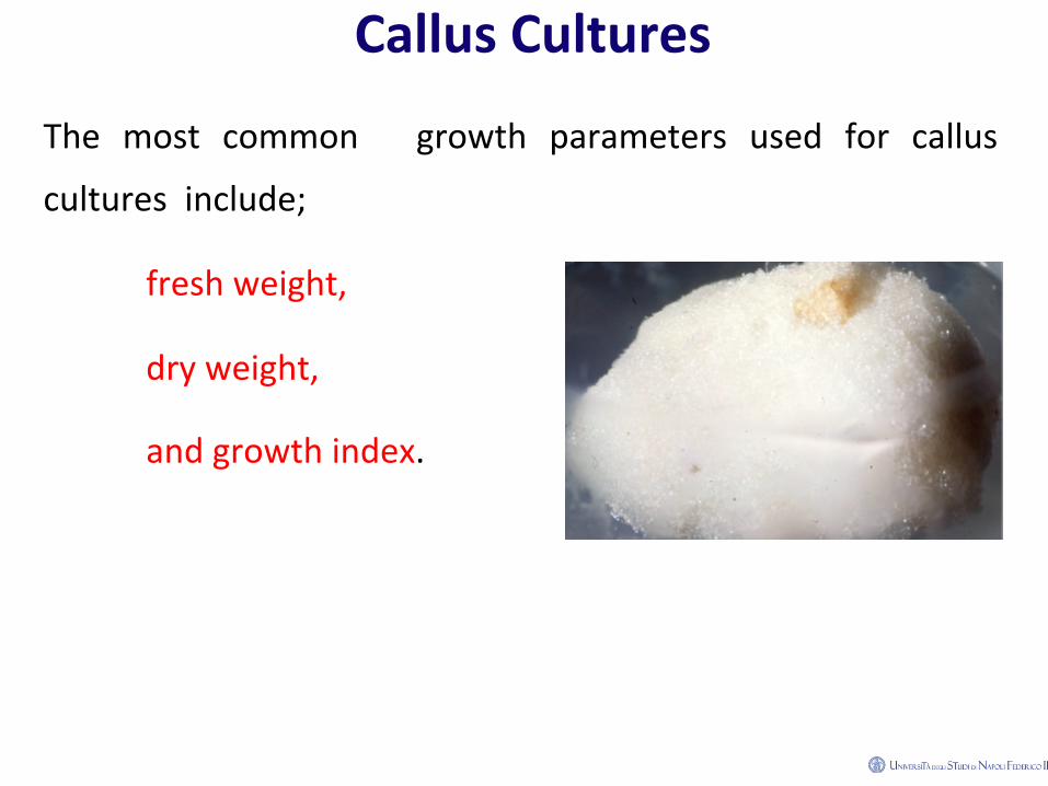
Callus Cultures
The most common growth parameters used for callus
cultures include;
fresh weight,
dry weight,
and growth index.
Dry-Weight Determination (Callus)
It can be es2mated by drying the 2ssue at 60°C in a convec2on oven, un2l of constant weight (usually 16 h).
1. Take a sample of the fresh 2ssue, weigh it on a pre-‐weighed
square of aluminum foil, and evaporate the water contained
in the 2ssue in the pre-‐heated oven at 60°C for 12 h.
2. Allow samples to cool down in a dessicator containing silica
gel for 15–20 min and then register the sample’s weight.

Dry-Weight Determination (Callus) 3. Put the 2ssue sample back into the oven and take a new
weight register aNer 2 h. If no varia2ons are detected, samples have reached constant weight. If varia2ons higher than 10% regarding the previous register are observed, return samples to the oven for another 2-‐h period.
4. Alterna2vely, dry weight can be obtained from lyophilized 2ssues. Once harvested, fresh 2ssues should be weighed, deposited in lyophilizer flasks, and frozen at –20°C for at least 12 h.
5. Flasks with frozen 2ssues are then connected to the vacuum line for 24 h and weight is registered

Monitoring cell suspension culture.
Cell vitality
Cell Growth
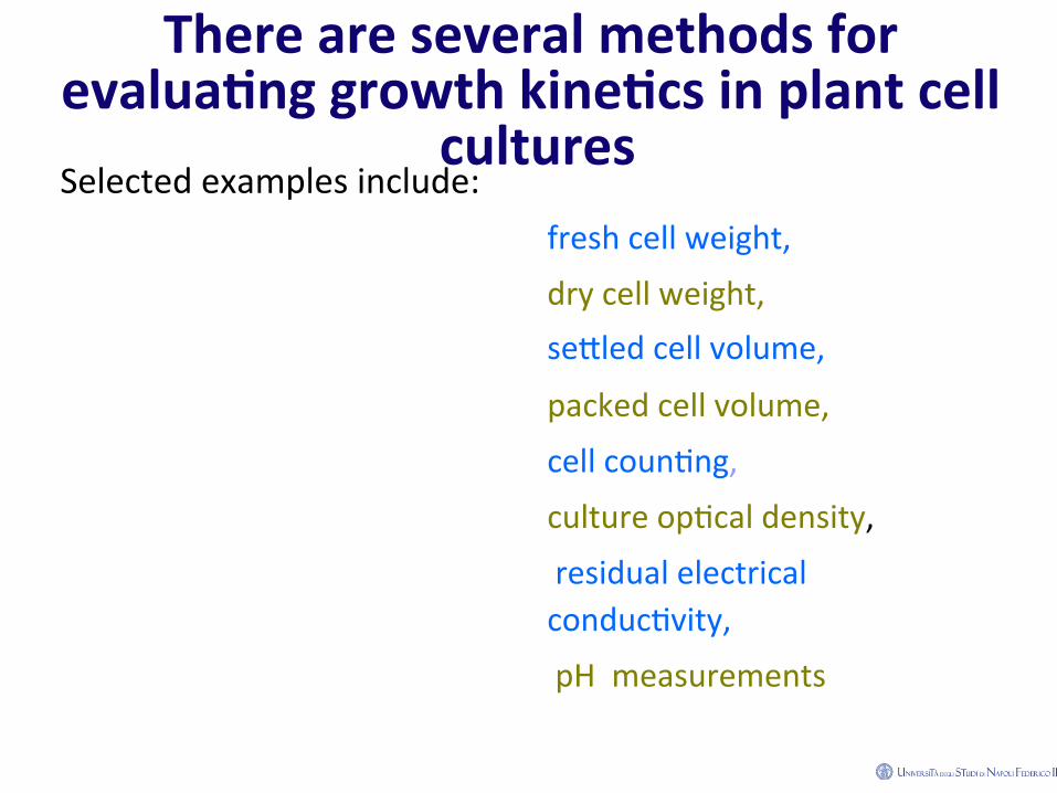
There are several methods for evalua&ng growth kine&cs in plant cell
cultures Selected examples include: fresh cell weight,
dry cell weight, se=led cell volume,
packed cell volume,
cell coun2ng,
culture op2cal density,
residual electrical conduc2vity,
pH measurements

Correla&ons among features
1. In cultures originated from different plant species, se=led volume, packed cell volume, as well as fresh weight, all show a very good linear correla2on with dry weight data. Thus, any of
these es2ma2ons can be used for measuring cell growth.
2. The measurement of cell concentra2on by cell coun2ng and turbidity (op2cal density) has also shown a reasonably good
correla2on with the dry-‐cell weight parameter.
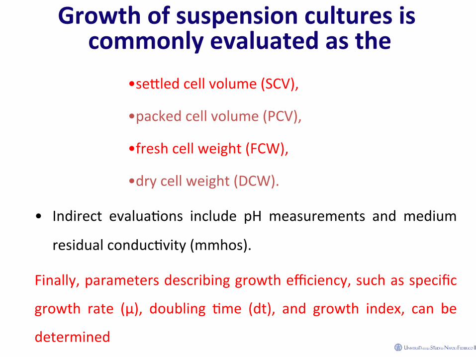
Growth of suspension cultures is commonly evaluated as the
• se=led cell volume (SCV),
• packed cell volume (PCV),
• fresh cell weight (FCW),
• dry cell weight (DCW).
• Indirect evalua2ons include pH measurements and medium
residual conduc2vity (mmhos).
Finally, parameters describing growth efficiency, such as specific
growth rate (μ), doubling 2me (dt), and growth index, can be
determined
SeTled Cell Volume (SCV) and Packed Cell Volume (PCV)
Both parameters allow the quick es2ma2on of culture growth, while maintaining sterile condi2ons.
These measurements are useful for monitoring growth in the same flasks along a culture cycle, because suspensions may be returned to prior culture condi2ons.
Care must be taken to maintain sterile condi2ons.

SeTled Cell Volume (SCV) and Packed Cell Volume (PCV)
Volume es2ma2on may not be an accurate way of monitoring growth, given its dependence on cell morphology (cell and clump size, cell density, and other).
SCV is determined by allowing a cell suspension to sediment in graduated tubes. It is reported as the percentage of the total volume of suspension occupied by the cell mass.
The PCV is determined in a similar way, aNer it has been compacted by centrifuga2on.
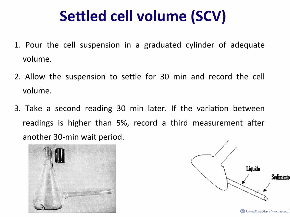
SeTled cell volume (SCV)
1. Pour the cell suspension in a graduated cylinder of adequate
volume.
2. Allow the suspension to se=le for 30 min and record the cell
volume.
3. Take a second reading 30 min later. If the varia2on between
readings is higher than 5%, record a third measurement aNer
another 30-‐min wait period.

4. The volume frac2on of the suspension occupied by the cells is
determined as the SCV.
PCV can be determined by centrifuging 10 mL of the
culture in a 15-‐mL graduated conical centrifuge tube at
200g for 5 min
SeTled cell volume (SCV)

Fresh Cell Weight and Dry Cell Weight
• Fresh and dry cell weight represent more precise measurements of cell growth than the sole culture volume.
• However, both require the manipula2on of samples in non
sterile condi2ons.
• Fresh weight es2ma2on involves less 2me than that required for
dry weight, but it may not reflect a real measurement of biomass
gain, par2cularly at the end of the culture period, when most of
the culture growth is because of water uptake.

Protocol for FW and DW
• Collect the cell mass by filtra2on, using a Büchner funnel
under vacuum.
• Wash the cell package with about 3 mL dis2lled water and
retain under vacuum for a fixed 2me period (e.g., 30 s).
• Weigh immediately to reduce varia2ons caused by water
evapora2on.
• Fresh and dry weights are determined as described earlier for
callus 2ssue.
Culture Cell Density In order to obtain a reliable value of the number of cells in a
suspension culture, clusters should be first disaggregated
This can be accomplished by incuba2ng the suspension with an 8% chromium trioxide solu2on, or with hydroly2c enzymes, such as cellulase and pec2nase. The chromium trioxide method is quicker and less complicated than the use of enzymes; however, it hinders the es2ma2on of cell viability in the same sample.
Because a careful use of enzymes maintains cells viable, the assessment of the number of living cells by the exclusion of vital stains can be performed in the same sample.

Cell cluster disaggregation by chromium and hydrolytic enzymes 1) Take 1 mL of the cell suspension and add 2 mL of 8%
chromium trioxide (CrO3).
2) Incubate the mixture for 15 min at 70°C.
3) Vortex the mixture vigorously for 15 min
A. Take 1 mL of the cell suspension and mix it with 0.5 mL of 10% cellulase and 0.5 mL 5% pec2nase.
B. Incubate 30 min at 25°C with rotatory agita2on (100 rpm)

Cell coun&ng
Although complicated and 2me consuming, cell coun2ng
represents the best way to assess culture growth in suspension
cultures.
Nevertheless, it oNen shows a good correla2on with other
parameters, such as electric conduc2vity.

Cell density is obtained by direct coun2ng of cells under the
microscope, using a cell coun2ng chamber, such as the Sedgewick
raNer cell (Gra2culates Limited, Tonbridge England) or the
Newbauer chamber (Sigma-‐Aldrich, St. Louis,MO). Such devices
hold a fixed volume of the suspension over a defined area.
The base of the chamber is divided in squares, frequently
containing a 1 mm3 (1 μL) volume.
By observing the suspension with a low magnifica2on objec2ve,
cells contained in such a volume are iden2fied and counted.
Cell coun&ng
• Fill the coun2ng cell chamber with the mixture, posi2on carefully the cover glass on top of the chamber, to avoid the forma2on of bubbles.
• Observe under the microscope with the ×10 objec2ve to locate the squared field.
• Count all the cells contained in 10 squares. Add the values of the 10 squares (do not obtain the average).
• This number represents the number of cells in 10 μL, so mul2ply by 100 to determine the cell number per milliliter.
• Depending on the culture’s cell density, further dilu2on may be required, which should be considered in the calcula2on.
Cell coun&ng
Electric conduc&vity of culture medium decreases inversely to biomass gain
This is a consequence of ion uptake by cells.
The monitoring of this decrease to assess cell growth offers several advantages over other methods, such as:
1) con2nuous and in situ or on-‐line monitoring of cell growth;
2) no sampling or wet chemical analysis is required;
3) it is economical and efficient;
4) it provides an accurate, reliable, and reproducible measurement of plant cell growth rate; and
5) it is independent of cell aggrega2on, growth morphology, and apparent viscosity
Parameter of growth efficiency Fresh and dry weight are measurements of 2ssue’s absolute biomass at a given sampling 2me.
Growth index (GI) is a rela2ve es2ma2on of such capacity as it correlates the biomass data at the sampling 2me to that of the ini2al condi2on.
It is calculated as the ra2o of the accumulated and the ini2al biomass. The accumulated biomass corresponds to the difference between the final and the ini2al masses.
GI= (WF-‐WO)/WO
Where GI represents growth index, and WF and W0, represent the final and ini2al masses, respec2vely (either as fresh or dry weight).
The specific growth rate (μ) refers to the steepness of such a curve, and it is defined as the rate of increase of biomass of a cell popula2on per unit of biomass concentra2on.
It can be calculated in batch cultures, since during a defined period of 2me, the rate of increase in biomass per unit of biomass concentra2on is constant and measurable.
This period of 2me occurs between the lag and sta2onary phases. During this period, the increase in the cell popula2on fits a straight-‐line equa2on
Ln X=µt+ ln x0 µ=(lnx-‐lnx0)/t
Where xo is the initial biomass (or cell density), x is the biomass (or cell density) at time t, and μ is the specific growth rate.
Specific Growth Rate
Measurement of Cell Viability in In Vitro Cultures
• The accurate assessment of the number of viable cells in a popula2on is very important to prevent the inclusion of low viable or dead cells in the calcula2ons of results per cell or on a fresh weight basis or to indicate the maximal a=ainable cell density in produc2on processes.

Viable Cell A cell is considered viable if it has the ability to grow and develop
Viability assays are based on either the physical proper2es of viable cells, such as membrane integrity or cytoplasmic streaming, or on their metabolic ac2vity, such as reduc2on of tetrazolium salts or hydrolysis of fluorogenic susbtrates.
• To assess cell membrane integrity, dyes such as Evans blue, methylene blue, Trypan blue, neutral red and phenosaphranin have been used.
• These compounds leak through the ruptured membranes and stain the contents of dead cells and then, are accounted for via microscopic observation or spectrometric estimation.
Assesment of cell viability
• To assess cell membrane integrity, dyes such as Evans blue, methylene blue, Trypan blue, neutral red and phenosaphranin have been used.
• These compounds leak through the ruptured membranes and stain the contents of dead cells and then, are accounted for via microscopic observa2on or spectrometric es2ma2on.

Other methods rely on the measurement of the ac&vity of some
enzymes REDUCTASE
MTT(3-‐[4,5-‐dimethylthiazol-‐2yl]-‐2,5-‐diphenyl tetrazolium bromide) and
TTC (2,3,5-‐ triphenyl tetrazolium chloride), accept electrons from the electron transport chain of the mitochondria;
as a result, these molecules are converted to insoluble formazan within viable cells with fully ac2ve mitochondria.
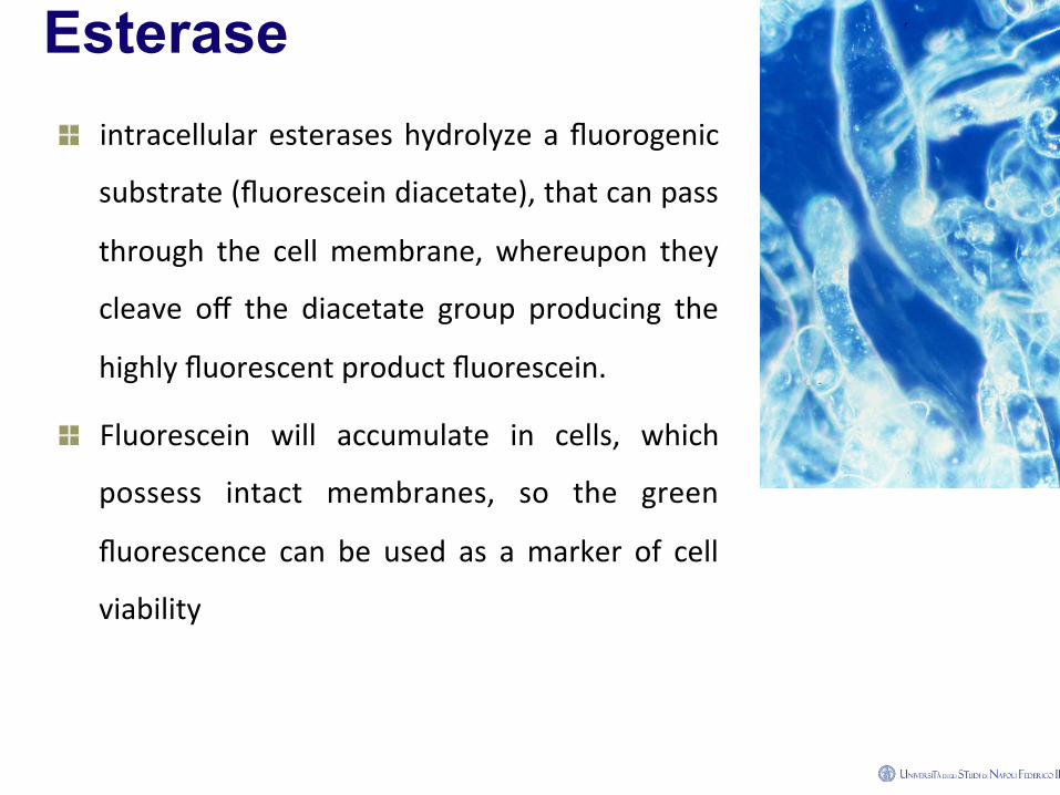
Esterase ! intracellular esterases hydrolyze a fluorogenic
substrate (fluorescein diacetate), that can pass
through the cell membrane, whereupon they
cleave off the diacetate group producing the
highly fluorescent product fluorescein.
! Fluorescein will accumulate in cells, which
possess intact membranes, so the green
fluorescence can be used as a marker of cell
viability
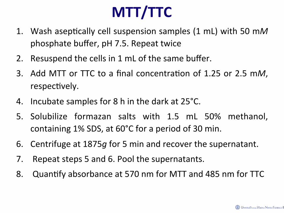
MTT/TTC 1. Wash asep2cally cell suspension samples (1 mL) with 50 mM
phosphate buffer, pH 7.5. Repeat twice
2. Resuspend the cells in 1 mL of the same buffer.
3. Add MTT or TTC to a final concentra2on of 1.25 or 2.5 mM, respec2vely.
4. Incubate samples for 8 h in the dark at 25°C.
5. Solubilize formazan salts with 1.5 mL 50% methanol, containing 1% SDS, at 60°C for a period of 30 min.
6. Centrifuge at 1875g for 5 min and recover the supernatant.
7. Repeat steps 5 and 6. Pool the supernatants. 8. Quan2fy absorbance at 570 nm for MTT and 485 nm for TTC

MTT assay in cells and protoplasts.
(a) Pink coloured cells (viables). (b) Cell of purple colour
(viable).
(c) Colourless cell (non-‐viable).
(d Pink and red coloured
protoplasts (viable).
(e) Protoplast with red
cytoplasm (viable).
(f) Protoplast with purple
cytoplasm (viable).
(g) Control of non-‐viable cells
dead with FAA.

TTC EXAMPLE
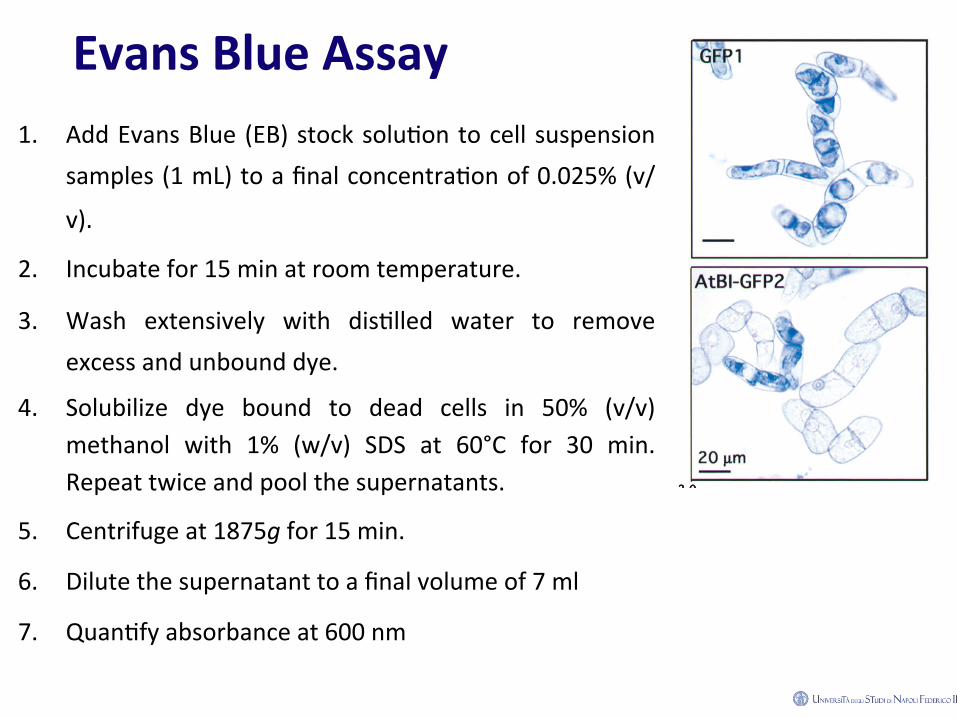
Evans Blue Assay 1. Add Evans Blue (EB) stock solu2on to cell suspension
samples (1 mL) to a final concentra2on of 0.025% (v/
v).
2. Incubate for 15 min at room temperature.
3. Wash extensively with dis2lled water to remove excess and unbound dye.
4. Solubilize dye bound to dead cells in 50% (v/v) methanol with 1% (w/v) SDS at 60°C for 30 min. Repeat twice and pool the supernatants.
5. Centrifuge at 1875g for 15 min.
6. Dilute the supernatant to a final volume of 7 ml
7. Quan2fy absorbance at 600 nm

Cells and protoplasts stained with Evans blue.
(a) Non-‐stained cells (viable). (b) Viable protoplast surrounded by blue cellular aggregates (c) Cell with blue cytoplasm. (d) Control of Non-‐viable cells dead with FAA.

FDA Assay 1. Mix cell samples (1 mL) with 10 μL of FDA stock solu2on.
2. Incubate for 15 min at room temperature in the dark.
3. Adjust the volume to 4 mL with dis2lled water.
4. Centrifuge at 1875g for 5 min. Resuspend the pellet in 1 mL 50 mM phosphate buffer, pH 7.5.
5. Freeze quickly in liquid nitrogen. Thaw and dilute samples to 3 mL with phosphate buffer.
6. Homogenize with a Brinkman polytron at high speed for 10 s.
7. Centrifuge at 1875g for 20 min.
8. Dilute a 100 μL sample of the supernatant to a final volume of 2 mL.
9. Determine fluorescence at 516 nm, using a 492 nm excita2on beam

Microscopic Assay 1. Stain cell samples with FDA by mixing the samples (1 mL) with 10
μL of the stock solu2on.
2. Incubate for 15 min at room temperature in the dark.
3. Wash with 50 mM phosphate buffer, pH 7.5.
4. Centrifuge at 1875g for 5 min.
5. Resuspend in phosphate buffer (1 mL).
6. Counterstain with EB, following steps 1–5 but using 10 μL of the EB stocksolu2on.
7. Determine the number of blue dead cells under a bright field and yellow-‐green fluorescent viable cells under ultraviolet light (excita2on: BP 450-‐490 nm and emission: LP 520 nm) in an Axioplan microscope in 10 randomized fields in Sedgewick chamber.