Embed Size (px)
Citation preview
1
Cell, Volume 137
Supplemental Data Cytokine/Jak/Stat Signaling Mediates Regeneration and Homeostasis in the Drosophila Midgut Huaqi Jiang, Parthive H. Patel, Alexander Kohlmaier, Marc O. Grenley, Donald G.
McEwen, and Bruce A. Edgar
Supplemental Experimental Procedures: Fly stocks UAS transgenes: UAS-Rpr, UAS-Hop, UAS-Upd26.2, UAS-Upd2, UAS-Stat92E,
UAS-HepAct, UAS-P35, UAS-Nintra, UAS-Dl20, UAS-E2F1Dp1, UAS-Puc and UAS-
BskDN. RNAi transgenes: UAS-Stat92E RNAi, UAS-Dome RNAi and USA-N
RNAi (NIG, Japan and VDRC, Austria). Mutants: hep1, hop25, hopTumL, FRT82B
stat85C9 and FRT82B stat397. Reporters and Gal4 drivers: MyoIANP0001Gal4,
esgNP7097Gal4, esglacZ, MyoIAlacZ, 10XStat-DGFP, (Gas)3lacZ, DomeGal4,
pucE69lacZ, updlacZ, upd3Gal4, GbeSu(H)lacZ and DllacZ. MARCM stocks:
FRT42D: y w hsflp UAS-GFP tubGal4; FRT42D tubGal80; +, FRT82B: y w hsflp
UAS-GFP tubGal4; +; FRT82B tubGal80. Midgut Gal4 drivers: MyoIAGal4ts: w;
MyoIAGal4 tubGal80ts UAS-GFP; +; esgGal4ts: w; esgGla4 tubGal80ts UAS-GFP;
+.
Generation of UAS-puc RNAi To deplete puckered (puc), a transgenic RNAi targeting nucleotides 1369-1932
(564 bp; Accession. NM_079549), was generated from puc cDNA with the
following primer pair: 5’-GCTCTAGAGCACCTGAATAGTCCTAGCAATCC-3’ and
5’-GCTCTAGAACATGGTTAATCCTGCTATCCGG-3’. The product was
subcloned, sequenced, and digested with XbaI prior to cloning, in the antisense
orientation, into the AvrII site of pWIZ (Lee and Carthew, 2003). This XbaI
fragment was cloned in the sense orientation into the downstream NheI site,
2
creating a tail-to-head, head-tail arrangement separated by white intron 2.
Transgenic lines were established through standard P-element mediated
transformation.
Gut turnover rate analysis For midgut turnover under normal lab culturing conditions, 3-10 days old female
adult flies (raised and aged at 18°C) with the genotype: w; tub>CD2>Gal4 UAS-
CD8-GFP/esgGal4; UAS-flp/tubGal80ts were shifted to 29°C (to induce the
transgenes) and adult midguts were dissected and examined at specific time
points after transfer.
For gut turnover in hopTumL mutants: hopTumL/Basc virgin flies were crossed to the
esgtsF/O tester males with the genotype: w; esgGal4 tubGal80ts UAS-GFP; UAS-
flp Act>CD2>Gal4. The hemizygous mutant males (hopTumL/Y, esgtsF/O) and
control males (Basc/Y, esgtsF/O) were aged at 18°C for 3-10 days and examined
after shifting cultures to 29°C for 8 days.
For Pe- induced gut turnover in hop mutant (hop25), hop25/FM7 virgin flies were
crossed to the esgtsF/O tester. The hemizygous mutant males (hop25/Y, esgtsF/O)
and control males (FM7/Y, esgtsF/O) were aged 3-10 days at 18ºC before shifted
to 29ºC and fed Pe-laced food (1/2 ml 10X concentrated overnight Pe culture) for
one day and then their gut turnover were analyzed.
Overexpression and mutant clone experiments For ectopic expression experiments, 3-10 days old adults (raised at 18°C) were
shifted to 29°C to activate UAS-transgene expression in progenitor cells (esgts) or
ECs (MyoIAts).
For mutant ISC clonal assay, 3-10 days old adults were heat shocked for 45
minutes at 37°C to induce clones, then the flies were kept at 25°C until
dissection. For Stat92E rescue experiment, adult flies with the following
genotype: y w hsflp tubGal4 UAS-GFP/+ or Y; UAS-Stat92E/+; FRT82B
Stat397/FRT82B tubGal80 were used.
3
RT-qPCR
The following were used to examine relative gene expression by RT-qPCR.
upd: Forward: 5’ CCACGTAAGTTTGCATGTTG 3’
ReverseN: 5’ CTAAACAGTAGCCAGGACTC 3’
upd2: Forward: 5’ ACTGTTGCATGTGGATGCTG 3’
ReverseN: 5’ CAGCCAAGGACGAGTTATCA 3’
upd3: Forward: 5’ GAGCACCAAGACTCTGGACA 3’
ReverseN: CCAGTGCAACTTGATGTTGC 3’
Socs36E: Forward: 5’ CAGTCAGCAATATGTTGTCG 3’
ReverseN: 5’ ACTTGCAGCATCGTCGCTTC 3’
Dl: Forward: 5’ TGTGAACATGGACATTGCGA 3’
ReverseN: 5’ GTCTGTGGTTGGTGCAGTAG 3’
puc: Forward: 5’ CATGTGGCTAGCAATTTGA 3’
ReverseN: 5’ GTCTTCGAAAAACGTACAGC 3’
E(spl): Forward: 5’ AGGTGGCTCAGGAAGAACAA 3’
ReverseN: 5’ CGTTGACGGCATTCATGTAG 3’
E(spl)mα: Forward: 5’ AGAATCCGTCGACAATCTGC 3’
ReverseN: 5’ GCTCCTCGACAATCTCGAAC 3’
E(spl)mδ: Forward: 5’ AATTGGGTCACCAGCTCAAG 3’
ReverseN: 5’ ATATGGAATGCCCTTCACCA 3’
E(spl)m7: Forward: 5’ GTGGCTTTTGGAACCACACT 3’
ReverseN: 5’ GACTGATGGAGGAGCAGGAG 3’
Quantification of adult gut cells Quantification of the marked cells, including Dl+, PH3+, GbeSu(H)lacZ+ and Pros+
cells were performed on double immunostained midguts from Rpr-induced EC
cell death or Pe-induced gut regeneration.
4
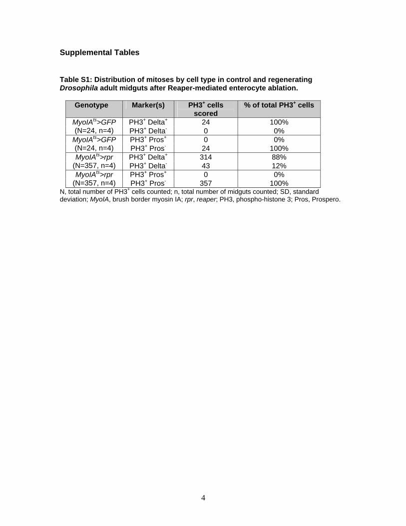
Supplemental Tables Table S1: Distribution of mitoses by cell type in control and regenerating Drosophila adult midguts after Reaper-mediated enterocyte ablation.
Genotype Marker(s) PH3+ cells scored
% of total PH3+ cells
PH3+ Delta+ 24 100% MyoIAts>GFP (N=24, n=4) PH3+ Delta- 0 0%
PH3+ Pros+ 0 0% MyoIAts>GFP (N=24, n=4) PH3+ Pros- 24 100%
PH3+ Delta+ 314 88% MyoIAts>rpr (N=357, n=4) PH3+ Delta- 43 12%
PH3+ Pros+ 0 0% MyoIAts>rpr (N=357, n=4) PH3+ Pros- 357 100%
N, total number of PH3+ cells counted; n, total number of midguts counted; SD, standard deviation; MyoIA, brush border myosin IA; rpr, reaper; PH3, phospho-histone 3; Pros, Prospero.
5
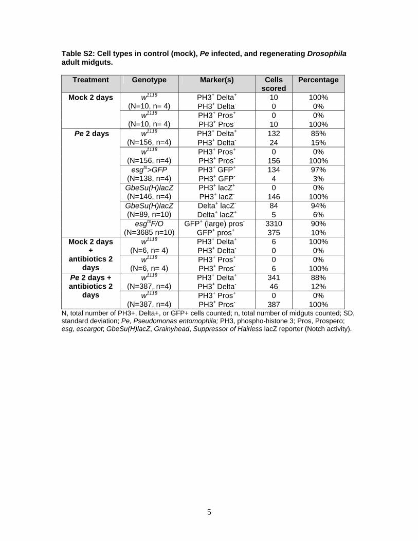
Table S2: Cell types in control (mock), Pe infected, and regenerating Drosophila adult midguts.
Treatment Genotype Marker(s) Cells scored
Percentage
PH3+ Delta+ 10 100% w1118 (N=10, n= 4) PH3+ Delta- 0 0%
PH3+ Pros+ 0 0%
Mock 2 days
w1118 (N=10, n= 4) PH3+ Pros- 10 100%
PH3+ Delta+ 132 85% w1118 (N=156, n=4) PH3+ Delta- 24 15%
PH3+ Pros+ 0 0% w1118 (N=156, n=4) PH3+ Pros- 156 100%
PH3+ GFP+ 134 97% esgts>GFP (N=138, n=4) PH3+ GFP- 4 3%
PH3+ lacZ+ 0 0% GbeSu(H)lacZ (N=146, n=4) PH3+ lacZ- 146 100%
Delta+ lacZ- 84 94% GbeSu(H)lacZ (N=89, n=10) Delta+ lacZ+ 5 6%
GFP+ (large) pros- 3310 90%
Pe 2 days
esgtsF/O (N=3685 n=10) GFP+ pros+ 375 10%
PH3+ Delta+ 6 100% w1118 (N=6, n= 4) PH3+ Delta- 0 0%
PH3+ Pros+ 0 0%
Mock 2 days +
antibiotics 2 days
w1118 (N=6, n= 4) PH3+ Pros- 6 100%
PH3+ Delta+ 341 88% w1118 (N=387, n=4) PH3+ Delta- 46 12%
PH3+ Pros+ 0 0%
Pe 2 days + antibiotics 2
days w1118 (N=387, n=4) PH3+ Pros- 387 100%
N, total number of PH3+, Delta+, or GFP+ cells counted; n, total number of midguts counted; SD, standard deviation; Pe, Pseudomonas entomophila; PH3, phospho-histone 3; Pros, Prospero; esg, escargot; GbeSu(H)lacZ, Grainyhead, Suppressor of Hairless lacZ reporter (Notch activity).
6
Supplemental Figure Legends
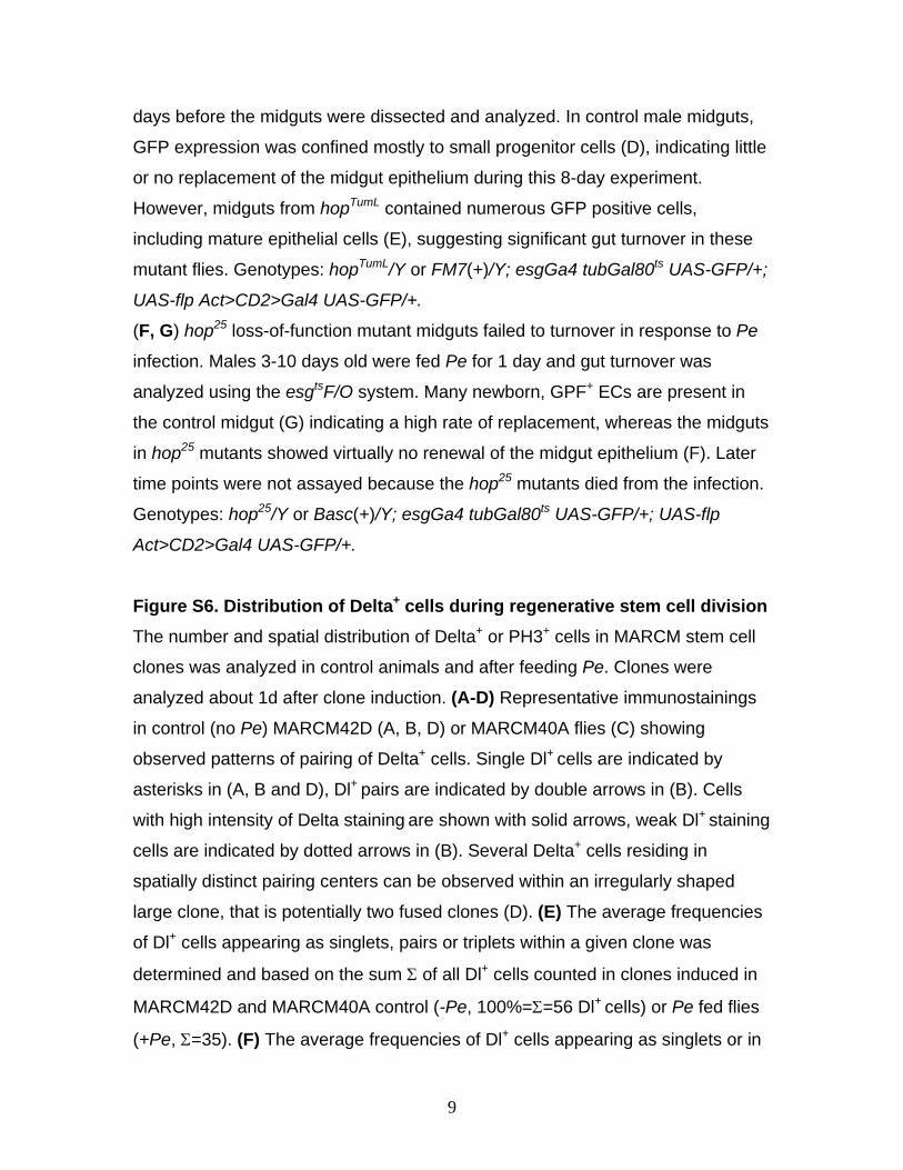
Figure S1. Recovery from midgut hyperplasia induced by JNK signaling. Punctate cytoplasmic Delta (Dl) staining (red) marks the ISCs and nuclear PH3
staining (red) marks the mitotic cells. Blue is DNA.
(A) Control midgut at day 0 (0 D) prior to HepAct induction.
(B) Hyperplastic midgut caused by prolonged JNK activation (MyoIAts>HepAct,
4D). The gut contains more large epithelial cells and small Dl+ progenitor cells.
(C) 2 days after transgene shutoff, the PH3 staining is still high in the midgut,
indicating that the progenitor cells continued to proliferate.
(D) 4 days after transgene shutoff, the midgut morphology reverted to normal and
the elevated proliferation of the progenitor cells ceased. The number of Dl+
progenitor cells also returned to normal.
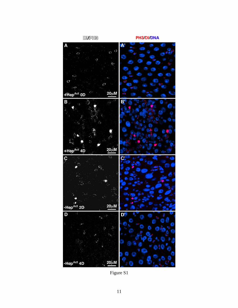
Figure S2. ISCs defective in Jak/Stat signaling fail to differentiate into mature ECs. MARCM clones were induced in young, 3-10 days old adults, and
midguts were dissected 8 days after clone induction. The clones, which occur
only in dividing cells (therefore ISCs) were marked by GFP expression (green).
Blue is DNA.
(A) FRT82B control. Asterisks indicate mature differentiated enterocytes (ECs) in
a control MARCM clone.
(B-E) ISC MARCM clones with defective Jak/Stat signaling were comprised
mostly of small progenitor-like cells. Clones were generated using the MARCM
FRT82B control and FRT82B stat85C9 (B), UAS-Dome RNAi (C) or UAS-Stat
RNAi (D) with FRT82B and FRT82B stat397 (E). Note the absence of large GFP
positive cells.
(F) The differentiation defects of Stat397 mutant ISCs were rescued by ectopic
expression of UAS-Stat92E. Asterisks indicate mature differentiated ECs in a
MARCM clone. Genotype: y w hsflp tubGal4 UAS-GFP/+ or Y; UAS-Stat92E/+;
FRT82B Stat397/FRT82B tubGal80.
7
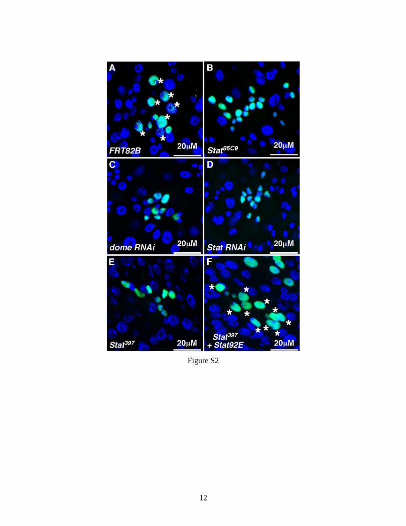
Figure S3. Ectopic Delta (Dl) does not rescue the differentiation defect of ISCs lacking Jak/Stat signaling. (A-C) The Dl gene expression pattern in the wildtype midgut epithelium, detected
using the DllacZ insertion reporter. DllacZ (red) is highly expressed in about half
of the esgGal4 positive progenitor cells (green, arrows), which we believe to be
ISCs. DllacZ expression is low or absent in the remaining esgGal4 positive cells,
which are usually paired with an ISC and are most likely EBs (arrowheads).
(D) FRT82B Control. 3-10 days old adult flies were heat shocked for 1 hour to
induce MARCM clones in ISCs, and midguts were analyzed after 8 days. A
typical MARCM clone containing one Dl+ ISC (arrow) is shown. Genotype: yw
hsflp UAS-GFP tubGal4; +; FRT82B tubGal80/FRT82B.
(E) A typical MARCM clone ectopically expressing Dl. In most such clones, Dl
was highly visible in small progenitor cells (indicated by arrows), but was low or
absent in differentiated ECs. Since UAS-Dl20 and UAS-GFP are being driven by
tubGal4 in this situation, this suggests that Dl is post-transcriptionally
downregulated in ECs. In this assay, ectopic Dl did not effect the ISC proliferation
or differentiation. Genotype: yw hsflp UAS-GFP tubGal4; UAS-Dl20/+; FRT82B
tubGal80/FRT82B.
(F) A typical stat85C9 MARCM clone overexpressing Dl. Such clones contained
many small progenitor cells that were all positive for ectopic Dl, indicating that Dl
is not sufficient to rescue the block to differentiation imposed by loss of Stat92E
function. Genotype: yw hsflp UAS-GFP tubGal4; UAS-Dl20/+; FRT82B
tubGal80/FRT82B stat85C9.
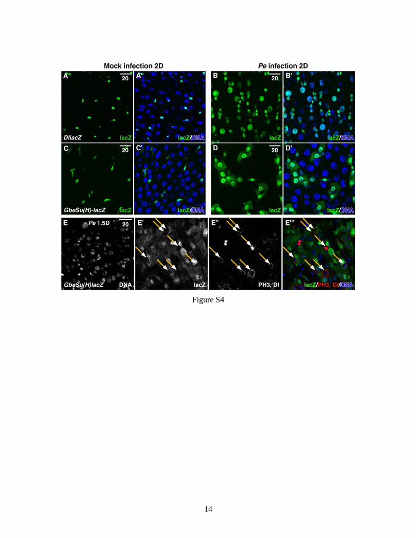
Figure S4. Induction of Delta and Notch activity in the midgut after Pseudomonas entomophila (Pe) ingestion. 3-10 days old adult flies were fed either food containing Pe (B, D) or
uncontaminated food (A, C) for 2 days, and then their midguts were dissected
and analyzed for expression of the Delta reporter DllacZ (A, B), or the Notch
reporter GbeSu(H)lacZ (C, D), or GbeSu(H)lacZ and endogenous Delta (E-E''').
8
(A, B) DllacZ is normally high in ISCs (A, S3), but after Pe ingestion it is highly
induced and expressed in many more gut epithelial cells (B).
(C, D) The N activity reporter, GbeSu(H)lacZ, is normally expressed in EBs (C),
but after Pe infection LacZ was detected in many more gut epithelial cells (D). In
both cases, the presence of LacZ in large differentiated cells could be due to
perdurance of this stable protein and the high regeneration rate of ECs during
infection.
(E-E''') During regeneration strong cytoplasmic punctuate Delta accumulation
and Notch activity are largely mutually exclusive (94%, see Table S2) as
measured by Delta and lacZ staining in GbeSu(H)lacZ reporter flies fed with PE
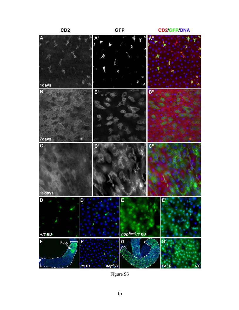
for 1.5 days. Figure S5. Midgut epithelial turnover as detected using the esgts F/O system. 3-10 day old female (A-C) or male (D, E) adult flies (raised at 18°C) were shifted
to 29°C for various times as indicated. Midguts were analyzed for CD2
expression (A-C), which is lost from progenitor cells following the temperature
shift, and GFP (A'-C') that is simultaneously activated. Genotype: w;
tub>CD2>Gal4 UAS-CD8-GFP/esgGal4; UAS-flp/tubGal80ts.
(A) One day after induction at 29°C, only small progenitor cells are GFP+. The
CD2 staining is also stronger in the progenitor cells. We attribute this to higher
activity of the tub>CD2>Gal4 transgene in progenitor cells.
(B) Due to Flp-mediated recombination, CD2 expression disappeared from
progenitor cells after 7 days. The large GFP+, CD2- cells are newly generated
ECs.
(C) By 12 days the majority of epithelial cells in the posterior midguts of females
were GFP+, indicating that the posterior midgut had largely renewed itself. The
epithelial replacement rate was much slower in wildtype males, as shown below
(D) and in Fig 7.
(D, E) Gut turnover in hop (Jak) gain-of-function mutants as measured using the
esgts F/O system. Control (D) and hopTumL/Y (E) males were shifted to 29°C for 8
9
days before the midguts were dissected and analyzed. In control male midguts,
GFP expression was confined mostly to small progenitor cells (D), indicating little
or no replacement of the midgut epithelium during this 8-day experiment.
However, midguts from hopTumL contained numerous GFP positive cells,
including mature epithelial cells (E), suggesting significant gut turnover in these
mutant flies. Genotypes: hopTumL/Y or FM7(+)/Y; esgGa4 tubGal80ts UAS-GFP/+;
UAS-flp Act>CD2>Gal4 UAS-GFP/+.
(F, G) hop25 loss-of-function mutant midguts failed to turnover in response to Pe
infection. Males 3-10 days old were fed Pe for 1 day and gut turnover was
analyzed using the esgtsF/O system. Many newborn, GPF+ ECs are present in
the control midgut (G) indicating a high rate of replacement, whereas the midguts
in hop25 mutants showed virtually no renewal of the midgut epithelium (F). Later
time points were not assayed because the hop25 mutants died from the infection.
Genotypes: hop25/Y or Basc(+)/Y; esgGa4 tubGal80ts UAS-GFP/+; UAS-flp
Act>CD2>Gal4 UAS-GFP/+.
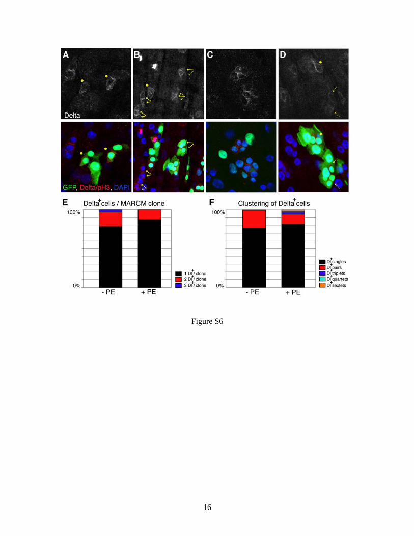
Figure S6. Distribution of Delta+ cells during regenerative stem cell division The number and spatial distribution of Delta+ or PH3+ cells in MARCM stem cell
clones was analyzed in control animals and after feeding Pe. Clones were
analyzed about 1d after clone induction. (A-D) Representative immunostainings
in control (no Pe) MARCM42D (A, B, D) or MARCM40A flies (C) showing
observed patterns of pairing of Delta+ cells. Single Dl+ cells are indicated by
asterisks in (A, B and D), Dl+ pairs are indicated by double arrows in (B). Cells
with high intensity of Delta staining are shown with solid arrows, weak Dl+ staining
cells are indicated by dotted arrows in (B). Several Delta+ cells residing in
spatially distinct pairing centers can be observed within an irregularly shaped
large clone, that is potentially two fused clones (D). (E) The average frequencies
of Dl+ cells appearing as singlets, pairs or triplets within a given clone was
determined and based on the sum Σ of all Dl+ cells counted in clones induced in
MARCM42D and MARCM40A control (-Pe, 100%=Σ=56 Dl+ cells) or Pe fed flies
(+Pe, Σ=35). (F) The average frequencies of Dl+ cells appearing as singlets or in

10
clusters was determined for all Dl+ staining cells in the posterior midgut region of
MARCM42D and hsFlp;Frt82b controls (-Pe, Σ=157 Dl+ cells) or Pe fed (+Pe,
Σ=254 Dl+ cells) flies irrespective of clone boundaries. Both analyses suggest that
Pe infection does not promote stem cell duplication.
11
Figure S1
12
Figure S2
13
Figure S3
14
Figure S4
15
Figure S5
16
Figure S6


![The value of cytokine levels in triage and risk prediction ......(STAT) pathway, which result in local epithelial cell proliferation and disease progression [11]. In this study, we](https://img.pdfslide.net/doc/110x75/608d4b682d4b1073450d4fdb/the-value-of-cytokine-levels-in-triage-and-risk-prediction-stat-pathway.jpg)