Embed Size (px)
Citation preview
CellsDevelopment of the Cell Theory
Introduction to Cells
Organelles
Cytoskeleton
Plasma Membrane
Cytomembrane System
Nucleus
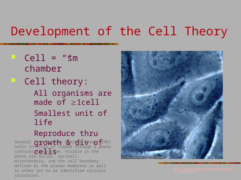
Development of the Cell Theory
Cell = “sm chamber” Cell theory:
1. All organisms are made of 1cell
2. Smallest unit of life
3. Reproduce thru growth & div of cells
http://www.flickr.com/photos/exothermic/2546338537/
Several live Chinese Hamster Ovary (CHO) cells in vitro as viewed through a phase contrast microscope. Visible in the photo are nuclei, nucleoli, mitochondria, and the cell boundary defined by the plasma membrane as well as other yet to be identified cellular structures.
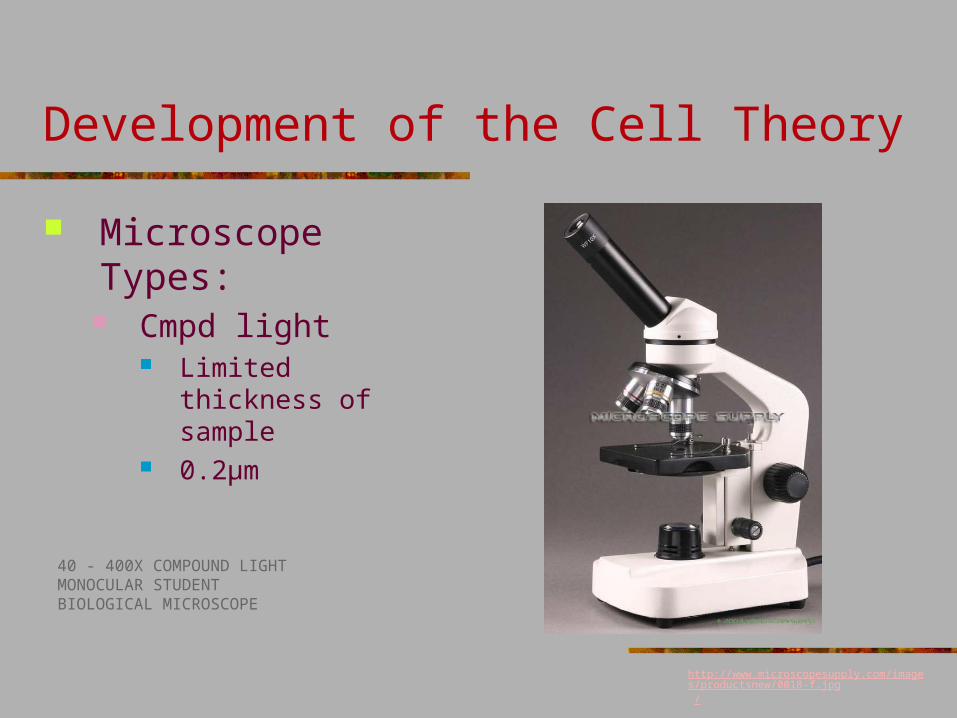
Development of the Cell Theory
Microscope Types: Cmpd light
Limited thickness of sample
0.2μm
http://www.microscopesupply.com/images/productsnew/0018-f.jpg /
40 - 400X COMPOUND LIGHTMONOCULAR STUDENTBIOLOGICAL MICROSCOPE
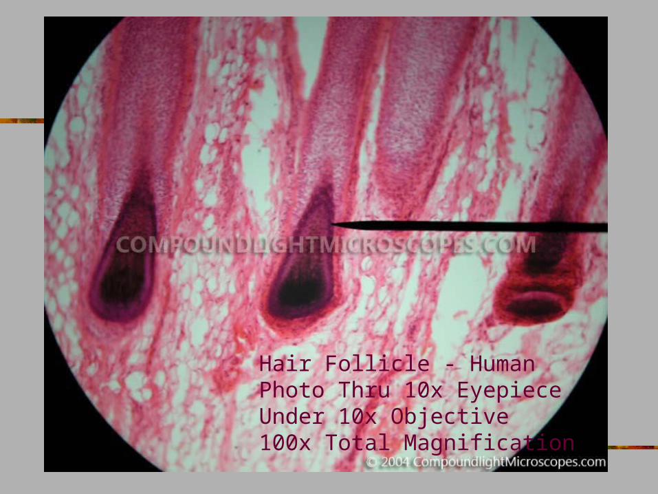
Hair Follicle - HumanPhoto Thru 10x EyepieceUnder 10x Objective100x Total Magnification
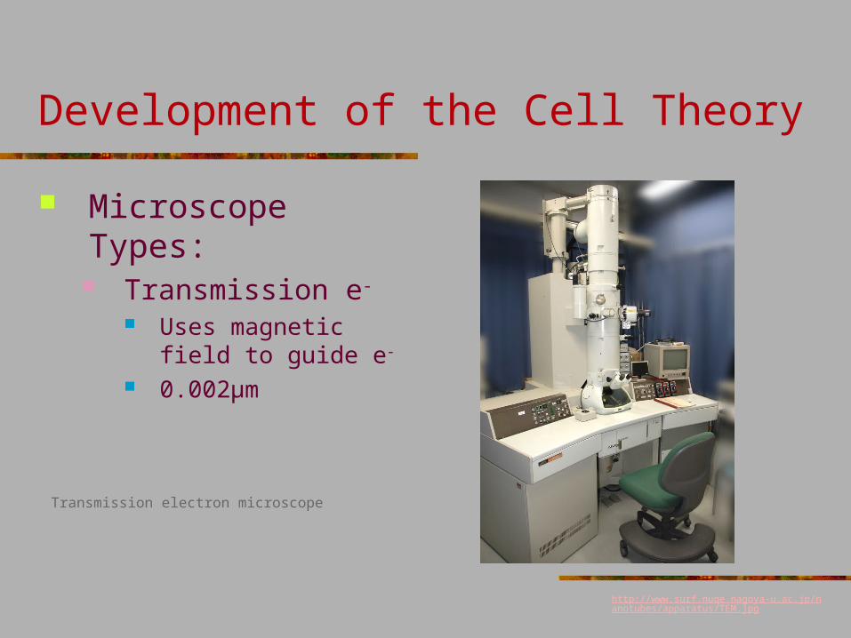
Development of the Cell Theory
Microscope Types: Transmission e-
Uses magnetic field to guide e-
0.002μm
http://www.surf.nuqe.nagoya-u.ac.jp/nanotubes/apparatus/TEM.jpg
Transmission electron microscope
http://neurocog.psy.tufts.edu/courses/Jan25/mitochondria.jpg
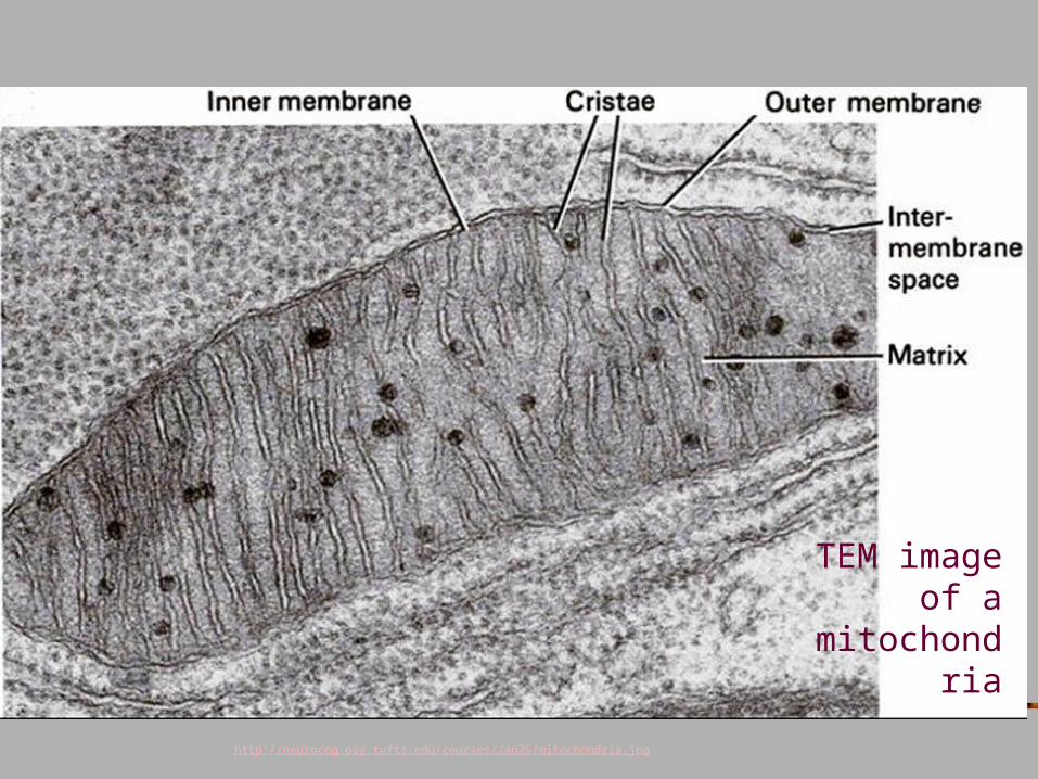
TEM image of a
mitochondria
Development of the Cell Theory
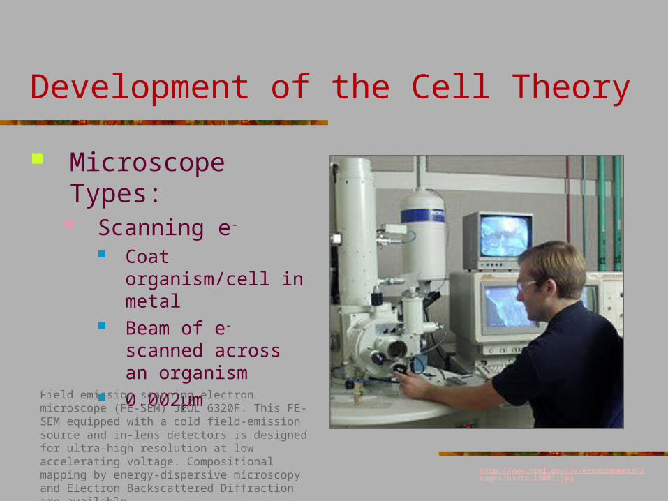
Microscope Types: Scanning e-
Coat organism/cell in metal
Beam of e- scanned across an organism
0.002μm
http://www.nrel.gov/pv/measurements/images/photo_14801.jpg
Field emission scanning electron microscope (FE-SEM) JEOL 6320F. This FE-SEM equipped with a cold field-emission source and in-lens detectors is designed for ultra-high resolution at low accelerating voltage. Compositional mapping by energy-dispersive microscopy and Electron Backscattered Diffraction are available.
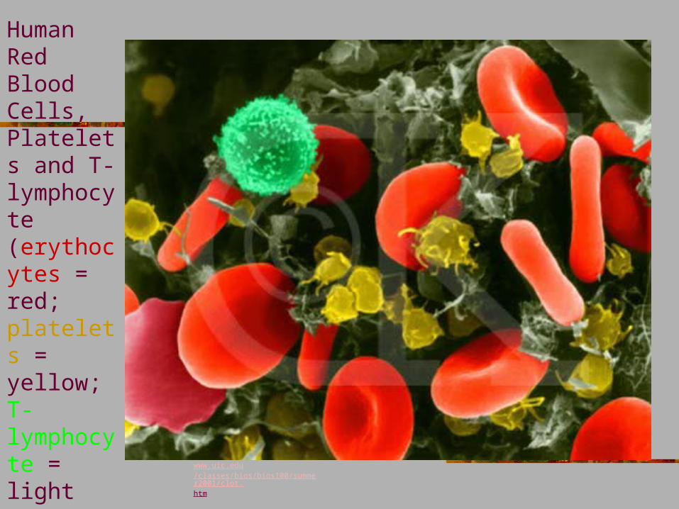
Human Red Blood Cells, Platelets and T-lymphocyte (erythocytes = red; platelets = yellow; T-lymphocyte = light green) (SEM x 9,900)
www.uic.edu/classes/bios/bios100/summer2001/clot.htm
How to use a scanning electron microscope: http://www.youtube.com/watch?v=lrXMIghANbg
Intro to Cells
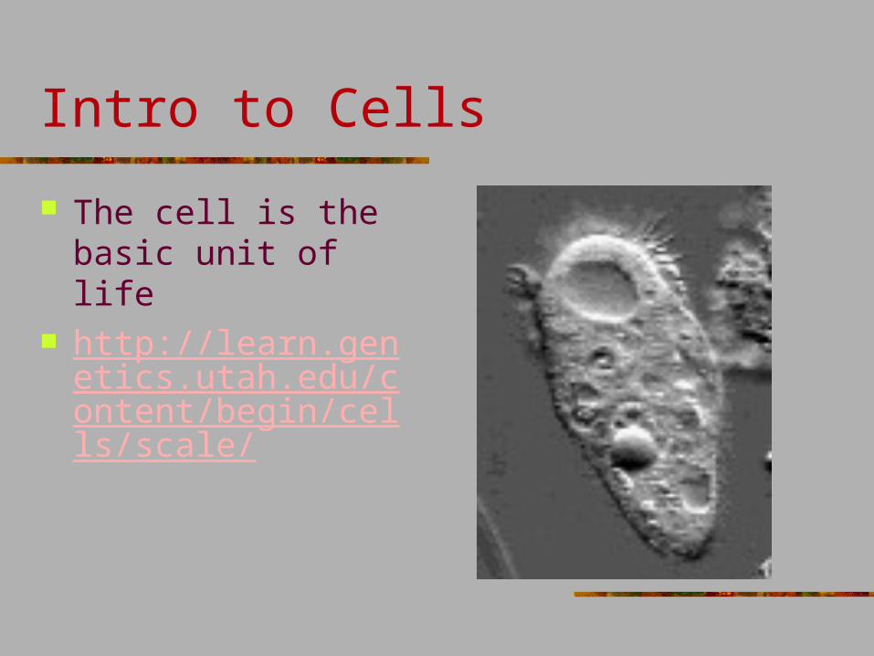
The cell is the basic unit of life
http://learn.genetics.utah.edu/content/begin/cells/scale/
Intro to Cells
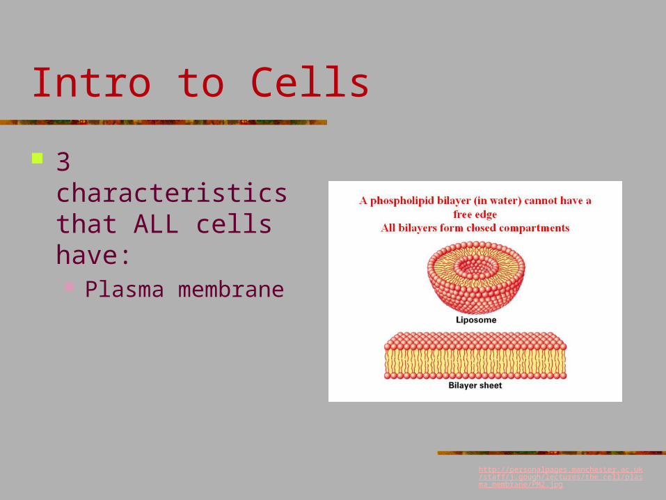
3 characteristics that ALL cells have: Plasma membrane
http://personalpages.manchester.ac.uk/staff/j.gough/lectures/the_cell/plasma_membrane/PM2.jpg
Intro to Cells
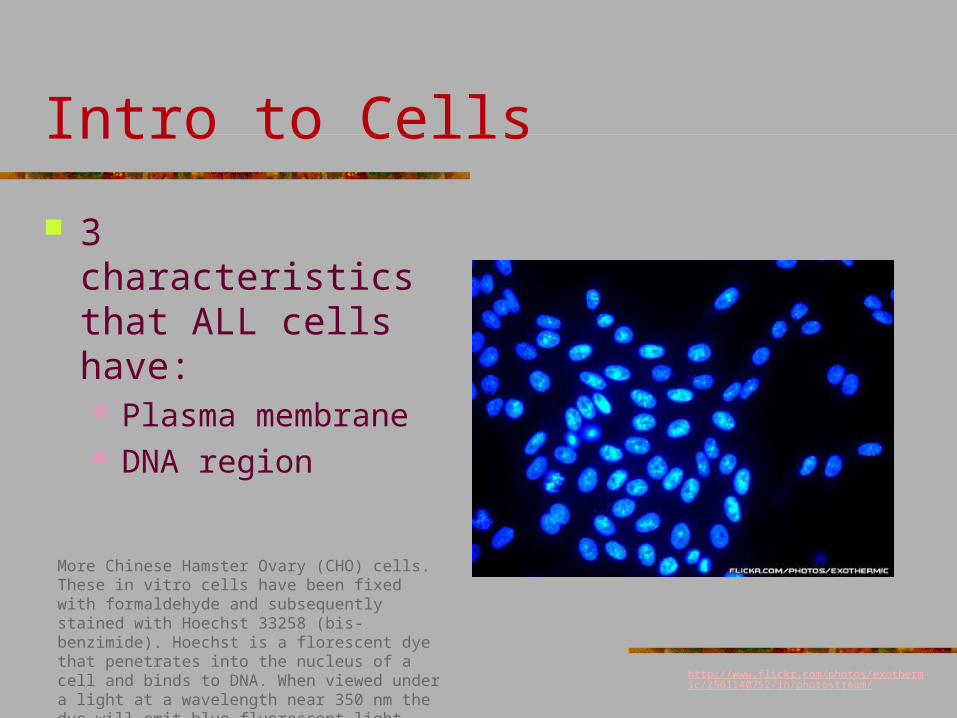
3 characteristics that ALL cells have: Plasma membrane DNA region
More Chinese Hamster Ovary (CHO) cells. These in vitro cells have been fixed with formaldehyde and subsequently stained with Hoechst 33258 (bis-benzimide). Hoechst is a florescent dye that penetrates into the nucleus of a cell and binds to DNA. When viewed under a light at a wavelength near 350 nm the dye will emit blue fluorescent light which effectively makes the DNA in the nucleus visible.
http://www.flickr.com/photos/exothermic/2561140752/in/photostream/
Intro to Cells
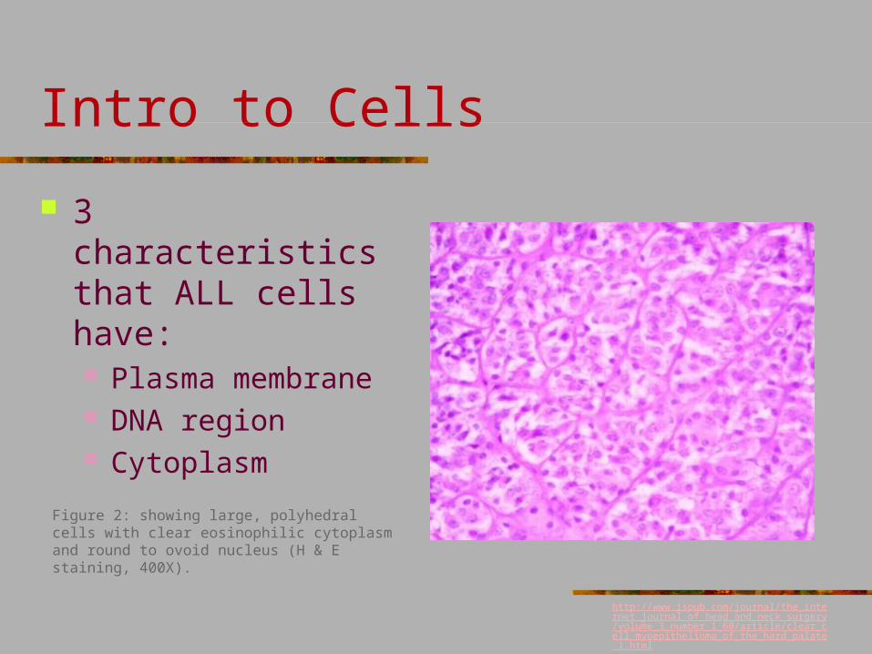
3 characteristics that ALL cells have: Plasma membrane DNA region Cytoplasm
Figure 2: showing large, polyhedral cells with clear eosinophilic cytoplasm and round to ovoid nucleus (H & E staining, 400X).
http://www.ispub.com/journal/the_internet_journal_of_head_and_neck_surgery/volume_3_number_1_60/article/clear_cell_myoepithelioma_of_the_hard_palate_1.html
Intro to Cells
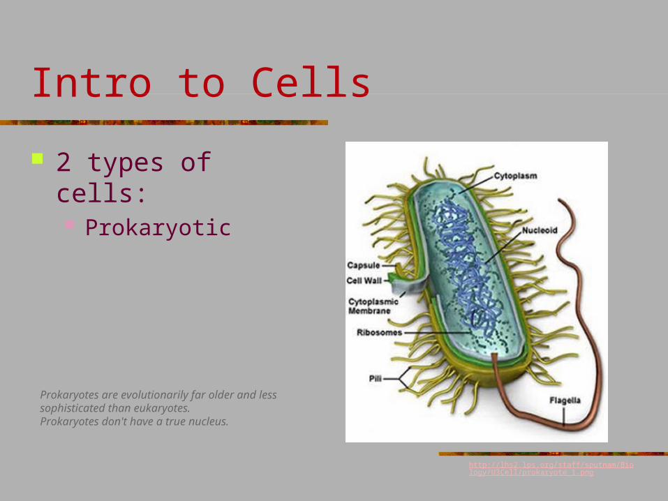
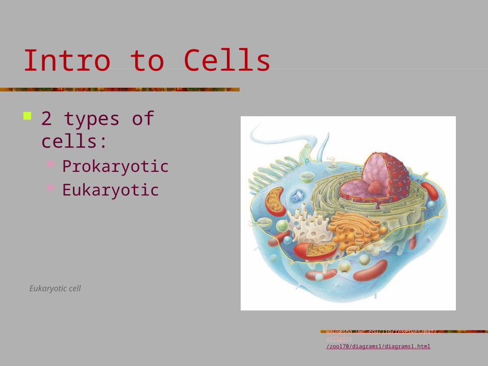
2 types of cells: Prokaryotic
Prokaryotes are evolutionarily far older and less sophisticated than eukaryotes. Prokaryotes don't have a true nucleus.
http://lhs2.lps.org/staff/sputnam/Biology/U3Cell/prokaryote_1.png
Intro to Cells
2 types of cells: Prokaryotic Eukaryotic
Eukaryotic cell
waukesha.uwc.edu/lib/reserves/pdf/zillgitt/zoo170/diagrams1/diagrams1.html
Intro to Cells
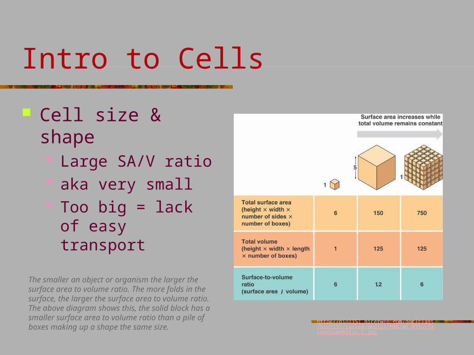
Cell size & shape Large SA/V ratio aka very small Too big = lack of easy
transport
The smaller an object or organism the larger the surface area to volume ratio. The more folds in the surface, the larger the surface area to volume ratio. The above diagram shows this, the solid block has a smaller surface area to volume ratio than a pile of boxes making up a shape the same size.
http://bio1151.nicerweb.com/doc/class/bio1151/Locked/media/ch06/06_07SurfaceVolumeRatio_L.jpg
7
1
2
3
456
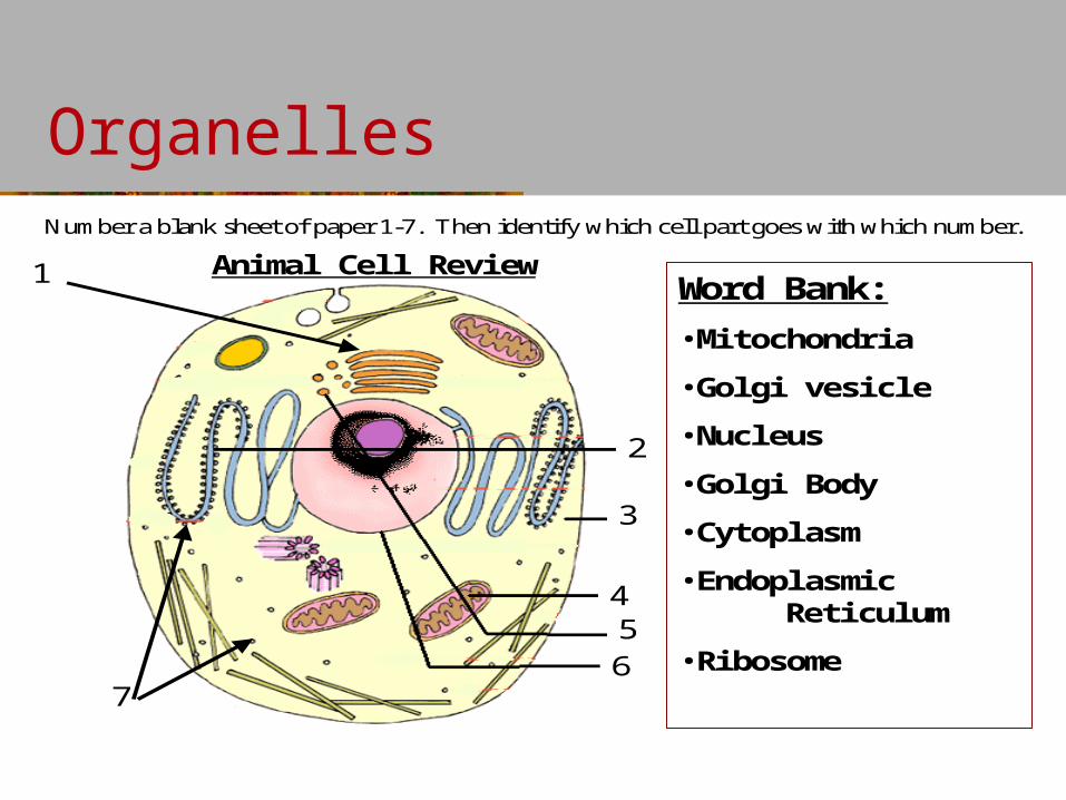
Word Bank:
•Mitochondria
•Golgi vesicle
•Nucleus
•Golgi Body
•Cytoplasm
•Endoplasmic Reticulum
•Ribosome
Animal Cell Review
Number a blank sheet of paper 1-7. Then identify which cell part goes with which number.
Organelles
Organelles
7
1
2
3
456
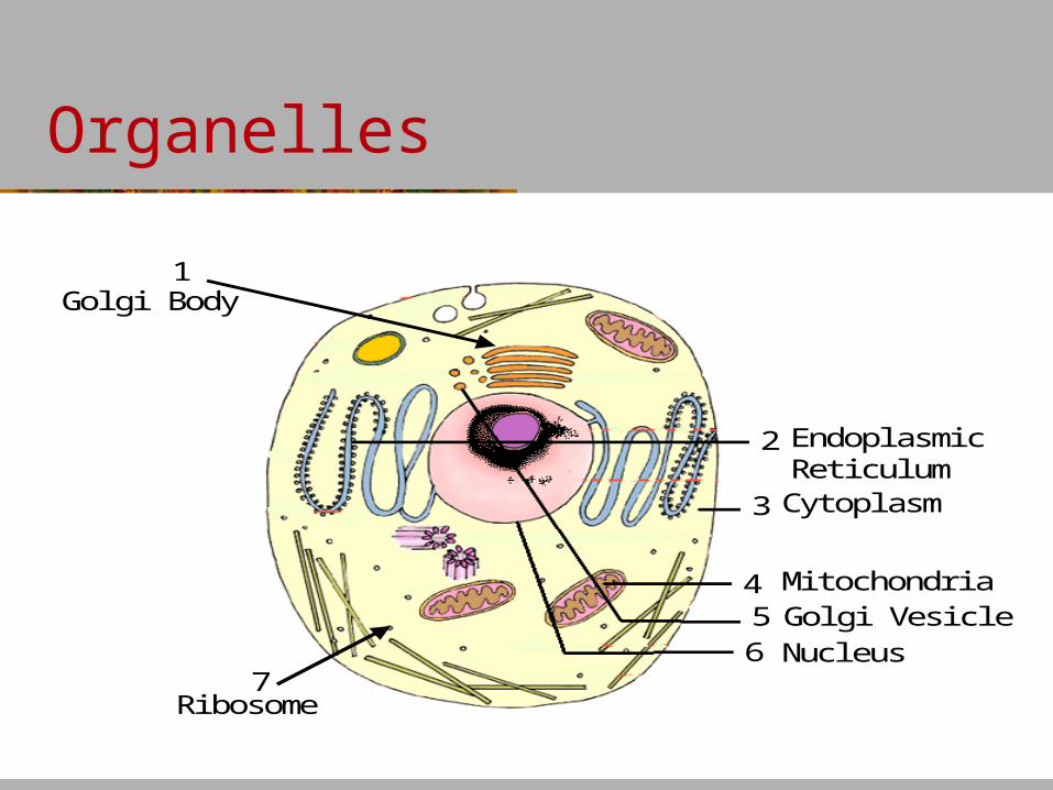
Gol gi Body
Endopl asmi c Reti cul umCytopl asm
Mi tochondri aGol gi Vesi cl eNucl eus
Ri bosome
Organelles
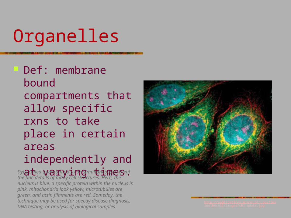
Def: membrane bound compartments that allow specific rxns to take place in certain areas independently and at varying times.
Dyes called quantum dots can simultaneously reveal the fine details of many cell structures. Here, the nucleus is blue, a specific protein within the nucleus is pink, mitochondria look yellow, microtubules are green, and actin filaments are red. Someday, the technique may be used for speedy disease diagnosis, DNA testing, or analysis of biological samples.
http://publications.nigms.nih.gov/insidethecell/images/ch1_qdots.jpg
Organelles
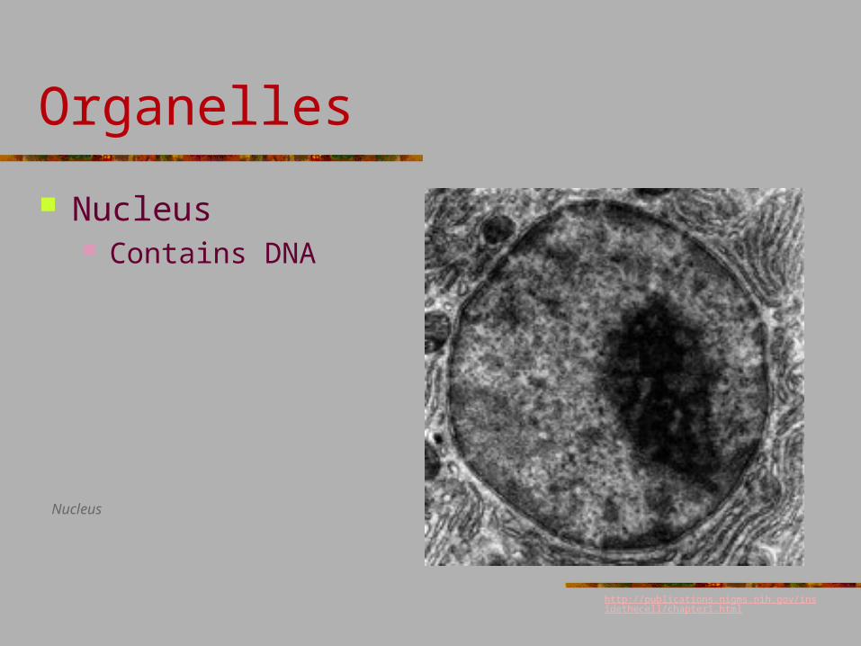
Nucleus Contains DNA
Nucleus
http://publications.nigms.nih.gov/insidethecell/chapter1.html
Organelles
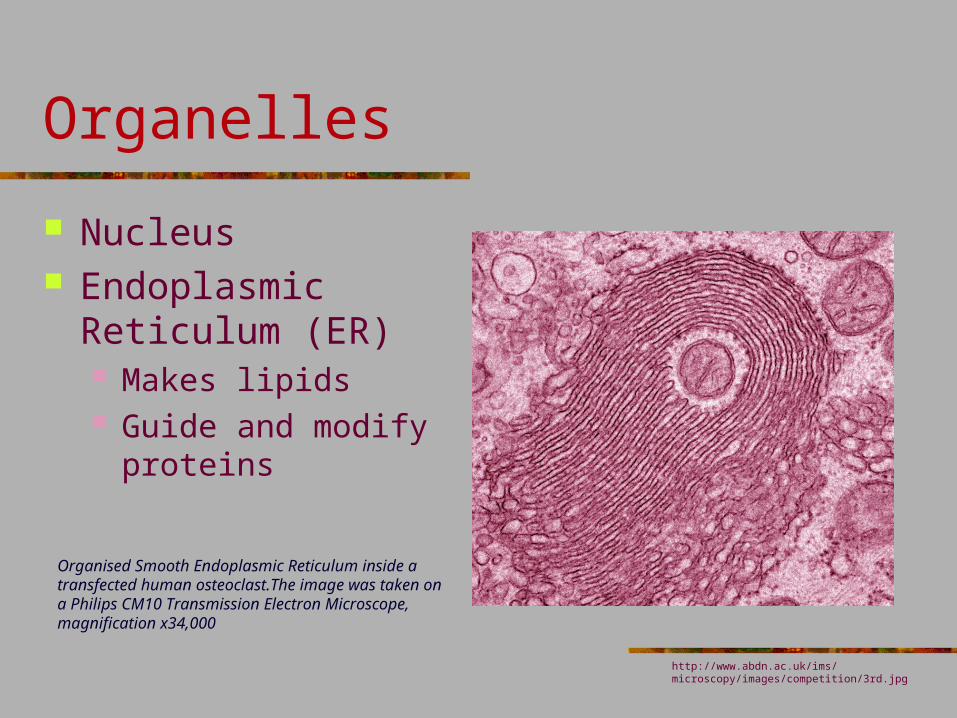
Nucleus Endoplasmic
Reticulum (ER) Makes lipids Guide and modify
proteins
Organised Smooth Endoplasmic Reticulum inside a transfected human osteoclast.The image was taken on a Philips CM10 Transmission Electron Microscope, magnification x34,000
http://www.abdn.ac.uk/ims/microscopy/images/competition/3rd.jpg
Organelles
Nucleus Endoplasmic
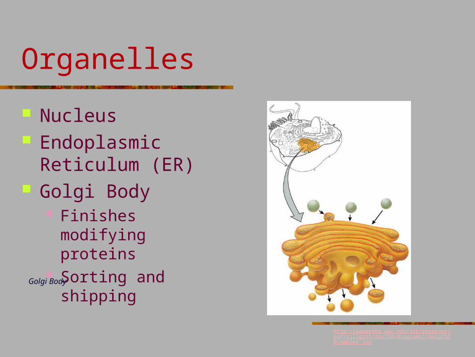
Reticulum (ER) Golgi Body
Finishes modifying proteins
Sorting and shippingGolgi Body
http://waukesha.uwc.edu/lib/reserves/pdf/zillgitt/zoo170/diagrams1/Golgi%20Complex.jpg
Organelles
Nucleus Endoplasmic
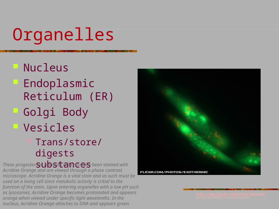
Reticulum (ER) Golgi Body Vesicles
Trans/store/digests substances
These progesterone treated CHO cells have been stained with Acridine Orange and are viewed through a phase contrast microscope. Acridine Orange is a vital stain and as such must be used on a living cell since metobolic activity is critial to the function of the stain. Upon entering organelles with a low pH such as lysosomes, Acridine Orange becomes protonated and appears orange when viewed under specific light wavelenths. In the nucleus, Acridine Orange attaches to DNA and appears green.
http://www.flickr.com/photos/exothermic/2611995050/in/photostream/
Organelles
Nucleus Endoplasmic
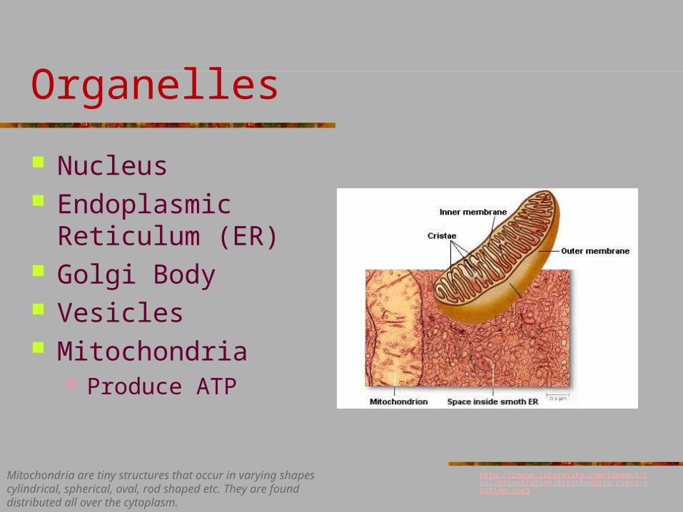
Reticulum (ER) Golgi Body Vesicles Mitochondria
Produce ATP
http://image.tutorvista.com/content/cell-organization/mitochondria-cross-section.jpeg
Mitochondria are tiny structures that occur in varying shapes cylindrical, spherical, oval, rod shaped etc. They are found distributed all over the cytoplasm.
Organelles
Nucleus Endoplasmic
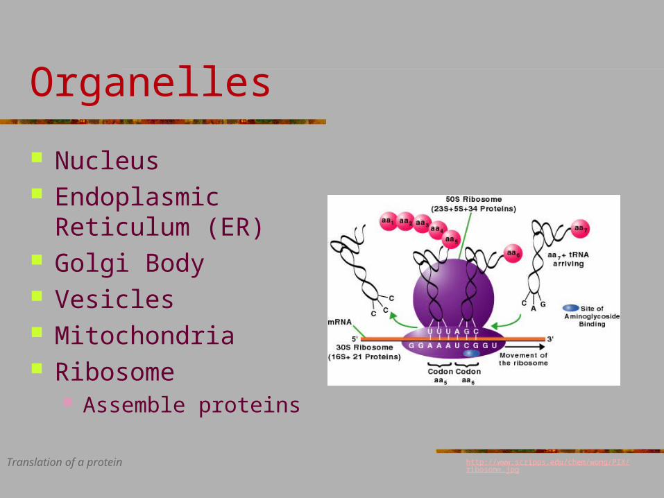
Reticulum (ER) Golgi Body Vesicles Mitochondria Ribosome
Assemble proteins
http://www.scripps.edu/chem/wong/PIX/ribosome.jpg Translation of a protein
Cytoskeleton
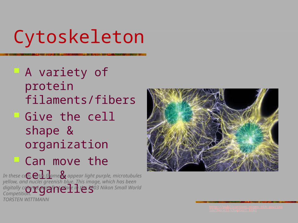
A variety of protein filaments/fibers
Give the cell shape & organization
Can move the cell & organelles
In these cells, actin filaments appear light purple, microtubules yellow, and nuclei greenish blue. This image, which has been digitally colored, won first place in the 2003 Nikon Small World Competition.TORSTEN WITTMANN
http://publications.nigms.nih.gov/insidethecell/chapter1.html
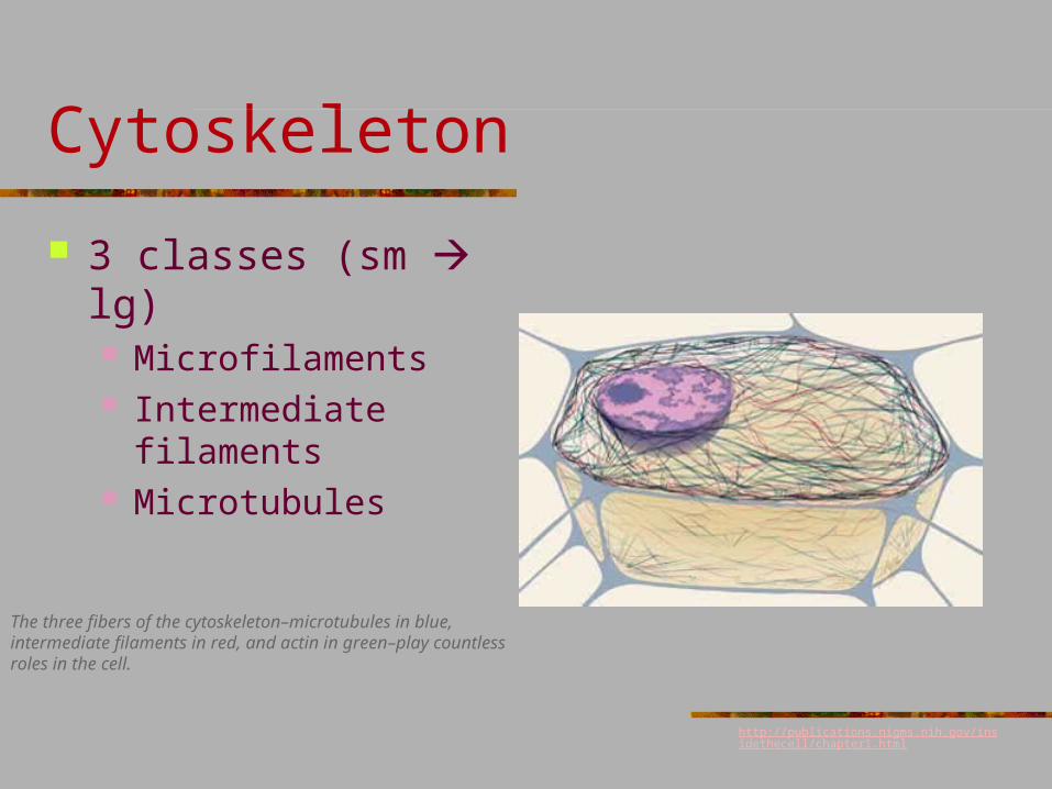
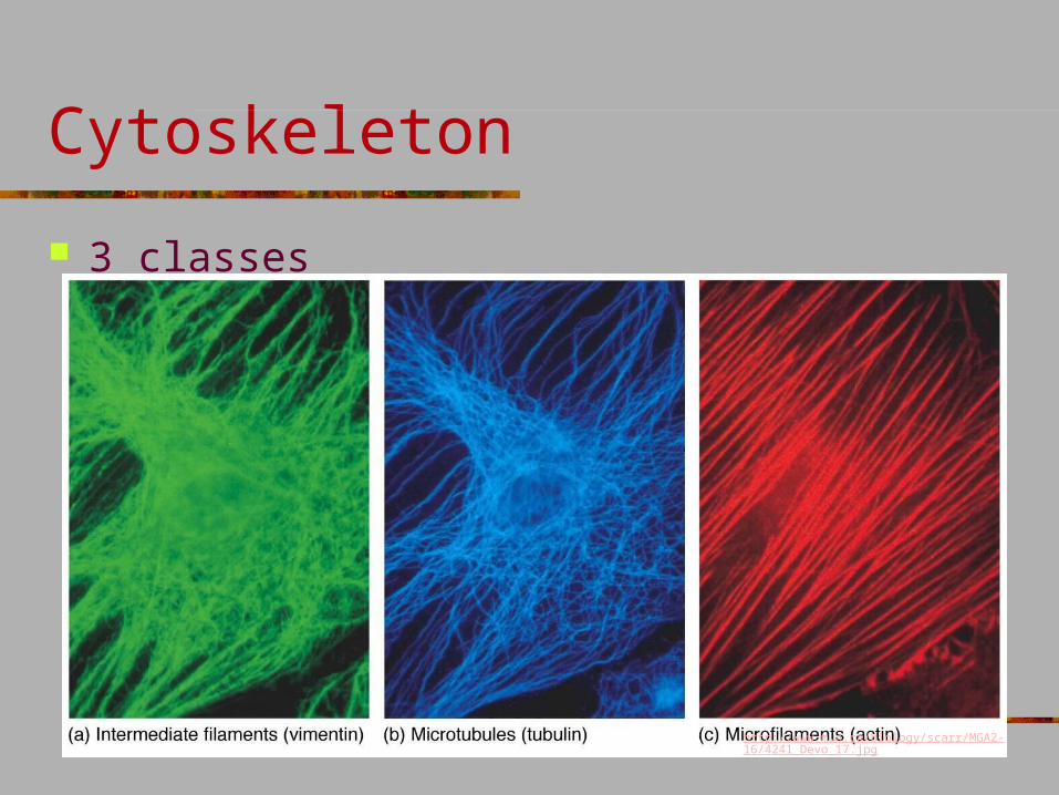
Cytoskeleton
3 classes (sm lg) Microfilaments Intermediate filaments Microtubules
The three fibers of the cytoskeleton–microtubules in blue, intermediate filaments in red, and actin in green–play countless roles in the cell.
http://publications.nigms.nih.gov/insidethecell/chapter1.html
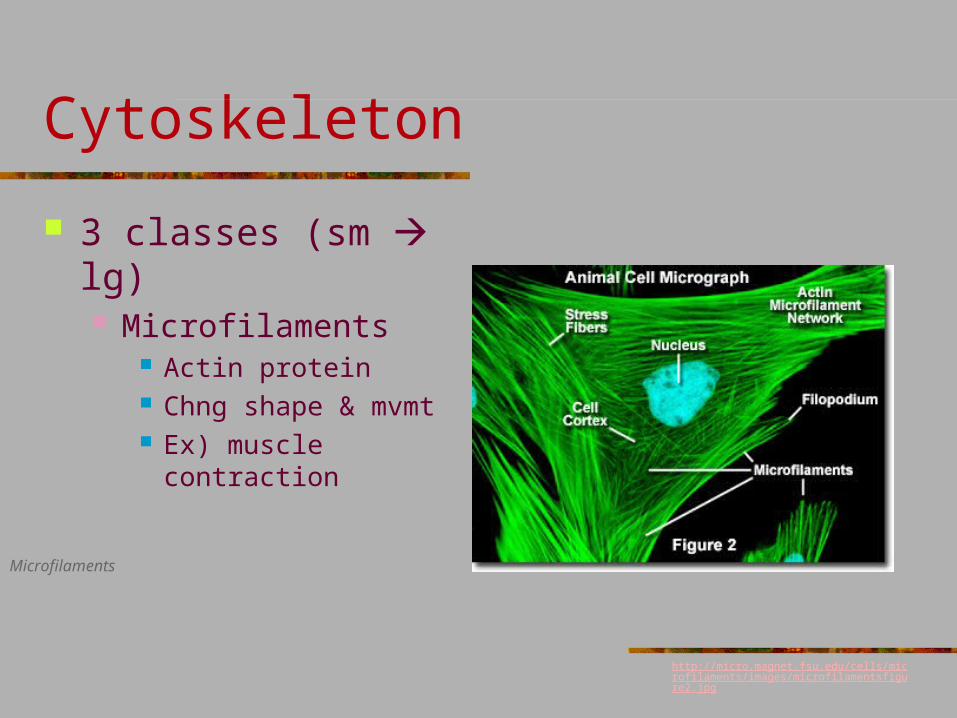
Cytoskeleton
3 classes (sm lg) Microfilaments
Actin protein Chng shape & mvmt Ex) muscle contraction
Microfilaments
http://micro.magnet.fsu.edu/cells/microfilaments/images/microfilamentsfigure2.jpg
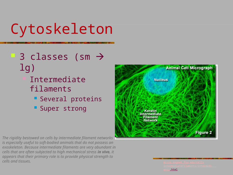
Cytoskeleton
3 classes (sm lg) Intermediate filaments
Several proteins Super strong
The rigidity bestowed on cells by intermediate filament networks is especially useful to soft-bodied animals that do not possess an exoskeleton. Because intermediate filaments are very abundant in cells that are often subjected to high mechanical stress in vivo, it appears that their primary role is to provide physical strength to cells and tissues. micro.magnet.fsu.edu/cellsintermediatefilaments/
intermedi...ents.html
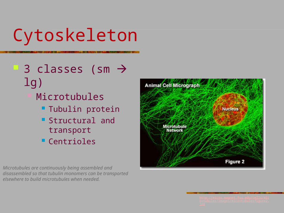
Cytoskeleton
3 classes (sm lg) Microtubules
Tubulin protein Structural and transport Centrioles
Microtubules are continuously being assembled and disassembled so that tubulin monomers can be transported elsewhere to build microtubules when needed.
http://micro.magnet.fsu.edu/cells/microtubules/images/microtubulesfigure2.jpg
Cytoskeleton
3 classes
http://www.mun.ca/biology/scarr/MGA2-16/4241_Devo_17.jpg
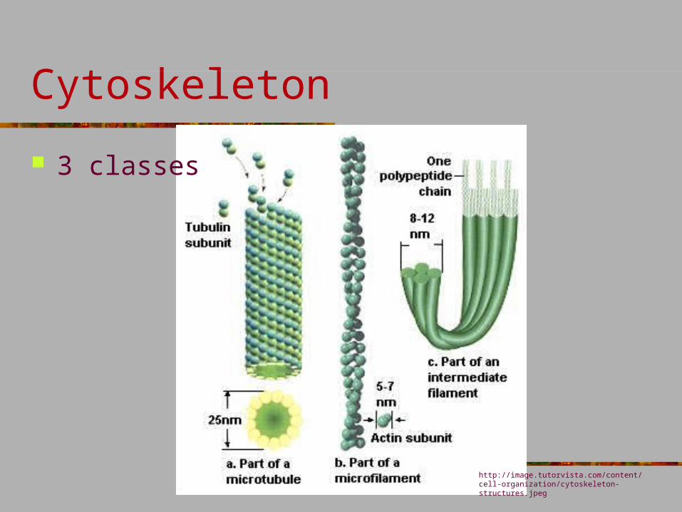
Cytoskeleton
3 classes
http://image.tutorvista.com/content/cell-organization/cytoskeleton-structures.jpeg
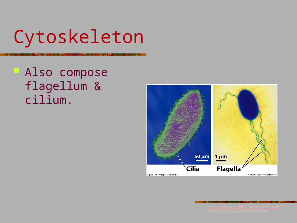
Cytoskeleton
Also compose flagellum & cilium.
http://www.uic.edu/classes/bios/bios100/lectures/cilia_flagella.jpg
The cytoskeleton: http://www.youtube.com/watch?v=5rqbmLiSkpk
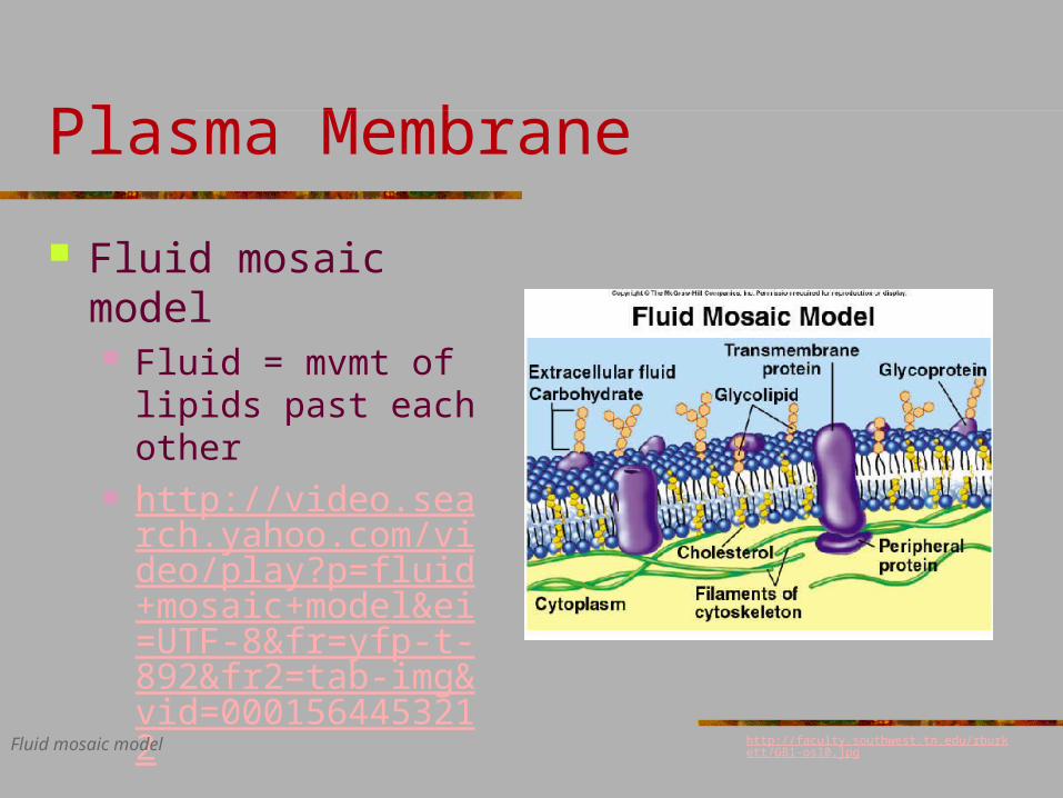
Plasma Membrane
Fluid mosaic model Fluid = mvmt of lipids
past each other http://video.search.yah
oo.com/video/play?p=fluid+mosaic+model&ei=UTF-8&fr=yfp-t-892&fr2=tab-img&vid=0001564453212
Fluid mosaic model http://faculty.southwest.tn.edu/rburkett/GB1-os10.jpg
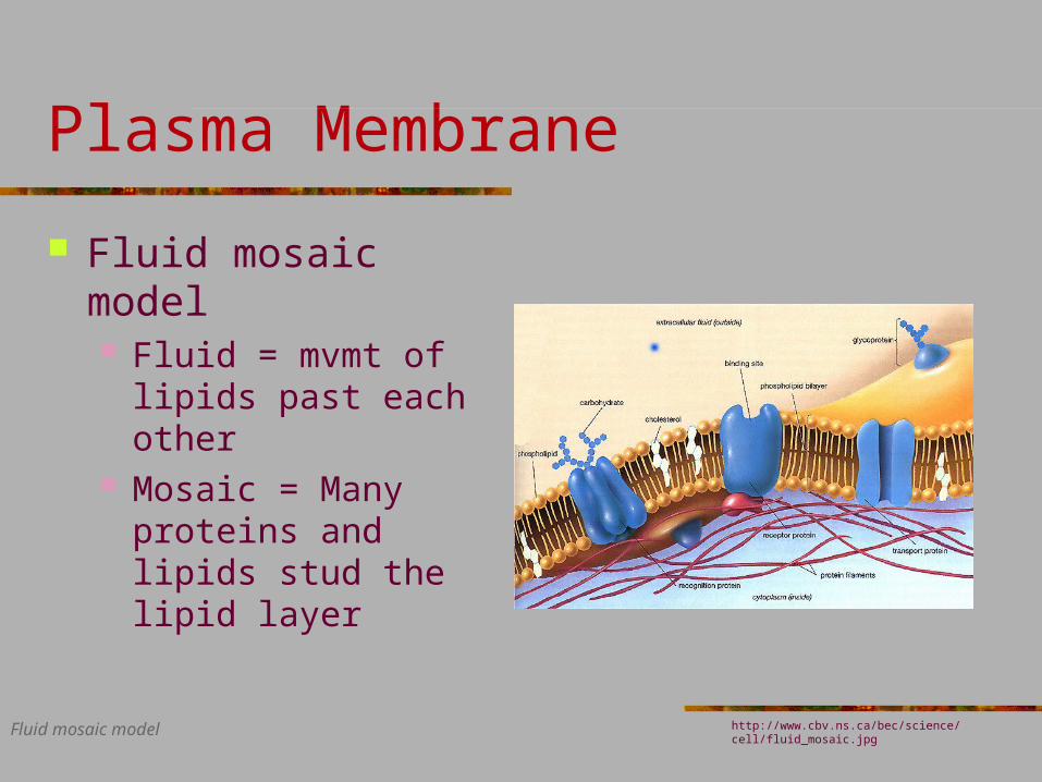
Plasma Membrane
Fluid mosaic model Fluid = mvmt of lipids
past each other Mosaic = Many
proteins and lipids stud the lipid layer
Fluid mosaic model http://www.cbv.ns.ca/bec/science/cell/fluid_mosaic.jpg
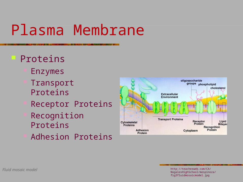
Plasma Membrane
Proteins Enzymes Transport Proteins Receptor Proteins Recognition Proteins Adhesion Proteins
Fluid mosaic model http://teacherweb.com/CA/NogalesHighSchool/mespinoza/fig3fluidmosaicmodel.jpg
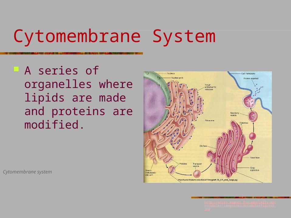
Cytomembrane System
A series of organelles where lipids are made and proteins are modified.
Cytomembrane system
http://micro.magnet.fsu.edu/cells/microtubules/images/microtubulesfigure2.jpg
Cytomembrane System
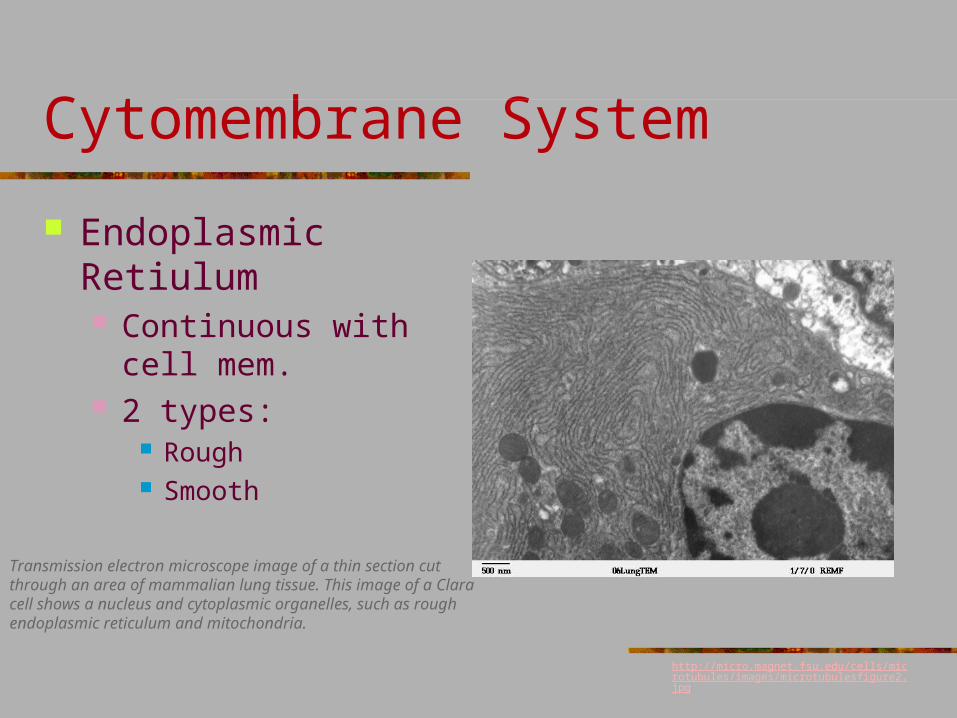
Endoplasmic Retiulum Continuous with cell
mem. 2 types:
Rough Smooth
Transmission electron microscope image of a thin section cut through an area of mammalian lung tissue. This image of a Clara cell shows a nucleus and cytoplasmic organelles, such as rough endoplasmic reticulum and mitochondria.
http://micro.magnet.fsu.edu/cells/microtubules/images/microtubulesfigure2.jpg
Cytomembrane System
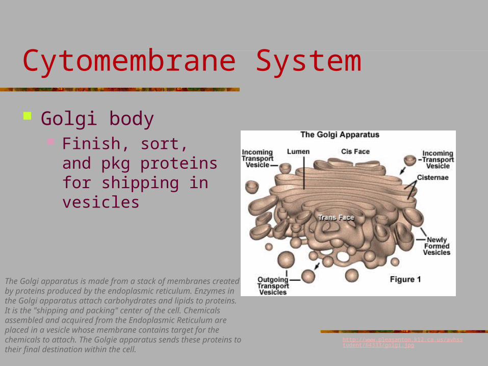
Golgi body Finish, sort, and pkg
proteins for shipping in vesicles
The Golgi apparatus is made from a stack of membranes created by proteins produced by the endoplasmic reticulum. Enzymes in the Golgi apparatus attach carbohydrates and lipids to proteins. It is the "shipping and packing" center of the cell. Chemicals assembled and acquired from the Endoplasmic Reticulum are placed in a vesicle whose membrane contains target for the chemicals to attach. The Golgie apparatus sends these proteins to their final destination within the cell.
http://www.pleasanton.k12.ca.us/avhsstudent/64333/golgi.jpg
Cytomembrane System
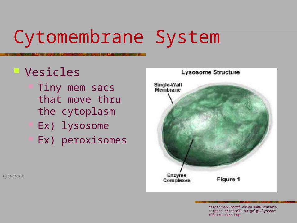
Vesicles Tiny mem sacs that
move thru the cytoplasm
Ex) lysosome Ex) peroxisomes
Lysosome
http://www.seorf.ohiou.edu/~tstork/compass.rose/cell.03/golgi/lysosme%20structure.bmp

Protein trafficking: http://www.youtube.com/watch?v=u38LjCOvDZU&feature=related Protein modification:http://www.youtube.com/watch?v=rvfvRgk0MfA&NR=1
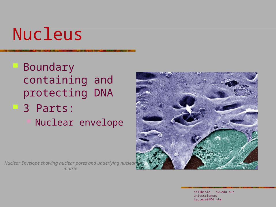
Nucleus
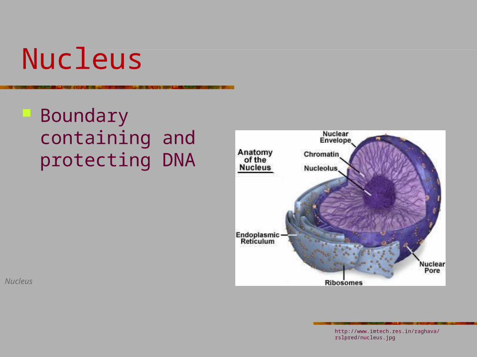
Boundary containing and protecting DNA
Nucleus
http://www.imtech.res.in/raghava/rslpred/nucleus.jpg
Nucleus
Boundary containing and protecting DNA
3 Parts: Nuclear envelope
Nuclear Envelope showing nuclear pores and underlying nuclear matrix
cellbiolo...sw.edu.au/unitsscience/lecture0804.htm
Nucleus
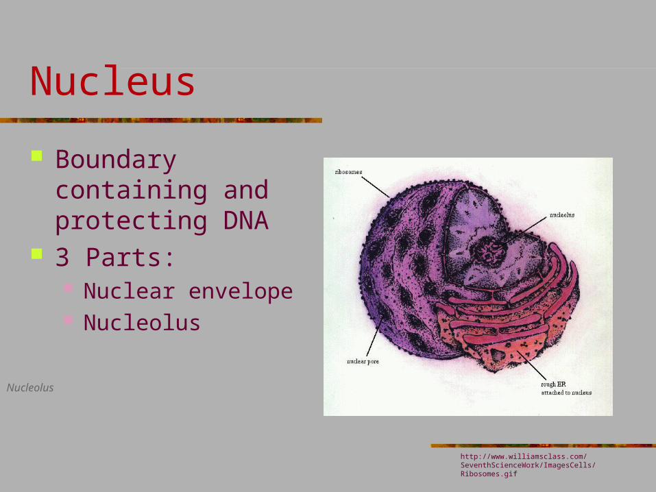
Boundary containing and protecting DNA
3 Parts: Nuclear envelope Nucleolus
Nucleolus
http://www.williamsclass.com/SeventhScienceWork/ImagesCells/Ribosomes.gif
Nucleus
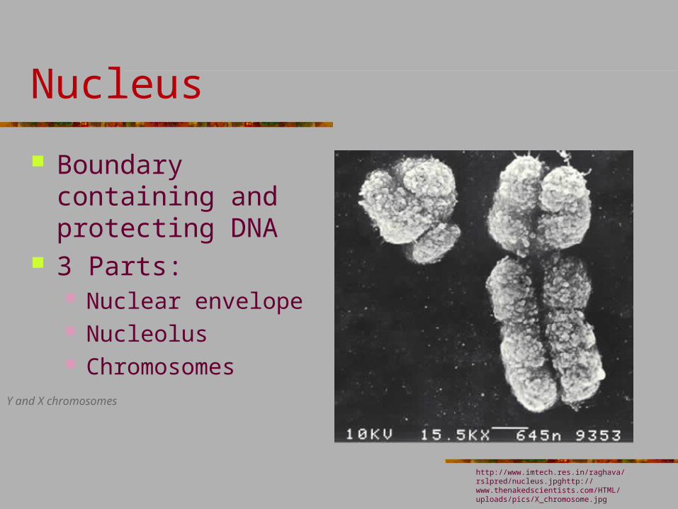
Boundary containing and protecting DNA
3 Parts: Nuclear envelope Nucleolus Chromosomes
Y and X chromosomes
http://www.imtech.res.in/raghava/rslpred/nucleus.jpghttp://www.thenakedscientists.com/HTML/uploads/pics/X_chromosome.jpg