Embed Size (px)
Citation preview
CELLULAR RESPIRATION 2018
Page 1 of 17
CELLULAR RESPIRATION A series of enzyme catalysed reactions in cells during which the chemical-bond energy of complex organic substances is
released and converted into the usable form called adenosine triphosphate (ATP).
Storage Of Chemical Energy In Organic Substances (Food) The C-H covalent bonds in organic substances (e.g. carbohydrates and lipids) form by sharing pairs of fast-moving
energetic electrons, and therefore contain potential energy. The catalytic breakage of the C-H bonds releases energy, some
of which powers the formation of ATP – a compound that can readily hydrolyse to provide energy that powers cellular
activities. The higher the C-H bonds, the more the energy yields. This explains why lipids yield twice more energy
than carbohydrates of same mass.
The Fate Of High Energy Electrons And Hydrogen Ions Released From Breaking C-H Bonds To avoid fatality, the electrons lost from compounds are prevented from joining other molecules by joining electron
carrier molecules which pass them along the electron transport chain until they get attached to oxygen, which becomes
negatively charged, O2-. As the electrons are transferred along the transport chain, energy is gradually extracted from them
to power ATP formation. To avoid pH becoming acidic, which would be fatal, hydrogen ions, H+ combine with O2- to
form neutral water.
WHAT IS ADENOSINE TRIPHOSPHATE (ATP) AND WHY IS IT CONSIDERED TO BE AN ENERGY
CARRIER?
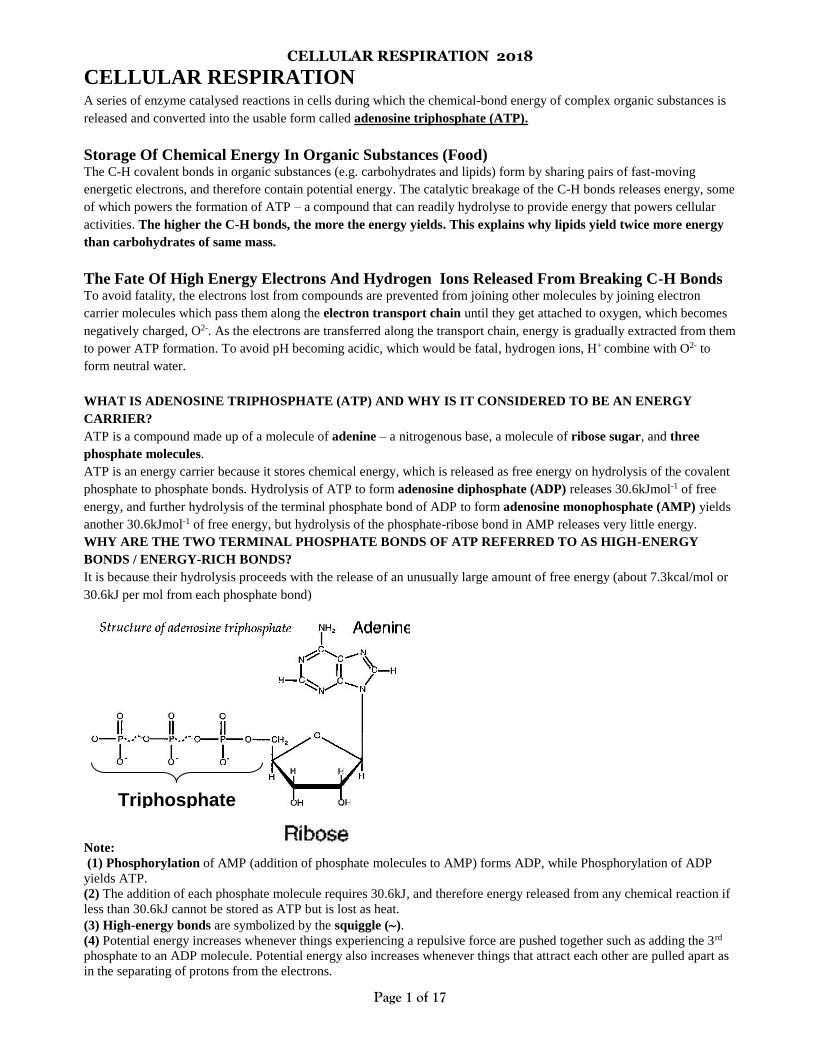
ATP is a compound made up of a molecule of adenine – a nitrogenous base, a molecule of ribose sugar, and three
phosphate molecules.
ATP is an energy carrier because it stores chemical energy, which is released as free energy on hydrolysis of the covalent
phosphate to phosphate bonds. Hydrolysis of ATP to form adenosine diphosphate (ADP) releases 30.6kJmol-1 of free
energy, and further hydrolysis of the terminal phosphate bond of ADP to form adenosine monophosphate (AMP) yields
another 30.6kJmol-1 of free energy, but hydrolysis of the phosphate-ribose bond in AMP releases very little energy.
WHY ARE THE TWO TERMINAL PHOSPHATE BONDS OF ATP REFERRED TO AS HIGH-ENERGY
BONDS / ENERGY-RICH BONDS?
It is because their hydrolysis proceeds with the release of an unusually large amount of free energy (about 7.3kcal/mol or
30.6kJ per mol from each phosphate bond)
Note:
(1) Phosphorylation of AMP (addition of phosphate molecules to AMP) forms ADP, while Phosphorylation of ADP
yields ATP.
(2) The addition of each phosphate molecule requires 30.6kJ, and therefore energy released from any chemical reaction if
less than 30.6kJ cannot be stored as ATP but is lost as heat.
(3) High-energy bonds are symbolized by the squiggle ().
(4) Potential energy increases whenever things experiencing a repulsive force are pushed together such as adding the 3rd
phosphate to an ADP molecule. Potential energy also increases whenever things that attract each other are pulled apart as
in the separating of protons from the electrons.
Triphosphate
CELLULAR RESPIRATION 2018
Page 2 of 17
HOW IS ATP FORMED IN CELLS?
1. Directly by substrate-level Phosphorylation i.e. direct transfer of a phosphate group from high energy
phosphorylated compounds to ADP. Examples of high energy phosphate compounds include
Phosphoenolpyruvate, 1, 3-Bisphosphoglycerate, acetyl phosphate and phosphocreatine.
2. Indirectly by use of energy supplied by transmembrane proton concentration gradients e.g. oxidative
Phosphorylation in the mitochondria and photophosphorylation during photosynthesis.
NOTE: ATP is regarded as a universal currency because its energy supplier molecule is the same in all living
organisms.
OTHER HIGH-ENERGY COMPOUND IN CELLS?
There are other compounds with even higher energy than ATP, only that ATP provides just the right amount of
energy at the right time and can be moved to any place when need arises. For example in muscles and nerve
cells where ATP is continually hydrolysed at a rate faster than respiration can provide due to high metabolic
activity, phosphocreatine provides the phosphate for regeneration of ATP from ADP. See the table below.
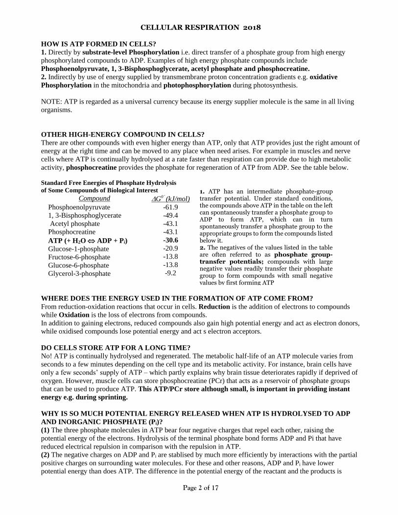
Standard Free Energies of Phosphate Hydrolysis
of Some Compounds of Biological Interest
Compound G0’ (kJ/mol)
Phosphoenolpyruvate
1, 3-Bisphosphoglycerate
Acetyl phosphate
Phosphocreatine
ATP (+ H2O ADP + Pi)
Glucose-1-phosphate
Fructose-6-phosphate
Glucose-6-phosphate
Glycerol-3-phosphate
-61.9
-49.4
-43.1
-43.1
-30.6
-20.9
-13.8
-13.8
-9.2
WHERE DOES THE ENERGY USED IN THE FORMATION OF ATP COME FROM?
From reduction-oxidation reactions that occur in cells. Reduction is the addition of electrons to compounds
while Oxidation is the loss of electrons from compounds.
In addition to gaining electrons, reduced compounds also gain high potential energy and act as electron donors,
while oxidised compounds lose potential energy and act s electron acceptors.
DO CELLS STORE ATP FOR A LONG TIME?
No! ATP is continually hydrolysed and regenerated. The metabolic half-life of an ATP molecule varies from
seconds to a few minutes depending on the cell type and its metabolic activity. For instance, brain cells have
only a few seconds’ supply of ATP – which partly explains why brain tissue deteriorates rapidly if deprived of
oxygen. However, muscle cells can store phosphocreatine (PCr) that acts as a reservoir of phosphate groups
that can be used to produce ATP. This ATP/PCr store although small, is important in providing instant
energy e.g. during sprinting.
WHY IS SO MUCH POTENTIAL ENERGY RELEASED WHEN ATP IS HYDROLYSED TO ADP
AND INORGANIC PHOSPHATE (Pi)?
(1) The three phosphate molecules in ATP bear four negative charges that repel each other, raising the
potential energy of the electrons. Hydrolysis of the terminal phosphate bond forms ADP and Pi that have
reduced electrical repulsion in comparison with the repulsion in ATP.
(2) The negative charges on ADP and Pi are stablised by much more efficiently by interactions with the partial
positive charges on surrounding water molecules. For these and other reasons, ADP and Pi have lower
potential energy than does ATP. The difference in the potential energy of the reactant and the products is
1. ATP has an intermediate phosphate-group transfer potential. Under standard conditions, the compounds above ATP in the table on the left can spontaneously transfer a phosphate group to ADP to form ATP, which can in turn spontaneously transfer a phosphate group to the appropriate groups to form the compounds listed below it. 2. The negatives of the values listed in the table are often referred to as phosphate group-transfer potentials; compounds with large negative values readily transfer their phosphate group to form compounds with small negative values by first forming ATP
CELLULAR RESPIRATION 2018
Page 3 of 17
released as heat, or visible light e.g. fireflies and certain bacteria are able to bioluminesce as some of this
chemical-bond energy is released as visible light, or some other form of energy.
USES OF ENERGY OF ATP IN CELLS:
(1) Enables transport of materials
(2) Enables movement of cilia or flagella and
(3) Allows active transport to be carried out (movement of substances against concentration gradient) e.g. ion
pumps
(4) Drives endergonic reactions e.g. assembly of amino acids into proteins, synthesis of polysaccharides from
monosaccharides, and DNA replication
(5) Activates chemicals to become more reactive e.g. Phosphorylation of sugar during Glycolysis
(6) Enables formation of vesicles during secretion of cell products.
(7) muscle contraction
AN ELEMENTARY UNDERSTANDING OF THE ROLE OF ATP
Each ATP molecule used in the cell is like a rechargeable AAA battery of 1.5v. Each contains just the right
amount of energy to power a small radio. When the power has been drained, the AAA battery can be recharged
many times using a small amount of energy from hydroelectric power, but the small radio cannot be plugged
directly into the high voltage hydroelectric power. Cells of organisms operate in much the same manner. When
the cell’s “batteries”, ATP, are drained while powering a job like muscle contraction, the discharged
“batteries”, ADP can be recharged back to full ATP power using (1) chemical-bond energy released from
cellular respiration (2) sunlight during photosynthesis in green plants. In cells therefore, ATP provides just the
right amount of energy at the right time and place, and is constantly used and synthesised.
WHERE DOES RESPIRATION OCCUR IN CELLS?
Cell type Location of pathway in cell
All prokaryotic cells Infoldings of cell membrane (mesosomes) and in cytoplasm
All eukaryotic cells Cytoplasm and inner membranes of mitochondria
STAGES OF CELLULAR RESPIRATION
(1) Glycolysis (2) Krebs cycle (3) Electron transfer system
GLYCOLYSIS (glyco = carbohydrate; lys = splitting; sis = the process of)
A series of enzymatically controlled reactions in the cytoplasm of cells during which one molecule of a six-
carbon sugar glucose, is split into two molecules of the three-carbon compound Pyruvate, with a net out put of
two ATP molecules.
CELLULAR RESPIRATION 2018
Page 4 of 17
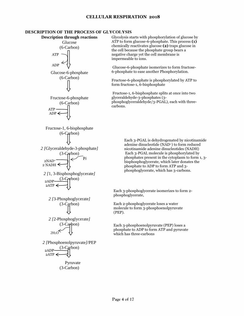
DESCRIPTION OF THE PROCESS OF GLYCOLYSIS
Description through reactions
Glucose
(6-Carbon)
Glucose-6-phosphate
(6-Carbon)
Fructose-6-phosphate
(6-Carbon)
Fructose-1, 6-bisphosphate
(6-Carbon)
2 [Glyceraldehyde-3-phosphate]
(3-Carbon)
Pi
2 [1, 3-Bisphosphoglycerate]
(3-Carbon)
2 [3-Phosphoglycerate]
(3-Carbon)
2 [2-Phosphoglycerate]
(3-Carbon) 2H2O
2 [Phosphoenolpyruvate]/PEP
(3-Carbon)
Pyruvate
(3-Carbon)
ADP
ATP
2NAD+
2 NADH
Each 3-phosphoglycerate isomerizes to form 2-phosphoglycerate, Each 2-phosphoglycerate loses a water molecule to form 3-phosphoenolpyruvate (PEP). Each 3-phosphoenolpyruvate (PEP) loses a phosphate to ADP to form ATP and pyruvate which has three-carbons
Glycolysis starts with phosphorylation of glucose by ATP to form glucose-6-phosphate. This process (1) chemically reactivates glucose (2) traps glucose in the cell because the phosphate group bears a negative charge yet the cell membrane is impermeable to ions. Glucose-6-phosphate isomerizes to form fructose-6-phosphate to ease another Phosphorylation. Fructose-6-phosphate is phosphorylated by ATP to form fructose-1, 6-bisphosphate Fructose-1, 6-bisphosphate splits at once into two glyceraldehyde-3-phosphates (3-phosphoglyceraldehyde/3-PGAL), each with three-carbons.
Each 3-PGAL is dehydrogenated by nicotinamide adenine dinucleotide (NAD+) to form reduced nicotinamide adenine dinucleotides (NADH) Each 3-PGAL molecule is phosphorylated by phosphates present in the cytoplasm to form 1, 3-bisphosphoglycerate, which later donates the phosphate to ADP to form ATP and 3-phosphoglycerate, which has 3-carbons.
ATP ADP
2ADP 2ATP
2ADP 2ATP
CELLULAR RESPIRATION 2018
Page 5 of 17
SIGNIFICANCE OF GLYCOLYSIS
Glycolysis forms (1) ATP which is used to power cell activities (2) NADH and Pyruvate which may be further
oxidized to generate additional ATP. However in oxygen deficiency, both NADH and pyruvate undergo
fermentation to regenerate NAD+.
NOTE:
Glycolytic degradation of glucose to two molecules of pyruvate yields only about 5.2% of the total energy that
can be released from glucose by complete oxidation. The two molecules of Pyruvate formed by glycolysis still
contain most of the chemical potential energy of the glucose molecule, energy that can be extracted by
oxidative reactions in the citric acid cycle
THE FATE OF PYRUVATE, NADH AND ATP PRODUCED FROM GLYCOLYSIS
1. ATP:
It is hydrolysed to release energy to power the cell’s needs.
2. NADH:
Under aerobic conditions (in the presence of oxygen), NADH is converted into FADH2 which is then shuttled
into the mitochondria where it donates electrons to a series of electron carriers until they reach the final
oxidizing agent oxygen in a process called electron transport system. During this process, the free energy of
electron transport drives the synthesis of ATP from ADP and NAD+ is regenerated such that it can participate
in further catalysis.
Under anaerobic conditions, NADH must be re-oxidised by other means in order to keep the glycolytic
pathway supplied with NAD+
3. PYRUVATE:
Under aerobic conditions, it is completely oxidised via the citric acid cycle to carbondioxide and water.
Under anaerobic conditions in the cytoplasm, pyruvate under goes fermentation.
Types of fermentation
There are many types of fermentation, but the two common types are given below:
(a) Alcoholic fermentation: pyruvate is decarboxylated to yield carbondioxide which is converted to a 2-
carbon compound acetaldehyde. Acetaldehyde is then reduced by NADH to ethanol and NAD+ also forms.
NAD+ enables the continuation of glycolysis. Alcoholic fermentation occurs in some bacteria and yeasts.
(b) Lactic acid fermentation: pyruvate is reduced directly by NADH to form lactic acid as the end product.
No carbondioxide is released. Lactic acid fermentation (1) is carried out by certain fungi and bacteria during
the formation of yoghurt and cheese (2) occurs during oxygen scarcity in human skeletal muscle cells during
sprinting. The lactic acid is gradually carried away by blood to the liver and converted back to pyruvate by
liver cells. If ATP is abundant, pyruvate and lactate can be used as a substrate in the synthesis of glucose.
Comparison of cellular respiration and fermentation
Similarities: Both (1) form ATP (2) use glycolysis to oxidise glucose to pyruvate (3) use NAD+ as the
oxidizing agent that accepts electrons from food during glycolysis (4) may be carried out by same cells (e.g.
muscle cells) or same organisms (e.g. yeasts and bacteria).
Differences: Cellular respiration Fermentation
• Final electron acceptor from is oxygen
• Harvests much more energy from each glucose
molecule i.e. up to 38 ATP per glucose molecule.
• Final electron acceptor is an organic molecule such
as pyruvate (lactic acid fermentation) or acetaldehyde
(alcohol fermentation)
• Harvests much less energy from each glucose
molecule i.e. 2 ATP per glucose molecule.
CELLULAR RESPIRATION 2018
Page 6 of 17
• Occurs in mitochondria. • Occurs in cytoplasm (cytosol).
Evolutionary significance of glycolysis
The role of glycolysis in both fermentation and respiration suggests that ancient prokaryotes probably used
glycolysis to make ATP long before oxygen was present in the atmosphere. This conclusion is based on the
following observations:
(1) The oldest bacterial fossils date back 3.5 billion years, yet oxygen accumulated about 2.7 billion years ago.
Therefore early prokaryotes may have generated ATP exclusively from glycolysis, which does not require
oxygen.
(2) Glycolysis is the most widespread metabolic pathway, which suggests that it evolved very early in the
history of life.
(3) Glycolysis is located in the cytoplasm where no membrane-bounded organelles are required in eukaryotic
cells, which evolved approximately 1 billion years after the prokaryotic cell.
FATE OF PYRUVATE IN AEROBIC CONDITIONS (TRANSITION STATE OF PYRUVATE)
Each pyruvate molecule produced by glycolysis in the cell cytoplasm is transported across the inner
mitochondrial membrane by active transport (since it is a charged molecule) into the matrix, where it is first
decarboxylated and then oxidised (dehydrogenated) to form a 2-C compound called acetate, carbondioxide
and NADH. Carbondioxide, a waste product is eventually excreted while NAD+ serves as a hydrogen carrier.
Finally, Acetate is attached to Coenzyme A to form acetyl coenzyme A, making the acetyl group very reactive.
Acetyl coenzyme A is now ready to feed its acetyl group into the citric acid cycle for further oxidation. (A –
stands for acetylation)
Note: the transition from pyruvate to acetyl coenzyme A is not usually considered as a separate phase and is
included with the first step of Krebs cycle.
THE ROLE OF CoA IN RESPIRATION
(1) Within the active centre of the enzyme citrate synthetase, CoA transfers the 2-carbon acetyl group to a 4-
carbon molecule of oxalocetate to make a molecule of citrate which enters the Krebs cycle. (2) it serves as a
link between many different pathways of metabolism to provide a wide range of carbon compounds needed in
the cell (3) during energy deficiency, amino acids from proteins and fatty acids from lipids can be broken
down to provide acetyl CoA for use in respiration.
Acetyl- Coenzyme A: a central metabolic intermediate
All proteins, lipids, and carbohydrates must be converted to Acetyl- Coenzyme A prior to participation in
cellular respiration.
The fate of acetyl-CoA is dependent upon ATP needs. When ATP is prevalent, acetyl-CoA serves as the basis
for fatty acid synthesis, which forms the basis of your body's long-term energy storage: triglycerides (i.e., fat).
Acetyl-CoA is the starting point for anabolic pathways that result in the synthesis of fatty acids.
Alternatively, acetyl-CoA may enter the Kreb's citric acid cycle.
KREBS CYCLE/ TRICARBOXYLIC ACID CYCLE / CITRIC ACID CYCLE
It is named:
1. Krebs cycle after the formulator Hans Krebs
2. Citric acid because citric acid is the first compound formed.
3. Tricarboxylic acid because citric acid which is the first compound formed has 3 carboxyl (-COOH) groups
It is a multi-step reaction in the mitochondrial matrix during which an acetyl group is completely oxidized to
CO2 with the generation of ATP and reducing hydrogens in the form of NADH and FADH2.
CELLULAR RESPIRATION 2018
Page 7 of 17
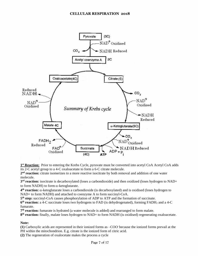
1st Reaction: Prior to entering the Krebs Cycle, pyruvate must be converted into acetyl CoA Acetyl CoA adds
its 2-C acetyl group to a 4-C oxaloacetate to form a 6-C citrate molecule.
2nd reaction: citrate isomerizes to a more reactive isocitrate by both removal and addition of one water
molecule.
3rd reaction: isocitrate is decarboxylated (loses a carbondioxide) and then oxidized (loses hydrogen to NAD+
to form NADH) to form -ketoglutarate.
4th reaction: -ketoglutarate loses a carbondioxide (is decarboxylated) and is oxidised (loses hydrogen to
NAD+ to form NADH) and attached to coenzyme A to form succinyl-CoA.
5th step: succinyl-CoA causes phosphorylation of ADP to ATP and the formation of succinate.
6th reaction: a 4-C succinate loses two hydrogens to FAD (is dehydrogenated), forming FADH2 and a 4-C
fumarate.
7th reaction: fumarate is hydrated (a water molecule is added) and rearranged to form malate.
8th reaction: finally, malate loses hydrogen to NAD+ to form NADH (is oxidised) regenerating oxaloacetate.
Note:
(1) Carboxylic acids are represented in their ionized forms as –COO- because the ionized forms prevail at the
PH within the mitochondrion. E.g. citrate is the ionized form of citric acid.
(2) The regeneration of oxalocetate makes the process a cycle
CELLULAR RESPIRATION 2018
Page 8 of 17
(3) For each acetyl group that enters the cycle, 3 NAD+ are reduced to NADH (reactions 3, 4, and 8)
(4) Most of the ATP out put of respiration results from oxidative phosphorylation, when the NADH and
FADH2 produced by the citric acid cycle relay the electrons extracted from food to the electron transport
chain.
Comparison of Krebs cycle and glycolysis
Similarities: In both: (1) reducing hydrogens are accumulated in NADH (2) ATP is generated (3) there is a
reduction in number of carbon atoms of organic compounds(4) pyruvate participates (5) both occur in living
cells
Differences:
Glycolysis Krebs cycle
• The electron acceptor FAD is not involved • The electron acceptor FAD is involved
• Carbondioxide doesn’t form • Carbondioxide is liberated
• Occurs in cell cytoplasm • Occurs in mitochondrial matrix
• Doesn’t necessarily depend on oxygen • Depends on oxygen availability to occur
ELECTRON TRANSPORT SYSTEM AND CHEMIOSMOTIC THEORY
Electron transport system: A process whereby a series of electron carriers operate together to transfer
electrons from donors to any of several different terminal electron acceptors to generate a transmembrane
electrochemical gradient in the mitochondrion
What are the components of the electron transport chain?
Complex Name
Complex I NADH Dehydrogenase
Complex II Succinate-Coenzyme Q Reductase Coenzyme Q (CoQ) (also called ubiquinone)
Complex III Cytochrome bc1 complex
Cytochrome c
Complex IV Cytochrome Oxidase
The production of ATP during electron transport involves two separate but connected processes i.e.
Chemiosmosis and oxidative phosphorylation
Description of the process of electron transport system
The electrons released during glycolysis and carried by NADH are converted to FADH2 in order to shuttle
them from the cytoplasm into the mitochondrial matrix.
In Complex I (also called NADH reductase), reduced nicotinamide adenine dinucleotide (NADH) donates
electrons to the coenzyme Flavin mononucleotide (FMN) which then passes electrons to an iron-sulphur (Fe-S)
protein and the electrons lose some energy. NADH is oxidized to NAD+, while FMN and Fe3+ are reduced to
FMNH2 and Fe2+ respectively. Each electron is transferred with a proton.
Electrons from the reduced Fe-S proteins are then passed to Coenzyme Q along with protons. Coenzyme Q is
thus reduced while the Fe-S proteins are oxidised back to Fe3+ state.
In complex II (succinate dehydrogenase), electrons from FADH2 are passed on to Fe-S proteins then to
Coenzyme Q which transfers them to complex III. FADH2 becomes oxidised to FAD+. During this process,
four protons (H+) are translocated across the inner mitochondrial membrane, from the matrix to the
intermembrane space. This creates a proton gradient that will be later used to generate ATP through oxidative
phosphorylation. During oxidation of FADH2 complex I is bypassed because complex II has only enough
reducing potential to pass electrons to Coenzyme Q.
CELLULAR RESPIRATION 2018
Page 9 of 17
Reduced coenzyme Q (CoQH2) transfers electrons to Complex III where they pass through several
cytochromes and Fe-S proteins and during the process Fe3+ is reduced to Fe2+. The electrons lose additional
energy and are passed on to cytochrome c which passes electrons to Complex IV (cytochrome c oxidase),
which finally transfers the electrons to reduce molecular oxygen to form water. O2 + 4 H+ + 4 e 2 H2O. At
the same time, complex IV moves protons (H+) across the membrane into the intermembrane space, producing
a proton gradient.
As electrons lose energy in complex I, III and IV, additional protons are pumped into the intermembrane space
producing a proton gradient. Complex II (succinate dehydrogenase) is not a proton pump. It only serves to
funnel additional electrons into coenzyme Q. Electron transfers involving Coenzyme Q and Cytochrome c do
not release enough free energy to pump any protons.
When the protons flow down the concentration gradient through the channels in the stalked particles, ATP
synthase enzymes are able to use the energy to generate ATP.
Note: If the oxygen supply is cut off, the electrons and hydrogen protons cease to flow through the electron
transport system. If this happens, the proton concentration gradient will not be sufficient to power the synthesis
of ATP. This is why we, and other species, are not able to survive for long without oxygen!
Is the ETS a sequence?
No! The complexes move in the fluid membrane independently of one another, and exchange electrons when
they are in mutual proximity. Although textbooks show the ETS as a physical sequence, the complexes and
carriers are not locked in place.
CHEMIOSMOTIC COUPLING HYPOTHESIS AND OXIDATIVE PHOSPHORYLATION
As proposed by Peter D. Mitchell, the chemiosmotic coupling hypothesis explains that the electron transport
chain and oxidative phosphorylation are coupled by a proton gradient across the inner mitochondrial
membrane.
The efflux of protons into the intermembrane space creates both a pH gradient and an electrochemical
gradient. This proton gradient is used by the ATP synthase complex to make ATP via oxidative
phosphorylation. The stalk component of ATP synthase complex acts as an ion channel for return of protons
back to mitochondrial matrix during which the free energy produced during the generation of the oxidized
forms of the electron carriers (NAD+) is released and used to drive ATP synthesis, catalyzed by the head
component of the ATP synthase complex.
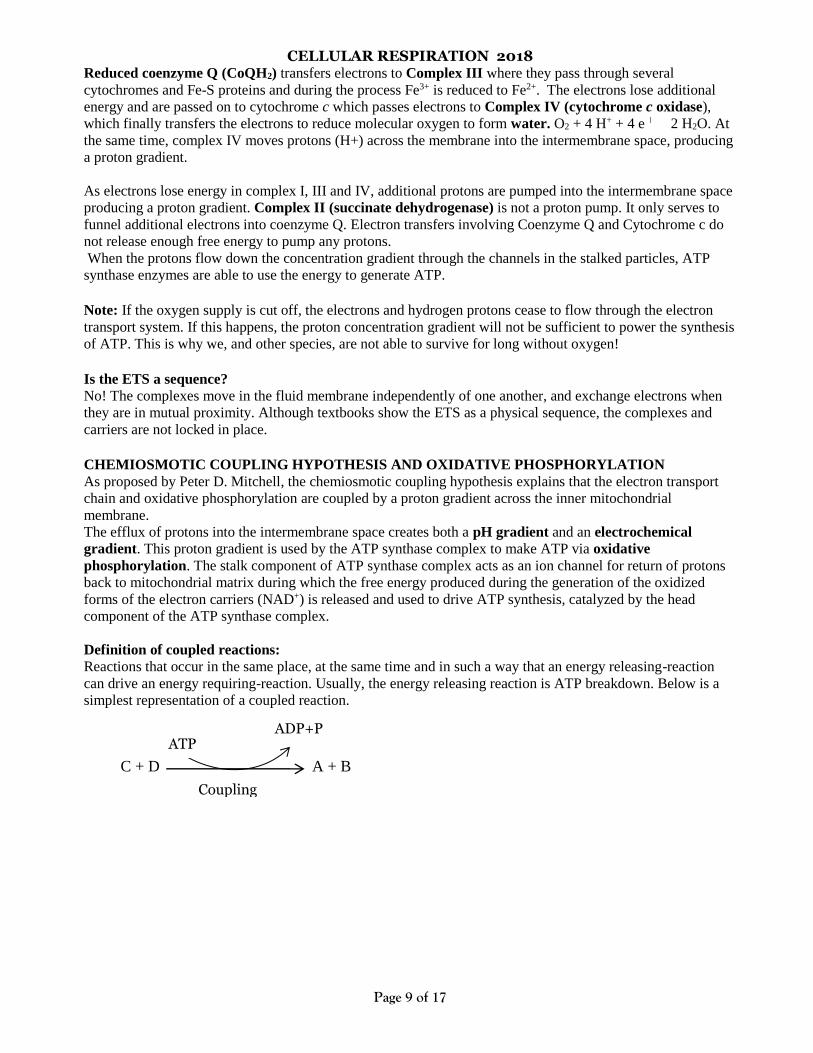
Definition of coupled reactions:
Reactions that occur in the same place, at the same time and in such a way that an energy releasing-reaction
can drive an energy requiring-reaction. Usually, the energy releasing reaction is ATP breakdown. Below is a
simplest representation of a coupled reaction.
C + D A + B
ATP ADP+P
Coupling
CELLULAR RESPIRATION 2018
Page 10 of 17
ACCOUNTING FOR THE ELECTRONS IN EUKARYOTIC ORGANISMS
Oxidation of NADH to NAD+ pumps 3 protons from the mitochondrial matrix into the intermembrane space,
which charges the electrochemical gradient with enough potential to generate 3 ATP. Oxidation of FADH2 to
FAD+ pumps 2 protons into the intermembrane space, which charges the electrochemical gradient with enough
potential to generate 2 ATP.
In some text books however, recent information suggests that 1NADH generates 2.5 ATP and 1FADH2
generates 1.5 ATP. The reasons for this are that not all of the energy stored in the proton gradient is used to
generate ATP. Some of the energy is used to power transport of ions in and out of the mitochondria.
A total of 12 pairs of electrons and hydrogens are transported to the electron transport system from glycolysis
and Krebs cycle for each glucose molecule that enters the process:
• 4 pairs are carried by NADH and were generated during glycolysis in the cytoplasm, 8 pairs are carried as
NADH and were generated within the mitochondrial matrix and 2 pairs are carried by FADH2 and were
generated within the mitochondrial matrix.
• For each of the 8 NADHs generated within the mitochondrial matrix, enough energy is released to produce 3
ATP molecules; therefore, 24 ATP molecules are released from these electrons carried by NADH.
• The electrons carried by FADH2 are lower in energy, so during the oxidation-reduction reactions, they
release energy to produce only 8 ATP molecules.
• Therefore, a grand total of 32 ATP molecules are produced from hydrogen electrons that enter the electron
transport system.
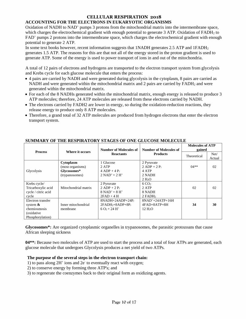
SUMMARY OF THE RESPIRATORY STAGES OF ONE GLUCOSE MOLECULE
Process Where it occurs Number of Molecules of
Reactants
Number of Molecules of
Products
Molecules of ATP
gained
Theoretical Net/
Actual
Glycolysis
Cytoplasm
(most organisms)
Glycosomes*
(trypanosomes)
1 Glucose
2 ATP
4 ADP + 4 Pi
2 NAD+ + 2 H+
2 Pyruvate
2 ADP + 2 Pi
4 ATP
2 NADH
2 H2O
04**
02
Krebs cycle/
Tricarboxylic acid
cycle / citric acid
cycle
Mitochondrial matrix
2 Pyruvate
2 ADP + 2 Pi
8 NAD+ + 8 H+
2FAD + 4 H
6 CO2
2 ATP
8 NADH
2 FADH2
02
02
Electron transfer
system &
chemiosmosis
(oxidative
Phosphorylation)
Inner mitochondrial
membrane
8NADH+24ADP+24Pi
2FADH2+8ADP+8Pi
6 O2 + 24 H+
8NAD++24ATP+16H
4FAD+8ATP+8H
12 H2O
34
30
Glycosomes*: Are organized cytoplasmic organelles in trypanosomes, the parasitic protozoans that cause
African sleeping sickness
04**: Because two molecules of ATP are used to start the process and a total of four ATPs are generated, each
glucose molecule that undergoes Glycolysis produces a net yield of two ATPs.
The purpose of the several steps in the electron transport chain:
1) to pass along 2H+ ions and 2e- to eventually react with oxygen;
2) to conserve energy by forming three ATP's; and
3) to regenerate the coenzymes back to their original form as oxidizing agents.
CELLULAR RESPIRATION 2018
Page 11 of 17
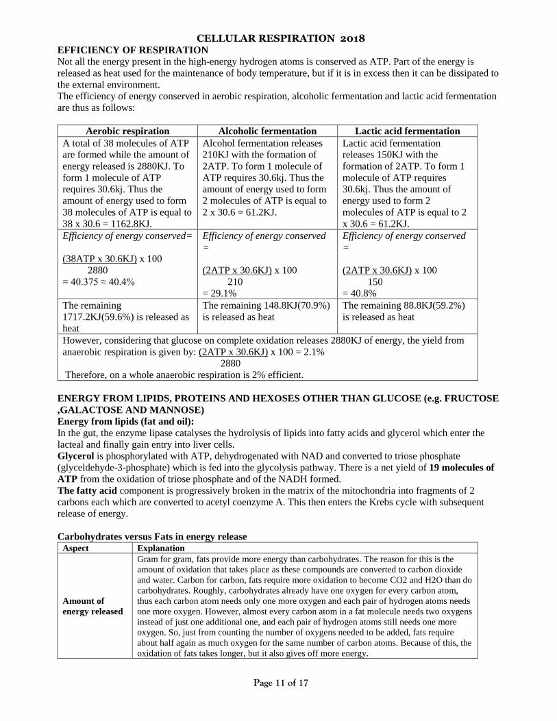
EFFICIENCY OF RESPIRATION
Not all the energy present in the high-energy hydrogen atoms is conserved as ATP. Part of the energy is
released as heat used for the maintenance of body temperature, but if it is in excess then it can be dissipated to
the external environment.
The efficiency of energy conserved in aerobic respiration, alcoholic fermentation and lactic acid fermentation
are thus as follows:
Aerobic respiration Alcoholic fermentation Lactic acid fermentation
A total of 38 molecules of ATP
are formed while the amount of
energy released is 2880KJ. To
form 1 molecule of ATP
requires 30.6kj. Thus the
amount of energy used to form
38 molecules of ATP is equal to
38 x 30.6 = 1162.8KJ.
Alcohol fermentation releases
210KJ with the formation of
2ATP. To form 1 molecule of
ATP requires 30.6kj. Thus the
amount of energy used to form
2 molecules of ATP is equal to
2 x 30.6 = 61.2KJ.
Lactic acid fermentation
releases 150KJ with the
formation of 2ATP. To form 1
molecule of ATP requires
30.6kj. Thus the amount of
energy used to form 2
molecules of ATP is equal to 2
x 30.6 = 61.2KJ.
Efficiency of energy conserved=
(38ATP x 30.6KJ) x 100
2880
= 40.375 ≈ 40.4%
Efficiency of energy conserved
=
(2ATP x 30.6KJ) x 100
210
= 29.1%
Efficiency of energy conserved
=
(2ATP x 30.6KJ) x 100
150
= 40.8%
The remaining
1717.2KJ(59.6%) is released as
heat
The remaining 148.8KJ(70.9%)
is released as heat
The remaining 88.8KJ(59.2%)
is released as heat
However, considering that glucose on complete oxidation releases 2880KJ of energy, the yield from
anaerobic respiration is given by: (2ATP x 30.6KJ) x 100 = 2.1%
2880
Therefore, on a whole anaerobic respiration is 2% efficient.
ENERGY FROM LIPIDS, PROTEINS AND HEXOSES OTHER THAN GLUCOSE (e.g. FRUCTOSE
,GALACTOSE AND MANNOSE)
Energy from lipids (fat and oil):
In the gut, the enzyme lipase catalyses the hydrolysis of lipids into fatty acids and glycerol which enter the
lacteal and finally gain entry into liver cells.
Glycerol is phosphorylated with ATP, dehydrogenated with NAD and converted to triose phosphate
(glyceldehyde-3-phosphate) which is fed into the glycolysis pathway. There is a net yield of 19 molecules of
ATP from the oxidation of triose phosphate and of the NADH formed.
The fatty acid component is progressively broken in the matrix of the mitochondria into fragments of 2
carbons each which are converted to acetyl coenzyme A. This then enters the Krebs cycle with subsequent
release of energy.
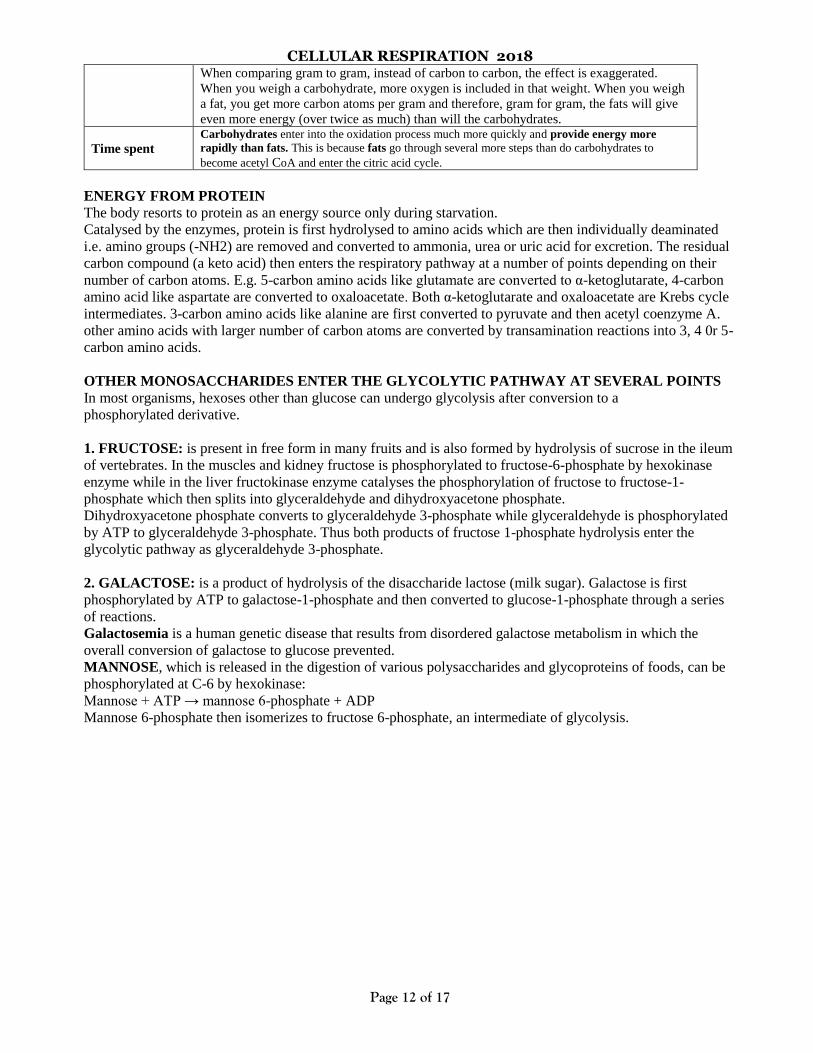
Carbohydrates versus Fats in energy release
Aspect Explanation
Amount of
energy released
Gram for gram, fats provide more energy than carbohydrates. The reason for this is the
amount of oxidation that takes place as these compounds are converted to carbon dioxide
and water. Carbon for carbon, fats require more oxidation to become CO2 and H2O than do
carbohydrates. Roughly, carbohydrates already have one oxygen for every carbon atom,
thus each carbon atom needs only one more oxygen and each pair of hydrogen atoms needs
one more oxygen. However, almost every carbon atom in a fat molecule needs two oxygens
instead of just one additional one, and each pair of hydrogen atoms still needs one more
oxygen. So, just from counting the number of oxygens needed to be added, fats require
about half again as much oxygen for the same number of carbon atoms. Because of this, the
oxidation of fats takes longer, but it also gives off more energy.
CELLULAR RESPIRATION 2018
Page 12 of 17
When comparing gram to gram, instead of carbon to carbon, the effect is exaggerated.
When you weigh a carbohydrate, more oxygen is included in that weight. When you weigh
a fat, you get more carbon atoms per gram and therefore, gram for gram, the fats will give
even more energy (over twice as much) than will the carbohydrates.
Time spent
Carbohydrates enter into the oxidation process much more quickly and provide energy more
rapidly than fats. This is because fats go through several more steps than do carbohydrates to
become acetyl CoA and enter the citric acid cycle.
ENERGY FROM PROTEIN
The body resorts to protein as an energy source only during starvation.
Catalysed by the enzymes, protein is first hydrolysed to amino acids which are then individually deaminated
i.e. amino groups (-NH2) are removed and converted to ammonia, urea or uric acid for excretion. The residual
carbon compound (a keto acid) then enters the respiratory pathway at a number of points depending on their
number of carbon atoms. E.g. 5-carbon amino acids like glutamate are converted to α-ketoglutarate, 4-carbon
amino acid like aspartate are converted to oxaloacetate. Both α-ketoglutarate and oxaloacetate are Krebs cycle
intermediates. 3-carbon amino acids like alanine are first converted to pyruvate and then acetyl coenzyme A.
other amino acids with larger number of carbon atoms are converted by transamination reactions into 3, 4 0r 5-
carbon amino acids.
OTHER MONOSACCHARIDES ENTER THE GLYCOLYTIC PATHWAY AT SEVERAL POINTS
In most organisms, hexoses other than glucose can undergo glycolysis after conversion to a
phosphorylated derivative.
1. FRUCTOSE: is present in free form in many fruits and is also formed by hydrolysis of sucrose in the ileum
of vertebrates. In the muscles and kidney fructose is phosphorylated to fructose-6-phosphate by hexokinase
enzyme while in the liver fructokinase enzyme catalyses the phosphorylation of fructose to fructose-1-
phosphate which then splits into glyceraldehyde and dihydroxyacetone phosphate.
Dihydroxyacetone phosphate converts to glyceraldehyde 3-phosphate while glyceraldehyde is phosphorylated
by ATP to glyceraldehyde 3-phosphate. Thus both products of fructose 1-phosphate hydrolysis enter the
glycolytic pathway as glyceraldehyde 3-phosphate.
2. GALACTOSE: is a product of hydrolysis of the disaccharide lactose (milk sugar). Galactose is first
phosphorylated by ATP to galactose-1-phosphate and then converted to glucose-1-phosphate through a series
of reactions.
Galactosemia is a human genetic disease that results from disordered galactose metabolism in which the
overall conversion of galactose to glucose prevented.
MANNOSE, which is released in the digestion of various polysaccharides and glycoproteins of foods, can be
phosphorylated at C-6 by hexokinase:
Mannose + ATP → mannose 6-phosphate + ADP
Mannose 6-phosphate then isomerizes to fructose 6-phosphate, an intermediate of glycolysis.
CELLULAR RESPIRATION 2018
Page 13 of 17
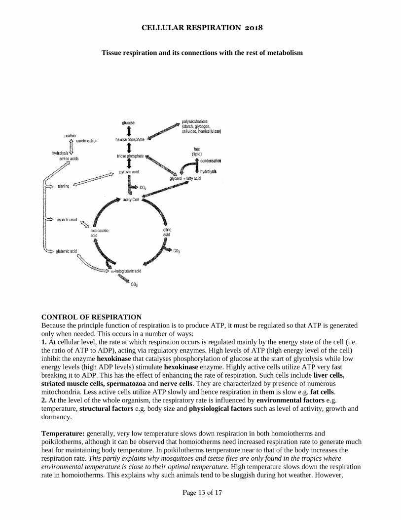
Tissue respiration and its connections with the rest of metabolism
CONTROL OF RESPIRATION
Because the principle function of respiration is to produce ATP, it must be regulated so that ATP is generated
only when needed. This occurs in a number of ways:
1. At cellular level, the rate at which respiration occurs is regulated mainly by the energy state of the cell (i.e.
the ratio of ATP to ADP), acting via regulatory enzymes. High levels of ATP (high energy level of the cell)
inhibit the enzyme hexokinase that catalyses phosphorylation of glucose at the start of glycolysis while low
energy levels (high ADP levels) stimulate hexokinase enzyme. Highly active cells utilize ATP very fast
breaking it to ADP. This has the effect of enhancing the rate of respiration. Such cells include liver cells,
striated muscle cells, spermatozoa and nerve cells. They are characterized by presence of numerous
mitochondria. Less active cells utilize ATP slowly and hence respiration in them is slow e.g. fat cells.
2. At the level of the whole organism, the respiratory rate is influenced by environmental factors e.g.
temperature, structural factors e.g. body size and physiological factors such as level of activity, growth and
dormancy.
Temperature: generally, very low temperature slows down respiration in both homoiotherms and
poikilotherms, although it can be observed that homoiotherms need increased respiration rate to generate much
heat for maintaining body temperature. In poikilotherms temperature near to that of the body increases the
respiration rate. This partly explains why mosquitoes and tsetse flies are only found in the tropics where
environmental temperature is close to their optimal temperature. High temperature slows down the respiration
rate in homoiotherms. This explains why such animals tend to be sluggish during hot weather. However,
CELLULAR RESPIRATION 2018
Page 14 of 17
excessively high temperatures trigger increased respiration rate and finally stop as a response by enzymes to
temperature.
Body size: small organisms with a large surface area to volume ratio lose heat faster and therefore respire
faster than large organisms.
Level of activity: animals engaging in vigorous physical exercise require much energy and so experience
faster respiration rate e.g. sprinting, flying, etc
Growth: actively growing organisms e.g. young animals and germinating seeds respire faster to generate
much energy required to drive metabolic processes
Dormancy during extreme cold and hot seasons: respiration rate is always slow to avoid depleting food
reserves before the unfavourable season ends.
RESPIRATORY QUOTIENT (RQ)
It is the ratio of the volume of Carbondioxide produced to the volume of oxygen used in respiration during the
same period of time
RQ = volume of Carbondioxide given out
Volume of oxygen taken in
Importance of RQ:
(1) it can indicate the kind of substrate being respired (2) it can indicate whether the respiration is aerobic or
anaerobic.
RQ can be measured using a spirometer or respirometer.
RQ FOR HEXOSE SUGAR: like glucose, the equation for its complete oxidation is:
C6H12O6 + 6O2 6CO2 + 6H2O
Hence RQ is: 6CO2 = 1.0 (one)
6O2
R.Q FOR FATS: For a lipid like tripalmitin, the equation for its complete oxidation is:
RQ is: 102CO2 = 0.7 (less than one)
145O2
NB: the R.Qs for different fats will of course show slight variations because of differences in molecular
composition
R.Q FOR PROTEINS: no concrete value can be calculated since (1) they vary so much in composition and
(2) are difficult to separate in the pure state. Estimates for protein vary between 0.5 and 0.8 for the complete
oxidation of proteins.
SUMMARY OF THE POSSIBLE INTERPRETATIONS OF R.Q VALUES:
Subject R.Q. Possible interpretations
Germinating starchy seeds
Leaves rich in carbohydrate
1.0
1.0
Complete oxidation of a carbohydrate substrate
Wheat seedlings in nitrogen ∞ Anaerobic respiration
Germinating linseeds 0.64 Oxidation of a fatty substance
Germinating peas 3.0 to 4.0 Slow entry oxygen causing some anaerobic
respiration
CELLULAR RESPIRATION 2018
Page 15 of 17
Germinating peas (testa removed) 1.5 to 2.5 More rapid entry of oxygen, but some
anaerobic respiration
Man (average) 0.8 to 0.85 Mixed fat and carbohydrates substrate
Lumbricus terrestris 0.75 Mainly fat substrate
Drosophila (at rest) 1.23 Conversion some carbohydrate to fat : excess
CO2 produce by decarboxylation
Drosophila (flying) 1.0 Complete oxidation carbohydrate
Nerve tissue (resting) 0.77 Possibly mainly fat substrate
Nerve tissue (active) 0.97 Almost entirely carbohydrate substrate
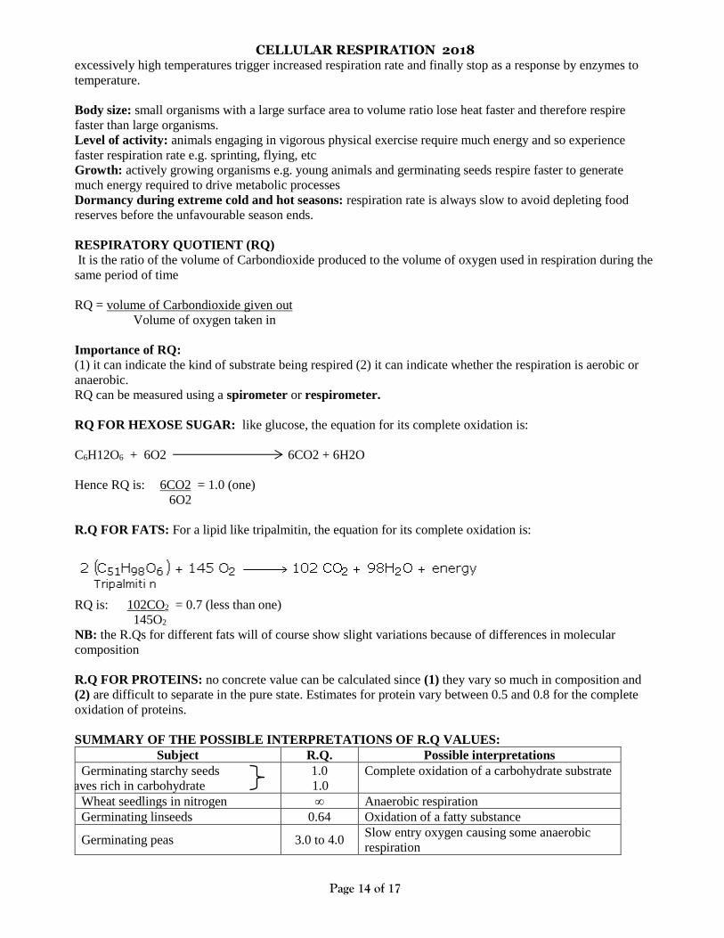
Measurement of R.Q. for small animals e.g. frog or a few earthworms
A, B, C, D and E are weighed. The animal(s) is/are placed in C and its weight determined. Then the apparatus
is connected, C is placed in a vessel of cold water to keep its temperature constant, and the pump is run for 10
to 20 minutes. After the experiment, the weight of Carbondioxide exhaled is the gain in the weight of E. the
weight of water given off by the animal is the gain in weight of D. the weight of substrate used is the loss in
weight of the animal. Hence, the weight of oxygen absorbed can be obtained thus:
Substrate + oxygen = Carbondioxide + water.
Oxygen used = (Carbondioxide + water) – substrate
= (gain in weight of E + gain in weight of D) – loss in weight of animal
The weights of Carbondioxide and oxygen thus obtained can be converted into volumes, and thus the
R.Q. obtained.
Precautions to be considered: (1) the soda-lime and sulphuric acid containers should be doubled in each case
(2) the animal container C must be carefully dried after the experiment and before weighing (3) all vessels
must not be touched by the hand but by strong wooden forceps.
COMPARISON OF RESPIRATION WITH PHOTOSYNTHESIS
Differences: Photosynthesis Respiration
Where they occur In chlorophyll-bearing cells In all cells
When they occur In the presence of light All the time
Input Carbon dioxide and water Reduced carbon compounds and oxygen
Output Reduced carbon compounds, oxygen, and water Carbon dioxide and water
Energy sources Light Chemical bonds
Energy result Energy stored Energy released
Chemical reaction Reduction of carbon compounds Oxidation of carbon compounds
Energy carrier(s) NADP NAD and FAD

CELLULAR RESPIRATION 2018
Page 16 of 17
Similarities
Both
1. involve converting energy from one form to another
2. occur in living cells
3. involve the formation of ATP
4. require energy to occur
5. involve a series of multi-enzyme catalysed reactions
6. involve flow of electrons along carriers.
ECONOMIC IMPORTANCE OF ANAEROBIC RESPIRATION
1. Fermentation is applied in the manufacture of alcoholic drinks like wine making, beer making and
manufacture of spirits.
2. Fermentation of yeast is used in leavening of bread i.e. production of raised bread
3. it is applied in the manufacture of milk products like sour milk, yoghurt and cheese
4. is applied in the manufacture of organic acids e.g. citric acid, oxalic acid and butyric acid all of which
have several industrial applications especially in food processing.
ATP PRODUCTION DURING EXERCISE
On average, a muscle contains only enough ATP to sustain about 15 seconds of intense exercise. For muscle
contractions to continue, massive amounts of ATP are required. Depending on the level of and duration of
activity, the muscles being exercised may produce the ATP they need by cellular respiration or by
fermentation.
Sustained periods of sub maximal activity like jogging are powered by aerobic respiration, but in contrast short
periods of intense activity like sprinting are powered by a combination of aerobic and anaerobic respiration.
“Anaerobic” here means a combination of glycolysis and stored ATP/Phosphocreatine release.
The table below shows the relative contributions of anaerobic and aerobic respiration to exercise during a
work out. Duration of maximal exercise
Seconds Minutes
10 30 60 2 4 10 30 60 120
Percent anaerobic 90 80 70 Percent anaerobic 50 35 15 5 2 1
Percent aerobic 10 20 30 Percent aerobic 50 65 85 95 98 99
[Quoted by Krogh David (2002): Biology; a guide to the natural world (2nd ed.), Prentice Hall; New Jersey, adapted from Astrand, P.
O., and Rodahl, K. (1977): textbook of work physiology; McGraw Hill, New York]
Observations:
(1) In the first minute, the energy supply from aerobic respiration is low but rapidly increases while that of
anaerobic respiration is high but rapidly decreases.
(2) In the second minute there is equal contribution to energy needs from both aerobic and anaerobic
respiration, followed by a rapid increase from aerobic respiration up to the 30th minute and a gradual increase
thereafter, while anaerobic respiration decreases rapidly up to the 30th minute and gradually thereafter.
Explanation:
(1) During the first minute, the small amounts of ATP and phosphocreatine stored in cells provide instant
energy. When glycolysis starts, it provides a proportionally smaller contribution, and a smaller contribution
yet comes from aerobic metabolism. These differences reflect the time it takes for each of these systems to get
going.
(2) As the duration of the exercise increases to the ATP/PCr reservoir reduce greatly while the aerobic
respiration now predominates.
CELLULAR RESPIRATION 2018
Page 17 of 17
NOTE:
Phosphocreatine (also called creatine phosphate), stores ~P bonds in nerve and muscle cells.
Creatine Kinase catalyzes: phosphocreatine + ADP ATP + creatine
This is a reversible reaction, though the equilibrium constant slightly favors phosphocreatine
formation. Phosphocreatine is produced when ATP levels are high. When ATP is depleted
during exercise in muscle, phosphate is transferred from phosphocreatine to ADP, to
replenish ATP.
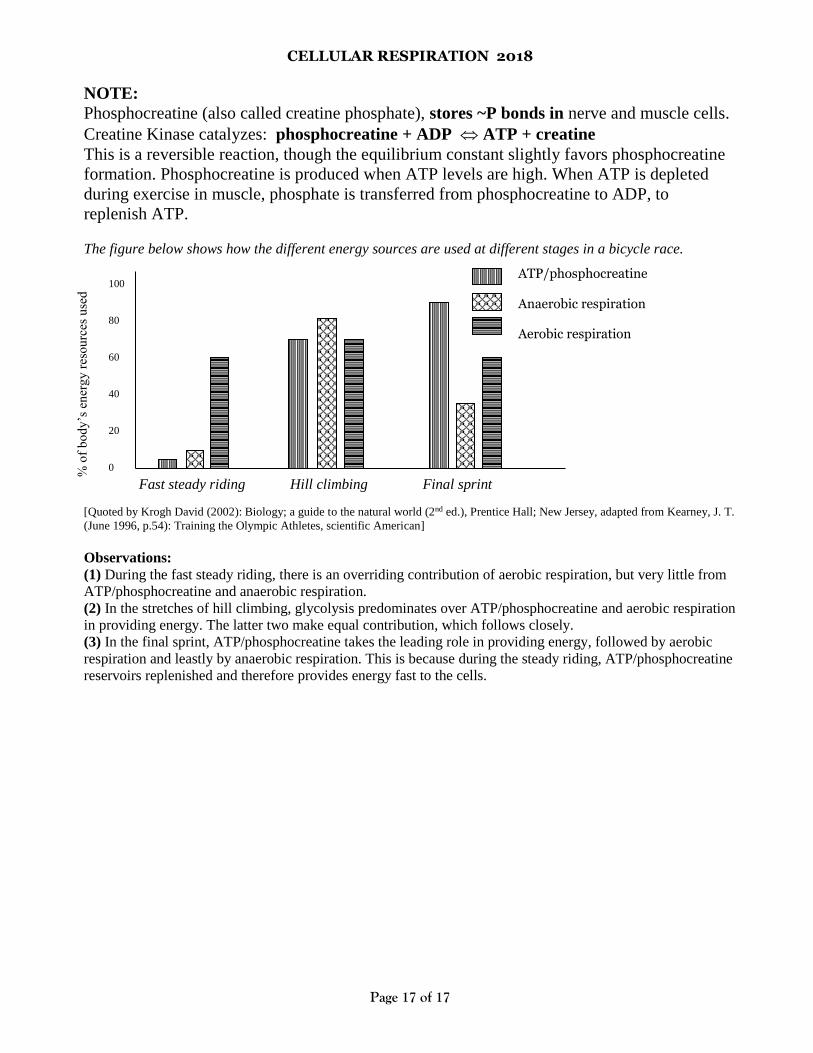
The figure below shows how the different energy sources are used at different stages in a bicycle race.
Fast steady riding Hill climbing Final sprint
[Quoted by Krogh David (2002): Biology; a guide to the natural world (2nd ed.), Prentice Hall; New Jersey, adapted from Kearney, J. T.
(June 1996, p.54): Training the Olympic Athletes, scientific American]
Observations:
(1) During the fast steady riding, there is an overriding contribution of aerobic respiration, but very little from
ATP/phosphocreatine and anaerobic respiration.
(2) In the stretches of hill climbing, glycolysis predominates over ATP/phosphocreatine and aerobic respiration
in providing energy. The latter two make equal contribution, which follows closely.
(3) In the final sprint, ATP/phosphocreatine takes the leading role in providing energy, followed by aerobic
respiration and leastly by anaerobic respiration. This is because during the steady riding, ATP/phosphocreatine
reservoirs replenished and therefore provides energy fast to the cells.
100
80
60
40
20
0
% o
f b
ody
’s e
ner
gy
res
ou
rces
use
d
ATP/phosphocreatine Anaerobic respiration Aerobic respiration