Embed Size (px)
Citation preview
Cells are supported in vivo by a three-dimensional extracellular
matrix (ECM) of nanofibers with different chemical ligands that
interact with cell surface receptors1-4. The interplay between cells
and the ECM is a dynamic and complex process where the physical
and chemical properties of the ECM elicit different cellular
responses1,5. An engineered substrate seeking to emulate the
functions of the native ECM should, therefore, recapitulate its
three dimensionality and nanofibrous topography, as well as its
plethora of chemical motifs6,7. Advances in nanotechnology in
recent years have enabled us to engineer novel biomaterials with
these levels of complexities7-9. The eventual use of such complex
biomaterials for specific applications will require an iterative
process of understanding the mechanisms guiding cell-matrix
interactions so that we can precisely control biomaterial
properties to elicit desirable cellular responses (Fig. 1). Here, we
emphasize the importance of the three-dimensional nanofibrous
features of extracellular environments in modulating cellular
responses with local or subcellular resolutions in space- and time-
dependent manners. We also highlight state-of-the-art
technologies to fabricate and characterize nanofibrous
environments for relevant applications.
Influence of the nanofibrous environmenton cell phenotype and signalingCells interact with their nanofibrous extracellular environment via cell
surface proteins, such as integrins, to activate various intracellular
signaling pathways that regulate cellular processes, e.g. cell shape,
mobility, and proliferation (Fig. 2). Understanding the specific biological
Cells respond profoundly to the mechanical rigidity and three-dimensionalnanotopology of substrates, as well as the spatial and temporalarrangements of extracellular cues. We summarize the latestdevelopments in probing and engineering biocompatible nanofibrousextracellular environments at the cell and molecular level for applicationsin tissue engineering and biological research. This will, in turn, guidefurther development of three-dimensional nanofibrous scaffolds in orderto elicit specific cellular responses for relevant applications.
Yi-Chin Toh1,2, Susanne Ng1,2, Yuet Mei Khong1,2, Xin Zhang2, Yajuan Zhu2, Pao-Chun Lin3, Chee-Min Te3, Wanxin Sun1, and Hanry
Yu1,2,3*1Institute of Bioengineering and Nanotechnology, 31 Biopolis Way, Singapore 1386992Graduate Program in Bioengineering, NUS Graduate School of Integrative Sciences and Engineering, National University of Singapore,
Singapore 1175973Department of Physiology, Yong Loo Lin School of Medicine, National University of Singapore, Singapore 117597
*E-mail: [email protected]
ISSN:1748 0132 © Elsevier Ltd 2006AUGUST 2006 | VOLUME 1 | NUMBER 3 34
Cellular responses to ananofibrous environment
NT103p34_43.qxd 07/06/2006 14:44 Page 34
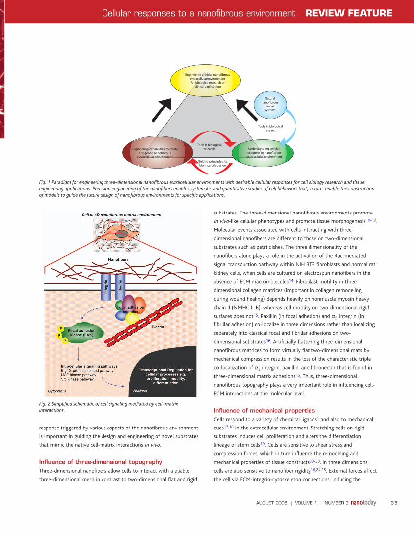
response triggered by various aspects of the nanofibrous environment
is important in guiding the design and engineering of novel substrates
that mimic the native cell-matrix interactions in vivo.
Influence of three-dimensional topographyThree-dimensional nanofibers allow cells to interact with a pliable,
three-dimensional mesh in contrast to two-dimensional flat and rigid
substrates. The three-dimensional nanofibrous environments promote
in vivo-like cellular phenotypes and promote tissue morphogenesis10-13.
Molecular events associated with cells interacting with three-
dimensional nanofibers are different to those on two-dimensional
substrates such as petri dishes. The three dimensionality of the
nanofibers alone plays a role in the activation of the Rac-mediated
signal transduction pathway within NIH 3T3 fibroblasts and normal rat
kidney cells, when cells are cultured on electrospun nanofibers in the
absence of ECM macromolecules14. Fibroblast motility in three-
dimensional collagen matrices (important in collagen remodeling
during wound healing) depends heavily on nonmuscle myosin heavy
chain II (NMHC II-B), whereas cell motility on two-dimensional rigid
surfaces does not15. Paxillin (in focal adhesion) and α5 integrin (in
fibrillar adhesion) co-localize in three dimensions rather than localizing
separately into classical focal and fibrillar adhesions on two-
dimensional substrates16. Artificially flattening three-dimensional
nanofibrous matrices to form virtually flat two-dimensional mats by
mechanical compression results in the loss of the characteristic triple
co-localization of α5 integrin, paxillin, and fibronectin that is found in
three-dimensional matrix adhesions16. Thus, three-dimensional
nanofibrous topography plays a very important role in influencing cell-
ECM interactions at the molecular level.
Influence of mechanical propertiesCells respond to a variety of chemical ligands1 and also to mechanical
cues17,18 in the extracellular environment. Stretching cells on rigid
substrates induces cell proliferation and alters the differentiation
lineage of stem cells19. Cells are sensitive to shear stress and
compression forces, which in turn influence the remodeling and
mechanical properties of tissue constructs20-23. In three dimensions,
cells are also sensitive to nanofiber rigidity16,24,25. External forces affect
the cell via ECM-integrin-cytoskeleton connections, inducing the
Tools in biologicalresearch
Engineered artificial nanofibrous extracellular environment for biological research or
clinical applications
Naturalnanofibrous
basedsystems
Understanding cellular responses to nanofibrous extracellular environment
Engineering capabilities to createand probe nanofibrous
extracellular environmentGuiding principles forbiomaterials design
Tools in biologicalresearch
AUGUST 2006 | VOLUME 1 | NUMBER 3 35
Cellular responses to a nanofibrous environment REVIEW FEATURE
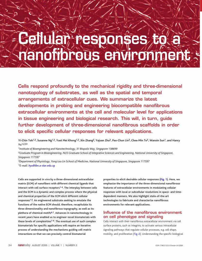
Fig. 1 Paradigm for engineering three-dimensional nanofibrous extracellular environments with desirable cellular responses for cell biology research and tissueengineering applications. Precision engineering of the nanofibers enables systematic and quantitative studies of cell behaviors that, in turn, enable the constructionof models to guide the future design of nanofibrous environments for specific applications.
Fig. 2 Simplified schematic of cell signaling mediated by cell-matrixinteractions.
NT103p34_43.qxd 07/06/2006 14:44 Page 35
assembly of focal adhesions that trigger tyrosine kinase/phosphatases
and Src family kinases (SFKs) with subsequent activation of small
G proteins and MAP kinases for the regulation of downstream cellular
events26. It will be important to quantify these events systematically
and establish models of cellular response to mechanical and chemical
properties of the nanofibers to guide the development of three-
dimensional nanofibrous environments for applications.
Influence of spatial and temporal arrangements ofextracellular cuesCellular responses to the mechanical and chemical properties of three-
dimensional nanofibers are often space- and time-dependent27-29.
Neural stem cells and smooth muscle cells, when cultured on aligned
electrospun nanofibers, elongate and orient themselves along the
fibers30,31. This sensitivity to the spatial arrangement of the nanofibers
AUGUST 2006 | VOLUME 1 | NUMBER 3 36
REVIEW FEATURE Cellular responses to a nanofibrous environment
Glossary α5 integrin One of the integrin family of transmembrane proteins that are involved in the adhesion of
cells to the ECM.
angiogenesis The generation of new blood vessels.
collagen Fibrous ECM protein rich in glycine and proline. At least 12 types of collagen have been
identified. Collagen I is commonly found in tendon and bone, while collagen II is commonly
found in cartilage.
complex coacervation The separation of two liquid phases in a colloidal system caused by the interaction of two
oppositely charged colloids.
DsRed2-calreticulin Calreticulin, a highly conserved Ca-binding protein within nonmuscle smooth muscle cell
ERs, conjugated to the fluorescent probe DsRed.
endoplasmic reticulum (ER) Membrane-bounded compartment in the cytoplasm of eukaryotic cells, responsible for lipid
synthesis, as well as the synthesis and sorting of membrane-bound and secretory proteins.
fluorescence resonance energy transfer (FRET) Describes the phenomenon of energy transfer between two fluorescent molecules. A
fluorescent donor is excited at one wavelength and its emission excites a neighboring
fluorescent acceptor, whose emission wavelength is detected.
mitogen-activated protein (MAP) kinases Protein kinases that perform a crucial step in relaying signals from the plasma membrane to
the nucleus. They regulate various cellular activities, such as gene expression, mitosis,
differentiation, and cell survival/apoptosis.
morphogenesis The process of forming a tissue or organ via regulated growth and differentiation of cells.
messenger ribonucleic acid (mRNA) An RNA molecule that specifies the amino acid sequence of a protein during protein synthesis.
NIH 3T3 fibroblasts A cell line derived from fibroblasts of disaggregated Swiss mouse embryos.
paxillin Focal adhesion protein involved in the binding of the actin cytoskeleton at sites of cell
attachment to the ECM.
phenotype The observable characteristics or traits of a cell or an organism, e.g. size, eye color.
Rac-mediated signal transduction pathway Signaling pathway regulated by Rac guanosine triphosphate (GTP)-binding protein, which is
involved in controlling the organization of cytoskeletal filaments.
RGD sites Peptide sequence consisting of three amino acids (arginine-glycine-aspartic acid) commonly
associated with cell attachment.
small G proteins A large family of monomeric GTP-binding proteins that activate target proteins on binding
GTP.
solid free-form fabrication (SFF) Three-dimensional, computer-aided designs are converted into stereolithography data and a
format recognizable by software for printing layer by layer. Reverse SFF fabricates the desired
construct by casting over a negative mould created via SFF.
Src family kinases A tyrosine kinase family of proteins, which interact with a variety of cell-surface receptors and
participate in intracellular signal transduction pathways.
tyrosine kinases/phosphatases Enzymes that catalyze the phosphorylation/dephosphorylation, respectively, of tyrosine
residues in proteins. Tyrosine kinase/phosphatase pairs regulate cellular signal transduction and
may play a role in cell growth control and carcinogenesis or cancer formation.
NT103p34_43.qxd 07/06/2006 14:44 Page 36
possibly results from localized interactions of cells with the
extracellular cues at subcellular resolutions. Most of the structural or
signaling proteins involved in rigidity sensing, ECM anchorage, and cell
migration are found in specific locations in a cell, such as the leading
tip of migrating fibroblasts or focal adhesion sites, but not in other
locations even 0.5 µm away28. A single ECM-integrin-cytoskeleton
linkage at one specific location can be dynamically modulated in
response to an extracellular force without affecting a nearby
linkage32,33. Clustering of integrin molecules to form focal adhesions
and stress fibers only occurs when sufficient RGD sites are clustered
within a discrete spot of less than 70 nm27-29.
Molecules associated with the integrin-cytoskeleton linkages
involved in cell-ECM adhesion also change in a time-dependent manner
throughout the life cycle of the adhesion sites34 and are sensitive to the
stiffness of the ECM nanofibers32,35-38. The formation of cell contacts
with the ECM is not a continuous process, but involves cycles of
contraction and relaxation. We have recently shown that presenting
extracellular nanofibers to hepatocytes at different times or sequences
in sandwich culture elicits different cellular responses39. Thus, cells are
sensitive to temporal as well as spatial arrangements of extracellular
cues in the environment. It seems that the sum of local responses to
extracellular cues over time helps determine cell morphology and gene
expression26,40. It is important therefore to understand and engineer the
local cellular responses to spatially and temporally distributed
extracellular cues in further development of three-dimensional
nanofibrous environments.
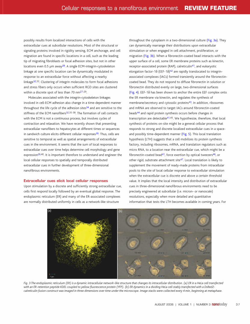
Extracellular cues elicit local cellular responsesUpon stimulation by a discrete and sufficiently strong extracellular cue,
cells first respond locally followed by an eventual global response. The
endoplasmic reticulum (ER) and many of the ER-associated complexes
are normally distributed uniformly in cells as a network-like structure
throughout the cytoplasm in a two-dimensional culture (Fig. 3a). They
can dynamically rearrange their distributions upon extracellular
stimulation or when engaged in cell attachment, proliferation, or
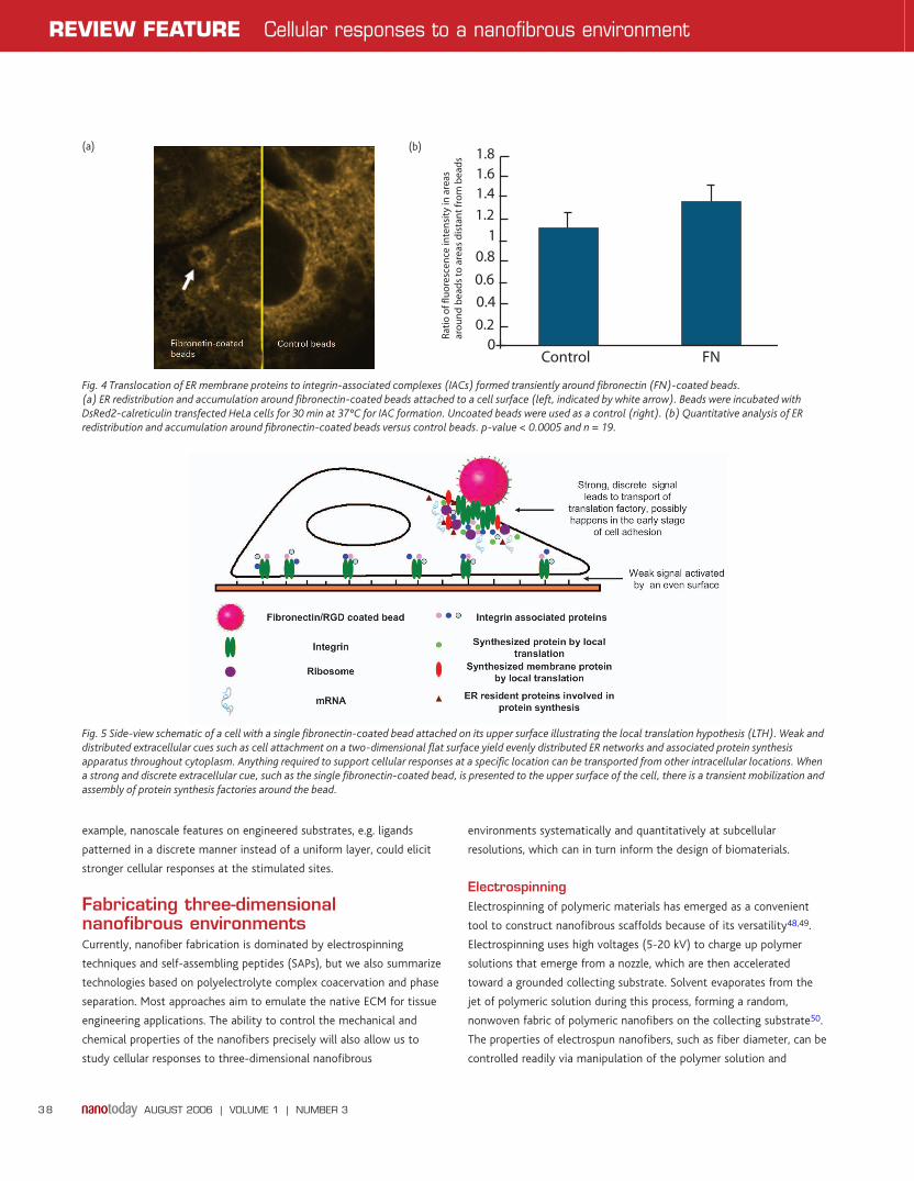
migration (Fig. 3b). When a fibronectin-coated bead interacts with the
upper surface of a cell, some ER membrane proteins such as kinectin,
receptor-associated protein (RAP), calreticulin41, and eukaryotic
elongation factor-1δ (EEF-1δ)42 are rapidly translocated to integrin-
associated complexes (IACs) formed transiently around the fibronectin-
coated bead. They do not respond to diffuse fibronectin in solution or
fibronectin distributed evenly on large, two-dimensional surfaces
(Fig. 4). EEF-1δ has been shown to anchor the entire EEF complex onto
the ER membrane via kinectin, and regulates the synthesis of
membrane/secretory and cytosolic proteins43. In addition, ribosomes
and mRNA are observed to target IACs around fibronectin-coated
beads44 and rapid protein synthesis occurs before changes in
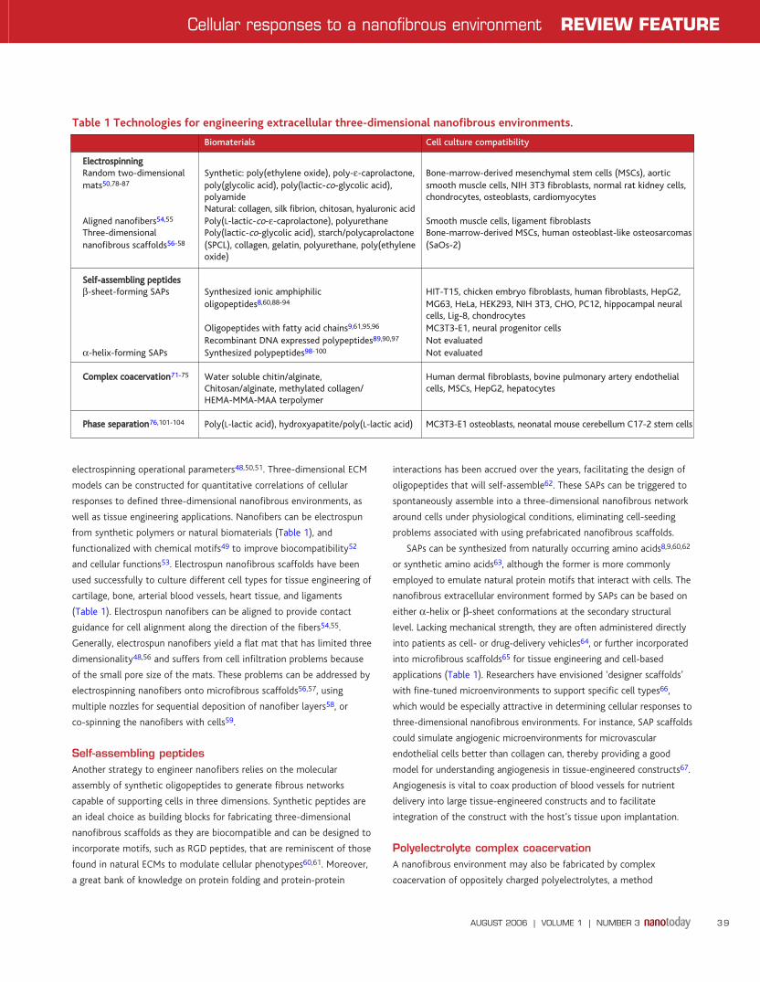
transcription are detectable41,45. We hypothesize, therefore, that local
synthesis of proteins on-site might be a general cellular process that
responds to strong and discrete localized extracellular cues in a space-
and possibly time-dependent manner (Fig. 5). This local translation
hypothesis (LTH) suggests that a cell mobilizes its protein synthesis
factory, including ribosomes, mRNA, and translation regulators such as
micro RNA, to a location near the extracellular cue, which might be a
fibronectin-coated bead41, force exertion by optical tweezers46, or
other rigid, substrate attachment site47. Local translation is likely to
supplement the movement of ready-made proteins from intracellular
pools to the site of local cellular response to extracellular stimulation
when the extracellular cue is discrete and above a certain threshold
value. It implies that the local intensity and distribution of extracellular
cues in three-dimensional nanofibrous environments need to be
precisely engineered at subcellular (i.e. micron- or nanoscale)
resolutions, especially when more detailed and quantitative
information that tests the LTH becomes available in coming years. For
AUGUST 2006 | VOLUME 1 | NUMBER 3 37
Cellular responses to a nanofibrous environment REVIEW FEATURE
(b)(a)
Fig. 3 The endoplasmic reticulum (ER) is a dynamic intracellular network-like structure that changes its intracellular distribution. (a) ER in a HeLa cell transfectedwith an ER-retention peptide KDEL coupled to yellow fluorescence protein (YFP). (b) ER dynamics in a dividing HeLa cell stably transfected with a DsRed2-calreticulin fusion construct was imaged in three dimensions over time under the microscope. Image stacks were collected every 4 min, beginning at metaphase.
NT103p34_43.qxd 07/06/2006 14:44 Page 37
example, nanoscale features on engineered substrates, e.g. ligands
patterned in a discrete manner instead of a uniform layer, could elicit
stronger cellular responses at the stimulated sites.
Fabricating three-dimensionalnanofibrous environmentsCurrently, nanofiber fabrication is dominated by electrospinning
techniques and self-assembling peptides (SAPs), but we also summarize
technologies based on polyelectrolyte complex coacervation and phase
separation. Most approaches aim to emulate the native ECM for tissue
engineering applications. The ability to control the mechanical and
chemical properties of the nanofibers precisely will also allow us to
study cellular responses to three-dimensional nanofibrous
environments systematically and quantitatively at subcellular
resolutions, which can in turn inform the design of biomaterials.
ElectrospinningElectrospinning of polymeric materials has emerged as a convenient
tool to construct nanofibrous scaffolds because of its versatility48,49.
Electrospinning uses high voltages (5-20 kV) to charge up polymer
solutions that emerge from a nozzle, which are then accelerated
toward a grounded collecting substrate. Solvent evaporates from the
jet of polymeric solution during this process, forming a random,
nonwoven fabric of polymeric nanofibers on the collecting substrate50.
The properties of electrospun nanofibers, such as fiber diameter, can be
controlled readily via manipulation of the polymer solution and
AUGUST 2006 | VOLUME 1 | NUMBER 3 38
REVIEW FEATURE Cellular responses to a nanofibrous environment
Fig. 5 Side-view schematic of a cell with a single fibronectin-coated bead attached on its upper surface illustrating the local translation hypothesis (LTH). Weak anddistributed extracellular cues such as cell attachment on a two-dimensional flat surface yield evenly distributed ER networks and associated protein synthesisapparatus throughout cytoplasm. Anything required to support cellular responses at a specific location can be transported from other intracellular locations. Whena strong and discrete extracellular cue, such as the single fibronectin-coated bead, is presented to the upper surface of the cell, there is a transient mobilization andassembly of protein synthesis factories around the bead.
Fig. 4 Translocation of ER membrane proteins to integrin-associated complexes (IACs) formed transiently around fibronectin (FN)-coated beads. (a) ER redistribution and accumulation around fibronectin-coated beads attached to a cell surface (left, indicated by white arrow). Beads were incubated withDsRed2-calreticulin transfected HeLa cells for 30 min at 37°C for IAC formation. Uncoated beads were used as a control (right). (b) Quantitative analysis of ERredistribution and accumulation around fibronectin-coated beads versus control beads. p-value < 0.0005 and n = 19.
(a) (b)
1.61.8
1.4
1.2
10.8
0.6
0.4
0.20
Control FN
Rat
io o
f flu
ore
scen
ce in
ten
sity
in a
reas
aro
un
d b
ead
s to
are
as d
ista
nt
fro
m b
ead
s
NT103p34_43.qxd 07/06/2006 14:44 Page 38
electrospinning operational parameters48,50,51. Three-dimensional ECM
models can be constructed for quantitative correlations of cellular
responses to defined three-dimensional nanofibrous environments, as
well as tissue engineering applications. Nanofibers can be electrospun
from synthetic polymers or natural biomaterials (Table 1), and
functionalized with chemical motifs49 to improve biocompatibility52
and cellular functions53. Electrospun nanofibrous scaffolds have been
used successfully to culture different cell types for tissue engineering of
cartilage, bone, arterial blood vessels, heart tissue, and ligaments
(Table 1). Electrospun nanofibers can be aligned to provide contact
guidance for cell alignment along the direction of the fibers54,55.
Generally, electrospun nanofibers yield a flat mat that has limited three
dimensionality48,56 and suffers from cell infiltration problems because
of the small pore size of the mats. These problems can be addressed by
electrospinning nanofibers onto microfibrous scaffolds56,57, using
multiple nozzles for sequential deposition of nanofiber layers58, or
co-spinning the nanofibers with cells59.
Self-assembling peptidesAnother strategy to engineer nanofibers relies on the molecular
assembly of synthetic oligopeptides to generate fibrous networks
capable of supporting cells in three dimensions. Synthetic peptides are
an ideal choice as building blocks for fabricating three-dimensional
nanofibrous scaffolds as they are biocompatible and can be designed to
incorporate motifs, such as RGD peptides, that are reminiscent of those
found in natural ECMs to modulate cellular phenotypes60,61. Moreover,
a great bank of knowledge on protein folding and protein-protein
interactions has been accrued over the years, facilitating the design of
oligopeptides that will self-assemble62. These SAPs can be triggered to
spontaneously assemble into a three-dimensional nanofibrous network
around cells under physiological conditions, eliminating cell-seeding
problems associated with using prefabricated nanofibrous scaffolds.
SAPs can be synthesized from naturally occurring amino acids8,9,60,62
or synthetic amino acids63, although the former is more commonly
employed to emulate natural protein motifs that interact with cells. The
nanofibrous extracellular environment formed by SAPs can be based on
either α-helix or β-sheet conformations at the secondary structural
level. Lacking mechanical strength, they are often administered directly
into patients as cell- or drug-delivery vehicles64, or further incorporated
into microfibrous scaffolds65 for tissue engineering and cell-based
applications (Table 1). Researchers have envisioned ‘designer scaffolds’
with fine-tuned microenvironments to support specific cell types66,
which would be especially attractive in determining cellular responses to
three-dimensional nanofibrous environments. For instance, SAP scaffolds
could simulate angiogenic microenvironments for microvascular
endothelial cells better than collagen can, thereby providing a good
model for understanding angiogenesis in tissue-engineered constructs67.
Angiogenesis is vital to coax production of blood vessels for nutrient
delivery into large tissue-engineered constructs and to facilitate
integration of the construct with the host’s tissue upon implantation.
Polyelectrolyte complex coacervationA nanofibrous environment may also be fabricated by complex
coacervation of oppositely charged polyelectrolytes, a method
AUGUST 2006 | VOLUME 1 | NUMBER 3 39
Cellular responses to a nanofibrous environment REVIEW FEATURE
Biomaterials Cell culture compatibility
EElleeccttrroossppiinnnniinnggRandom two-dimensional Synthetic: poly(ethylene oxide), poly-ε-caprolactone, Bone-marrow-derived mesenchymal stem cells (MSCs), aortic mats50,78-87 poly(glycolic acid), poly(lactic-co-glycolic acid), smooth muscle cells, NIH 3T3 fibroblasts, normal rat kidney cells,
polyamide chondrocytes, osteoblasts, cardiomyocytesNatural: collagen, silk fibrion, chitosan, hyaluronic acid
Aligned nanofibers54,55 Poly(L-lactic-co-ε-caprolactone), polyurethane Smooth muscle cells, ligament fibroblastsThree-dimensional Poly(lactic-co-glycolic acid), starch/polycaprolactone Bone-marrow-derived MSCs, human osteoblast-like osteosarcomas nanofibrous scaffolds56-58 (SPCL), collagen, gelatin, polyurethane, poly(ethylene (SaOs-2)
oxide)
SSeellff--aasssseemmbblliinngg ppeeppttiiddeessβ-sheet-forming SAPs Synthesized ionic amphiphilic HIT-T15, chicken embryo fibroblasts, human fibroblasts, HepG2,
oligopeptides8,60,88-94 MG63, HeLa, HEK293, NIH 3T3, CHO, PC12, hippocampal neural cells, Lig-8, chondrocytes
Oligopeptides with fatty acid chains9,61,95,96 MC3T3-E1, neural progenitor cellsRecombinant DNA expressed polypeptides89,90,97 Not evaluated
α-helix-forming SAPs Synthesized polypeptides98-100 Not evaluated
CCoommpplleexx ccooaacceerrvvaattiioonn71-75 Water soluble chitin/alginate, Human dermal fibroblasts, bovine pulmonary artery endothelial Chitosan/alginate, methylated collagen/ cells, MSCs, HepG2, hepatocytesHEMA-MMA-MAA terpolymer
PPhhaassee sseeppaarraattiioonn76,101-104 Poly(L-lactic acid), hydroxyapatite/poly(L-lactic acid) MC3T3-E1 osteoblasts, neonatal mouse cerebellum C17-2 stem cells
Table 1 Technologies for engineering extracellular three-dimensional nanofibrous environments.
NT103p34_43.qxd 07/06/2006 14:44 Page 39
originally developed for the macro- or microencapsulation of cells68-70.
Polyelectrolyte complex coacervation can take place under mild
aqueous conditions, and is therefore favorable for in situ formation of
nanofibers in the presence of cells. A plethora of polyelectrolytes have
been developed and evaluated for cell encapsulation68,69. Complex
coacervated nanofibers generally lack mechanical strength; therefore, it
is difficult to cast them into three-dimensional scaffolds for tissue
engineering applications. The nanofibers can be readily incorporated
into cell culture constructs such as three-dimensional microfibrous
scaffolds and microfluidic channels to immobilize and support sensitive,
anchorage-dependent cells71,72. Alternatively, nanofibers may be drawn
directly into microfibers, with nanofibrous features to support cultures
of various mammalian cells73-75.
Phase separationPhase separation, an approach commonly used to fabricate
microfibrous scaffolds, can also be used to fabricate nanocomposite
scaffolds. Polymer- and solvent-rich domains of a polymer solution are
separated either by cooling the solution or exchanging a
nonsolvent for the solvent. A nanofibrous (fibers with diameters of
50-500 nm) three-dimensional scaffold has been constructed with
poly(L-lactic acid) via thermally induced phase separation76. The
scaffold has controllable high porosity (up to 98.5%) and surface-to-
volume ratios, as well as defined mechanical properties (Table 1).
A reverse solid freeform fabrication (SFF) technique to control
scaffold architecture and dimensions by integrating paraffin spheres for
pore connectivity has recently been developed77, enabling the
fabrication of scaffolds of any desired shape or size. Nanofiber
distribution and uniformity is subject to the controllability of the
processing. Scaffold fabrication also involves a five-step process and
has, so far, only been applied to a few polymers such as
poly(L-lactic acid) and its blends49. This technique can be readily
applied in tissue engineering applications.
Characterizing the three-dimensionalnanofibrous environmentNew developments in microscopy techniques and fluorescence probes
have enabled dynamic imaging of three-dimensional nanofibrous
matrices and their interactions with cells.
Introduction of fluorescence probes normally interferes with the
biological system under scrutiny to a certain extent by changing the
matrix properties or interacting with the cells in the system. Intensive
efforts are being made to develop less invasive fluorescence dyes
and proteins with high specificity to characterize the chemical
properties of the cellular environment. Several imaging techniques can
be used to quantify structural ECM dynamics without fluorescence
probes, and force-sensing methods such as atomic force microscopy and
optical tweezers have great potential for mechanical property
measurements.
Structural characterization using imaging techniquesCellular interactions with three-dimensional nanofibrous networks have
been monitored using differential interference contrast (DIC)
microscopy, which is a contrast-enhancing imaging technique105. Two-
dimensional images are obtained at different depths, though it is
intrinsically difficult to construct three-dimensional images from DIC
images even after image deconvolution processing.
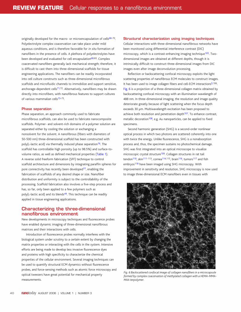
Reflection or backscattering confocal microscopy exploits the light
scattering properties of nanofibrous ECM molecules to construct images.
It has been used to image collagen fibers and cell-ECM interactions5,106.
Fig. 6 is a projection of a three-dimensional collagen matrix obtained by
backscattering confocal microscopy with an illumination wavelength of
488 nm. In three-dimensional imaging, the resolution and image quality
deteriorate greatly because of light scattering when the focus depth
exceeds 30 µm. Multiwavelength excitation has been proposed to
achieve both resolution and penetration depth107. To enhance contrast,
metallic decoration108, e.g. Au nanoparticles, can be applied to fixed
specimens.
Second harmonic generation (SHG) is a second-order nonlinear
optical process in which two photons are scattered coherently into one
with twice the energy. Unlike fluorescence, SHG is a nonabsorption
process and, thus, the specimen sustains no photochemical damage.
SHG was first integrated into an optical microscope to visualize
microscopic crystal structure109. Collagen structures in rat tail
tendon110, skin111-113, cornea114,115, brain116, tumors117 and fish
embryos118 have been imaged using SHG microscopy. With
improvement in sensitivity and resolution, SHG microscopy is now used
to image three-dimensional ECM nanofibers even in tissues with
AUGUST 2006 | VOLUME 1 | NUMBER 3 40
REVIEW FEATURE Cellular responses to a nanofibrous environment
Fig. 6 Backscattered confocal image of collagen nanofibers in a microcapsuleformed by complex coacervation of methylated collagen with a HEMA-MMA-MAA terpolymer.
NT103p34_43.qxd 07/06/2006 14:44 Page 40
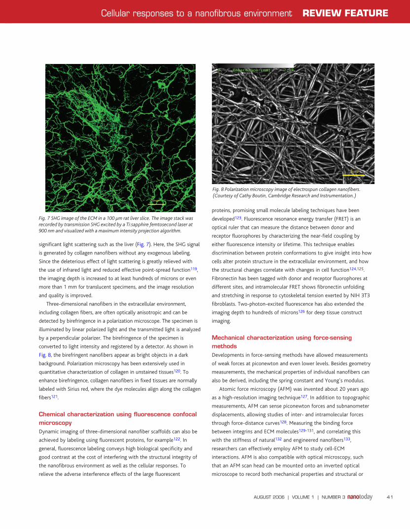
significant light scattering such as the liver (Fig. 7). Here, the SHG signal
is generated by collagen nanofibers without any exogenous labeling.
Since the deleterious effect of light scattering is greatly relieved with
the use of infrared light and reduced effective point-spread function119,
the imaging depth is increased to at least hundreds of microns or even
more than 1 mm for translucent specimens, and the image resolution
and quality is improved.
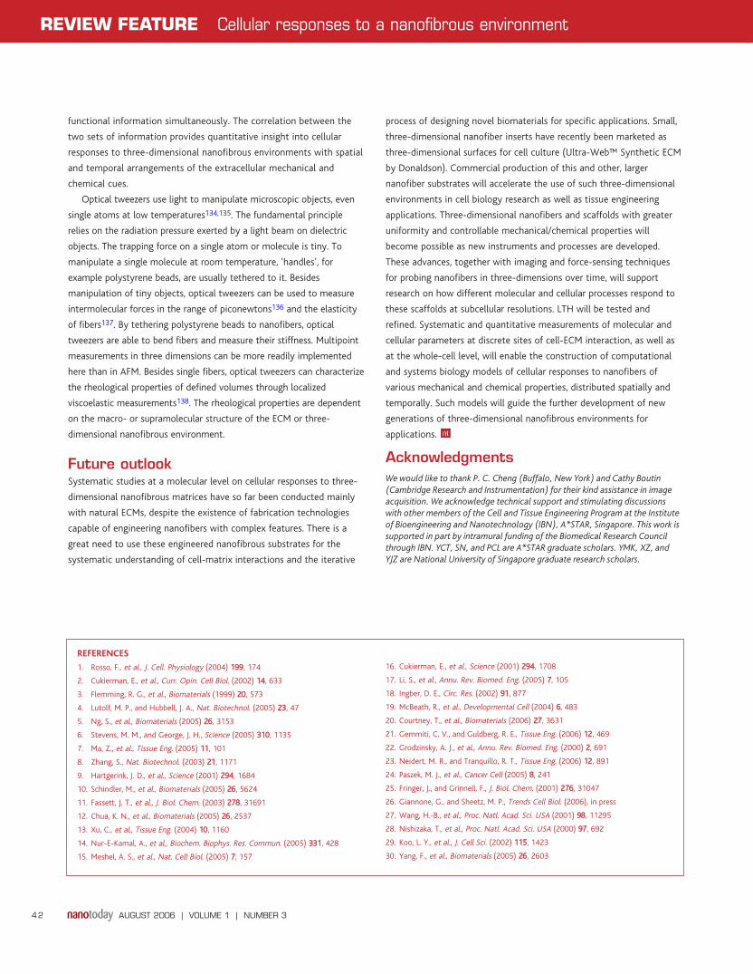
Three-dimensional nanofibers in the extracellular environment,
including collagen fibers, are often optically anisotropic and can be
detected by birefringence in a polarization microscope. The specimen is
illuminated by linear polarized light and the transmitted light is analyzed
by a perpendicular polarizer. The birefringence of the specimen is
converted to light intensity and registered by a detector. As shown in
Fig. 8, the birefringent nanofibers appear as bright objects in a dark
background. Polarization microscopy has been extensively used in
quantitative characterization of collagen in unstained tissues120. To
enhance birefringence, collagen nanofibers in fixed tissues are normally
labeled with Sirius red, where the dye molecules align along the collagen
fibers121.
Chemical characterization using fluorescence confocalmicroscopyDynamic imaging of three-dimensional nanofiber scaffolds can also be
achieved by labeling using fluorescent proteins, for example122. In
general, fluorescence labeling conveys high biological specificity and
good contrast at the cost of interfering with the structural integrity of
the nanofibrous environment as well as the cellular responses. To
relieve the adverse interference effects of the large fluorescent
proteins, promising small molecule labeling techniques have been
developed123. Fluorescence resonance energy transfer (FRET) is an
optical ruler that can measure the distance between donor and
receptor fluorophores by characterizing the near-field coupling by
either fluorescence intensity or lifetime. This technique enables
discrimination between protein conformations to give insight into how
cells alter protein structure in the extracellular environment, and how
the structural changes correlate with changes in cell function124,125.
Fibronectin has been tagged with donor and receptor fluorophores at
different sites, and intramolecular FRET shows fibronectin unfolding
and stretching in response to cytoskeletal tension exerted by NIH 3T3
fibroblasts. Two-photon-excited fluorescence has also extended the
imaging depth to hundreds of microns126 for deep tissue construct
imaging.
Mechanical characterization using force-sensingmethodsDevelopments in force-sensing methods have allowed measurements
of weak forces at piconewton and even lower levels. Besides geometry
measurements, the mechanical properties of individual nanofibers can
also be derived, including the spring constant and Young’s modulus.
Atomic force microscopy (AFM) was invented about 20 years ago
as a high-resolution imaging technique127. In addition to topographic
measurements, AFM can sense piconewton forces and subnanometer
displacements, allowing studies of inter- and intramolecular forces
through force-distance curves128. Measuring the binding force
between integrins and ECM molecules129-131, and correlating this
with the stiffness of natural132 and engineered nanofibers133,
researchers can effectively employ AFM to study cell-ECM
interactions. AFM is also compatible with optical microscopy, such
that an AFM scan head can be mounted onto an inverted optical
microscope to record both mechanical properties and structural or
AUGUST 2006 | VOLUME 1 | NUMBER 3 41
Cellular responses to a nanofibrous environment REVIEW FEATURE
Fig. 8 Polarization microscopy image of electrospun collagen nanofibers.(Courtesy of Cathy Boutin, Cambridge Research and Instrumentation.)
Fig. 7 SHG image of the ECM in a 100 µm rat liver slice. The image stack wasrecorded by transmission SHG excited by a Ti:sapphire femtosecond laser at900 nm and visualized with a maximum intensity projection algorithm.
NT103p34_43.qxd 07/06/2006 14:44 Page 41
AUGUST 2006 | VOLUME 1 | NUMBER 3 42
REVIEW FEATURE Cellular responses to a nanofibrous environment
REFERENCES
1. Rosso, F., et al., J. Cell. Physiology (2004) 119999, 174
2. Cukierman, E., et al., Curr. Opin. Cell Biol. (2002) 1144, 633
3. Flemming, R. G., et al., Biomaterials (1999) 2200, 573
4. Lutolf, M. P., and Hubbell, J. A., Nat. Biotechnol. (2005) 2233, 47
5. Ng, S., et al., Biomaterials (2005) 2266, 3153
6. Stevens, M. M., and George, J. H., Science (2005) 331100, 1135
7. Ma, Z., et al., Tissue Eng. (2005) 1111, 101
8. Zhang, S., Nat. Biotechnol. (2003) 2211, 1171
9. Hartgerink, J. D., et al., Science (2001) 229944, 1684
10. Schindler, M., et al., Biomaterials (2005) 2266, 5624
11. Fassett, J. T., et al., J. Biol. Chem. (2003) 227788, 31691
12. Chua, K. N., et al., Biomaterials (2005) 2266, 2537
13. Xu, C., et al., Tissue Eng. (2004) 1100, 1160
14. Nur-E-Kamal, A., et al., Biochem. Biophys. Res. Commun. (2005) 333311, 428
15. Meshel, A. S., et al., Nat. Cell Biol. (2005) 77, 157
16. Cukierman, E., et al., Science (2001) 229944, 1708
17. Li, S., et al., Annu. Rev. Biomed. Eng. (2005) 77, 105
18. Ingber, D. E., Circ. Res. (2002) 9911, 877
19. McBeath, R., et al., Developmental Cell (2004) 66, 483
20. Courtney, T., et al., Biomaterials (2006) 2277, 3631
21. Gemmiti, C. V., and Guldberg, R. E., Tissue Eng. (2006) 1122, 469
22. Grodzinsky, A. J., et al., Annu. Rev. Biomed. Eng. (2000) 22, 691
23. Neidert, M. R., and Tranquillo, R. T., Tissue Eng. (2006) 1122, 891
24. Paszek, M. J., et al., Cancer Cell (2005) 88, 241
25. Fringer, J., and Grinnell, F., J. Biol. Chem. (2001) 227766, 31047
26. Giannone, G., and Sheetz, M. P., Trends Cell Biol. (2006), in press
27. Wang, H.-B., et al., Proc. Natl. Acad. Sci. USA (2001) 9988, 11295
28. Nishizaka, T., et al., Proc. Natl. Acad. Sci. USA (2000) 9977, 692
29. Koo, L. Y., et al., J. Cell Sci. (2002) 111155, 1423
30. Yang, F., et al., Biomaterials (2005) 2266, 2603
functional information simultaneously. The correlation between the
two sets of information provides quantitative insight into cellular
responses to three-dimensional nanofibrous environments with spatial
and temporal arrangements of the extracellular mechanical and
chemical cues.
Optical tweezers use light to manipulate microscopic objects, even
single atoms at low temperatures134,135. The fundamental principle
relies on the radiation pressure exerted by a light beam on dielectric
objects. The trapping force on a single atom or molecule is tiny. To
manipulate a single molecule at room temperature, ‘handles’, for
example polystyrene beads, are usually tethered to it. Besides
manipulation of tiny objects, optical tweezers can be used to measure
intermolecular forces in the range of piconewtons136 and the elasticity
of fibers137. By tethering polystyrene beads to nanofibers, optical
tweezers are able to bend fibers and measure their stiffness. Multipoint
measurements in three dimensions can be more readily implemented
here than in AFM. Besides single fibers, optical tweezers can characterize
the rheological properties of defined volumes through localized
viscoelastic measurements138. The rheological properties are dependent
on the macro- or supramolecular structure of the ECM or three-
dimensional nanofibrous environment.
Future outlookSystematic studies at a molecular level on cellular responses to three-
dimensional nanofibrous matrices have so far been conducted mainly
with natural ECMs, despite the existence of fabrication technologies
capable of engineering nanofibers with complex features. There is a
great need to use these engineered nanofibrous substrates for the
systematic understanding of cell-matrix interactions and the iterative
process of designing novel biomaterials for specific applications. Small,
three-dimensional nanofiber inserts have recently been marketed as
three-dimensional surfaces for cell culture (Ultra-Web™ Synthetic ECM
by Donaldson). Commercial production of this and other, larger
nanofiber substrates will accelerate the use of such three-dimensional
environments in cell biology research as well as tissue engineering
applications. Three-dimensional nanofibers and scaffolds with greater
uniformity and controllable mechanical/chemical properties will
become possible as new instruments and processes are developed.
These advances, together with imaging and force-sensing techniques
for probing nanofibers in three-dimensions over time, will support
research on how different molecular and cellular processes respond to
these scaffolds at subcellular resolutions. LTH will be tested and
refined. Systematic and quantitative measurements of molecular and
cellular parameters at discrete sites of cell-ECM interaction, as well as
at the whole-cell level, will enable the construction of computational
and systems biology models of cellular responses to nanofibers of
various mechanical and chemical properties, distributed spatially and
temporally. Such models will guide the further development of new
generations of three-dimensional nanofibrous environments for
applications.
AcknowledgmentsWe would like to thank P. C. Cheng (Buffalo, New York) and Cathy Boutin(Cambridge Research and Instrumentation) for their kind assistance in imageacquisition. We acknowledge technical support and stimulating discussionswith other members of the Cell and Tissue Engineering Program at the Instituteof Bioengineering and Nanotechnology (IBN), A*STAR, Singapore. This work issupported in part by intramural funding of the Biomedical Research Councilthrough IBN. YCT, SN, and PCL are A*STAR graduate scholars. YMK, XZ, andYJZ are National University of Singapore graduate research scholars.
NT103p34_43.qxd 07/06/2006 14:44 Page 42
31. Venugopal, J., et al., Cell Biol. Int. (2005) 2299, 861
32. Jiang, G., et al., Nature (2003) 442244, 334
33. Jiang, G., et al., Biophys. J. (2006) 9900, 1804
34. Webb, D. J., et al., Nat. Cell. Biol. (2004) 66, 154
35. Zaidel-Bar, R., et al., J. Cell Sci. (2003) 111166, 4605
36. Giannone, G., et al., J. Cell Biol. (2003) 116633, 409
37. Laukaitis, C. M., et al., J. Cell Biol. (2001) 115533, 1427
38. Zamir, E., et al., Nat. Cell Biol. (2000) 22, 191
39. Ng, S., et al., Tissue Eng. (2006), in press
40. Vogel, V., and Sheetz, M. P., Nat. Rev. Cell Mol. Biol. (2006), in press
41. Tran, H., et al., J. Cell Sci. (2002) 111155, 2031
42. Ong, L.-L., et al., J. Biol. Chem. (2003) 227788, 32115
43. Ong, L. L., et al., J. Biol. Chem. (2006), in press
44. Chicurel, M. E., et al., Nature (1998) 339922, 730
45. Benecke, B. J., et al., Cell (1978) 1144, 931
46. Choquet, D., et al., Cell (1997) 8888, 39
47. Lo, C.-M., et al., Biophys. J. (2000) 7799, 144
48. Ma, Z., et al., Tissue Eng. (2005) 1111, 101
49. Zhang, Y., et al., J. Mater.Sci.: Mater. Med. (2005) 1166, 933
50. Dieitzel, J. M., et al., Polymer (2001) 4422, 8163
51. Shin, Y. M., et al., Polymer (2001) 4422, 9955
52. Kenawy, E.-R., et al., J. Controlled Release (2002) 8811, 57
53. Ma, Z., et al., Tissue Eng. (2005) 1111, 1149
54. Xu, C. Y., et al., Biomaterials (2004) 2255, 877
55. Lee, C. H., et al., Biomaterials (2005) 2266, 1261
56. Sahoo, S., et al., Tissue Eng. (2006) 1122, 91
57. Tuzlakoglu, K., et al., J. Mater. Sci.: Mater. Med. (2005) 1166, 1099
58. Kidoaki, S., et al., Biomaterials (2005) 2266, 37
59. Stankus, J. J., et al., Biomaterials (2006) 2277, 735
60. Holmes, T. C., Trends Biotechnol. (2002) 2200, 16
61. Beniash, E., et al., Acta Biomater. (2005) 11, 387
62. Rajagopal, K., and Schneider, J. P., Curr. Opin. Struct. Biol. (2004) 1144, 480
63. Lashuel, H. A., et al., J. Am. Chem. Soc. (2000) 112222, 5262
64. Hsieh, P. C. H., et al., J. Clinical Investigation (2006) 1166, 237
65. Harrington, D. A., et al., J. Biomed. Mater. Res. Part A (2006), in press
66. Zhang, S., et al., Seminars Cancer Biol. (2005) 1155, 413
67. Narmoneva, D. A., et al., Biomaterials (2005) 2266, 4837
68. Uludag, H., et al., Adv. Drug Delivery Rev. (2000) 4422, 29
69. Bhatia, S. R., et al., Curr. Opin. Colloid Interface Sci. (2005) 1100, 45
70. Chia, S.-M., et al., Tissue Eng. (2000) 66, 481
71. Toh, Y. C., et al., Biomaterials (2005) 2266, 4149
72. Toh, Y.-C., et al., Assay Drug Development Technol. (2005) 33, 169
73. Wan, A. C. A., et al., Macromolecules (2004) 3377, 7019
74. Wan, A. C. A., et al., Adv. Mater. (2006) 1188, 641
75. Wan, A. C. A., et al., J. Biomed. Mater. Res. Part A (2004) 7711AA, 586
76. Ma, P. X., and Zhang, R., J. Biomed. Mater. Res. (1999) 4466, 59
77. Chen, V. J., et al., Biomaterials (2006) 2277, 3973
78. Bognitzki, M., et al., Polymer Eng. Sci. (2001) 4411, 982
79. Boland, E. D., et al., J. Macromol. Sci.-Pure Appl. Chem. (2001) 3388, 1231
80. Matthews, J. A., et al., Biomacromolecules (2002) 33, 232
81. Min, B.-M., et al., Biomaterials (2004) 2255, 1289
82. Bhattarai, N., et al., Biomaterials (2005) 2266, 6176
83. Um, I. C., et al., Biomacromolecules (2004) 55, 1428
84. Schindler, M., et al., Biomaterials (2005) 2266, 5624
85. Li, W.-J., et al., Biomaterials (2005) 2266, 599
86. Zong, X., et al., Biomaterials (2005) 2266, 5330
87. Yoshimoto, H., et al., Biomaterials (2003) 2244, 2077
88. Zhang, S., et al., Biomaterials (1995) 1166, 1385
89. Petka, W. A., et al., Science (1998) 228811, 389
90. Nowak, A. P., et al., Nature (2002) 441177, 424
91. Holmes, T. C., et al., Proc. Natl. Acad. Sci USA (2000) 9977, 6728
92. Semino, C. E., et al., Tissue Eng. (2004) 1100, 643
93. Semino, C. E., et al., Differentiation (2003) 7711, 262
94. Kisiday, J., et al., Proc. Natl. Acad. Sci USA (2002) 9999, 9996
95. Hartgerink, J. D., et al., Proc. Natl. Acad. Sci USA (2002) 9999, 5133
96. Silva, G. A., et al., Science (2004) 330033, 1352
97. Schneider, J. P., et al., J. Am. Chem. Soc. (2002) 112244, 15030
98. Potekhin, S. A., et al., Chem. Biol. (2001) 88, 1025
99. Ryadnov, M. G., and Woolfson, D. N., Nat. Mater. (2003) 22, 329
100. Zhou, M., et al., J. Am. Chem. Soc. (2004) 112266, 734
101. Wei, G. B., and Ma, P. X., Biomaterials (2004) 2255, 4749
102. Liu, X., et al., Biomaterials (2006) 2277, 3980
103. Woo, K. M., et al., J. Biomed. Mater. Res. Part A (2003) 6677AA, 531
104. Yang, F., et al., Biomaterials (2004) 2255, 1891
105. Petroll, W. M., and Ma, L., Cell Motil. Cytoskeleton (2003) 5555, 254
106. Brightman, A. O., et al., Biopolymers (2000) 5544, 222
107. Friedl, P., Histochem. Cell Biol. (2004) 112222, 183
108. Cheng, P. C., and Kriete, A., In Handbook of Biological Confocal Microscopy, Pawley, J. B., (ed.), Plenum Press, New York, (1995), 281
109. Hellwarth, R., and Christensen, P., Optics Commun. (1974) 1122, 318
110. Freund, I., et al., Biophys. J. (1986) 5500, 693
111. König, K., and Riemann, I., J. Biomed. Opt. (2003) 88, 432
112. Yasui, T., et al., J. Biomed. Opt. (2004) 99, 259
113. Lin, X. S., et al., Prog. Biochem. Biophys. (2004) 3311, 83
114. Yeh, A. T., et al., Opt. Lett. (2002) 2277, 2082
115. Han, M., et al., J. Biomed. Opt. (2004) 99, 760
116. Dombeck, D. A., et al., Proc. Natl. Acad. Sci. USA (2003) 110000, 7081
117. Brown, E., et al., Nat. Med. (2003) 99, 796
118. Sun, C. K., et al., J. Struct. Biol. (2004) 114477, 19
119. Gan, X., and Gu, M., Opt. Lett. (1999) 2244, 741
120. Király, K., et al., Histochem. J. (1997) 2299, 317
121. Kocsis, K., et al., Microsc. Res. Tech. (1998) 4433, 511
122. Heim, R., and Tsien, R. Y., Curr. Biol. (1996) 66, 178
123. Griffin, B. A., et al., Science (1998) 228811, 269
124. Baneyx, G., et al., Proc. Natl. Acad. Sci. USA (2001) 9988, 14464
125. Baneyx, G., et al., Proc. Natl. Acad. Sci. USA (2002) 9999, 5139
126. Denk, W., et al., Science (1990) 224488, 73
127. Binnig, G., et al., Phys. Rev. Lett. (1986) 5566, 930
128. Cappella, B., et al., IEEE Eng. Med. Biol. (1997) 1166, 58
129. Lehenkari, P. P., and Horton, M. A., Biochem. Biophys. Res. Commun. (1999) 225599, 645
130. Moy, V. T., et al., Science (1994) 226666, 257
131. Sun, Z., et al., Am. J. Physiol. Heart Circ. Physiol. (2005) 228899, H2526
132. Graham, J. S., et al., Exp. Cell Res. (2004) 229999, 335
133. Lee, S. H., et al., Mater. Sci. Eng. A (2005) 339988, 77
134. Ashkin, A., Phys. Rev. Lett. (1970) 2244, 156
135. Chu, S., Science (1991) 225533, 861
136. Kuo, S. C., and Sheetz, M. P., Science (1993) 226600, 232
137. Smith, S. B., et al., Science (1996) 227711, 795
138. Velegol, D., and Frederick, L., Biophys. J. (2001) 8811, 1786.
AUGUST 2006 | VOLUME 1 | NUMBER 3 43
Cellular responses to a nanofibrous environment REVIEW FEATURE
NT103p34_43.qxd 07/06/2006 14:44 Page 43