Embed Size (px)
Citation preview
Cephalic Disorders
Katelyn Anderson
Adapted From: National Institute of Neurological Disorders and Stroke
http://www.ninds.nih.gov/disorders/cephalic_disorders/detail_cephalic_disorders.htm
http://magisgroup.com/wp-content/uploads/2011/03/BlueBrainFrontNew.jpg
Cephalic Disorders
• Abnormal development of the nervous system
• Neurological disorders that are present from birth (i.e. congenital)
• Genetic and environmental – Exposure to medication, infection, and
radiation before birth • Most caused early in development

Prevention, Treatment, and Prognosis
• Adding folic acid to a mother’s diet before and during pregnancy helps decrease the probability of a fetus developing neural tube defects – Women of child-bearing age should take 0.4 mg
of folic acid daily
• Treatment is often times limited to symptomatic and supportive help
• Prognosis is dependent upon the disease; some people with Cephalic Disorders function almost normally, but most are either disabled or die soon after birth
• http://my.clevelandclinic.org/disorders/cephalic_disorders/hic_cephalic_disorders.aspx
Nervous System Development: 4 major processes
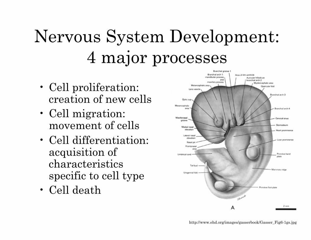
• Cell proliferation: creation of new cells
• Cell migration: movement of cells
• Cell differentiation: acquisition of characteristics specific to cell type
• Cell death
http://www.ehd.org/images/gasserbook/Gasser_Fig6-1gs.jpg
Anencephaly
• Defect of neural tube where the head of the neural tube does not close
• Happens between day 23 and 26 of pregnancy
• Results: missing major parts of the brain, skull, and scalp
• Infants born with no forebrain, and what is left of the brain tissue is exposed
Anencephaly
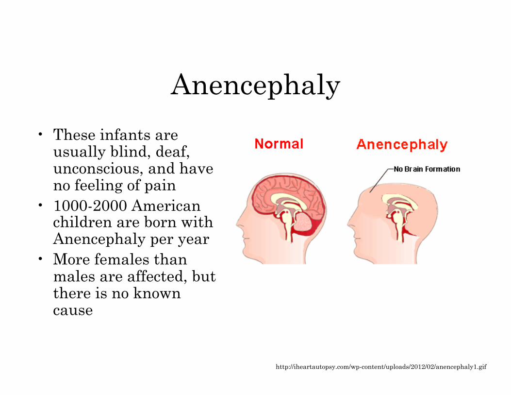
• These infants are usually blind, deaf, unconscious, and have no feeling of pain
• 1000-2000 American children are born with Anencephaly per year
• More females than males are affected, but there is no known cause
http://iheartautopsy.com/wp-content/uploads/2012/02/anencephaly1.gif
Colpocephaly
• Enlargement of occipital horns (posterior part of lateral ventricles)
• This happens when white matter fails to thicken in the cerebrum
• Infants have microcephaly (small head) and mental retardation
• Other possible conditions include motor problems, muscle spasms, and seizures
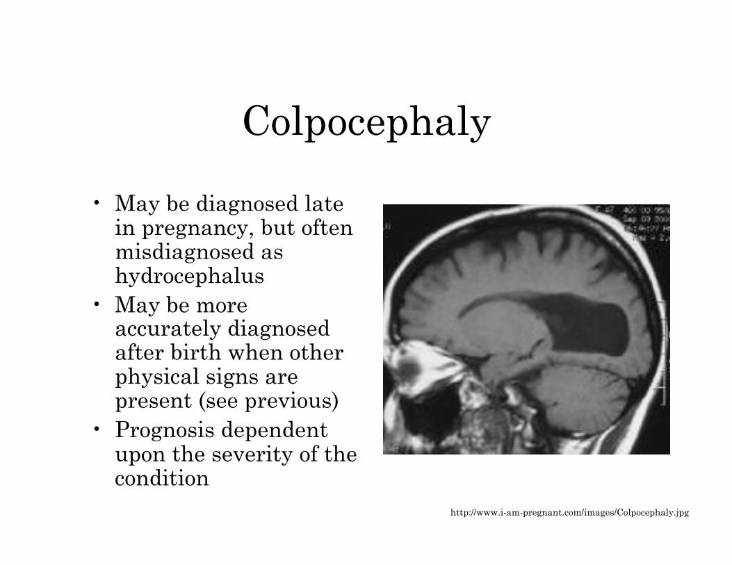
Colpocephaly
• May be diagnosed late in pregnancy, but often misdiagnosed as hydrocephalus
• May be more accurately diagnosed after birth when other physical signs are present (see previous)
• Prognosis dependent upon the severity of the condition
http://www.i-am-pregnant.com/images/Colpocephaly.jpg
Holoprosencephaly
• No development of prosencephalon (forebrain)
• Forebrain fails to divide (therefore, no bilateral hemispheres)
• Causes abnormal face and brain structure • A spectrum of defects, from serious
malformations that cause death, to facial defects (including cyclopia and median cleft lip)
• Seizures and mental retardation can occur
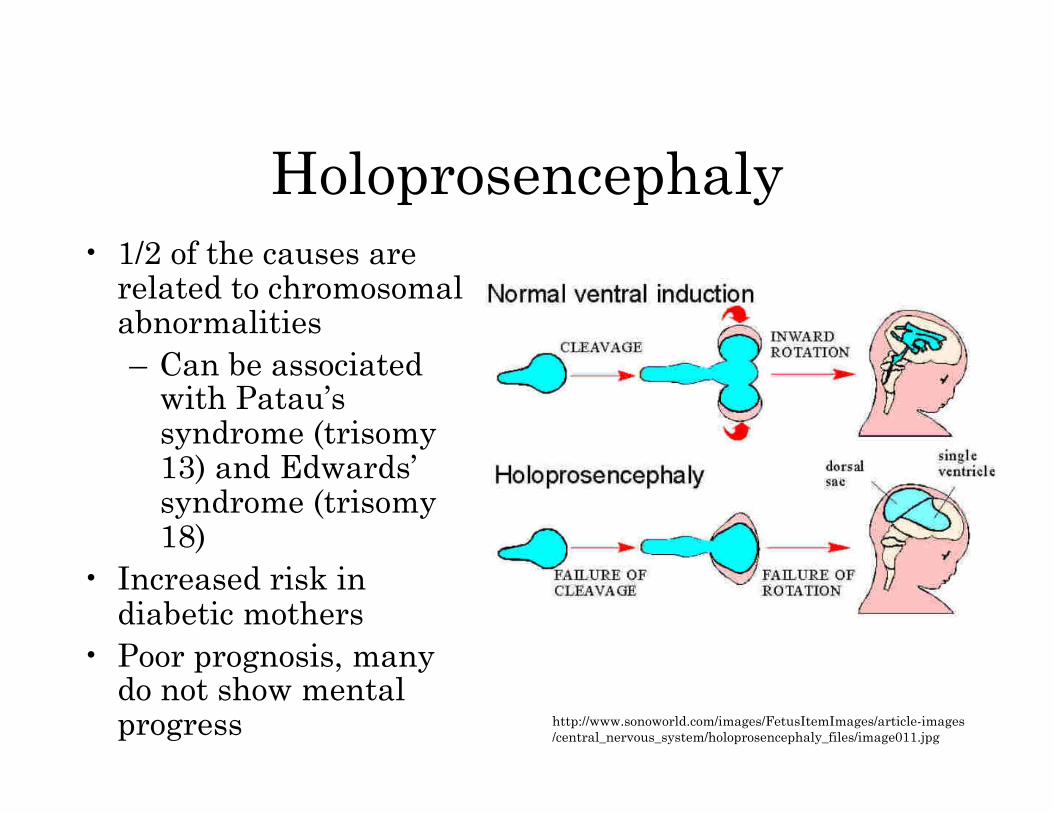
Holoprosencephaly • 1/2 of the causes are
related to chromosomal abnormalities – Can be associated
with Patau’s syndrome (trisomy 13) and Edwards’ syndrome (trisomy 18)
• Increased risk in diabetic mothers
• Poor prognosis, many do not show mental progress http://www.sonoworld.com/images/FetusItemImages/article-images
/central_nervous_system/holoprosencephaly_files/image011.jpg
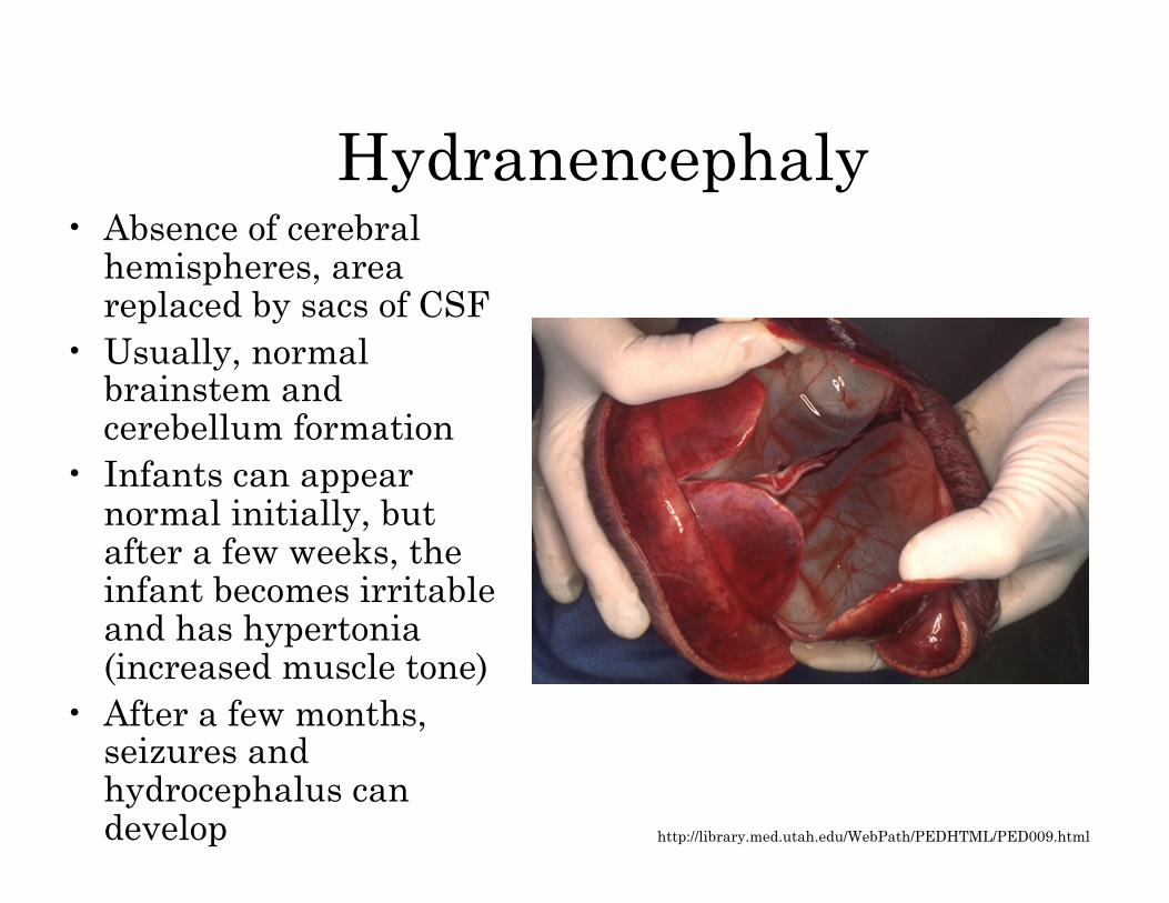
Hydranencephaly • Absence of cerebral
hemispheres, area replaced by sacs of CSF
• Usually, normal brainstem and cerebellum formation
• Infants can appear normal initially, but after a few weeks, the infant becomes irritable and has hypertonia (increased muscle tone)
• After a few months, seizures and hydrocephalus can develop http://library.med.utah.edu/WebPath/PEDHTML/PED009.html
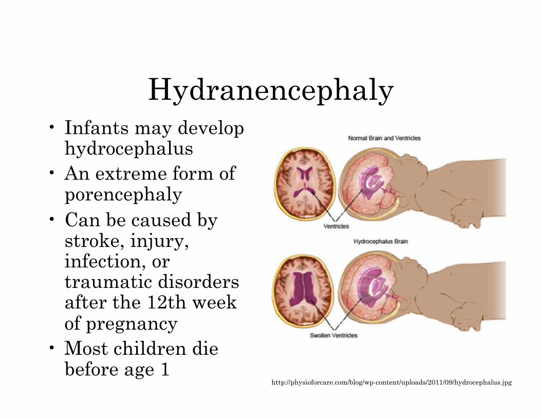
Hydranencephaly • Infants may develop
hydrocephalus • An extreme form of
porencephaly • Can be caused by
stroke, injury, infection, or traumatic disorders after the 12th week of pregnancy
• Most children die before age 1
http://physioforcare.com/blog/wp-content/uploads/2011/09/hydrocephalus.jpg
Iniencephaly
• Neural tube defect; includes extreme retroflexion of the head (head bends backwards) and defects of the spine
• Infant is usually short with a very large head
• The skin of the face is connected to the chest, and the skin of the scalp is connected to the back; generally there is no neck
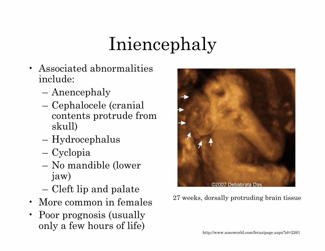
Iniencephaly • Associated abnormalities
include: – Anencephaly – Cephalocele (cranial
contents protrude from skull)
– Hydrocephalus – Cyclopia – No mandible (lower
jaw) – Cleft lip and palate
• More common in females • Poor prognosis (usually
only a few hours of life)
27 weeks, dorsally protruding brain tissue
http://www.sonoworld.com/fetus/page.aspx?id=2261
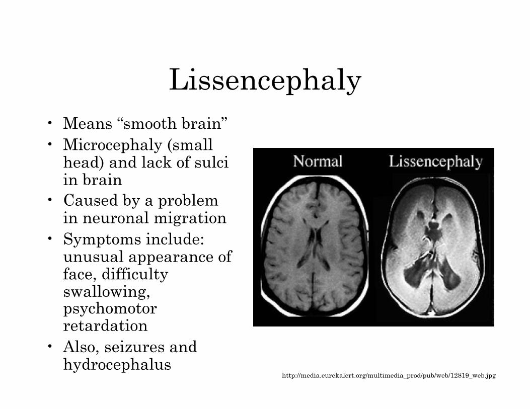
Lissencephaly • Means “smooth brain” • Microcephaly (small
head) and lack of sulci in brain
• Caused by a problem in neuronal migration
• Symptoms include: unusual appearance of face, difficulty swallowing, psychomotor retardation
• Also, seizures and hydrocephalus
http://media.eurekalert.org/multimedia_prod/pub/web/12819_web.jpg

Lissencephaly • Can be caused by viral infections of the mother of fetus during
the first trimester, insufficient blood supply to brain in early pregnancy, or a genetic disorder (X-linked, and chromosome 17-linked)
• Associated with Miller-Dieker syndrome and Walker-Warburg syndrome
• Prognosis varies; some develop almost normally, some fail to develop beyond the 5-month level, and many die before 2 years
• Most common causes of death are respiratory problems
http://webspace.webring.com/people/dl/lfurlotte99/brain.gif
Megalencephaly
• Also called macrencephaly • Abnormally large and usually
malfunctioning brain • Head can be large at birth or can become
abnormally large in early years of life • May be related to a problem in the
regulation of cell proliferation and reproduction
• Symptoms include development delay, corticospinal dysfunction, seizures, and convulsive disorders
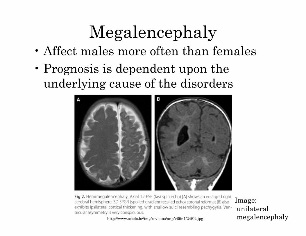
Megalencephaly • Affect males more often than females • Prognosis is dependent upon the
underlying cause of the disorders
http://www.scielo.br/img/revistas/anp/v69n1/24f02.jpg
Image: unilateral megalencephaly
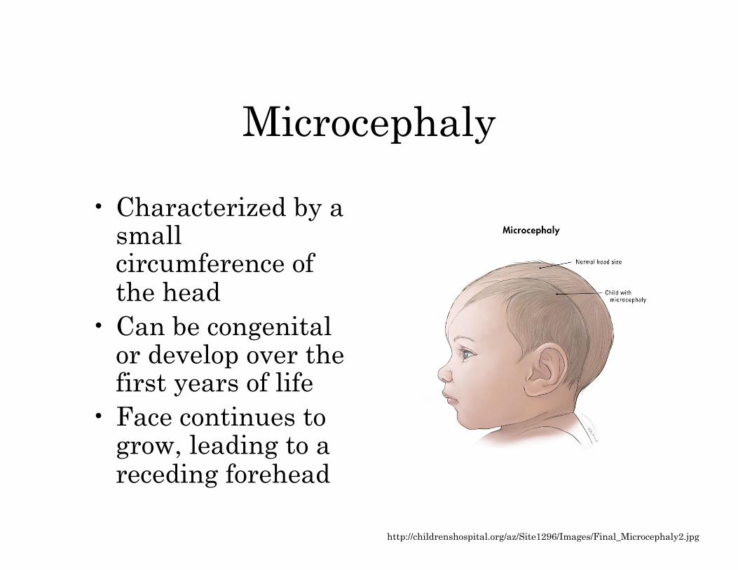
Microcephaly
• Characterized by a small circumference of the head
• Can be congenital or develop over the first years of life
• Face continues to grow, leading to a receding forehead
http://childrenshospital.org/az/Site1296/Images/Final_Microcephaly2.jpg
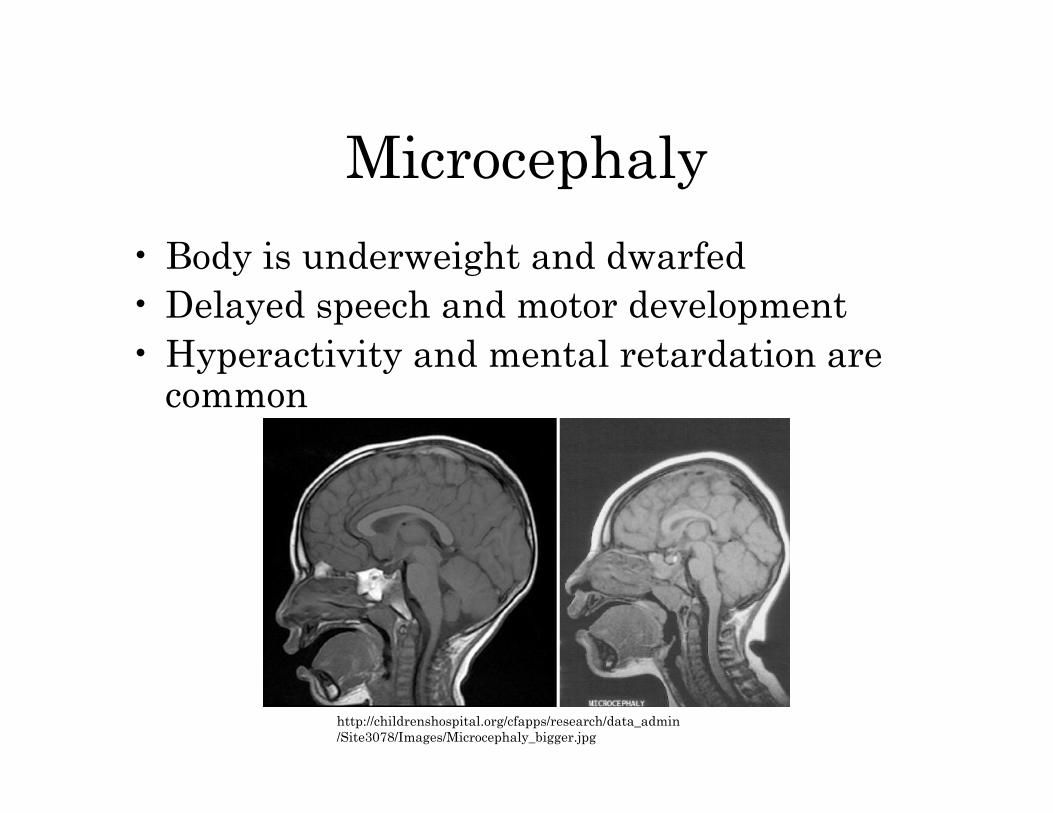
Microcephaly
• Body is underweight and dwarfed • Delayed speech and motor development • Hyperactivity and mental retardation are
common
http://childrenshospital.org/cfapps/research/data_admin/Site3078/Images/Microcephaly_bigger.jpg
Porencephaly
• Cyst or cavity in a cerebral hemisphere – Remnants of destructive lesions – Or result of abnormal development
• Can occur before or after birth • Diagnosis usually made before age 1 • Delayed growth an development, spastic
paresis (slight/incomplete paralysis), hypotonia (decreased muscle tone), seizures, and macrocephaly or microcephaly
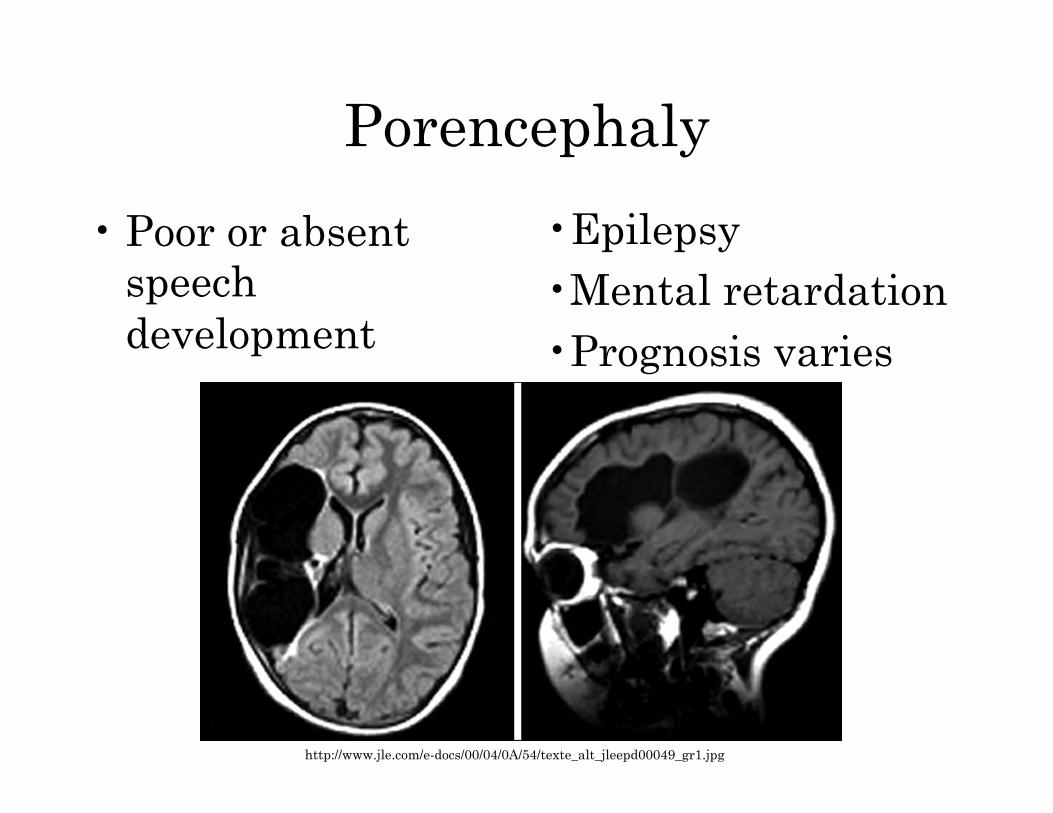
Porencephaly
• Poor or absent speech development
http://www.jle.com/e-docs/00/04/0A/54/texte_alt_jleepd00049_gr1.jpg
• Epilepsy • Mental retardation • Prognosis varies
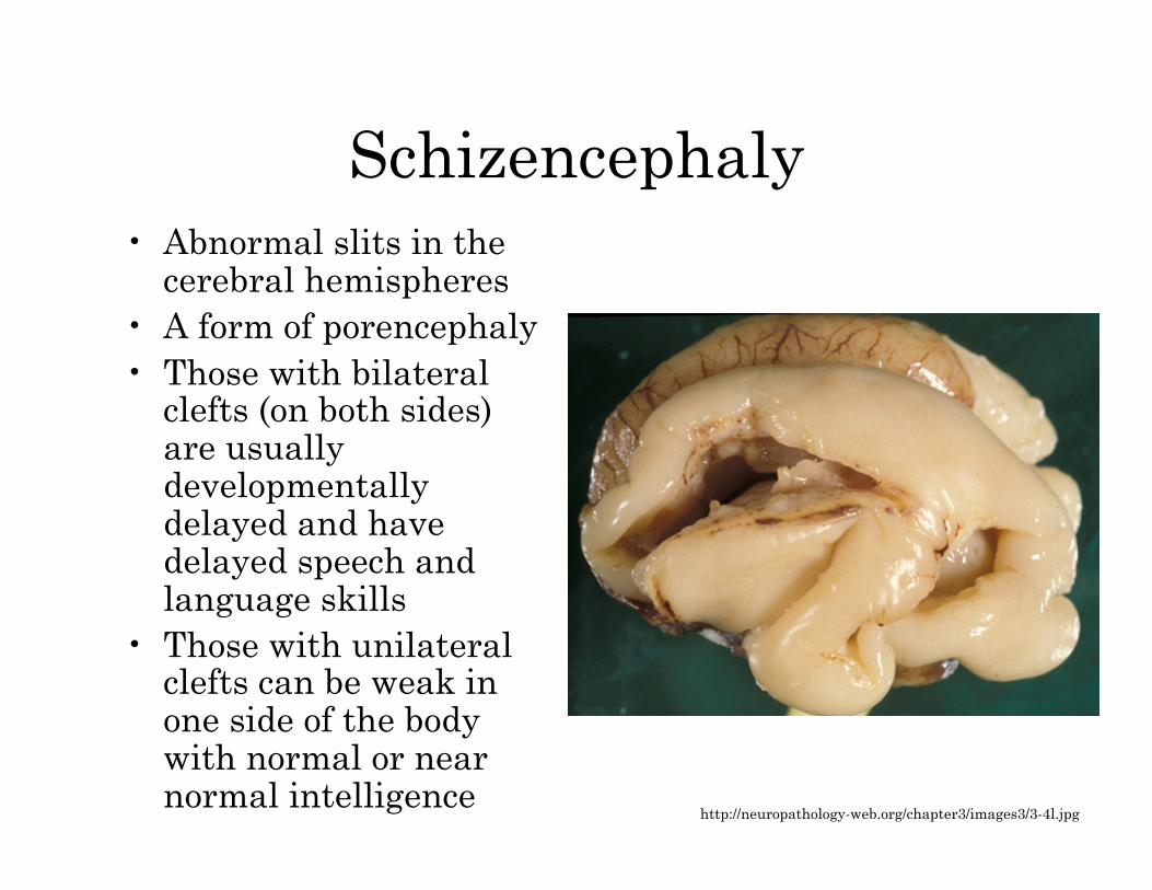
Schizencephaly • Abnormal slits in the
cerebral hemispheres • A form of porencephaly • Those with bilateral
clefts (on both sides) are usually developmentally delayed and have delayed speech and language skills
• Those with unilateral clefts can be weak in one side of the body with normal or near normal intelligence
http://neuropathology-web.org/chapter3/images3/3-4l.jpg
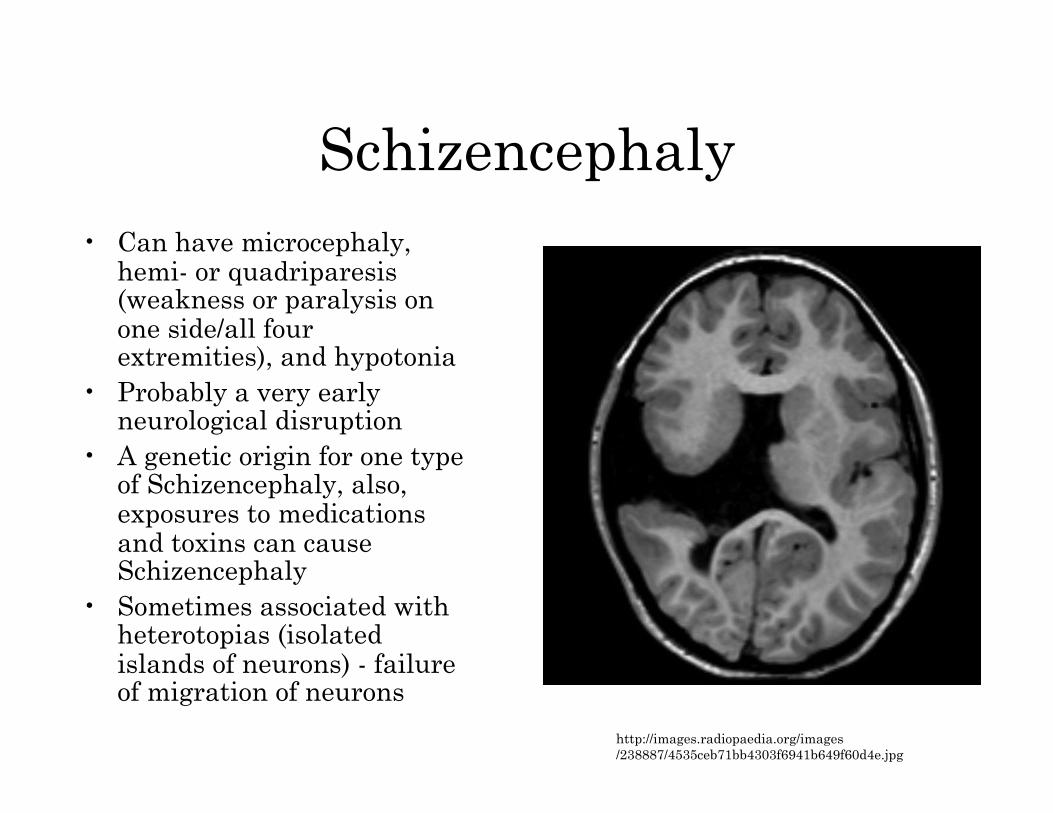
Schizencephaly • Can have microcephaly,
hemi- or quadriparesis (weakness or paralysis on one side/all four extremities), and hypotonia
• Probably a very early neurological disruption
• A genetic origin for one type of Schizencephaly, also, exposures to medications and toxins can cause Schizencephaly
• Sometimes associated with heterotopias (isolated islands of neurons) - failure of migration of neurons
http://images.radiopaedia.org/images/238887/4535ceb71bb4303f6941b649f60d4e.jpg
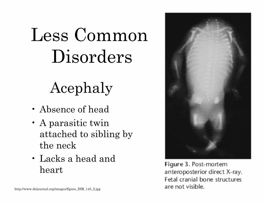
Acephaly • Absence of head • A parasitic twin
attached to sibling by the neck
• Lacks a head and heart
http://www.dirjournal.org/images/figure_DIR_145_2.jpg
Less Common Disorders
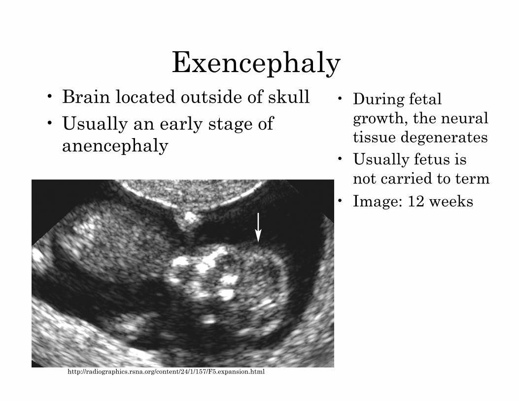
Exencephaly • Brain located outside of skull • Usually an early stage of
anencephaly
• During fetal growth, the neural tissue degenerates
• Usually fetus is not carried to term
• Image: 12 weeks
http://radiographics.rsna.org/content/24/1/157/F5.expansion.html
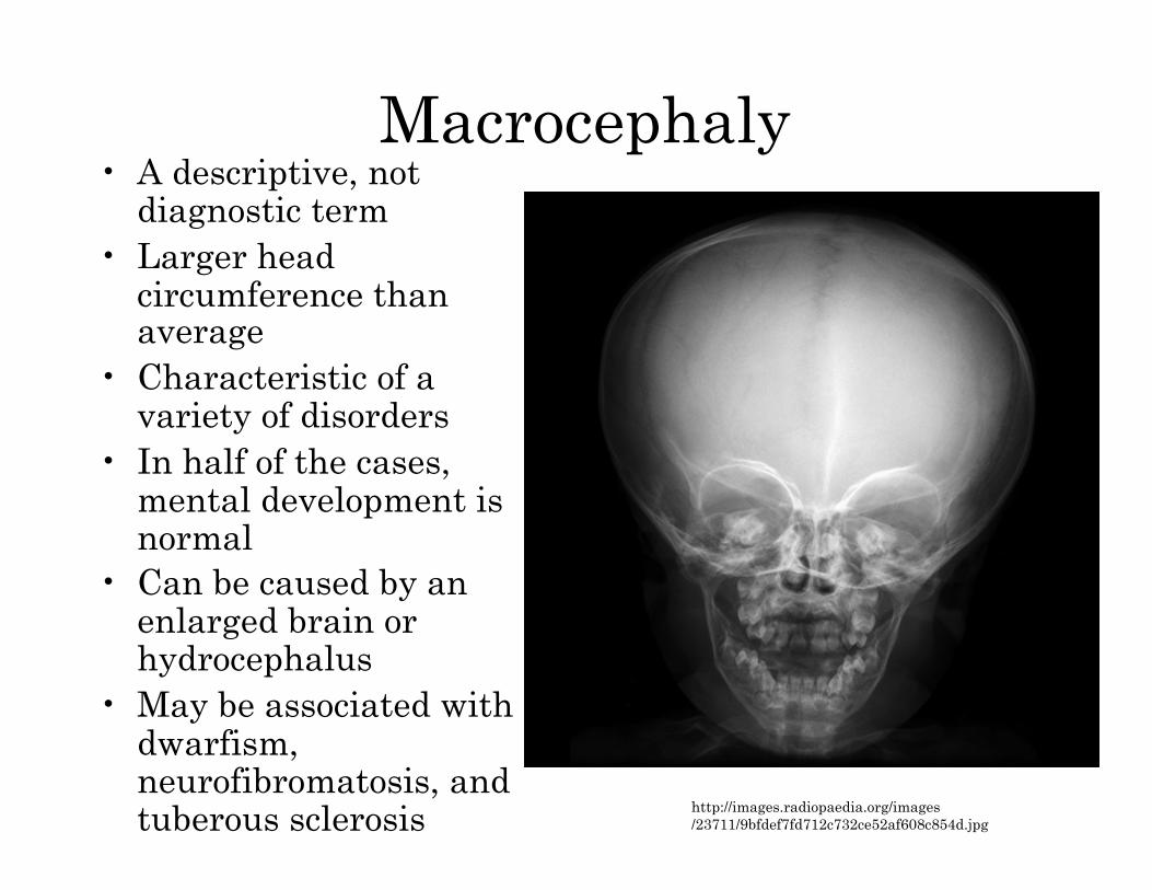
Macrocephaly • A descriptive, not
diagnostic term • Larger head
circumference than average
• Characteristic of a variety of disorders
• In half of the cases, mental development is normal
• Can be caused by an enlarged brain or hydrocephalus
• May be associated with dwarfism, neurofibromatosis, and tuberous sclerosis http://images.radiopaedia.org/images
/23711/9bfdef7fd712c732ce52af608c854d.jpg
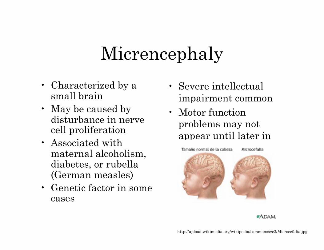
Micrencephaly
• Characterized by a small brain
• May be caused by disturbance in nerve cell proliferation
• Associated with maternal alcoholism, diabetes, or rubella (German measles)
• Genetic factor in some cases
• Severe intellectual impairment common
• Motor function problems may not appear until later in life
http://upload.wikimedia.org/wikipedia/commons/c/c3/Microcefalia.jpg
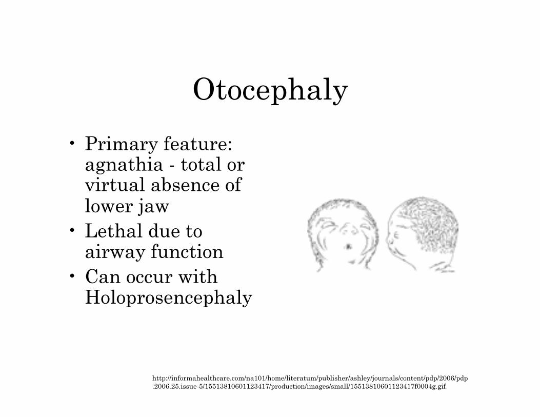
Otocephaly
• Primary feature: agnathia - total or virtual absence of lower jaw
• Lethal due to airway function
• Can occur with Holoprosencephaly
http://informahealthcare.com/na101/home/literatum/publisher/ashley/journals/content/pdp/2006/pdp.2006.25.issue-5/15513810601123417/production/images/small/15513810601123417f0004g.gif
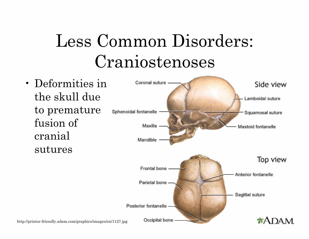
Less Common Disorders: Craniostenoses
• Deformities in the skull due to premature fusion of cranial sutures
http://printer-friendly.adam.com/graphics/images/en/1127.jpg
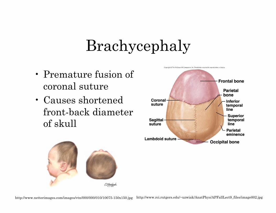
Brachycephaly
• Premature fusion of coronal suture
• Causes shortened front-back diameter of skull
http://www.rci.rutgers.edu/~uzwiak/AnatPhys/APFallLect9_files/image002.jpg http://www.netterimages.com/images/vtn/000/000/010/10075-150x150.jpg
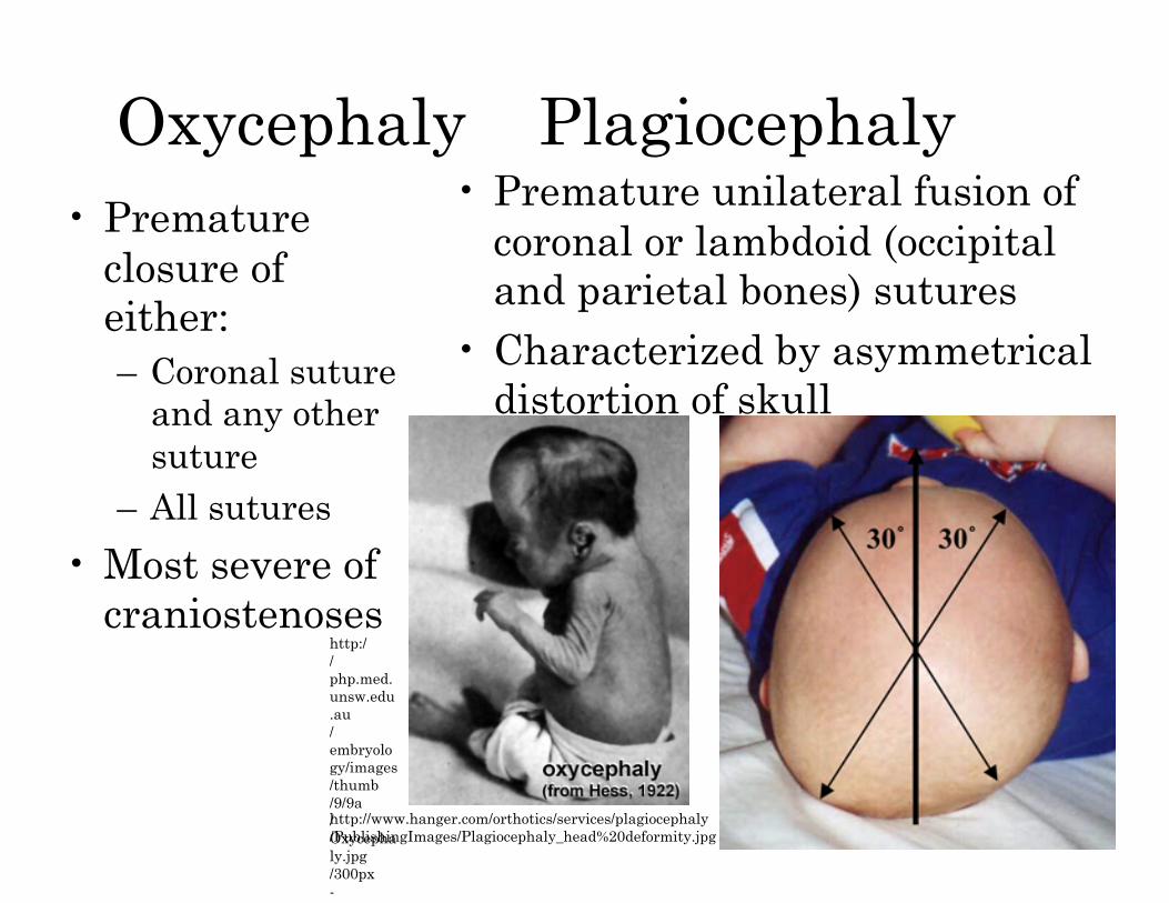
Oxycephaly Plagiocephaly • Premature
closure of either: – Coronal suture
and any other suture
– All sutures
• Most severe of craniostenoses
• Premature unilateral fusion of coronal or lambdoid (occipital and parietal bones) sutures
• Characterized by asymmetrical distortion of skull
http://php.med.unsw.edu.au/embryology/images/thumb/9/9a/Oxycephaly.jpg/300px-Oxycephaly.jpg
http://www.hanger.com/orthotics/services/plagiocephaly/PublishingImages/Plagiocephaly_head%20deformity.jpg
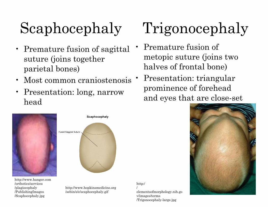
Scaphocephaly Trigonocephaly • Premature fusion of sagittal
suture (joins together parietal bones)
• Most common craniostenosis • Presentation: long, narrow
head
• Premature fusion of metopic suture (joins two halves of frontal bone)
• Presentation: triangular prominence of forehead and eyes that are close-set
http://www.hanger.com/orthotics/services/plagiocephaly/PublishingImages/Scaphocephaly.jpg
http://www.hopkinsmedicine.org/sebin/s/e/scaphocephaly.gif
http://elementsofmorphology.nih.gov/images/terms/Trigonocephaly-large.jpg