Embed Size (px)
Citation preview
Cerebellar Arteriovenous Malformations:Anatomic Subtypes, Surgical Results, and IncreasedPredictive Accuracy of the SupplementaryGrading System
BACKGROUND: Anatomic diversity among cerebellar arteriovenous malformations(AVMs) calls for a classification that is intuitive and surgically informative. Selection toolslike the Spetzler-Martin grading system are designed to work best with cerebral AVMsbut have shortcomings with cerebellar AVMs.OBJECTIVE: To define subtypes of cerebellar AVMs that clarify anatomy and surgicalmanagement, to determine results according to subtypes, and to compare predictiveaccuracies of the Spetzler-Martin and supplementary systems.METHODS: From a consecutive surgical series of 500 patients, 60 had cerebellar AVMs,39 had brainstem AVMs and were excluded, and 401 had cerebral AVMs.RESULTS: Cerebellar AVM subtypes were as follows: 18 vermian, 13 suboccipital,12 tentorial, 12 petrosal, and 5 tonsillar. Patients with tonsillar and tentorial AVMs faredbest. Cerebellar AVMs presented with hemorrhage more than cerebral AVMs (P, .001).Cerebellar AVMs were more likely to drain deep (P = .04) and less likely to be eloquent(P, .001). The predictive accuracy of the supplementary grade was better than that ofthe Spetzler-Martin grade with cerebellar AVMs (areas under the receiver-operatingcharacteristic curve, 0.74 and 0.59, respectively). The predictive accuracy of the sup-plementary system was consistent for cerebral and cerebellar AVMs, whereas that of theSpetzler-Martin system was greater with cerebral AVMs.CONCLUSION: Patients with cerebellar AVMs present with hemorrhage more oftenthan patients with cerebral AVMs, justifying an aggressive treatment posture. Thesupplementary system is better than the Spetzler-Martin system at predicting outcomesafter cerebellar AVM resection. Key components of the Spetzler-Martin system such asvenous drainage and eloquence are distorted by cerebellar anatomy in ways thatcomponents of the supplementary system are not.
KEYWORDS: Arteriovenous malformation, Cerebellum, Microsurgical resection, Spetzler-Martin grading scale,
Supplementary grading scale
Neurosurgery 71:1111–1124, 2012 DOI: 10.1227/NEU.0b013e318271c081 www.neurosurgery-online.com
Cerebellar arteriovenous malformations(AVMs) are a small group of AVMs thatdiffer from cerebral AVMs in their hemor-
rhagic behavior, clinical presentation, and surgi-cal outcomes. They make up, 15% of all brainAVMs,1 and this relatively low incidence has led
authors to combine them with brainstem AVMsin reports on infratentorial or posterior fossaAVMs.2-11 This contamination confuses theclinical description of cerebellar AVMs. In thisreport, we examined the anatomy, surgical strat-egies, and operative results with only cerebellarAVMs.Few authors have tried to categorize cerebellar
AVMsintodistinct subtypes.Yasargil12 categorized58 cerebellar AVMs into 7 types: hemispheric(superior and inferior, divided by the horizontalfissure), vermian (superior and inferior, divided
Ana Rodrıguez-Hernandez,MD*
Helen Kim, MPH, PhDद
Tony Pourmohamad, MA‡¶
William L. Young, MD*‡¶kMichael T. Lawton, MD*¶
for the University of California,
San Francisco Arteriovenous
Malformation Study Project
*Department of Neurological Surgery; ‡De-
partments of Anesthesia and Perioperative
Care; §Department of Epidemiology and
Biostatistics; ¶Center for Cerebrovascular
Research; kDepartment of Neurology, Uni-
versity of California, San Francisco, San
Francisco, California
Correspondence:
Michael T. Lawton, MD,
Department of Neurological Surgery,
University of California at San Francisco,
505 Parnassus Ave, M780,
San Francisco, CA 94143-0112.
E-mail: [email protected]
Received, March 13, 2012.
Accepted, August 16, 2012.
Published Online, September 14, 2012.
Copyright ª 2012 by the
Congress of Neurological Surgeons
SANS LifeLong Learning and
NEUROSURGERY offer CME for subscribers
that complete questions about featured
articles. Questions are located on the SANS
website (http://sans.cns.org/). Please read
the featured article and then log into SANS
for this educational offering.
ABBREVIATIONS: AICA, anterior inferior cerebellar
artery; AVM, arteriovenous malformation; mRS,
modified Rankin Scale; PICA, posterior inferior
cerebellar artery; ROC, receiver-operating charac-
teristic; SCA, superior cerebellar artery
RESEARCH—HUMAN—CLINICAL STUDIESTOPIC RESEARCH—HUMAN—CLINICAL STUDIES
NEUROSURGERY VOLUME 71 | NUMBER 6 | DECEMBER 2012 | 1111
Copyright © Congress of Neurological Surgeons. Unauthorized reproduction of this article is prohibited.
Copyright © Congress of Neurological Surgeons. Unauthorized reproduction of this article is prohibited.
by the horizontal fissure), cerebellopontine, giant, and fistula.Rhoton13 described 3 cerebellar surfaces (suboccipital, tentorial,and petrosal) in anatomic studies but did not apply this to AVMsspecifically. We adapted these efforts to define 5 distinct subtypesof cerebellar AVMs: suboccipital, vermian, tonsillar, tentorial, andpetrosal. We found that these subtypes offer an intuitive appre-ciation of their anatomy and surgical management and are usefulin describing surgical results. A focused analysis of outcomes aftercerebellar AVM resection enables assessment of our patient selec-tion. Some neurosurgeons have expressed dissatisfaction with theSpetzler-Martin grading scale as a tool for surgical selection withcerebellar AVMs because deep nuclei are the only eloquent struc-tures in the cerebellum and galenic venous drainage is not a goodindicator of cerebellar AVM depth.14 We compared the predictiveaccuracy of this grading scale with that of the supplementarygrading scale, which combines patient age, bleeding, and com-pactness with the assignment of points in a manner analogous tothe Spetzler-Martin grading scale. We hypothesized that key com-ponents of the Spetzler-Martin scale such as venous drainage andeloquence are distorted by cerebellar anatomy in ways that thecomponents of the supplementary scale are not, resulting in greaterpredictive accuracy with the supplementary scale.
PATIENTS AND METHODS
The study was approved by the University of California, San FranciscoCommittee on Human Research and was conducted in compliance withHIPAA (Health Insurance Portability and Accountability Act) regulations.
Five Types of Cerebellar AVMs
Suboccipital
AVMs are based on the posterior cerebellar surface facing the occipitalbone, located below and between the transverse and sigmoid sinuses. Thehemispheric portion of the suboccipital surface is made up of the superiorsemilunar, inferior semilunar, and biventral lobules. The suboccipitalsurface is divided into superior and inferior parts by its major fissure, thesuboccipital fissure. Minor fissures on the suboccipital surface include thepetrosal or horizontal fissure (between the superior and inferior semilunarlobules), the prebiventral fissure (between the inferior semilunar andbiventral lobules), and the tonsillobiventral fissure. AVMs based on thevermian portion of the suboccipital surface are categorized as vermian.
Tentorial
AVMs are based on the tentorial surface. The hemispheric part ofthe tentorial surface comprises the quadrangular, simple, and superiorsemilunar lobules. The tentorial surface is divided into anterior andposterior parts by its major fissure, the tentorial or primary fissure. Thisfissure separates the quadrangular and simple lobules on the hemisphereand the culmen and declive on the vermis. The postclival fissure separatesthe simple and superior semilunar lobules. AVMs based on the vermianportion of the tentorial surface are categorized as vermian.
Petrosal
AVMs are based on the petrosal surface, the anterior cerebellum that facesthe posterior petrous bone. The cerebellopontine angle is the V-shaped
cerebellopontine fissure formed where the hemispheric lobules wraparound the pons and middle cerebellar peduncle. The petrosal surface isdivided into superior and inferior parts by the petrosal or horizontalfissure, which extends onto the suboccipital surface between the superiorand inferior semilunar lobules. The superior and inferior limbs of thecerebellopontine fissure meet laterally at the apex of the cerebellopontineangle, at the anterior end of the petrosal fissure. The petrosal surface isformed by the anterior surfaces of the quadrangular, simple, semilunar,and biventral lobules and the flocculi.
Vermian
AVMs lie in the midline and may be on the tentorial surface or on thesuboccipital surface. The inferior vermis lies in a deep vertical depressionin the suboccipital surface called the posterior cerebellar incisura, whichalso contains the falx cerebelli. The inferior vermis forms the posteriorcortical surface within this incisura. In contrast, the superior vermis is thehighest point on the cerebellum, occupying the space under the straightsinus where the tentorial leaflets intersect with the falx cerebri. The supe-rior vermis slopes downward from its apex anteriorly to the posteriorcerebellar incisura. The tentorial part of the vermian surface includes (fromanterior to posterior) the culmen, declive, and folium. The suboccipitalpart of the vermian surface includes (from superior to inferior) the tuber,pyramid, uvula, and nodule. The nodule is hidden deep to the uvula.
Tonsillar
AVMs lie in the tonsils, which are ovoid structures on the inferomedialaspect of the cerebellar hemispheres that attach to cerebellum super-olaterally through the tonsillar peduncles. The other tonsillar surfaces arefree, with the inferior pole and posterior surfaces in the cisterna magna.The anterior tonsil faces the posterior medulla and is separated by thecerebellomedullary fissure. The medial tonsils face each other and areseparated by the vallecula, a cleft that leads into the fourth ventricle. Theventral aspect of the superior tonsil faces the lower half of the roof of thefourth ventricle, which is formed by the tela choroidea, inferior medullaryvelum, and nodule. The lateral tonsil is separated from the hemisphere bythe tonsillobiventral fissure.Computed tomographic scans, magnetic resonance imaging, digital sub-
traction angiograms, operative reports, intraoperative photographs, andsurgeon notes were systematically reviewed to classify cerebellar AVMsaccording to the 5 types.
Patients
During a 13-year period, a total of 500 patients with brain AVMs weretreated surgically at the University of California, San Francisco MedicalCenter by 1 neurosurgeon (M.T.L.) and recorded in the prospectiveregistry of theUniversity of California, San Francisco Brain ArteriovenousMalformation Study Project. The registry was searched for patients withcerebellar AVMs, and 60 patients were identified for inclusion in this study.Surgical patients with AVMs in the midbrain, pons, and medulla were notincluded (39 patients). For comparison, the remainder of surgical patientswith cerebral AVMs were analyzed (401 patients).
Outcome Evaluation
Neurological outcome was assessed with the modified Rankin Scale(mRS). Neurological assessments were performed by a nurse-clinicianunder the supervision of a neurologist preoperatively, postoperatively, andup to 2 years postoperatively. Follow-up information was obtained during
RODRIGUEZ-HERNANDEZ ET AL
1112 | VOLUME 71 | NUMBER 6 | DECEMBER 2012 www.neurosurgery-online.com
Copyright © Congress of Neurological Surgeons. Unauthorized reproduction of this article is prohibited.
Copyright © Congress of Neurological Surgeons. Unauthorized reproduction of this article is prohibited.

routine clinic visits or telephone interviews. Good outcomes were definedas a final mRS score of 0 to 2, and poor outcomes were defined as a finalmRS. 2. Improvement was defined as a decrease in mRS score (changein the mRS of# 0), and deterioration was defined as an increase in mRSscore (change in the mRS of . 0).
Statistical Analysis
Characteristics of cerebellar vs cerebral AVMs were evaluated by use ofdescriptive statistics, including t tests for continuous variables and x2 testsfor categorical variables. Logistic regression analysis was performed usingchange in mRS score (outcome) vs log time in combination with either theSpetzler-Martin score or the supplementary score (predictors). Predictiveaccuracy of the Spetzler-Martin and supplementary grading scales wasquantified by measuring the area under receiver-operating characteristic(ROC) curves based on our logistic regression models. An area under theROC curve of 1.0 indicates perfect discrimination; an area of 0.5 indicatesno discrimination; and an area $ 0.70 was considered clinically useful. Avalue of P, .05 was considered significant for all tests.
RESULTS
Cerebellar AVM Patients
There were 32 women (53%) and 28 men (47%) with a meanage of 41 years (range, 6-84 years). Forty-seven patients (78%)presented with hemorrhage; 7 patients (12%) presented withheadaches or neurological deficits; and 6 patients (10%) hadincidental, asymptomatic AVMs. None presented with seizures.Hemorrhagic presentation was more frequent in women (90%)than in men (69%).
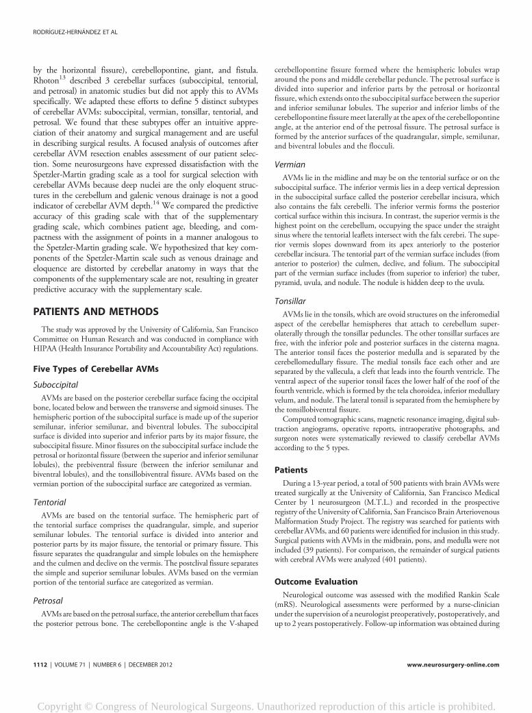
The mean cerebellar AVM size was 2.5 cm. The medianSpetzler-Martin gradewas II, and themedian supplementary gradewas III (Table 1). Small AVMs (, 3 cm) were more likely topresent with hemorrhage than larger AVMs (. 3 cm; 91% vs65%; P, .05). In decreasing order of frequency, cerebellar AVMtypes were as follows: 18 vermian (30%), 13 suboccipital (22%),12 tentorial (20%), 12 petrosal (20%), and 5 tonsillar (8%;Table 2). Vermian, tentorial, and tonsillar AVMs had the highestfrequency of hemorrhagic presentation (all . 93%).
One patient with hereditary hemorrhagic telangiectasia had acoexisting supratentorial AVM. Twelve patients (20%) hadintracranial aneurysms, including 10 feeding artery aneurysmsand 2 intranidal aneurysms. All 12 aneurysms were diagnosed inpatients presenting with hemorrhage. Nine of these patients pre-sented with intracerebellar hemorrhage, and the rupture wasattributed to the AVM. Three of these patients presented withsubarachnoid hemorrhage without intracerebellar hemorrhage,and the rupture was attributed to the aneurysm (unrupturedAVM). Therefore, of the 47 patients presenting with posteriorfossa bleeding, 44 patients had bleeding from AVM ruptures and3 from aneurysm ruptures.
Surgical Management
Patients with suspected cerebellar AVMs were evaluated withpreoperative angiography. However, 6 patients (10%) with largehematomas and brainstem compression were operated on
emergently. Four of these patients underwent hematoma evac-uation at outside hospitals before transferring to our institution,deferring AVM resection for later. The other 2 patients presented toour institution and underwent emergency hematoma evacuationand AVM resection without angiography.Preoperative embolization was performed in 33 patients (55%;
Table 3) with a variety of agents, including polyvinyl alcoholparticles, n-butyl cyanoacrylate glue, and Onyx. Embolic compli-cations occurred in 1 patient: an anterior inferior cerebellar artery(AICA) perforation resulting in subarachnoid hemorrhage anda pontine infarct with new facial numbness and contralateralhemisensory deficit.Seven different approaches were used for cerebellar AVMs, con-
sisting of mainly suboccipital craniotomy, far lateral craniotomy,retrosigmoid craniotomy, or combination craniotomies (Table 2).Vermian AVMs were typically resected through torcular craniotomies
TABLE 1. Summary of Cerebellar Arteriovenous Malformation
Gradinga
Characteristics
Cerebellar
(n = 60),
n (%)
Cerebral
(n = 401),
n (%)
Overall
(n = 461),
n (%) P
AVM size, cm 1.00
0-3 42 (70) 276 (69) 318 (69)
3-6 17 (28) 115 (29) 132 (29)
. 6 1 (2) 10 (2) 11 (2)
Venous drainage .04
Superficial 26 (43) 235 (59) 261 (57)
Deep 34 (57) 166 (41) 200 (43)
Eloquence , .001
No 42 (70) 158 (39) 200 (43)
Yes 18 (30) 243 (61) 261 (57)
Spetzler-Martin grade .10
I 19 (32) 68 (17) 87 (19)
II 18 (30) 146 (36) 164 (36)
III 15 (25) 136 (34) 151 (33)
IV 8 (13) 46 (11) 54 (12)
V 0 (0) 5 (1) 5 (1)
Age at treatment, y .18
, 20 8 (14) 65 (16) 73 (16)
20-40 17 (28) 153 (38) 170 (37)
. 40 35 (58) 183 (46) 218 (47)
Compactness .32
Compact 48 (80) 341 (85) 389 (84)
Diffuse 12 (20) 60 (15) 72 (16)
Hemorrhagic
presentation
, .001
Ruptured 44 (73) 196 (49) 240 (52)
Unruptured 16 (27) 205 (51) 221 (48)
Supplementary grade .30
I 6 (10) 40 (10) 46 (10)
II 14 (23) 88 (22) 102 (22)
III 25 (41) 141 (35) 166 (36)
IV 13 (22) 109 (27) 122 (26)
V 2 (3) 23 (6) 25 (5)
aAVM, arteriovenous malformation.
CEREBELLAR ARTERIOVENOUS MALFORMATIONS
NEUROSURGERY VOLUME 71 | NUMBER 6 | DECEMBER 2012 | 1113
Copyright © Congress of Neurological Surgeons. Unauthorized reproduction of this article is prohibited.
Copyright © Congress of Neurological Surgeons. Unauthorized reproduction of this article is prohibited.
(89%), improving access to the superior vermian surface relativeto the standard suboccipital craniotomy by removing the ledge ofoverhanging bone and lifting the transverse sinuses with tackingsutures on the dural flap. Tentorial AVMs were also resectedthrough torcular craniotomies (58%) because of this improvedaccess to the tentorial cerebellar surface. Petrosal AVMs wereresected through extended retrosigmoid craniotomies (67%),with skeletonization of the sigmoid sinus from transverse-sigmoidjunction to the jugular bulb for wider access to the petrosalsurface and cerebellopontine angle. Tonsillar AVMs were resectedthrough standard midline suboccipital craniotomies (80%).Suboccipital AVMs had the widest variety of surgical approaches,the most common being a lateral suboccipital craniotomy (31%).Combination approaches were used for larger AVMs at the marginsof the suboccipital surface, with the far lateral approach adding to theinferolateral exposure, the extended retrosigmoid approach adding tothe superolateral exposure, and the torcular approach adding to thesuperior exposure.Complete AVM resection was achieved in all 60 patients and
confirmed angiographically (surgical obliteration rate, 100%). AllAVMs were resected in a single stage. Complications included apostoperative intracerebellar hematoma that required evacuation in1patient and a superficial wound infection that required debridementin another patient. All 3 ruptured aneurysms were treated: 2 superiorcerebellar artery (SCA) aneurysms with preoperative endovascularcoiling and 1 AICA aneurysm with simultaneous clipping.
Patient Outcomes
Three patients died in the perioperative period (surgicalmortality, 5%). Two of these patients had ruptured AVMs,
TABLE 2. Summary of Anatomic Subtypes of Cerebellar Arteriovenous Malformations, Approaches, and Outcomesa
Total Suboccipital Vermian Tonsillar Tentorial Petrosal
n % n % n % N % n % n %
Patients 60 13 22 18 30 5 8 12 20 12 20
Craniotomy
Suboccipital, midline 8 13 1 8 2 11 4 80 1 8 0 0
Suboccipital, lateral 6 10 4 31 0 0 0 0 2 17 0 0
Torcular 25 42 2 15 16 89 0 0 7 58 0 0
Far lateral 2 3 0 0 0 0 1 20 0 0 1 8
Extended retrosigmoid 11 18 1 8 0 0 0 0 2 17 8 67
Far lateral–retrosigmoid 6 10 3 23 0 0 0 0 0 0 3 25
Far lateral–retrosigmoid–torcular 2 3 2 15 0 0 0 0 0 0 0 0
AVM grades
Spetzler-Martin, median/mean 2.3/II 2.6/III 2.3/II 1.8/I 1.6/I 2.7/III
Supplementary, median/mean 2.9/III 2.8/II 2.8/III 2.0/II 2.7/III 3.5/III.5
Combined grade 5.1/V 5.5/V 5.2/V 4.0/IV 4.3/IV 6.0/V.5
Outcome (change in mRS)
Improved/unchanged 44 77 10 77 13 72 4 100 11 92 6 60
Worse/dead 13 23 3 23 5 28 0 0 1 8 4 40
Lost 3 0 0 1 0 2
aAVM, arteriovenous malformation; mRS, modified Rankin Scale.
TABLE 3. Summary of Cerebellar Arteriovenous Malformation
Management and Patient Outcomesa
Characteristics
Cerebellar
(n = 60),
n (%)
Cerebral
(n = 401),
n (%)
Overall
(n = 461),
n (%) P
Embolization before AVM resection .68
Yes 33 (55) 209 (52) 242 (52)
No 27 (45) 192 (48) 219 (48)
Staged AVM resection .03
No 60 (100) 366 (91) 426 (92)
Yes 0 (0) 35 (9) 35 (8)
Presurgery mRS , .001
0 11(18) 112 (28) 123 (27)
1 7 (12) 97 (24) 104 (23)
2 1 (2) 53 (13) 54 (12)
3 16 (27) 63 (16) 79 (17)
4 10 (17) 39 (10) 49 (11)
5 15 (25) 37 (9) 52 (11)
Postsurgery mRS .01
0 11 (19) 124 (32) 135 (31)
1 14 (25) 123 (32) 137 (31)
2 17 (30) 62 (16) 79 (18)
3 7 (12) 28 (7) 35 (8)
4 2 (4) 24 (6) 26 (6)
5 0 (0) 1 (,1) 1 (, 1)
6 6 (11) 22 (6) 28 (6)
mRS change .76
Improved or unchanged 44 (77) 292 (76) 336 (76)
Worsened or dead 13 (23) 92 (24) 105 (24)
Lost 3 (5) 17 (4) 20 (4)
aAVM, arteriovenous malformation; mRS, modified Rankin Scale.
RODRIGUEZ-HERNANDEZ ET AL
1114 | VOLUME 71 | NUMBER 6 | DECEMBER 2012 www.neurosurgery-online.com
Copyright © Congress of Neurological Surgeons. Unauthorized reproduction of this article is prohibited.
Copyright © Congress of Neurological Surgeons. Unauthorized reproduction of this article is prohibited.
presented in coma, and failed to improve after aggressive man-agement. One of these patients had a Spetzler-Martin grade IVAVM, difficult intraoperative bleeding, and significant post-operative deficits, and support was withdrawn. Twelve patientshad neurological deficits perioperatively that resolved completelyat late follow-up (transient neurological morbidity, 20%).
Follow-up evaluations (mean duration, 1.1 years) were per-formed in all but 3 patients. Of the remaining patients, 3 patientsdied. One death was caused by a delayed cerebellar abscess in apatientwho had donewell with surgery (unchanged at early follow-up). Two deaths occurred 1 and 5 years after surgery in patientswho were worse postoperatively but living dependently. Overall,42 patients (74%) had good outcomes (mRS score, 0-2), 9 patients(16%) had poor outcomes (mRS score, 3-4), and 6 patients (11%)died (Table 3). Relative to preoperative neurological condition,44 patients (77%) were improved or unchanged after surgery, and13 patients (23%) were worsened or dead. Of the 7 patients whowere worsened, 6 patients had good outcomes (2 patients, mRSscore 1; 4 patients, mRS score 2).
Patients with tonsillar and tentorial AVMs fared best(improved/unchanged in 100% and 92%, respectively). Thesepatients also had the lowest Spetzler-Martin and supplementarygrades (Table 2). Patients with petrosal and vermian AVMs faredworst (improved/unchanged in 60% and 72%, respectively).Patients with petrosal AVMs had the highest Spetzler-Martin andsupplementary grades (Table 2).
Comparison of Cerebellar and Cerebral AVMs
Differences in patient age and sex were not significant withcerebellar and cerebral AVMs (P = .54). However, cerebellarAVMs are more likely than cerebral AVMs to present withhemorrhage (78% vs 53%; P, .001). Increased hemorrhagicpresentation accounted for worse presurgical mRS scores inpatients with cerebellar AVMs (P, .001; Table 3). Hemorrhagicpresentation with cerebellar AVMs is not attributable to dif-ferences in AVM size as measured by mean diameter or by sizescores on the Spetzler-Martin scale (Table 1). However, cerebellarAVMs are more likely than cerebral AVMs to have deep venousdrainage (57% vs 41%; P = .04) and less likely to have eloquence(39% vs 70%; P, .001). Overall differences in Spetzler-Martingrades were not significant (P = .10).
Cerebellar AVMs were no more compact or diffuse than cerebralAVMs(P = .32). Overall differences in supplementary AVM gradeswere not significant (P = .30). Deep perforator supply was twice asfrequent with cerebral AVMs than with cerebellar AVMs (21% vs10%; P = .05). Cerebral AVMs were more likely to requiremultiple stages than cerebellar AVMs (9% vs 0%; P = .03).Patients with cerebral AVMs tended to have more favorableoutcomes than those with cerebellar AVMs (P = .01; Table 3).
Predictive Accuracy of Grading Scales WithCerebellar AVMs
The predictive accuracy of the supplementary AVM grade wasbetter than that of the Spetzler-Martin grade for both cerebellar
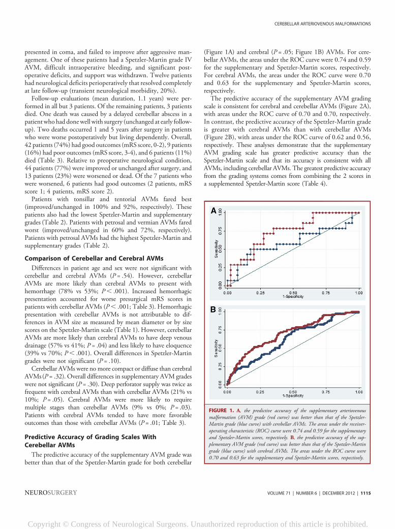
(Figure 1A) and cerebral (P = .05; Figure 1B) AVMs. For cere-bellar AVMs, the areas under the ROC curve were 0.74 and 0.59for the supplementary and Spetzler-Martin scores, respectively.For cerebral AVMs, the areas under the ROC curve were 0.70and 0.63 for the supplementary and Spetzler-Martin scores,respectively.The predictive accuracy of the supplementary AVM grading
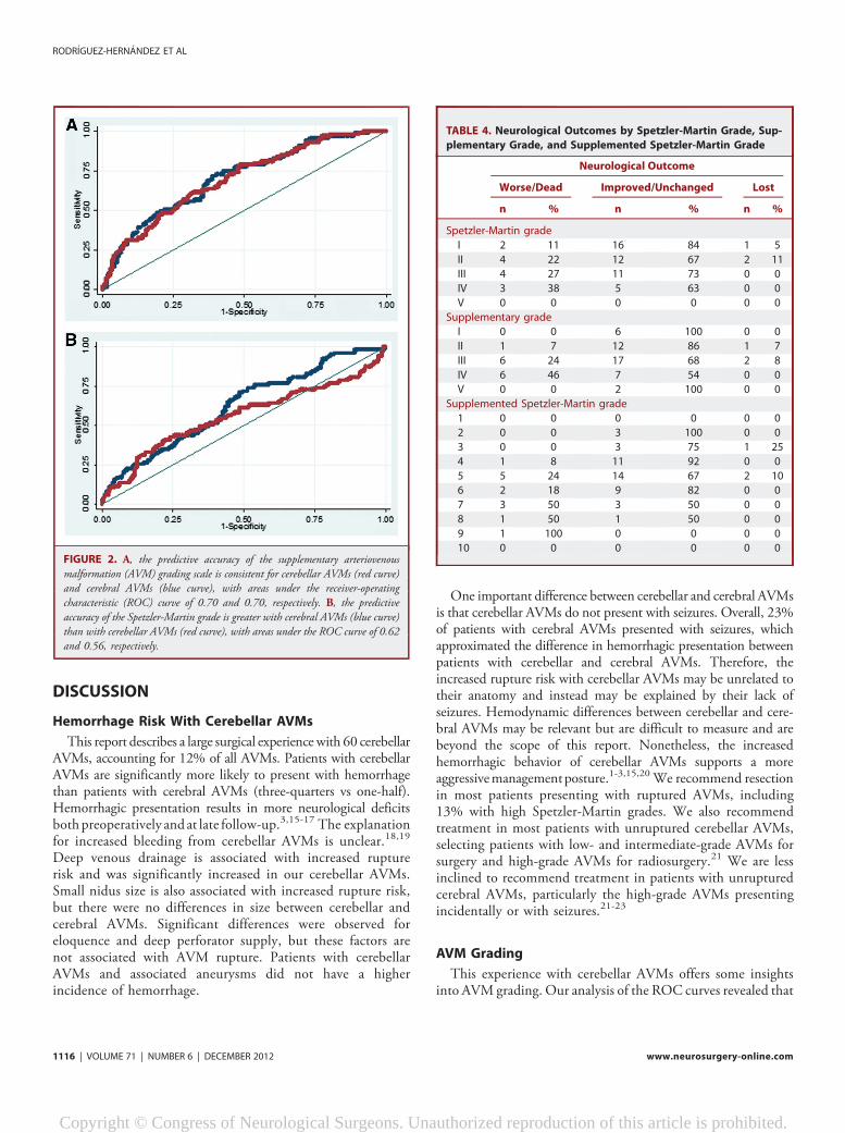
scale is consistent for cerebral and cerebellar AVMs (Figure 2A),with areas under the ROC curve of 0.70 and 0.70, respectively.In contrast, the predictive accuracy of the Spetzler-Martin gradeis greater with cerebral AVMs than with cerebellar AVMs(Figure 2B), with areas under the ROC curve of 0.62 and 0.56,respectively. These analyses demonstrate that the supplementaryAVM grading scale has greater predictive accuracy than theSpetzler-Martin scale and that its accuracy is consistent with allAVMs, including cerebellar AVMs. The greatest predictive accuracyfrom the grading systems comes from combining the 2 scores ina supplemented Spetzler-Martin score (Table 4).
FIGURE 1. A, the predictive accuracy of the supplementary arteriovenousmalformation (AVM) grade (red curve) was better than that of the Spetzler-Martin grade (blue curve) with cerebellar AVMs. The areas under the receiver-operating characteristic (ROC) curve were 0.74 and 0.59 for the supplementaryand Spetzler-Martin scores, respectively. B, the predictive accuracy of the sup-plementary AVM grade (red curve) was better than that of the Spetzler-Martingrade (blue curve) with cerebral AVMs. The areas under the ROC curve were0.70 and 0.63 for the supplementary and Spetzler-Martin scores, respectively.
CEREBELLAR ARTERIOVENOUS MALFORMATIONS
NEUROSURGERY VOLUME 71 | NUMBER 6 | DECEMBER 2012 | 1115
Copyright © Congress of Neurological Surgeons. Unauthorized reproduction of this article is prohibited.
Copyright © Congress of Neurological Surgeons. Unauthorized reproduction of this article is prohibited.
DISCUSSION
Hemorrhage Risk With Cerebellar AVMs
This report describes a large surgical experience with 60 cerebellarAVMs, accounting for 12% of all AVMs. Patients with cerebellarAVMs are significantly more likely to present with hemorrhagethan patients with cerebral AVMs (three-quarters vs one-half).Hemorrhagic presentation results in more neurological deficitsboth preoperatively and at late follow-up.3,15-17 The explanationfor increased bleeding from cerebellar AVMs is unclear.18,19
Deep venous drainage is associated with increased rupturerisk and was significantly increased in our cerebellar AVMs.Small nidus size is also associated with increased rupture risk,but there were no differences in size between cerebellar andcerebral AVMs. Significant differences were observed foreloquence and deep perforator supply, but these factors arenot associated with AVM rupture. Patients with cerebellarAVMs and associated aneurysms did not have a higherincidence of hemorrhage.
One important difference between cerebellar and cerebral AVMsis that cerebellar AVMs do not present with seizures. Overall, 23%of patients with cerebral AVMs presented with seizures, whichapproximated the difference in hemorrhagic presentation betweenpatients with cerebellar and cerebral AVMs. Therefore, theincreased rupture risk with cerebellar AVMs may be unrelated totheir anatomy and instead may be explained by their lack ofseizures. Hemodynamic differences between cerebellar and cere-bral AVMs may be relevant but are difficult to measure and arebeyond the scope of this report. Nonetheless, the increasedhemorrhagic behavior of cerebellar AVMs supports a moreaggressivemanagementposture.1-3,15,20 We recommend resectionin most patients presenting with ruptured AVMs, including13% with high Spetzler-Martin grades. We also recommendtreatment in most patients with unruptured cerebellar AVMs,selecting patients with low- and intermediate-grade AVMs forsurgery and high-grade AVMs for radiosurgery.21 We are lessinclined to recommend treatment in patients with unrupturedcerebral AVMs, particularly the high-grade AVMs presentingincidentally or with seizures.21-23
AVM Grading
This experience with cerebellar AVMs offers some insightsinto AVM grading. Our analysis of the ROC curves revealed that
FIGURE 2. A, the predictive accuracy of the supplementary arteriovenousmalformation (AVM) grading scale is consistent for cerebellar AVMs (red curve)and cerebral AVMs (blue curve), with areas under the receiver-operatingcharacteristic (ROC) curve of 0.70 and 0.70, respectively. B, the predictiveaccuracy of the Spetzler-Martin grade is greater with cerebral AVMs (blue curve)than with cerebellar AVMs (red curve), with areas under the ROC curve of 0.62and 0.56, respectively.
TABLE 4. Neurological Outcomes by Spetzler-Martin Grade, Sup-
plementary Grade, and Supplemented Spetzler-Martin Grade
Neurological Outcome
Worse/Dead Improved/Unchanged Lost
n % n % n %
Spetzler-Martin grade
I 2 11 16 84 1 5
II 4 22 12 67 2 11
III 4 27 11 73 0 0
IV 3 38 5 63 0 0
V 0 0 0 0 0 0
Supplementary grade
I 0 0 6 100 0 0
II 1 7 12 86 1 7
III 6 24 17 68 2 8
IV 6 46 7 54 0 0
V 0 0 2 100 0 0
Supplemented Spetzler-Martin grade
1 0 0 0 0 0 0
2 0 0 3 100 0 0
3 0 0 3 75 1 25
4 1 8 11 92 0 0
5 5 24 14 67 2 10
6 2 18 9 82 0 0
7 3 50 3 50 0 0
8 1 50 1 50 0 0
9 1 100 0 0 0 0
10 0 0 0 0 0 0
RODRIGUEZ-HERNANDEZ ET AL
1116 | VOLUME 71 | NUMBER 6 | DECEMBER 2012 www.neurosurgery-online.com
Copyright © Congress of Neurological Surgeons. Unauthorized reproduction of this article is prohibited.
Copyright © Congress of Neurological Surgeons. Unauthorized reproduction of this article is prohibited.
the Spetzler-Martin grading system tended not to be as good asthe supplementary grading system at predicting patient out-comes after resection of cerebellar AVMs (Figure 1). Areas underthe ROC curve exceeded thresholds for clinical utility with thesupplementary scale but not with the Spetzler-Martin scale.This was also true with cerebral AVMs. The predictive accuracyof the supplementary scale was the same for cerebellar and cerebralAVMs, demonstrating consistency with all AVMs. In contrast, thepredictive accuracy of the Spetzler-Martin scale decreased withcerebellar AVMs (Figure 2).
The Spetzler-Martin grading system is well designed for cerebralAVMs inwhich venous drainage to the galenic system is an excellentindicator of AVM depth.24 Similarly, eloquence is an importantdeterminant of surgical risk in the cerebral hemispheres in whichthere are numerous eloquent areas in the motor cortex, sensorycortex, visual cortex, basal ganglia, thalamus, and hypothalamus.25
These 2 components of the Spetzler-Martin grading scale aredistorted by cerebellar anatomy. First, venous drainage to thegalenic system is not a reliable indicator of cerebellar AVM depth.Superior vermian and precentral cerebellar veins are the onlycerebellar veins draining to the galenic complex, and these veins aresuperficial relative to the cerebellum as a whole. Cerebellar AVMsdrained frequently through the galenic complex (57%), which wassignificantly increased compared with cerebral AVMs (41%).Assessment of deep venous drainage specifically and cerebellarAVM depth generally is artificially elevated by the Spetzler-Martinsystem, which defines deep venous drainage as everything except“cerebellar hemispheric veins that drain directly into the straightsinus or transverse sinus.”24 Conversely, draining veins at the depthof the cerebellar nuclei often connected to nongalenic veins thatcourse to the straight sinus, torcula, and transverse sinuses, whichare considered superficial and would not be assigned a point onthe scale. Therefore, the conventional assignments of venousdrainage scores can misjudge cerebellar AVM depth. Second, thedeep nuclei are the only eloquent structures in the cerebellum. As aresult, eloquence with cerebellar AVMs was half that of cerebralAVMs (30% and 61%, respectively). Therefore, the importanceof eloquence as a risk predictor with cerebellar AVMs is reducedin the Spetzler-Martin scale. It is interesting to note that therewere no Spetzler-Martin grade V AVMs in the cerebellum. Only1 AVM was. 6 cm in diameter, and this low likelihood of largeAVMs limits Spetzler-Martin grades in the posterior fossa togrades I to IV.
In contrast, the supplementary grading systemembodies 3 factors(patient age, bleeding, and compactness) that are not susceptibleto anatomic differences between the cerebellum and cerebrum.Compactness (or diffuseness) relates the surgical plane of dissec-tion around the AVM to patient outcome and does not depend onAVM location.22,26 Age is a clinical factor that is also indepen-dent of AVM location.22,27 The supplementary scale incorporatesthe increased incidence of hemorrhagic presentation already dis-cussed. Therefore, it is not surprising that the supplementaryscale outperformed the Spetzler-Martin scale in the cerebellumand was consistently accurate for all AVMs and all locations.
Cerebellar AVM Types
The suboccipital surface is the largest and most accessible ofthe cerebellar surfaces. AVM exposure depends on its location onthe surface: medial or lateral, superior or inferior. SuboccipitalAVMs (Figure 3) had the widest variety of surgical approach, themost common being a lateral suboccipital craniotomy (31%).Combination approaches were used for larger AVMs at themargins of the suboccipital surface, with the far lateral approachadding to the inferolateral exposure, the extended retrosigmoidapproach adding to the superolateral exposure, and the torcularapproach adding to the superior exposure. The skin incision wastypically a linear midline incision for more medial craniotomies,and a “hockey stick” or lateral horseshoe incision was usedfor more lateral or combination craniotomies. The approachis perpendicular, with good access to all sides of the nidus.Subarachnoid dissection is minimal because suboccipital AVMsare supplied by distal cortical branches from the SCA superiorly,posterior inferior cerebellar artery (PICA) inferiorly, and AICAlaterally, with the relative contributions of each depending onthe nidus size and location in the hemisphere. SCA feeders areidentified as they course inferiorly over the superior semilunarlobule. AICA feeders are identified as they course medially overthe superior and inferior semilunar lobules. PICA feeders areidentified as they emerge from the tonsillobiventral fissure andcourse superiorly over the biventral lobule. Cortical feeders areoccluded with the circumscribing incision around the AVM andare typically unilateral. Deep perforating arteries are encoun-tered along the deep plane of the nidus. The draining veinsare typically superficial, including the inferior vermian veins andinferior hemispheric veins that drain to the torcula or transversesinuses. Larger AVMs can have draining veins that cross to thecontralateral hemisphere or travel anteriorly to the vein ofGalen. Suboccipital AVMs have no associated cranial nerves andare noneloquent unless they are large and extend down to thedeep cerebellar nuclei.Vermian AVMs (Figure 4) are located in the midline and
exposed with a torcular craniotomy to gain access to both thesuboccipital and tentorial surfaces. The suboccipital part of thevermis (tuber, pyramid, uvula, and nodule) is superficial and easilyaccessed, but the tentorial part (culmen, declive, and folium) isdeep and requires subarachnoid dissection to open the supra-cerebellar-infratentorial plane. The ascending slope of the tentorialpart of the vermis requires significant neck flexion when posi-tioning the head, tucking the chin 2 finger breadths from themanubrium in the prone position to align the tentorium vertically.Alternatively, small AVMs at the apex of the vermis or anteriorly inthe quadrigeminal cistern can be approached with the patient inthe sitting position, which allows gravity to retract the cerebellumand open the supracerebellar-infratentorial plane. Vermian AVMsattract bilateral feeding arteries, with superior vermian AVMssupplied by SCAs and inferior vermian AVMs supplied by PICAs.Superior vermian AVMs are much more common than inferiorvermian AVMs (90% and 10%, respectively). Surgical exposure is
CEREBELLAR ARTERIOVENOUS MALFORMATIONS
NEUROSURGERY VOLUME 71 | NUMBER 6 | DECEMBER 2012 | 1117
Copyright © Congress of Neurological Surgeons. Unauthorized reproduction of this article is prohibited.
Copyright © Congress of Neurological Surgeons. Unauthorized reproduction of this article is prohibited.
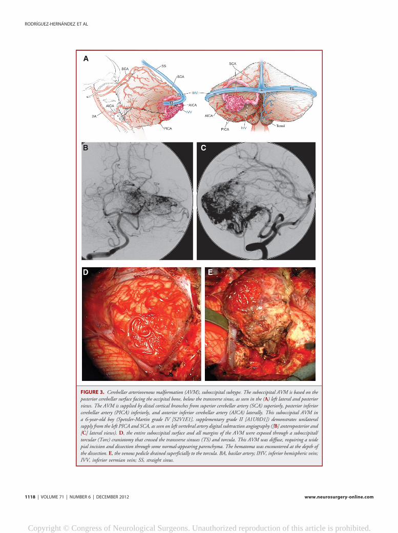
FIGURE 3. Cerebellar arteriovenous malformation (AVM), suboccipital subtype. The suboccipital AVM is based on theposterior cerebellar surface facing the occipital bone, below the transverse sinus, as seen in the (A) left lateral and posteriorviews. The AVM is supplied by distal cortical branches from superior cerebellar artery (SCA) superiorly, posterior inferiorcerebellar artery (PICA) inferiorly, and anterior inferior cerebellar artery (AICA) laterally. This suboccipital AVM ina 6-year-old boy (Spetzler-Martin grade IV [S2V1E1], supplementary grade II [A1U0D1]) demonstrates unilateralsupply from the left PICA and SCA, as seen on left vertebral artery digital subtraction angiography ([B] anteroposterior and[C] lateral views). D, the entire suboccipital surface and all margins of the AVM were exposed through a suboccipital/torcular (Torc) craniotomy that crossed the transverse sinuses (TS) and torcula. This AVM was diffuse, requiring a widepial incision and dissection through some normal-appearing parenchyma. The hematoma was encountered at the depth ofthe dissection. E, the venous pedicle drained superficially to the torcula. BA, basilar artery; IHV, inferior hemispheric vein;IVV, inferior vermian vein; SS, straight sinus.
RODRIGUEZ-HERNANDEZ ET AL
1118 | VOLUME 71 | NUMBER 6 | DECEMBER 2012 www.neurosurgery-online.com
Copyright © Congress of Neurological Surgeons. Unauthorized reproduction of this article is prohibited.
Copyright © Congress of Neurological Surgeons. Unauthorized reproduction of this article is prohibited.
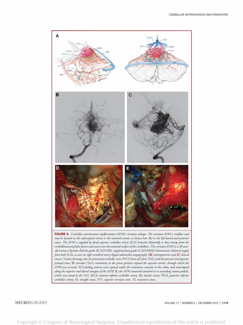
FIGURE 4. Cerebellar arteriovenous malformation (AVM), vermian subtype. The vermian AVM is midline andmay be located on the suboccipital vermis or the tentorial vermis, as shown here (A) in the left lateral and posteriorviews. The AVM is supplied by distal superior cerebellar artery (SCA) branches bilaterally as they emerge from thecerebellomesencephalic fissure and course over the tentorial surface of the cerebellum. This vermian AVM in a 28-year-old woman (Spetzler-Martin grade III [S2V1E0], supplementary grade II [A2U0D0]) demonstrates bilateral supplyfrom both SCAs, as seen on right vertebral artery digital subtraction angiography ([B] anteroposterior and [C] lateralviews). Venous drainage into the precentral cerebellar vein (PcCV)/vein of Galen (VoG) and the petrosal vein/superiorpetrosal sinus. D, torcular (Torc) craniotomy in the prone position exposed the superior vermis, through which theAVM was accessed. SCA feeding arteries were exposed under the tentorium, anterior to the nidus, and interruptedalong the superior and lateral margins of the AVM. E, the AVM remained attached to its ascending venous pedicle,which was traced to the VoG. AICA, anterior inferior cerebellar artery; BA, basilar artery; PICA, posterior inferiorcerebellar artery; SS, straight sinus; SVV, superior vermian vein; TS, transverse sinus.
CEREBELLAR ARTERIOVENOUS MALFORMATIONS
NEUROSURGERY VOLUME 71 | NUMBER 6 | DECEMBER 2012 | 1119
Copyright © Congress of Neurological Surgeons. Unauthorized reproduction of this article is prohibited.
Copyright © Congress of Neurological Surgeons. Unauthorized reproduction of this article is prohibited.
perpendicular with inferior vermian AVMs but tangential withsuperior vermian AVMs, requiring some transgression of the pos-terior vermis to access the inferior margins. The SCA feeders areidentified by incising the posterior arachnoid of the quad-rigeminal cistern on both sides of the vermian apex and openingthe cerebellomesencephalic fissure where the cortical branches(s4 segments) emerge.28 Feeders are traced to the AVM marginand coagulated, carefully preserving arteries to the tectum andposterior midbrain. PICA feeders originate beyond its cranial loopalong the distal telovelotonsillar (p4) and cortical (p5) segments.28
Venous drainage is through superior vermian veins, which drain tothe galenic complex (unlike inferior vermian veins). Vermian AVMsare not considered eloquent unless they extend to the cerebellarnuclei and can be near but not associated with the trochlear nerve.
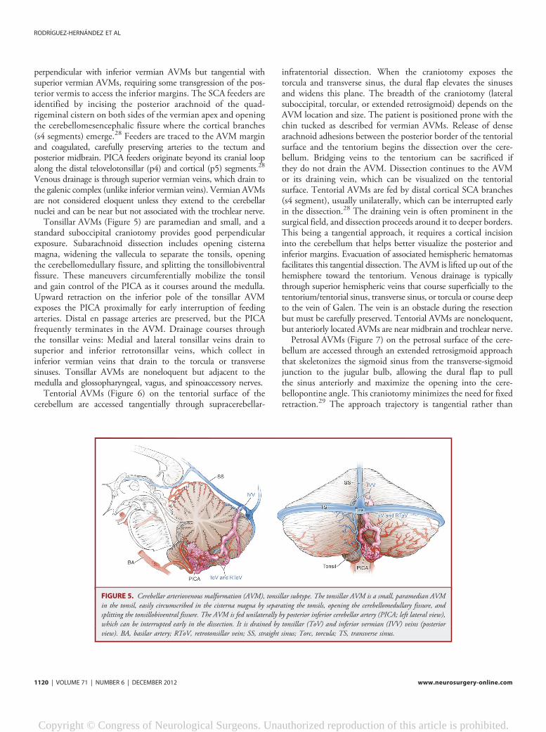
Tonsillar AVMs (Figure 5) are paramedian and small, and astandard suboccipital craniotomy provides good perpendicularexposure. Subarachnoid dissection includes opening cisternamagna, widening the vallecula to separate the tonsils, openingthe cerebellomedullary fissure, and splitting the tonsillobiventralfissure. These maneuvers circumferentially mobilize the tonsiland gain control of the PICA as it courses around the medulla.Upward retraction on the inferior pole of the tonsillar AVMexposes the PICA proximally for early interruption of feedingarteries. Distal en passage arteries are preserved, but the PICAfrequently terminates in the AVM. Drainage courses throughthe tonsillar veins: Medial and lateral tonsillar veins drain tosuperior and inferior retrotonsillar veins, which collect ininferior vermian veins that drain to the torcula or transversesinuses. Tonsillar AVMs are noneloquent but adjacent to themedulla and glossopharyngeal, vagus, and spinoaccessory nerves.
Tentorial AVMs (Figure 6) on the tentorial surface of thecerebellum are accessed tangentially through supracerebellar-
infratentorial dissection. When the craniotomy exposes thetorcula and transverse sinus, the dural flap elevates the sinusesand widens this plane. The breadth of the craniotomy (lateralsuboccipital, torcular, or extended retrosigmoid) depends on theAVM location and size. The patient is positioned prone with thechin tucked as described for vermian AVMs. Release of densearachnoid adhesions between the posterior border of the tentorialsurface and the tentorium begins the dissection over the cere-bellum. Bridging veins to the tentorium can be sacrificed ifthey do not drain the AVM. Dissection continues to the AVMor its draining vein, which can be visualized on the tentorialsurface. Tentorial AVMs are fed by distal cortical SCA branches(s4 segment), usually unilaterally, which can be interrupted earlyin the dissection.28 The draining vein is often prominent in thesurgical field, and dissection proceeds around it to deeper borders.This being a tangential approach, it requires a cortical incisioninto the cerebellum that helps better visualize the posterior andinferior margins. Evacuation of associated hemispheric hematomasfacilitates this tangential dissection. The AVM is lifted up out of thehemisphere toward the tentorium. Venous drainage is typicallythrough superior hemispheric veins that course superficially to thetentorium/tentorial sinus, transverse sinus, or torcula or course deepto the vein of Galen. The vein is an obstacle during the resectionbut must be carefully preserved. Tentorial AVMs are noneloquent,but anteriorly located AVMs are near midbrain and trochlear nerve.Petrosal AVMs (Figure 7) on the petrosal surface of the cere-
bellum are accessed through an extended retrosigmoid approachthat skeletonizes the sigmoid sinus from the transverse-sigmoidjunction to the jugular bulb, allowing the dural flap to pullthe sinus anteriorly and maximize the opening into the cere-bellopontine angle. This craniotomy minimizes the need for fixedretraction.29 The approach trajectory is tangential rather than
FIGURE 5. Cerebellar arteriovenous malformation (AVM), tonsillar subtype. The tonsillar AVM is a small, paramedian AVMin the tonsil, easily circumscribed in the cisterna magna by separating the tonsils, opening the cerebellomedullary fissure, andsplitting the tonsillobiventral fissure. The AVM is fed unilaterally by posterior inferior cerebellar artery (PICA; left lateral view),which can be interrupted early in the dissection. It is drained by tonsillar (ToV) and inferior vermian (IVV) veins (posteriorview). BA, basilar artery; RToV, retrotonsillar vein; SS, straight sinus; Torc, torcula; TS, transverse sinus.
RODRIGUEZ-HERNANDEZ ET AL
1120 | VOLUME 71 | NUMBER 6 | DECEMBER 2012 www.neurosurgery-online.com
Copyright © Congress of Neurological Surgeons. Unauthorized reproduction of this article is prohibited.
Copyright © Congress of Neurological Surgeons. Unauthorized reproduction of this article is prohibited.
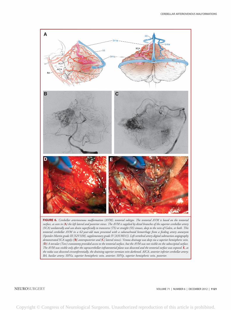
FIGURE 6. Cerebellar arteriovenous malformation (AVM), tentorial subtype. The tentorial AVM is based on the tentorialsurface, as seen in (A) the left lateral and posterior views. The AVM is supplied by distal branches of the superior cerebellar artery(SCA) unilaterally and can drain superficially to transverse (TS) or straight (SS) sinuses, deep to the vein of Galen, or both. Thistentorial cerebellar AVM in a 62-year-old man presented with a subarachnoid hemorrhage from a feeding artery aneurysm(Spetzler-Martin grade III [S2V1E0], supplementary grade IV [A3U0D1]). Left vertebral artery digital subtraction angiographydemonstrated SCA supply ([B] anteroposterior and [C] lateral views). Venous drainage was deep via a superior hemispheric vein.(D) A torcular (Torc) craniotomy provided access to the tentorial surface, but the AVM was not visible on the suboccipital surface.The AVM was visible only after the supracerebellar-infratentorial plane was dissected and the tentorial surface was exposed. E, asthe nidus was dissected circumferentially, the draining superior vermian vein darkened. AICA, anterior inferior cerebellar artery;BA, basilar artery; SHVa, superior hemispheric vein, anterior; SHVp, superior hemispheric vein, posterior.
CEREBELLAR ARTERIOVENOUS MALFORMATIONS
NEUROSURGERY VOLUME 71 | NUMBER 6 | DECEMBER 2012 | 1121
Copyright © Congress of Neurological Surgeons. Unauthorized reproduction of this article is prohibited.
Copyright © Congress of Neurological Surgeons. Unauthorized reproduction of this article is prohibited.
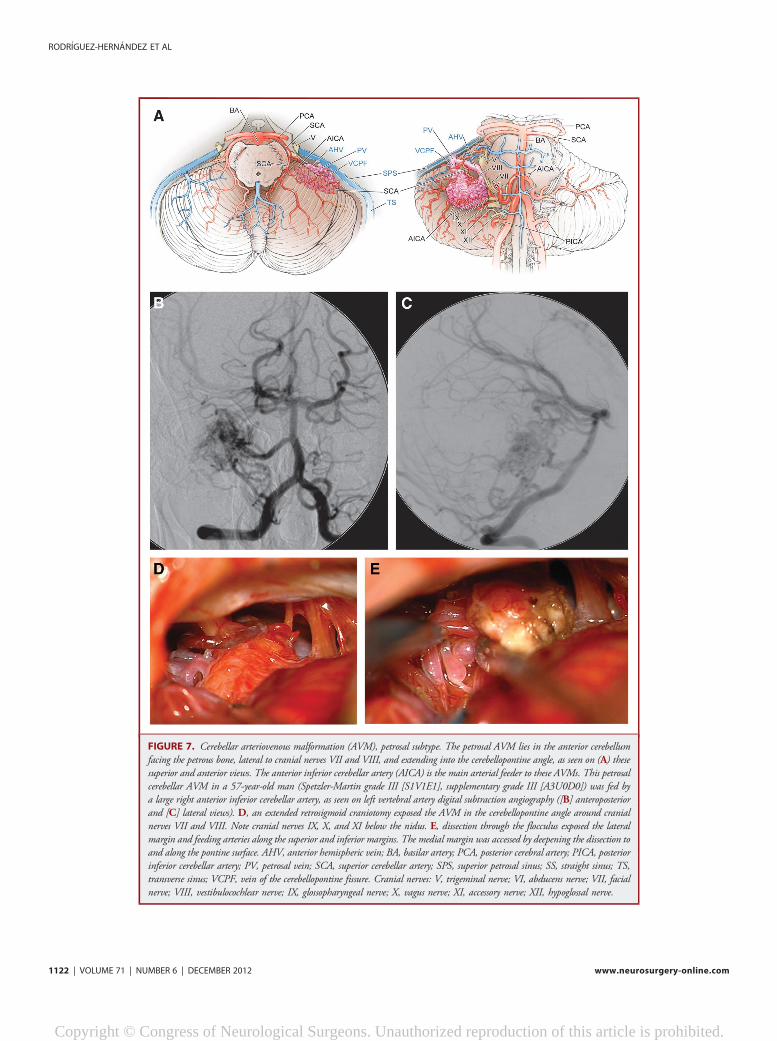
FIGURE 7. Cerebellar arteriovenous malformation (AVM), petrosal subtype. The petrosal AVM lies in the anterior cerebellumfacing the petrous bone, lateral to cranial nerves VII and VIII, and extending into the cerebellopontine angle, as seen on (A) thesesuperior and anterior views. The anterior inferior cerebellar artery (AICA) is the main arterial feeder to these AVMs. This petrosalcerebellar AVM in a 57-year-old man (Spetzler-Martin grade III [S1V1E1], supplementary grade III [A3U0D0]) was fed bya large right anterior inferior cerebellar artery, as seen on left vertebral artery digital subtraction angiography ([B] anteroposteriorand [C] lateral views). D, an extended retrosigmoid craniotomy exposed the AVM in the cerebellopontine angle around cranialnerves VII and VIII. Note cranial nerves IX, X, and XI below the nidus. E, dissection through the flocculus exposed the lateralmargin and feeding arteries along the superior and inferior margins. The medial margin was accessed by deepening the dissection toand along the pontine surface. AHV, anterior hemispheric vein; BA, basilar artery; PCA, posterior cerebral artery; PICA, posteriorinferior cerebellar artery; PV, petrosal vein; SCA, superior cerebellar artery; SPS, superior petrosal sinus; SS, straight sinus; TS,transverse sinus; VCPF, vein of the cerebellopontine fissure. Cranial nerves: V, trigeminal nerve; VI, abducens nerve; VII, facialnerve; VIII, vestibulocochlear nerve; IX, glossopharyngeal nerve; X, vagus nerve; XI, accessory nerve; XII, hypoglossal nerve.
RODRIGUEZ-HERNANDEZ ET AL
1122 | VOLUME 71 | NUMBER 6 | DECEMBER 2012 www.neurosurgery-online.com
Copyright © Congress of Neurological Surgeons. Unauthorized reproduction of this article is prohibited.
Copyright © Congress of Neurological Surgeons. Unauthorized reproduction of this article is prohibited.
perpendicular, which means that the lateral and posterior AVMmargins are accessed at the expense of some overlying cerebellum.Subarachnoid dissection opens the lateral arachnoid of the pre-pontine cistern to identify the vestibulocochlear and facial nerves(cranial nerves VII and VIII) and AICA, the main feeder to thesepetrosal AVMs. Subarachnoid dissection extends superiorly to thetrigeminal nerve (cranial nerve V) when SCA contributes to theAVM and inferiorly to the lower cranial nerves (cranial nerves IX,X, and XI) when the PICA contributes. Petrosal AVMs arecerebellar rather than pontine and therefore reside lateral tocranial nerves VII and VIII. AICA feeders arising from theflocculopeduncular (a3) and cortical (a4) segments are inter-rupted early by dissecting medial to the nidus, but many ofthese medial feeders cannot be visualized early in the resection.28
The tangential nature of this exposure requires an incision inthe cerebellar cortex lateral to the AVM and resection of someintervening lobule to reach the lateral AVM margin. In hem-orrhagic cases, this route can access hemispheric hematomas forearly evacuation and relaxation of swollen cerebellum. The AVMis then circumscribed, dissecting around the back side and rollingit anteriorly away from the middle cerebellar peduncle and cranialnerves deep to the nidus. Scooping the AVM from behind leavesdeep feeders until the end of the resection, and draining veinsmust be meticulously preserved. AVM drainage is via anteriorhemispheric veins and the vein of the cerebellopontine fissure,which course to the petrosal vein (the Dandy vein) and superiorpetrosal sinus. Petrosal AVMs are noneloquent but adjacent topons, middle cerebellar peduncle, and cranial nerves VII andVIII. The dissection must remain lateral to the cranial nerves toavoid entry into the brainstem.
CONCLUSION
Patients with cerebellar AVMs are significantly more likely topresent with hemorrhage than patients with cerebral AVMs,resulting in more neurological deficits preoperatively and at latefollow-up and justifying a more aggressive treatment posture.The supplementary AVM grading system is better than theSpetzler-Martin system at predicting outcomes after cerebellarAVM resection, and its accuracy is consistent with all AVMs,cerebellar and cerebral. Key components of the Spetzler-Martinsystem, eg, venous drainage and eloquence, are distorted bycerebellar anatomy in ways that the components of thesupplementary system are not, resulting in greater predictiveaccuracy with supplementary grades.
Disclosures
This research is funded in part by the National Institutes of Health, R01NS034949 (Dr Young) and P01 NS044155 (Center for CerebrovascularResearch). Dr Rodríguez-Hernández is supported by a grant from Obra SocialLa Caixa. Dr Lawton receives a royalty for microsurgical instruments from MizuhoAmerica, Inc. The other authors have no personal or financial interests in the drugs,materials, or devices described in this article.
REFERENCES
1. Arnaout OM, Gross BA, Eddleman CS, Bendok BR, Getch CC, Batjer HH.Posterior fossa arteriovenous malformations. Neurosurg Focus. 2009;26(5):E12.
2. Batjer H, Samson D. Arteriovenous malformations of the posterior fossa: clinicalpresentation, diagnostic evaluation and surgical treatment. Neurosurg Rev. 1986;9(4):287-296.
3. da Costa L, Thines L, Dehdashti AR, et al. Management and clinical outcome ofposterior fossa arteriovenous malformations: report on a single-centre 15-yearexperience. J Neurol Neurosurg Psychiatry. 2009;80(4):376-379.
4. Drake CG, Friedman AH, Peerless SJ. Posterior fossa arteriovenous malforma-tions. J Neurosurg. 1986;64(1):1-10.
5. Kelly ME, Guzman R, Sinclair J, et al. Multimodality treatment of posterior fossaarteriovenous malformations. J Neurosurg. 2008;108(6):1152-1161.
6. Khaw AV, Mohr JP, Sciacca RR, et al. Association of infratentorial brainarteriovenous malformations with hemorrhage at initial presentation. Stroke. 2004;35(3):660-663.
7. Neacsu A, Ciurea AV. General considerations on posterior fossa arteriovenousmalformations (clinics, imaging and therapy). Actual concepts and literaturereview. J Med Life. 2010;3(1):26-35.
8. O’Shaughnessy BA, Getch CC, Bendok BR, Batjer HH. Microsurgicalresection of infratentorial arteriovenous malformations. Neurosurg Focus.2005;19(2):E5.
9. Sinclair J, Kelly ME, Steinberg GK. Surgical management of posterior fossaarteriovenous malformations. Neurosurgery. 2006;58(4 suppl 2):ONS-189-ONS-201.
10. Symon L, Tacconi L, Mendoza N, Nakaji P. Arteriovenous malformations of theposterior fossa: a report on 28 cases and review of the literature. Br J Neurosurg.1995;9(6):721-732.
11. Vilalta J, Topezewski T, Añez JD, Arikan F, Guitart JM, Rubio E. Arteriovenousmalformations of the posterior fossa. Clinical features, treatment and results [inSpanish]. Rev Neurol. 2001;32(12):1124-1128.
12. Yasargil MG. Microneurosurgery. Vol IIIB. New York, NY: Thieme MedicalPublishers, Inc.; 1988.
13. Rhoton AL Jr. Cerebellum and fourth ventricle. Neurosurgery. 2000;47(3 suppl):S7-S27.
14. Fine AD, Beauregard CL, Day AL. Arteriovenous malformations of the cerebellarvermis and hemispheres. In: Stieg PE, Batjer HH, Samsom D, eds. IntracranialArteriovenous Malformations. New York, NY: Informa Health Care; 2007:285-297.
15. de Oliveira E, Tedeschi H, Raso J. Comprehensive management of arteriovenousmalformations. Neurol Res. 1998;20(8):673-683.
16. Lawton MT, Du R, Tran MN, et al. Effect of presenting hemorrhage on outcomeafter microsurgical resection of brain arteriovenous malformations. Neurosurgery.2005;56(3):485-493.
17. Muñoz F, Clavel P, Molet J, et al. Current management of arteriovenousmalformations: retrospective study of 31 cases and literature review [in Spanish].Neurocirugia (Astur). 2007;18(5):394-404.
18. Fleetwood IG, Steinberg GK. Arteriovenous malformations. Lancet. 2002;359(9309):863-873.
19. Stapf C, Mast H, Sciacca RR, et al. Predictors of hemorrhage in patientswith untreated brain arteriovenous malformation. Neurology. 2006;66(9):1350-1355.
20. Halim AX, Johnston SC, Singh V, et al. Longitudinal risk of intracranialhemorrhage in patients with arteriovenous malformation of the brain withina defined population. Stroke. 2004;35(7):1697-1702.
21. Sanchez-Mejia RO, McDermott MW, Tan J, Kim H, Young WL, Lawton MT.Radiosurgery facilitates resection of brain arteriovenous malformations and reducessurgical morbidity. Neurosurgery. 2009;64(2):231-238.
22. Lawton MT, Kim H, McCulloch CE, Mikhak B, Young WL. A supplementarygrading scale for selecting patients with brain arteriovenous malformations forsurgery. Neurosurgery. 2010;66(4):702-713.
23. Lawton MT; UCSF AVM Study Project. Spetzler-Martin grade III arteriovenousmalformations: surgical results and a modification of the grading scale. Neurosurgery.2003;52(4):740-748.
24. Spetzler RF, Martin NA. A proposed grading system for arteriovenousmalformations. J Neurosurg. 1986;65(4):476-483.
CEREBELLAR ARTERIOVENOUS MALFORMATIONS
NEUROSURGERY VOLUME 71 | NUMBER 6 | DECEMBER 2012 | 1123
Copyright © Congress of Neurological Surgeons. Unauthorized reproduction of this article is prohibited.
Copyright © Congress of Neurological Surgeons. Unauthorized reproduction of this article is prohibited.
25. Vates GE, Lawton MT, Wilson CB, et al. Magnetic source imaging demonstratesaltered cortical distribution of function in patients with arteriovenous malformations.Neurosurgery. 2002;51(3):614-623.
26. Du R, Keyoung HM, DowdCF, YoungWL, LawtonMT. The effects of diffusenessand deep perforating artery supply on outcomes after microsurgical resection of brainarteriovenous malformations. Neurosurgery. 2007;60(4):638-646.
27. Sanchez-Mejia RO, Chennupati SK, Gupta N, Fullerton H, Young WL,Lawton MT. Superior outcomes in children compared with adults after
microsurgical resection of brain arteriovenous malformations. J Neurosurg. 2006;105(2 suppl):82-87.
28. Rodríguez-Hernández A, Rhoton AL Jr, Lawton MT. Segmental anatomy ofcerebellar arteries: a proposed nomenclature: laboratory investigation. J Neurosurg.2011;115(2):387-397.
29. Quiñones-Hinojosa A, Chang EF, Lawton MT. The extended retrosigmoidapproach: an alternative to radical cranial base approaches for posterior fossalesions. Neurosurgery. 2006;58(4 suppl 2):ONS-208-ONS-214.
RODRIGUEZ-HERNANDEZ ET AL
1124 | VOLUME 71 | NUMBER 6 | DECEMBER 2012 www.neurosurgery-online.com
Copyright © Congress of Neurological Surgeons. Unauthorized reproduction of this article is prohibited.
Copyright © Congress of Neurological Surgeons. Unauthorized reproduction of this article is prohibited.


![RESEARCH Open Access Monoclonal antibodies to 65kDa ......Stiff Person Syndrome (SPS) [2] and certain subtypes of Cerebellar Ataxia (CA) [3-5]. GAD65Ab in these three disorders show](https://img.pdfslide.net/doc/110x75/60f8116f22d0e6639f6115f8/research-open-access-monoclonal-antibodies-to-65kda-stiff-person-syndrome.jpg)