Embed Size (px)
Citation preview
Cerebellar Disorders
• Dr Manesh Pillay • Neurologist • January 2012
Objectives
• Neurological examination To understand how to localize lesions within the
cerebellum
• Disorders of Cerebellum
Presentation
• Ataxia - syndrome of imbalance and incoordination involving gait and limbs
• Cerebellar vs Sensory ataxia vs motor vs spinal cord disease vs basal ganglia vs vestibular disease vs Cortical ataxia
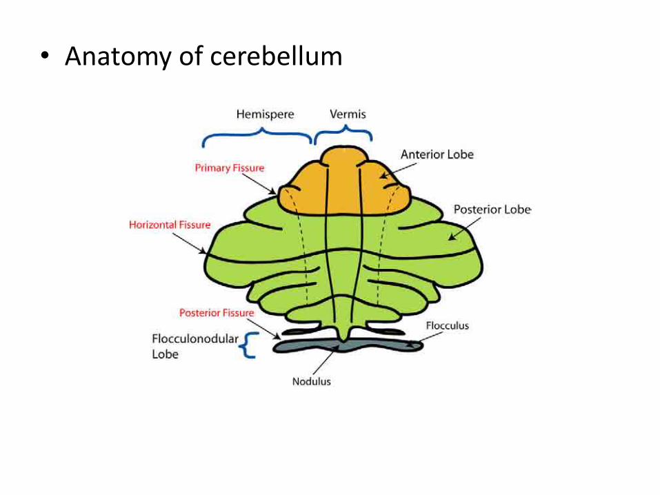
• Anatomy of cerebellum
Cerebellar Syndromes
• In general, precise clinical localization is difficult in the cerebellum • Some syndromes can be classified anatomically:
– Rostral vermis syndrome (anterior lobe)
– Caudal vermis syndrome (flocculonodular, posterior lobe)
– Hemispheric syndrome (posterior lobe, variably anterior too)
– Pancerebellar syndrome
Cerebellar Syndromes- rostral vermis
– Wide-based stance and gait – Ataxia of gait, proportionally little ataxia on heel-shin with pt lying
down – Normal or slightly impaired arm cooordination – Infrequent hypotonia, nystagmus, dysarthria
• alcoholics (restricted form of cerebellar cortical degeneration)
Cerebellar Syndromes- caudal vermis
– Axial dysequilibrium, staggering gait – Little or no limb ataxia – Sometimes spontaneous nystagmus – Rotated postures of head
• Seen in diseases that damage the flocculonodular lobe (esp
medulloblastoma in children) — as tumor grows, a hemispheric cerebellar syndrome may be superimposed
• Need to also consider other signs of ICP (obstruction of CSF)
Cerebellar Syndromes - hemispheric
– Incoordination of ipsilateral limb movements
– More noticeable with fine motor skills
– Incoordination affects most noticeably muscles involved in speech and finger movements
• Etiologies include infarcts, neoplasms, abscesses
Cerebellar Syndromes- pancerebellar
• Combination of all the other syndromes
• Bilateral signs of cerebellar dysfunction involving trunk, limbs, cranial musculature
• Etiologies usually infectious/parainfectious processes, hypoglycemia, paraneoplastic disorders, toxic-metabolic disorders,hereditary
Localisation in cerebellum
• Lateralized cerebellar lesions - ipsilateral symptoms and signs
• Generalized cerebellar lesions - symmetrical symptomatology.
• Vestibulocerebellar lesions cause disequilibrium and an ataxic gait.
• Vermis, ―spinocerebellar‖ organ, truncal and gait ataxia with relative sparing of the limbs.
Time period
• Acute - severe abnormalities early, but recovery with time.
• Chronic - progressive diseases gradually declining balance with longer lasting effects.
Symptoms
Gait Disturbances • Insecurity while walking, especially -
turning or balancing on a narrow ledge. • Specialized skills such as skiing,
bicycling, or climbing. • Report the sense of imbalance as
dizziness. • Increase imbalance when visual cues
removed = sensory
Symptoms
• Limb Ataxia - clumsiness and tremor, slow movements to be more accurate. symptoms one-sided with lateralized lesions .
• Truncal Ataxia - head tremor and truncal instability
Symptoms
• Dysarthria and Bulbar Symptoms - slurred speech and abnormalities of pitch and volume control (scanning speech).
• Dysphagia - incoordination of swallowing muscles. Ineffectiveness of cough may also be a symptom
Symptoms
• Visual Symptoms - blurriness or a sense of environmental movements as a result of cerebellar ocular oscillations
Symptoms in Sensory Ataxia
• Patients with a sensory basis for ataxia usually do not experience dysarthria or visual symptoms.
• Symptoms of sensory pathway disease such as parasthesias and numbness.
Signs in Cerebellar Ataxia
• Deficits involving gait and stance, limb incoordination, muscle tone, speech, and the oculomotor system.
• Subtle cognitive deficits.
Stance and Gait
• Increase in body sway,feet are placed together. Stance more than 12cm.
• Early - tandem or stand on one foot; 30Sec
• Romberg test - prominent in proprioceptive or vestibular lesions.
Stance and Gait
• Straight path-widened base and an irregular staggering appearance - alcoholic.
• Speed not impaired.
• Steps are irregular and the patient may lurch in unpredictable ways.
• Rhythmic oscillations trunk and head - titubation.
• Truncal ataxia - inability to sit upright without back support.
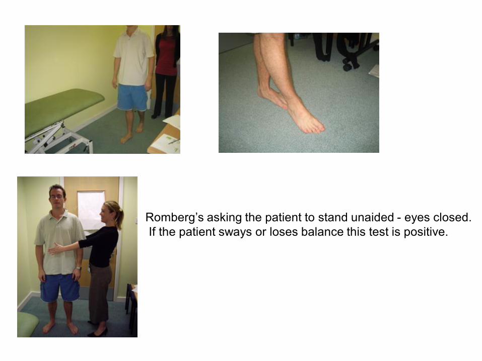
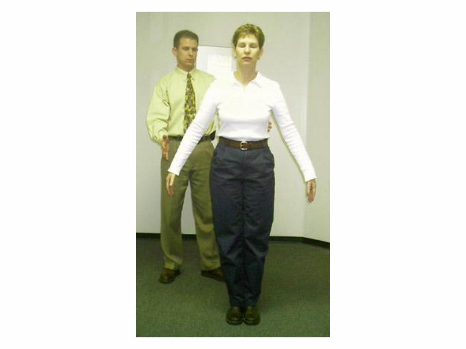
Romberg’s asking the patient to stand unaided - eyes closed. If the patient sways or loses balance this test is positive.
Dysrhythmia
• Inability to tap and keep a rhythm. It can be tested by tapping the table with a hand (or the floor with a foot) and asking the patient to repeat the maneuver.
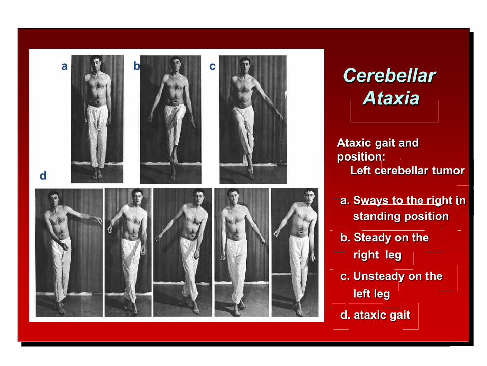
Cerebellar Ataxia
Ataxic gait and position: Left cerebellar tumor a. Sways to the right in standing position
b. Steady on the right leg
c. Unsteady on the left leg
d. ataxic gait
a b c
d
Cerebellar examination
• Rebound
• examined by allowing the patient to flex the elbow against the examiner's hand and then abruptly removing the resistance and assessing the patient's ability to arrest the sudden flexion movement.
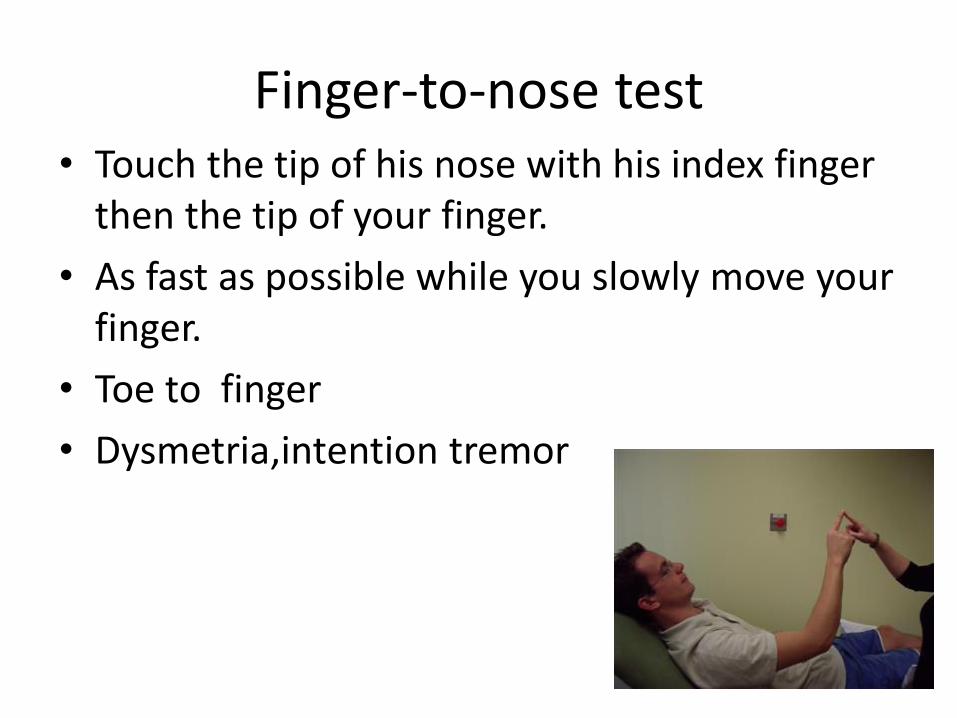
Finger-to-nose test • Touch the tip of his nose with his index finger
then the tip of your finger.
• As fast as possible while you slowly move your finger.
• Toe to finger
• Dysmetria,intention tremor
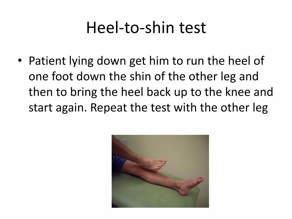
Heel-to-shin test
• Patient lying down get him to run the heel of one foot down the shin of the other leg and then to bring the heel back up to the knee and start again. Repeat the test with the other leg
Dysmetria
• Inaccuracy of movement so that the desired target is either under-reached (hypometria) or over-reached (hypermetria).
• Dysmetria is evident in the finger-chase and toe-to-finger tests.
• Disturbance of the rate, range, and force of movement.
• Increased by adding a mass to the hand

Kinetic (Intention) Tremor
• Oscillations during a voluntary movement intended to reach a target increasing in amplitude as the target is reached .
• Result from instability at the proximal, rather than the distal, portions of the limb typically perpendicular to axis of motion.
• Finger-to-nose and the heel-to-shin maneuvers detect the kinetic tremor.
• Kinetic tremor is better evaluated when mass is added to the hand
Other Tremors
• Cerebellar lesions can give rise to a postural tremor initiated
by keeping the arms outstretched or pointing the fingers steadily at each other. In the legs, maintaining one heel on the opposite knee can bring out such tremor.
• Axial tremor involving the head and shoulders. Also, a severe tremor in the upper limbs that has both an intention and a postural component can appear in cerebellar outflow tract disease. “rubral” or “wing-beating” tremor.
• ETIOLOGY - multiple sclerosis, Wilson's disease, and midbrain strokes.
Dysdiadochokinesia
• Irregularity of the rhythm and amplitude of rapid alternating movements.
• Simple tapping tasks such as the index finger on the thumb crease or the feet on the floor can also detect the disturbance in rhythm (dysrhythmokinesis).
Dysdiadochokinesia
• Rapid alternating movements - supinate and pronate the forearm in the unsupported position.
OR
• clap one hand on the palm of the other (stationary) hand alternately with the palm and dorsum of the clapping hand
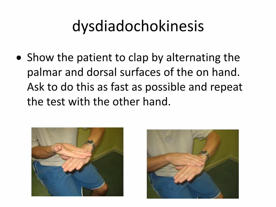
dysdiadochokinesis
Show the patient to clap by alternating the palmar and dorsal surfaces of the on hand. Ask to do this as fast as possible and repeat the test with the other hand.
Hypotonia
• Decrease in the normal resistance offered by muscles to passive manipulation.
• When an affected limb is shaken,flapping movements of the hands appear of wider excursion than normal.
• Also, a forced flexion of the arm at the elbow may obliterate the space between the volar aspect of the wrist and the deltoid.
• Not consistent sign
Oculomotor Disturbances • Fixation abnormalities are examined by asking the
patient to maintain sustained gaze at the examiner's finger held about 2 feet in front.
• Follow finger as it is moved slowly in all directions of gaze (pursuit).
• Eccentric gaze is maintained (at about 30 degrees deviation) to check for nystagmus.
• Saccades are examined by having the patient shift gaze quickly between an eccentrically held finger and the examiner's nose in the middle.
Oculomotor dysfunction
• Nystagmus frequently seen in cerebellar disorders • Gaze-evoked nystagmus, upbeat nystagmus, rebound nystagmus,
opticokinetic nystagmus may all be seen in midline cerebellar lesions
• Other ocular lesions seen include opsoclonus, skew deviation, ocular bobbing
• Most of the disorders giving rise to these affect brainstem structures, too: cerebellar role in their onset not well-defined
• Overall, most ―cerebellar‖ eye signs cannot be localized to specific areas of the cerebellum
Nystagmus
• Gaze-evoked - eccentric gaze is maintained at 30 degrees . Eyes repetitively drift toward midline followed by saccades to eccentric position. Fast phase of the nystagmus is always to the side of gaze.
• Gaze-evoked nystagmus fatigues and reverses direction after a few seconds, rebound nystagmus
• Downbeat nystagmus characterized by rapid phase in down direction- downgaze or gaze to the side- craniovertebral junction
• Upbeat primary position nystagmus - anterior vermis.
Speech and Bulbar Function
• Listening to spoken words - speak standard phrases. • Slowness, slurring of the words, and a general
inability to control the process of articulation, leading to unnecessary hesitations and stops, omissions of pauses when needed, and an accentuation of syllables when not needed.
• Variability in volume and pitch of words and inappropriate control of the breathing needed for speech- scanning dysarthria.
• Mild dysphagia is not uncommon
Sensory Ataxia
• Defective proprioception.
• Impaired position and vibration sense - deep tendon reflexes are often lost - afferent fiber pathology.
• Romberg test is positive
• Degenerative ataxic syndromes combine features of cerebellar and proprioceptive deficits in variable proportion.
Ataxic sensory gait
a) brisk leg movements;
b) legs placed far apart to correct instability;
c) steps of variable length;
d) need for carefully watching the ground.
e)Incoordination enhanced when deprived of visual information.
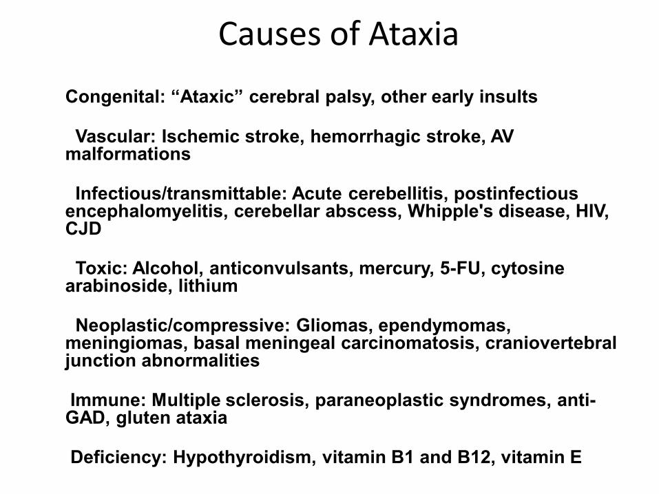
Causes of Ataxia
Congenital: “Ataxic” cerebral palsy, other early insults
Vascular: Ischemic stroke, hemorrhagic stroke, AV malformations
Infectious/transmittable: Acute cerebellitis, postinfectious encephalomyelitis, cerebellar abscess, Whipple's disease, HIV, CJD
Toxic: Alcohol, anticonvulsants, mercury, 5-FU, cytosine arabinoside, lithium
Neoplastic/compressive: Gliomas, ependymomas, meningiomas, basal meningeal carcinomatosis, craniovertebral junction abnormalities
Immune: Multiple sclerosis, paraneoplastic syndromes, anti-GAD, gluten ataxia
Deficiency: Hypothyroidism, vitamin B1 and B12, vitamin E
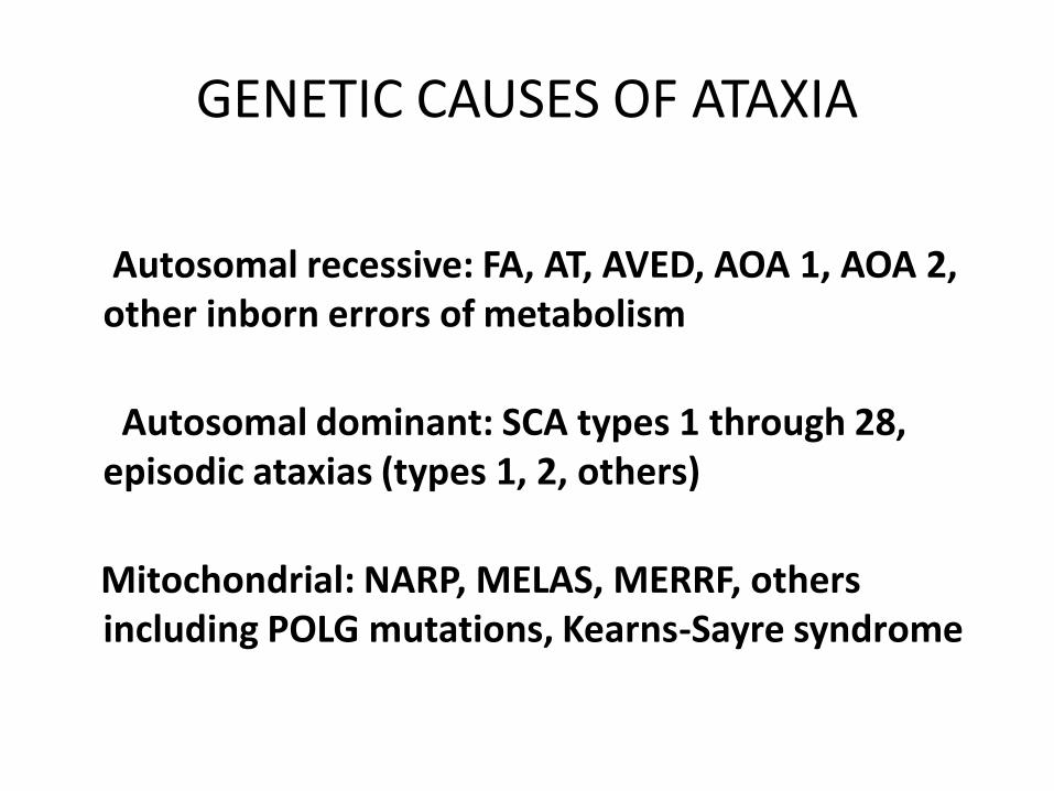
GENETIC CAUSES OF ATAXIA
Autosomal recessive: FA, AT, AVED, AOA 1, AOA 2, other inborn errors of metabolism
Autosomal dominant: SCA types 1 through 28, episodic ataxias (types 1, 2, others)
Mitochondrial: NARP, MELAS, MERRF, others including POLG mutations, Kearns-Sayre syndrome
Causes of ataxia
• Investigation in patients with ataxia
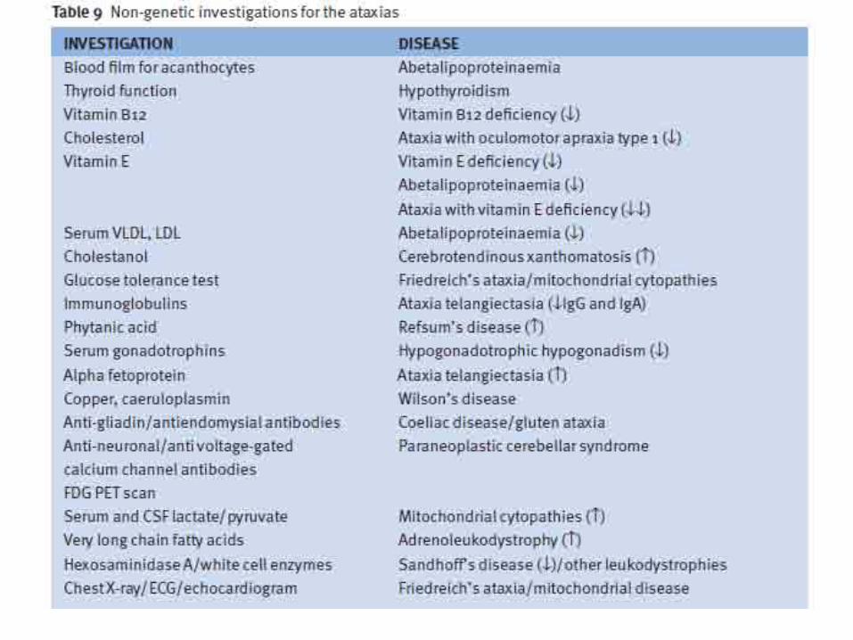
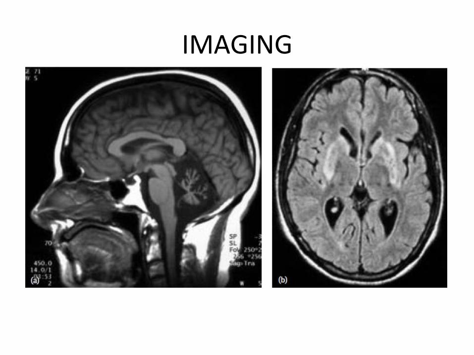
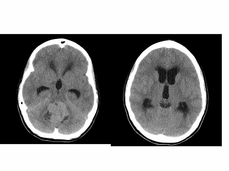
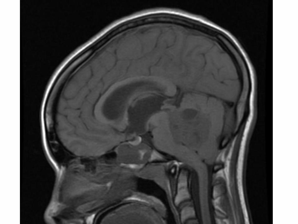
IMAGING
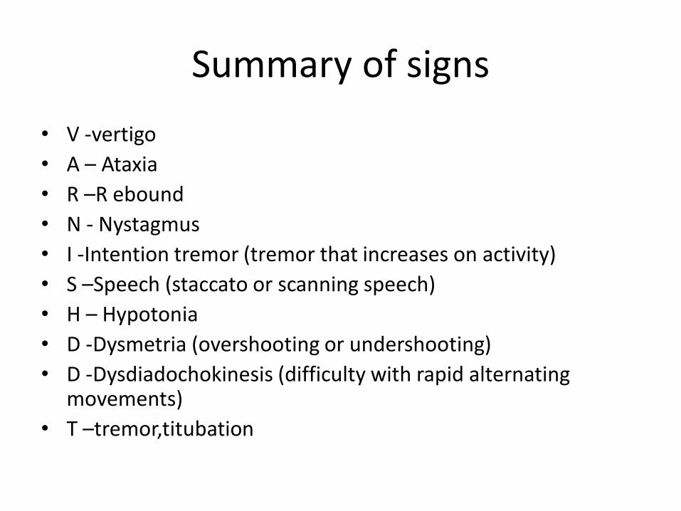
Summary of signs
• V -vertigo
• A – Ataxia
• R –R ebound
• N - Nystagmus
• I -Intention tremor (tremor that increases on activity)
• S –Speech (staccato or scanning speech)
• H – Hypotonia
• D -Dysmetria (overshooting or undershooting)
• D -Dysdiadochokinesis (difficulty with rapid alternating movements)
• T –tremor,titubation
THE END