Embed Size (px)
Citation preview
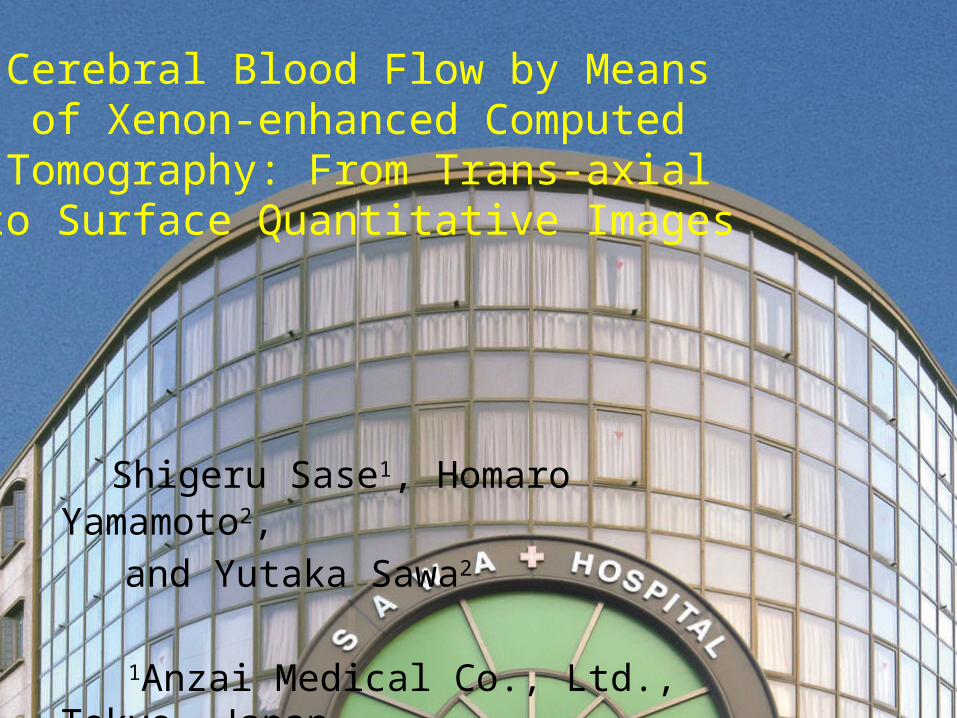
Cerebral Blood Flow by Means of Xenon-enhanced Computed Tomography: From Trans-axial
to Surface Quantitative Images
Shigeru Sase1, Homaro Yamamoto2,
and Yutaka Sawa2
1Anzai Medical Co., Ltd., Tokyo, Japan
2Sawa Hospital, Osaka, Japan
[Purpose]
• To create brain surface images by stacking thin tomographic images obtained by xenon-enhanced computed tomography (Xe-CT).
• To demonstrate usefulness of layer-by-layer spherical analysis of blood flow and lambda for patients with dementia.
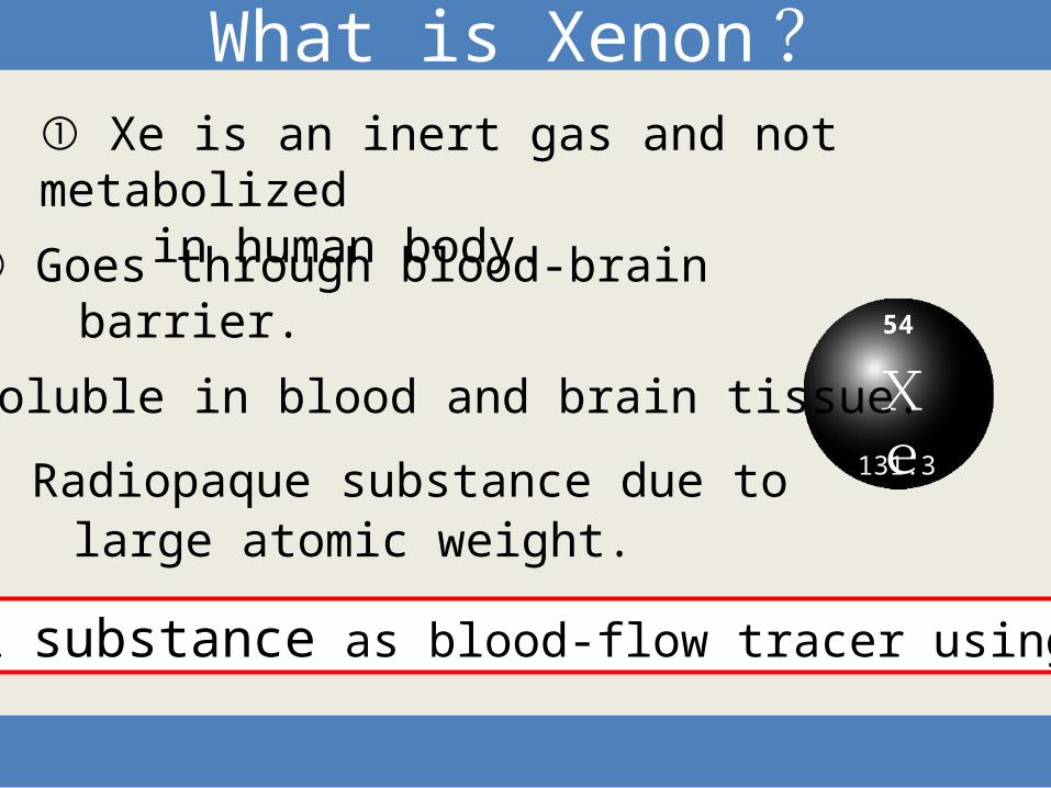
④ Radiopaque substance due to large atomic weight.
Xe
What is Xenon ?
③ Soluble in blood and brain tissue.
② Goes through blood-brain barrier.
① Xe is an inert gas and not metabolized in human body.
Ideal substance as blood-flow tracer using CT.
54
131.3
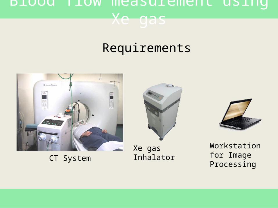
Blood flow measurement using Xe gas
Workstation for Image Processing
Xe gas InhalatorCT System
Requirements
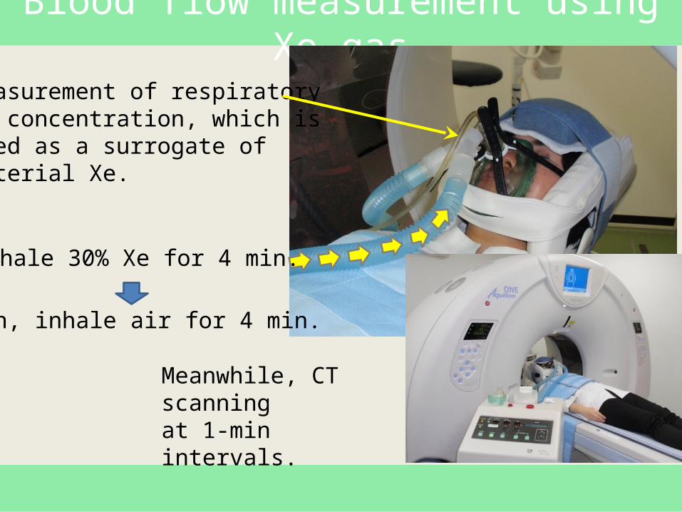
Blood flow measurement using Xe gas
Inhale 30% Xe for 4 min.
Then, inhale air for 4 min.
Measurement of respiratoryXe concentration, which isused as a surrogate ofarterial Xe.
Meanwhile, CT scanningat 1-min intervals.
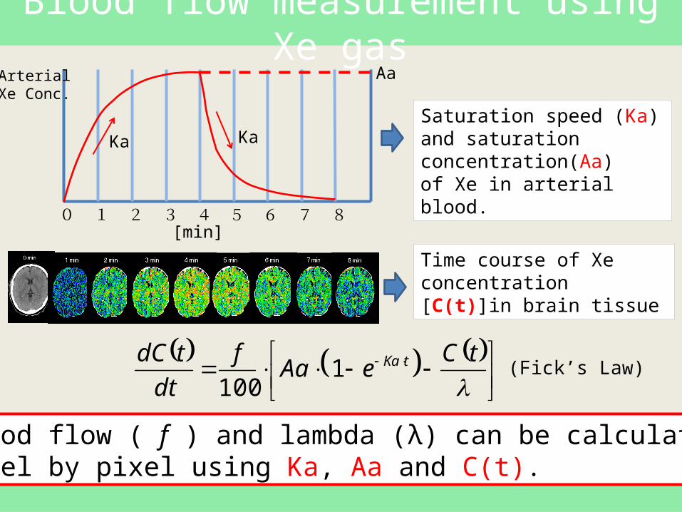
Blood flow measurement using Xe gas
0 1 2 3 4 5 6 7 8[min]
Saturation speed (Ka) and saturation concentration(Aa)of Xe in arterial blood.
Time course of Xe concentration [C(t)]in brain tissue
Blood flow ( f ) and lambda (λ) can be calculatedpixel by pixel using Ka, Aa and C(t).
Ka
Aa
tC
eAaf
dt
tdC tKa1100
Ka
(Fick’s Law)
ArterialXe Conc.
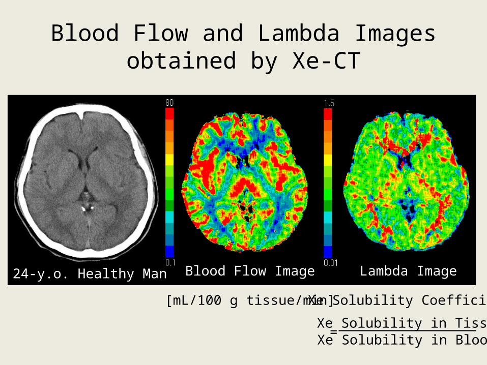
Blood Flow and Lambda Imagesobtained by Xe-CT
24-y.o. Healthy Man Blood Flow Image Lambda Image
[mL/100 g tissue/min]
Xe Solubility in TissueXe Solubility in Blood
Xe Solubility Coefficient
=
Lambda Image
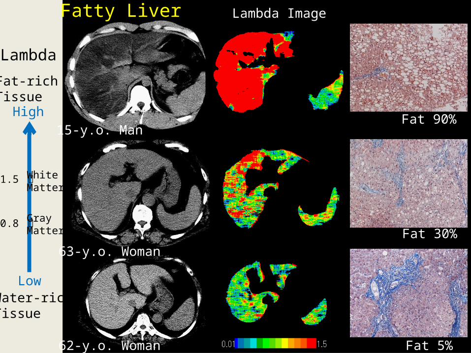
Fat 5%
Fat 30%
62-y.o. Woman
63-y.o. Woman
Fat 90%15-y.o. Man
Fatty Liver
Lambda
Fat-rich Tissue
Water-richTissue
High
Low
WhiteMatter
GrayMatter
1.5 ー
0.8 ー
[Methods]
• CT: Aquilion ONE (Toshiba, Japan): Area-detector CT capable of volume scan of the brain.
• Xe gas inhalator: AZ-725 (Anzai Medical, Japan).
• Subjects: Patients with dementia, Age-matched healthy controls.
• Creation of brain surface images, and layer-by-layer analyses (layer thickness: 5mm)
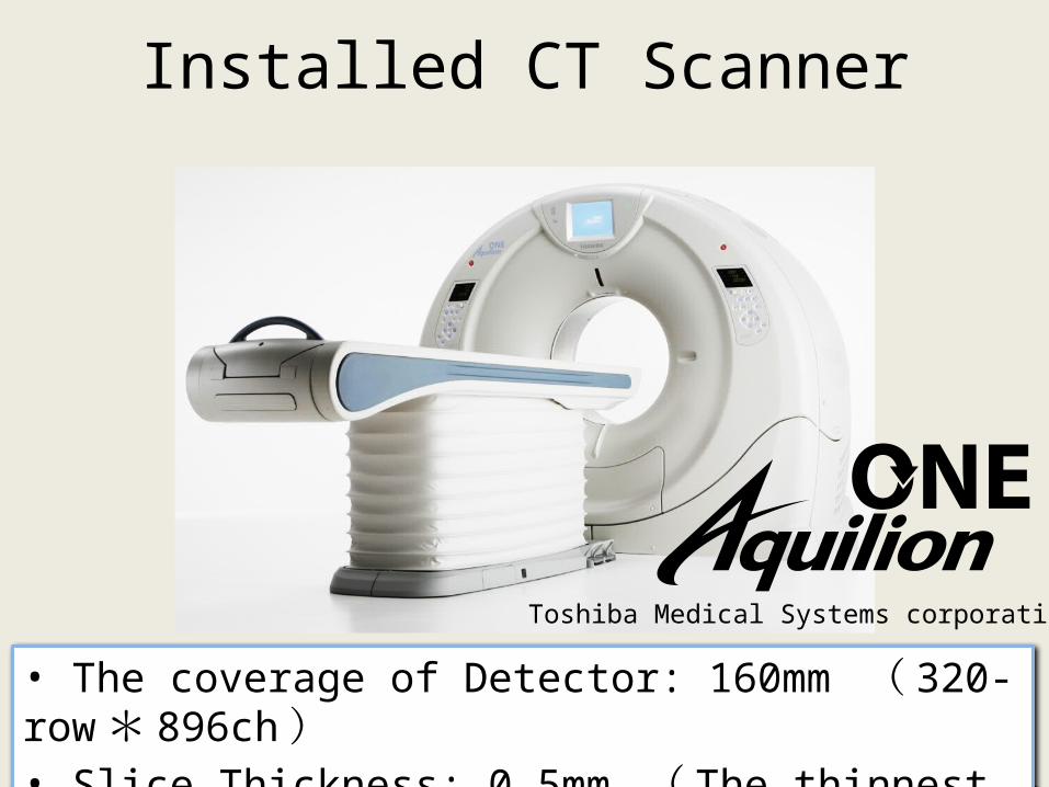
Installed CT Scanner
• The coverage of Detector: 160mm ( 320-row *896ch )• Slice Thickness: 0.5mm ( The thinnest in the industry )
Toshiba Medical Systems corporation
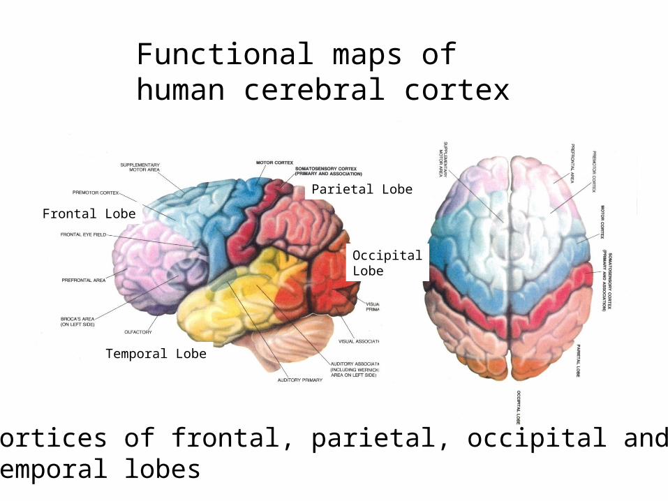
Functional maps ofhuman cerebral cortex
Cortices of frontal, parietal, occipital andtemporal lobes
Frontal Lobe
Parietal Lobe
OccipitalLobe
Temporal Lobe

[Results]
• Surface images of blood flow and lambda for healthy volunteer
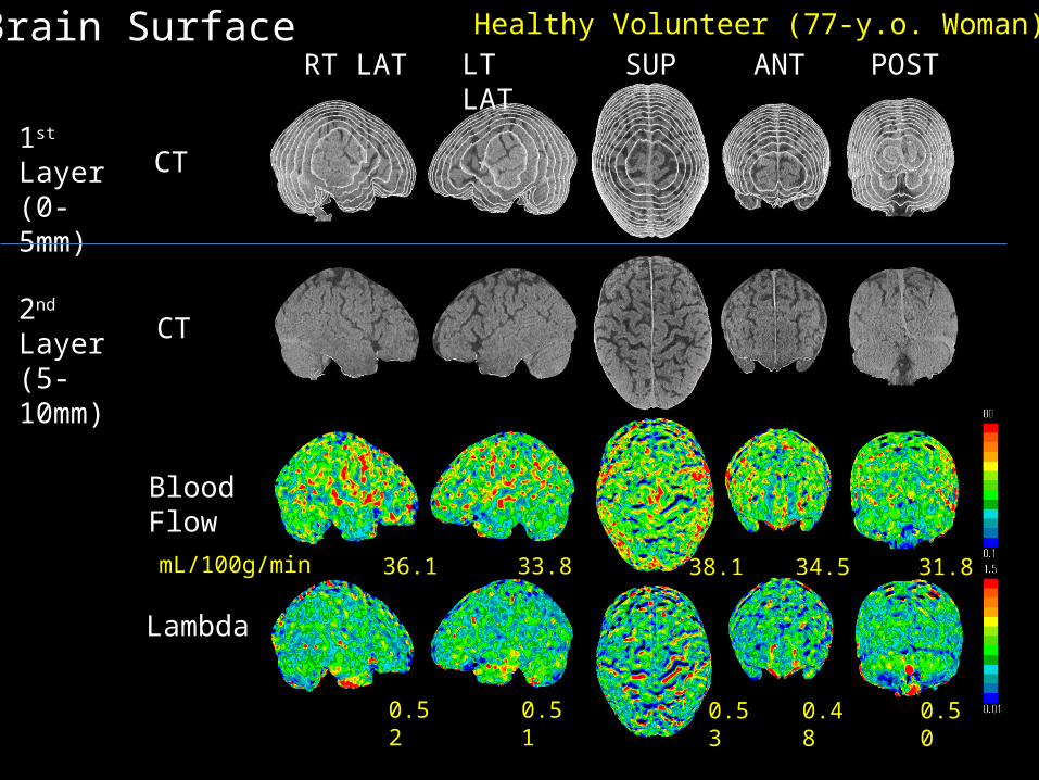
36.1 33.8 34.5 31.838.1
RT LAT LT LAT SUP ANT POST
CT
BloodFlow
Brain Surface
0.52 0.51 0.48 0.500.53
Lambda
1st Layer(0-5mm)
2nd Layer(5-10mm) CT
Healthy Volunteer (77-y.o. Woman)
mL/100g/min
• Comparison of Xe-CT and SPECT
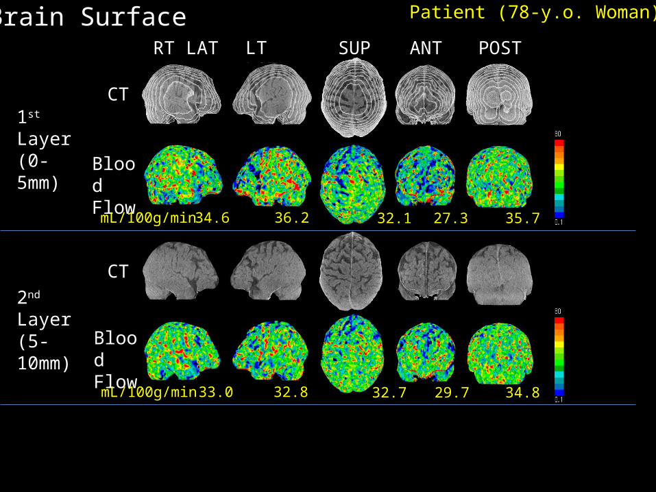
RT LAT LT LAT SUP ANT POST
Brain Surface
34.6 36.2 27.3 35.732.1
33.0 32.8 29.7 34.832.7
1st Layer(0-5mm)
2nd Layer(5-10mm)
CT
CT
BloodFlow
BloodFlow
Patient (78-y.o. Woman)
mL/100g/min
mL/100g/min
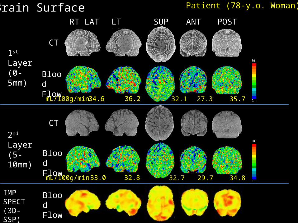
RT LAT LT LAT SUP ANT POST
Brain Surface
34.6 36.2 27.3 35.732.1
33.0 32.8 29.7 34.832.7
1st Layer(0-5mm)
2nd Layer(5-10mm)
IMPSPECT(3D-SSP)
CT
CT
BloodFlow
BloodFlow
BloodFlow
Patient (78-y.o. Woman)
mL/100g/min
mL/100g/min
RT LAT LT LAT SUP ANT POST
Brain Surface
34.6 36.2 27.3 35.732.1
33.0 32.8 29.7 34.832.7
1st Layer(0-5mm)
2nd Layer(5-10mm)
IMPSPECT(3D-SSP)
CT
CT
BloodFlow
BloodFlow
FlowReductionRegions
Patient (78-y.o. Woman)
mL/100g/min
mL/100g/min
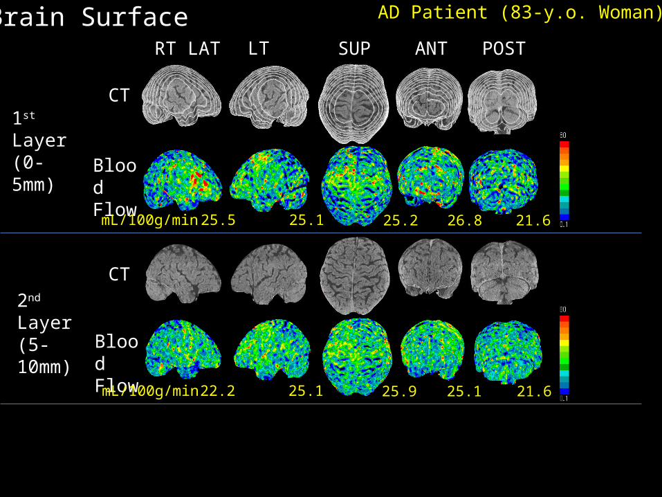
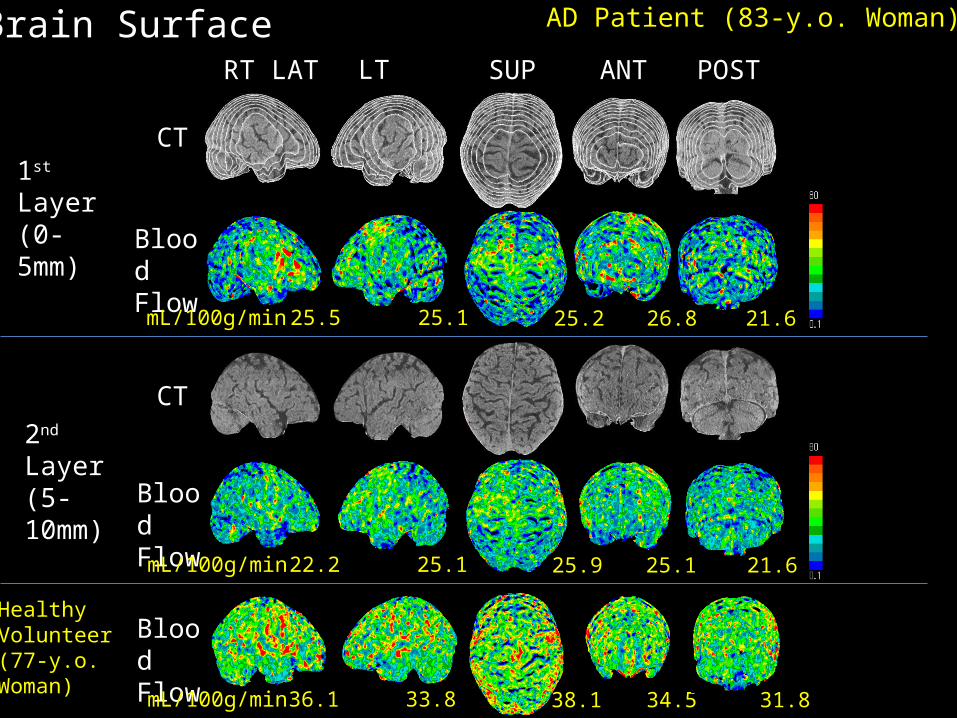
• Comparison of AD patient and healthy volunteer
RT LAT LT LAT SUP ANT POST
Brain Surface
1st Layer(0-5mm)
2nd Layer(5-10mm)
CT
CT
BloodFlow
BloodFlow
AD Patient (83-y.o. Woman)
25.5 25.1 25.2 26.8 21.6
22.2 25.1 25.9 25.1 21.6
mL/100g/min
mL/100g/min
RT LAT LT LAT SUP ANT POST
Brain Surface
1st Layer(0-5mm)
2nd Layer(5-10mm)
CT
CT
BloodFlow
BloodFlow
25.5 25.1 25.2 26.8 21.6
22.2 25.1 25.9 25.1 21.6
33.8 34.538.136.1 31.8
HealthyVolunteer(77-y.o.Woman)
BloodFlow
AD Patient (83-y.o. Woman)
mL/100g/min
mL/100g/min
mL/100g/min
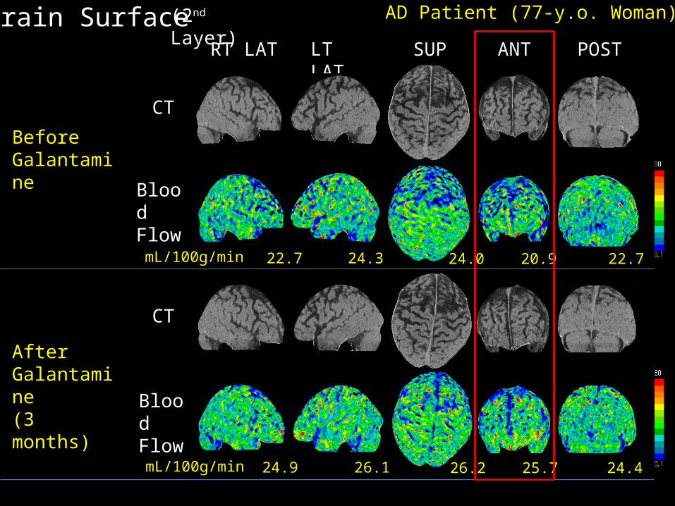
• Effect of drug administration to AD patient
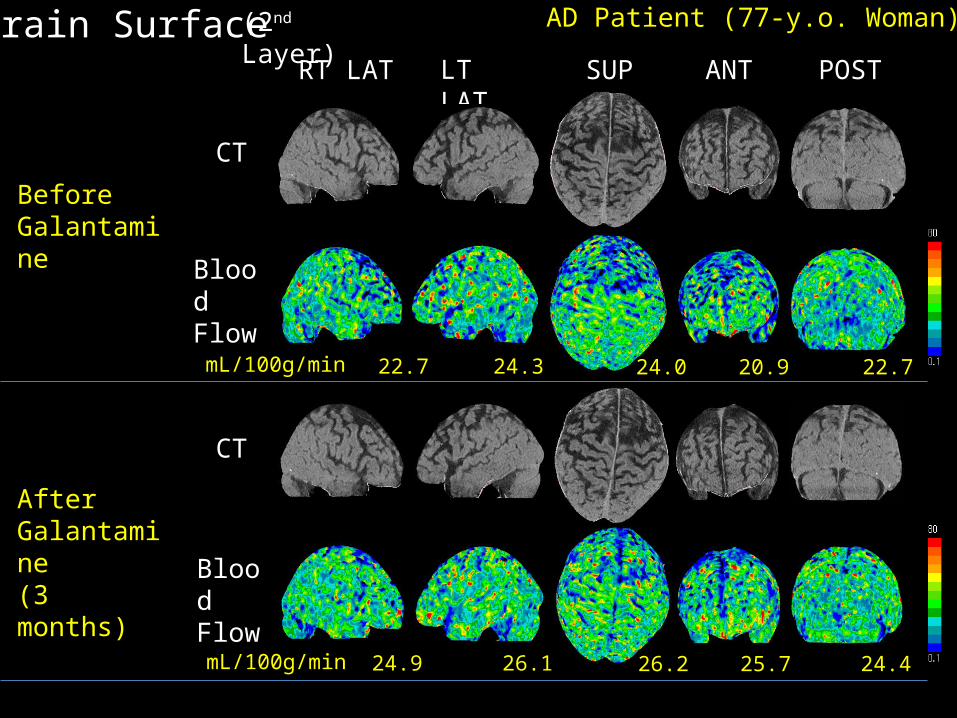
RT LAT LT LAT SUP ANT POST
Brain Surface
BeforeGalantamine
CT
CT
BloodFlow
BloodFlow
AD Patient (77-y.o. Woman)
AfterGalantamine(3 months)
22.7 24.3 20.9 22.724.0
24.9 26.1 25.7 24.426.2
(2nd Layer)
mL/100g/min
mL/100g/min
RT LAT LT LAT SUP ANT POST
Brain Surface
BeforeGalantamine
CT
CT
BloodFlow
BloodFlow
AD Patient (77-y.o. Woman)
AfterGalantamine(3 months)
22.7 24.3 20.9 22.724.0
24.9 26.1 25.7 24.426.2
(2nd Layer)
mL/100g/min
mL/100g/min
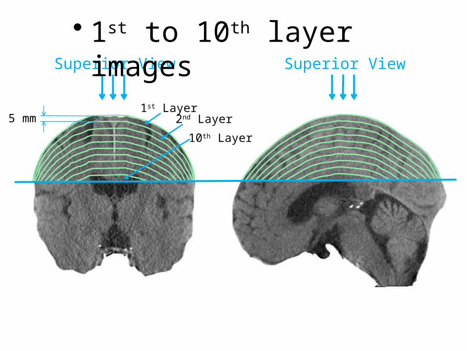
1st Layer
Superior View
• 1st to 10th layer images
2nd Layer
10th Layer
5 mm
Superior View
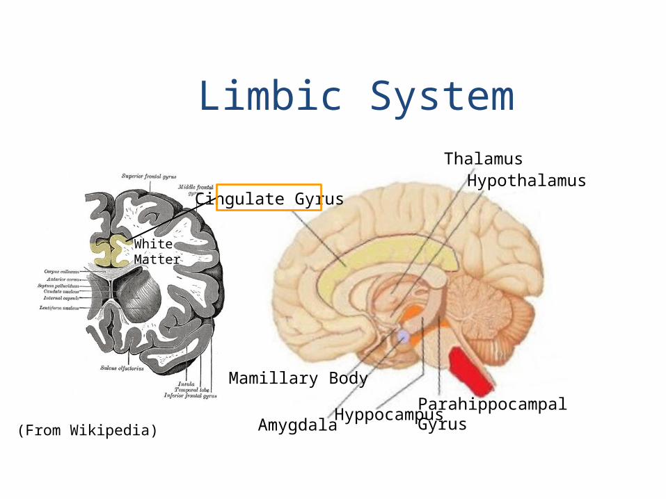
(From Wikipedia)
Cingulate Gyrus
ThalamusHypothalamus
ParahippocampalGyrusHyppocampus
Amygdala
Mamillary Body
Limbic System
WhiteMatter
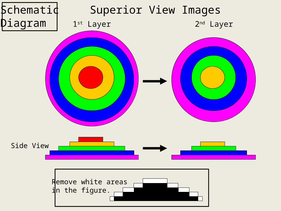
Remove white areasin the figure.
Side View
1st Layer
Superior View Images2nd Layer
SchematicDiagram
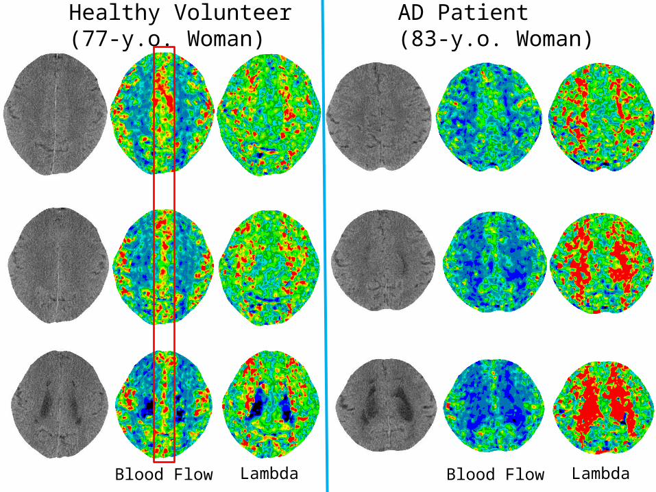
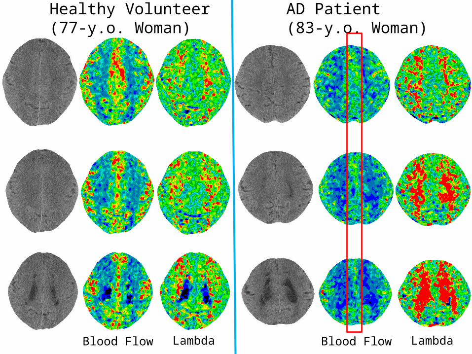
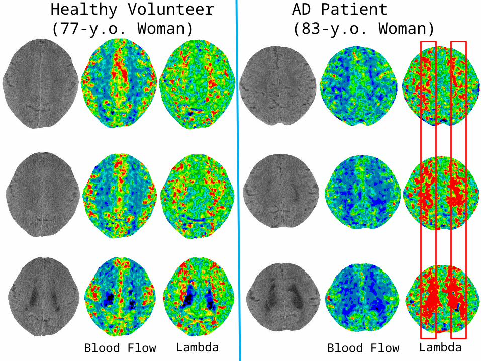
• Comparison of healthy volunteer and AD patient
Blood Flow Lambda
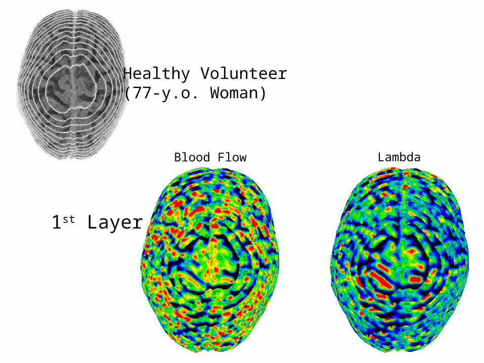
1st Layer
Healthy Volunteer(77-y.o. Woman)
1st LayerBlood Flow Lambda
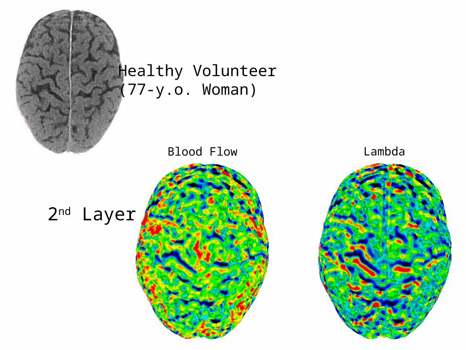
2nd Layer
Healthy Volunteer(77-y.o. Woman)

Blood Flow Lambda
3rd Layer
Healthy Volunteer(77-y.o. Woman)
Blood Flow Lambda
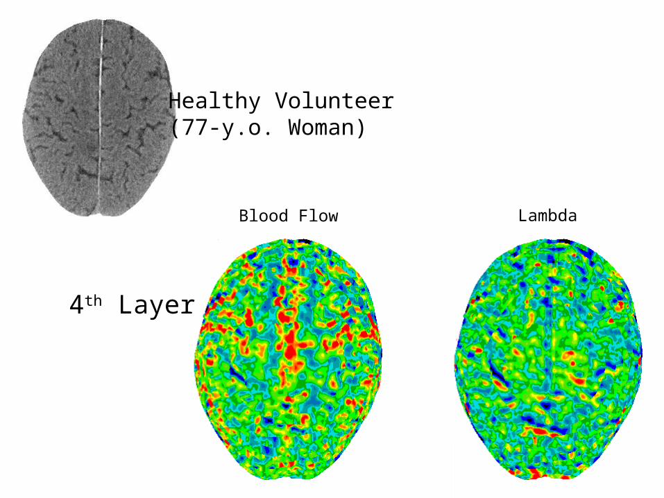
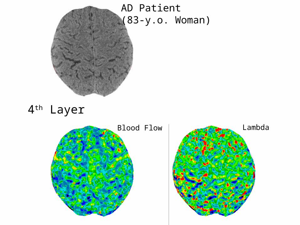
4th Layer
Healthy Volunteer(77-y.o. Woman)

Blood Flow Lambda
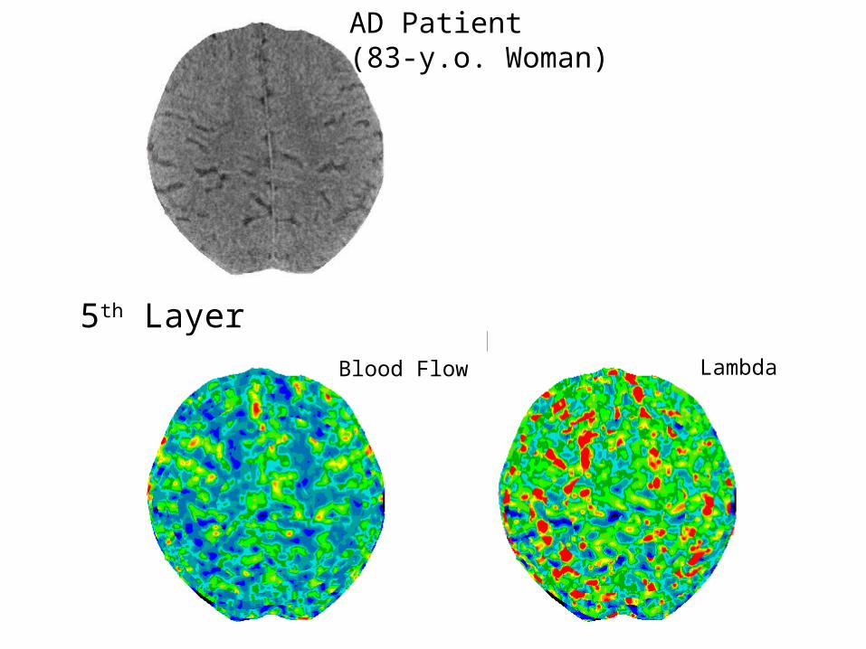
5th Layer
Healthy Volunteer(77-y.o. Woman)
Blood Flow Lambda
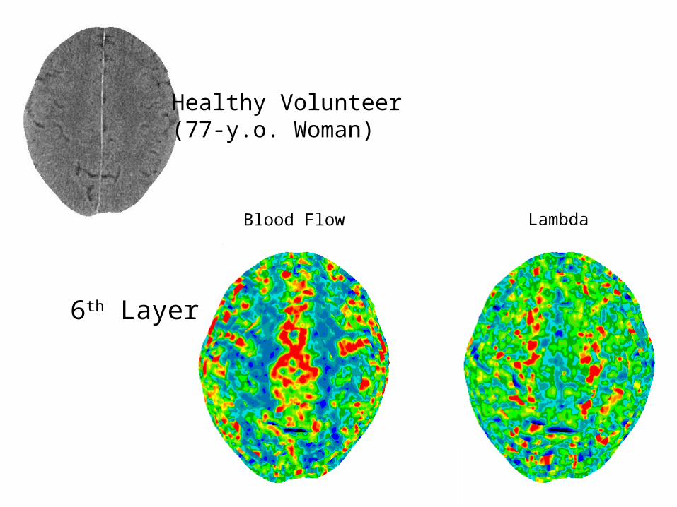
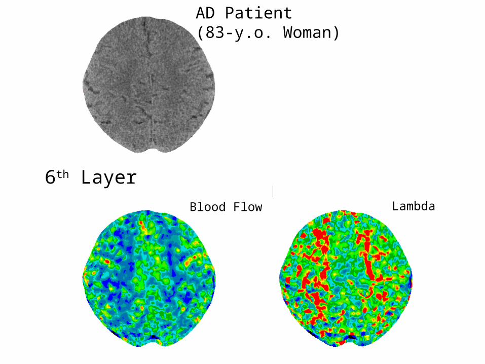
6th Layer
Healthy Volunteer(77-y.o. Woman)
Blood Flow Lambda
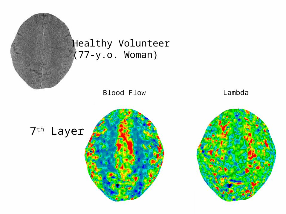
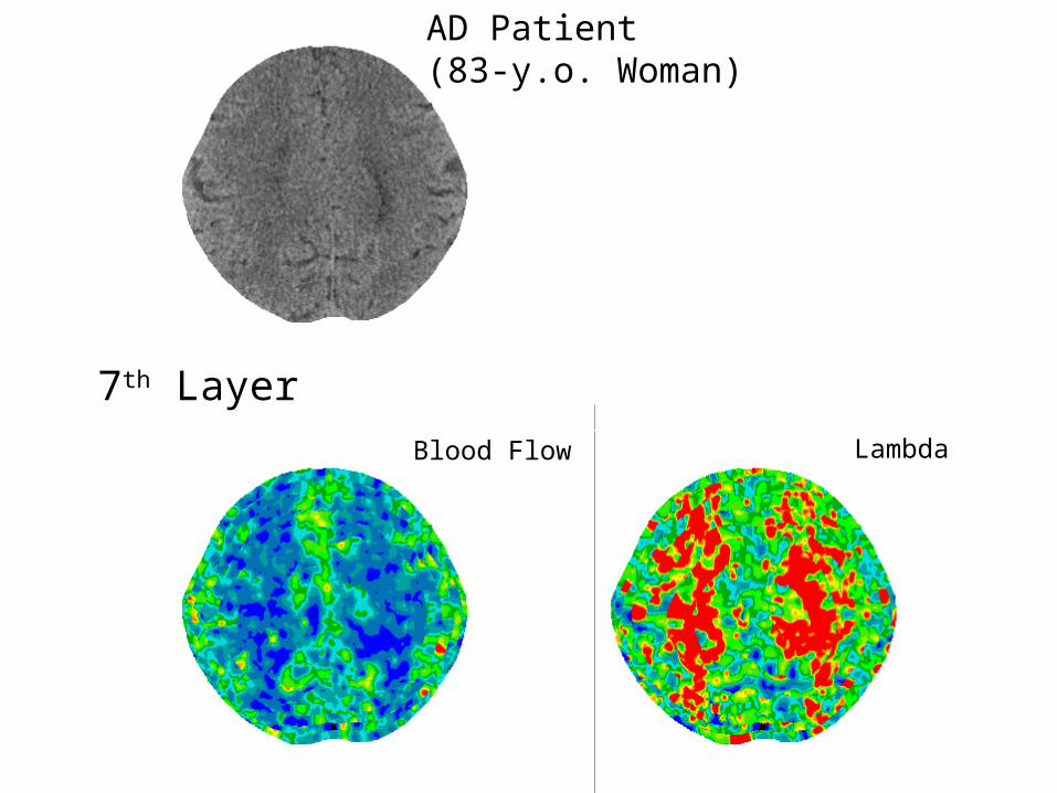
7th Layer
Healthy Volunteer(77-y.o. Woman)
Blood Flow Lambda
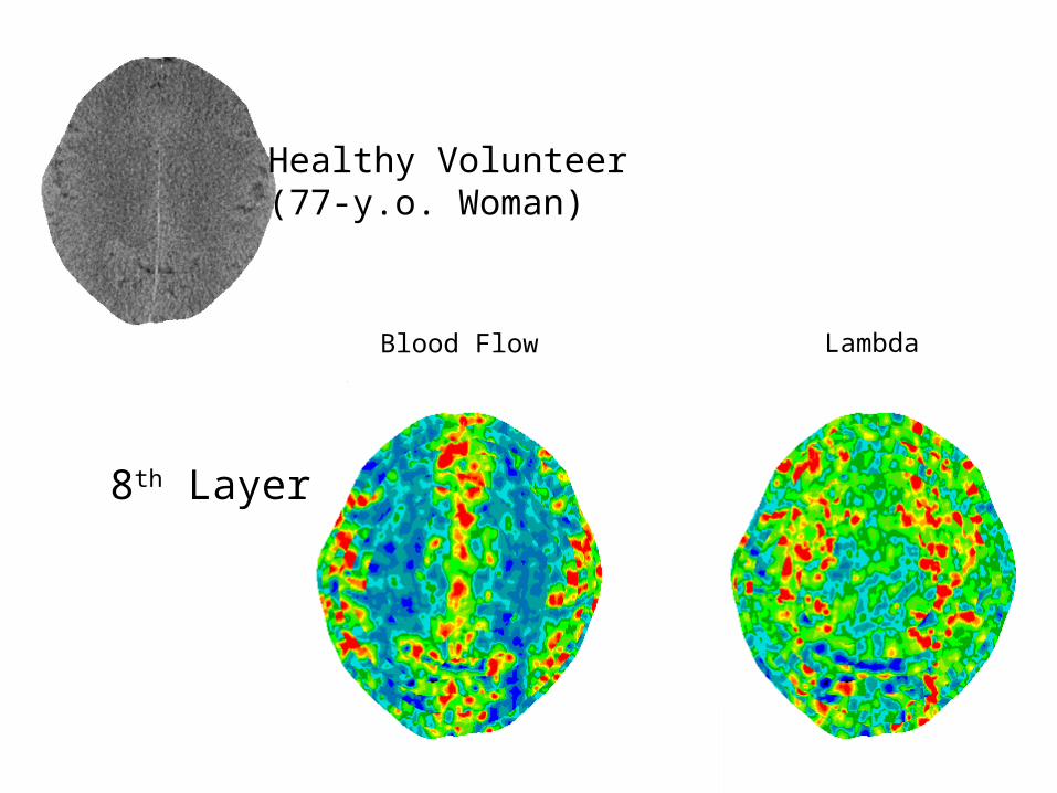
8th Layer
Healthy Volunteer(77-y.o. Woman)
Blood Flow Lambda
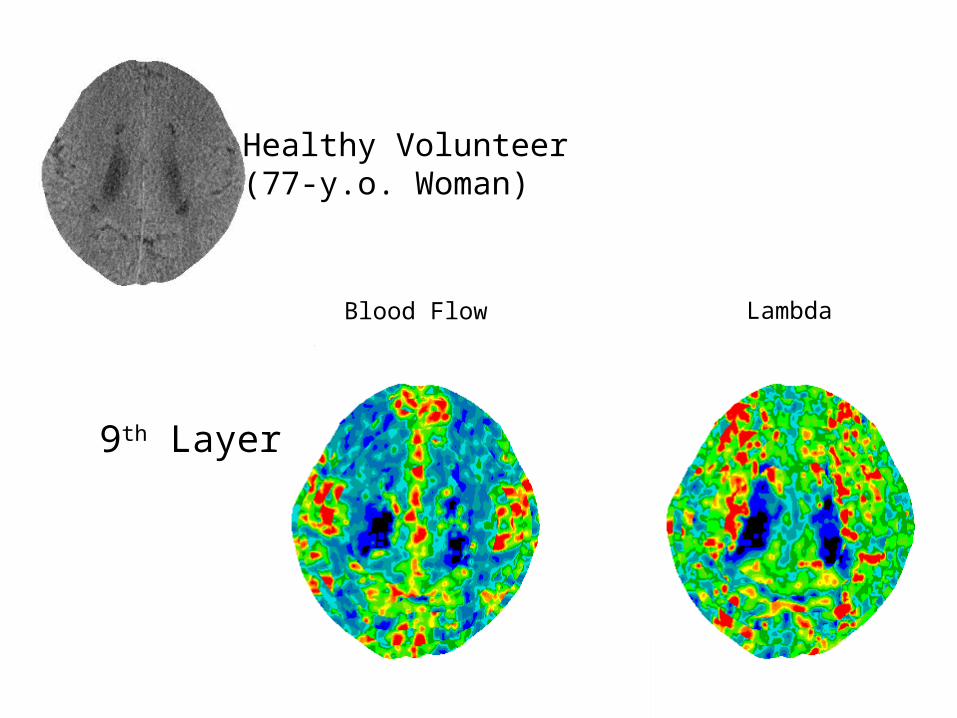
9th Layer
Healthy Volunteer(77-y.o. Woman)
Blood Flow Lambda
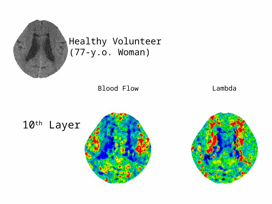
10th Layer
Healthy Volunteer(77-y.o. Woman)

Blood Flow Lambda
1st Layer
AD Patient(83-y.o. Woman)
Blood Flow Lambda
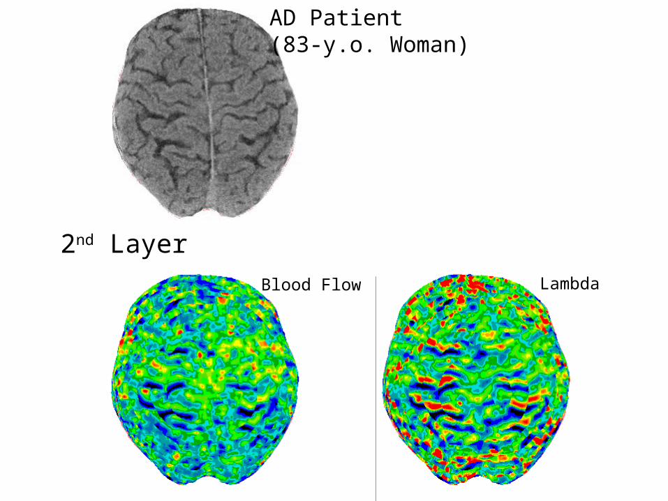
2nd Layer
AD Patient(83-y.o. Woman)
Blood Flow Lambda
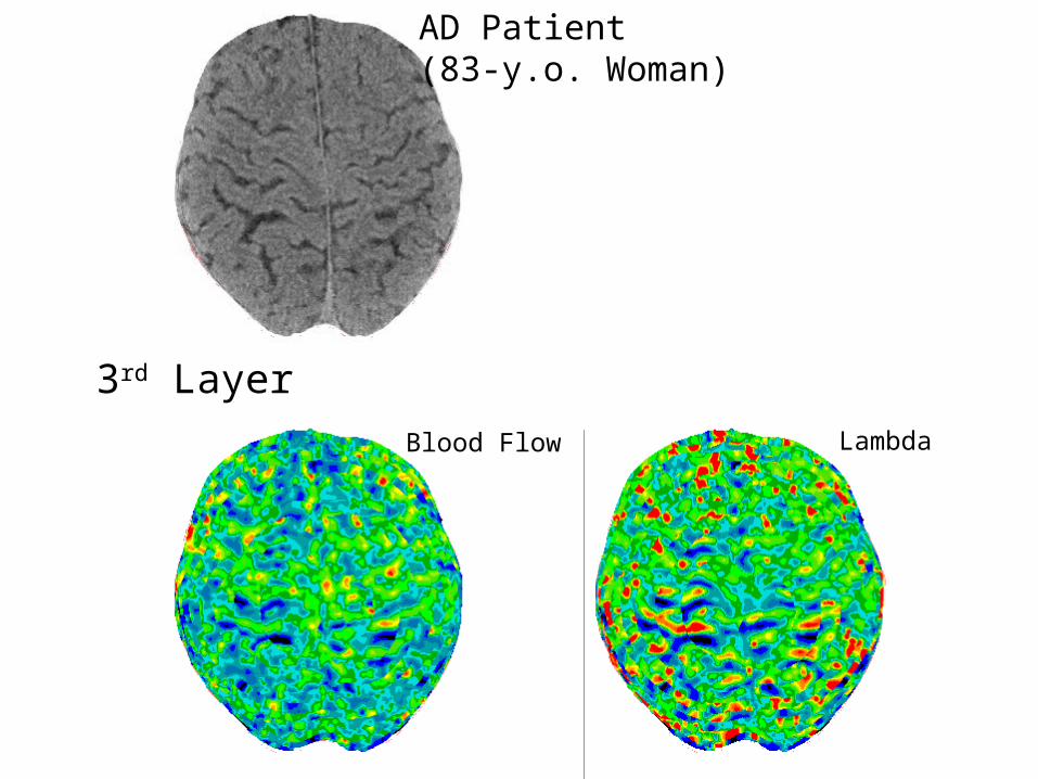
3rd Layer
AD Patient(83-y.o. Woman)
Blood Flow Lambda
4th Layer
AD Patient(83-y.o. Woman)
Blood Flow Lambda
5th Layer
AD Patient(83-y.o. Woman)
Blood Flow Lambda
6th Layer
AD Patient(83-y.o. Woman)
Blood Flow Lambda
7th Layer
AD Patient(83-y.o. Woman)
Blood Flow Lambda
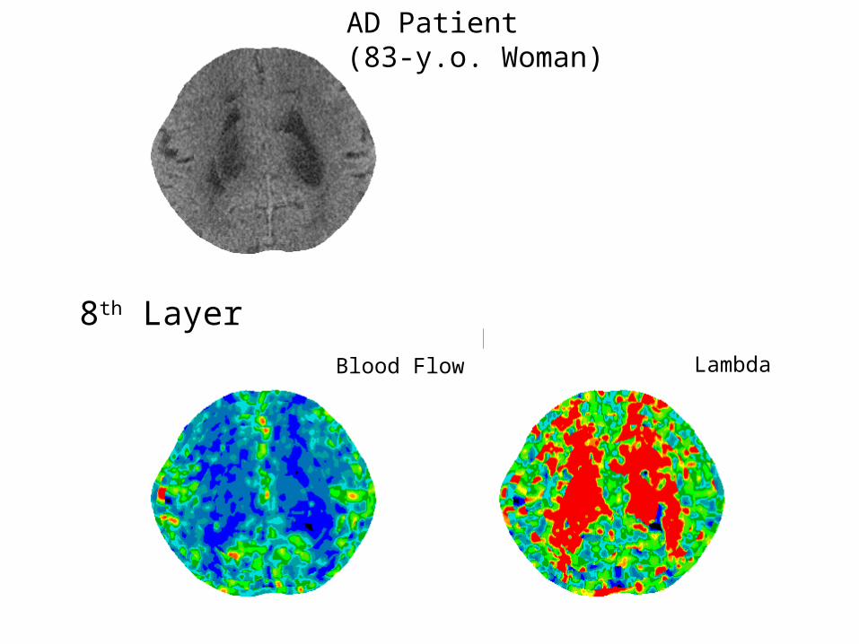
8th Layer
AD Patient(83-y.o. Woman)
Blood Flow Lambda
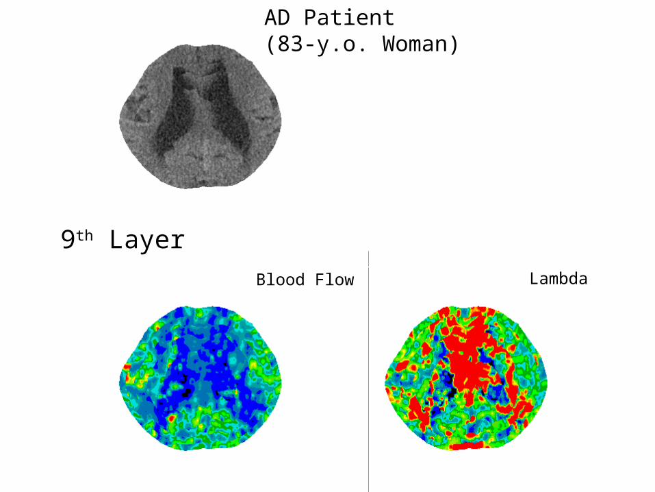
9th Layer
AD Patient(83-y.o. Woman)
Blood Flow Lambda
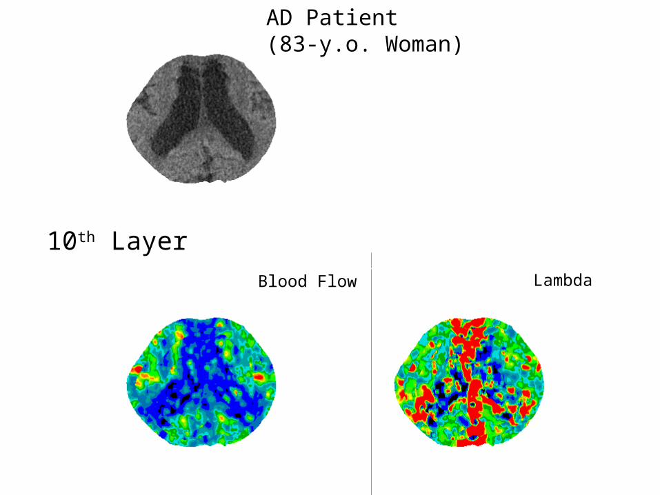
10th Layer
AD Patient(83-y.o. Woman)
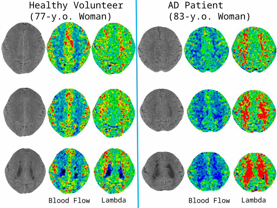
Healthy Volunteer(77-y.o. Woman)
AD Patient(83-y.o. Woman)
Blood Flow Lambda Blood Flow Lambda
Healthy Volunteer(77-y.o. Woman)
AD Patient(83-y.o. Woman)
Blood Flow Lambda Blood Flow Lambda
Healthy Volunteer(77-y.o. Woman)
AD Patient(83-y.o. Woman)
Blood Flow Lambda Blood Flow Lambda
Healthy Volunteer(77-y.o. Woman)
AD Patient(83-y.o. Woman)
Blood Flow Lambda Blood Flow Lambda
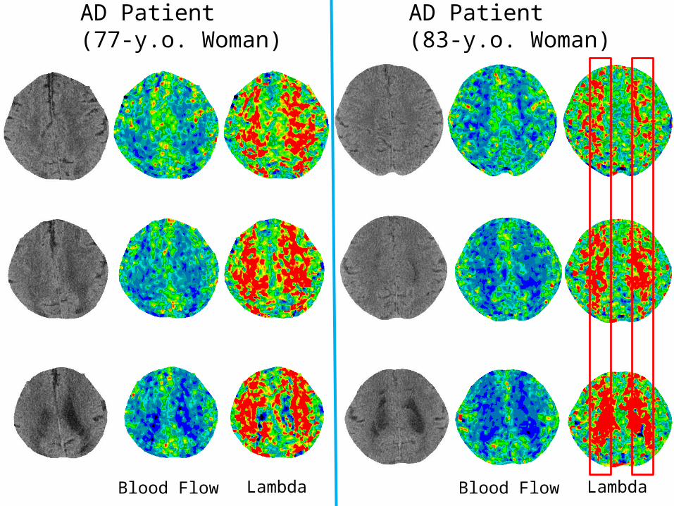
AD Patient(77-y.o. Woman)
AD Patient(83-y.o. Woman)
Blood Flow Lambda Blood Flow Lambda
[Conclusions]
• Method of creating quantitative blood flow images for the brain surface was established.
• Layer-by-layer spherical analysis would provide useful information which could not be obtained from tomographic images.
• High lambda suggests accumulation of substances in which xenon is highly soluble.
[Future]
• When the function of head movement correction is much more accomplished, radiation exposure will be reduced by nearly half by decreasing the number of CT scans (from 9 to 5).
• Improvement of CT image processing by TOSHIBA could reduce mAs with little deterioration of the quality of CT images and also reduce the radiation exposure.
• Quantitative judgment of treatment effectiveness, specifying the form of dementia, and detecting the dementia in its early stage, by means of Xe-CT.

Thank you very much.