Embed Size (px)
Citation preview
Cervical Myelopathy
Extrait du L'encyclopédie neurochirurgicale
http://neurochirurgica.org/spip.php?article2
Cervical Myelopathy- Pathology -
Date de mise en ligne : Friday 13 September 2013
Description :
Myelopathy is the term designated to all chronic cord lesions regardless of their etiologic origins which may be vascular, inflammatory, nutritional deficiency,
post-radiation etc ... but the term myelopathy is more restrictive and should be reserved for a chronic cord lesion resulting from the reduction in the dimensions of
the cervical spinal canal of which the main etiology is cervical spondylosis.
L'encyclopédie neurochirurgicale
Copyright © L'encyclopédie neurochirurgicale Page 1/33
Cervical Myelopathy
DEFINITION
Myelopathy is the term designated to all chronic cord lesions regardless of their etiologic origins which may bevascular, inflammatory, nutritional deficiency, post-radiation etc ... but the term myelopathy is more restrictive andshould be reserved for a chronic cord lesion resulting from the reduction in the dimensions of the cervical spinal canalof which the main etiology is cervical spondylosis.
HISTORY• 1823 : Charles Prosper Ollivier d'Angers in "From the spinal cord and its diseases" suggests a link between a
protruding intervertebral disc and the myelopathy.• 1888 : Strumpell and Pierre-Marie (1898) studying the "spinal cord lesions from chronic cervical osteoarthritis"
reported the the relationship between spondylosis and paraplegia.• 1892 : Victor Horsely did a cervical laminectomy for a patient with cervical spondylotic myelopathy in TaylorJ,
CollierJ. The occurrence of lesions in optic neuritis of the spinal cord-Injury, tumor, myelitis. (An account oftwelve cases and one autopsy). Brain1901; 24:532.
• 1928 : JA Barré YC Liu and specified the role of cervical spondylosis in the genesis of certain myelopathies.• 1952 : W. R. Brain, D. Northfield and Mr Wilkinsonin The neurological manifestation of cervical spondylosis.
Brain (1952) 75 (2): 187-225 published the first work on the neurological manifestations of cervical spondylosis.• 1955 : Robinson RA, Smith GW. Anterolateral cervical disc removal and interbody fusion for cervical disk
syndrom. Bull Johns Hopkins Hosp 1955; 96: 223 (Abstract)• 1956 : Wolf B, Khilnani M, Malis L. Emphasized on the importance of narrowing of the cervical canal in the
genesis of cord complications in The sagittal diameter of the bony cervical spinal canal and its significance incervical spondylosis. J. Mount Sinai Hosp 1956; 23:283 -292.
EPIDEMIOLOGY
Cervical spondylotic myelopathy represents 55% of cervical myelopathies in adults (7). The condition is observedmostly after the age of 50 years, with men more affected than women. Its incidence increases with age and it is aleading cause of functional disability in the elderly (25). ++++
NATURAL HISTORY
The natural history of cervical myelopathies is not well understood. The natural history of cervical spondylotic myelopathy. Matz PG, Anderson PA, Holly LT, Groff MW, Heary RF,Kaiser MG, Mummaneni PV, Ryken TC, Choudhri TF, Vresilovic EJ, Resnick DK. J Neurosurg Spine. 2009; 11 (2):104-11.
ETIOLOGIES
Copyright © L'encyclopédie neurochirurgicale Page 2/33

Cervical Myelopathy
1. Constitutional Stenoses
Pure constitutional stenosis rarely is a cause of cervical myelopathy except in extreme cases of severe stenosis asin achondroplasia.It is more often a predisposing factor.
2. Cervical Spondylosis
Spinal degenerative diseases start as early as the age of 20 years and constitute the main etiology of cervicalmyelopathy(25).They seem to be favored by the load on the spine in certain professions, previous trauma (rugbyplayers) and occur earlier and more frequently in patients with abnormal movements (spasmodic torticollis disease,Tourette's, choreoathetosis ...). Decompensation of congenital cervical blocks leads to early onset of degenerationfrom adjacent segment "overload". The initial lesions start in the disc and correspond to a reduction of the water content of the nucleus pulposus(nuclear desiccation), an increase of collagen, and a decrease in the mucopolysaccharides and chondroitin sulfatecontent. Anatomically, there are annular fissures which engage in displaced fragments of the nucleus. There is a lossof disc height (narrowing of the disc space). These anatomical changes alter the mechanical properties of the discand are responsible for its degeneration (26, 76).
The degenerative cascade successively involves the disc ("soft" herniated disc, degenerative disc disease, herniatedcalcified "hard disc"), unco-vertebral joints (uncarthrosis), the facet joints (zygapophysial or facet joint osteoarthritis).The ligaments (posterior longitudinal ligament, ligamentum flavum) hypertrophy, lose their mechanical properties,thicken and calcify. All these lesions aggravated by osteophytic reaction reduce the size of the cervical spinal canaland are much more pathogenic than the constitutional narrow canal.
The lesions may sometimes be limited to one or two adjacent segments, at the most mobile segments of the lowercervical spine (C5/C6 and C6/C7), but sometimes extend through the whole sub-axial cervical spine (C3-C7). Degenerative lesions can also be the source of static spinal disorders (loss of physiological lordosis, sometimeskyphosis or rarely degenerative scoliosis), being initiated by the loss of disc height, chronic instability or degenerativespondylolisthesis secondary to a change in the orientation of the joint surfaces. During flexion-extension movements,the ligaments that have lost their elasticity can jut into the lumen of the spinal canal, contributing to the compromiseof the spinal cord (76).
3. Ossification of the posterior longitudinal ligament(OPLL)
OPLL is a disease found mainly in the Far East and could correspond to a specific anatomic type of degenerativecervical spine lesion in Asian populations, suggesting a hypothetical genetic predisposition, corroborated by theincreased prevalence in certain families and in identical twins (3).
Significant lesions of the posterior longitudinal ligament are observed in 11% of patients in the sixth decade in the FarEast. The disease has an incidence of 1.4%. Few cases have been observed in Caucasian populations in Europeand the United States. The frequency of this disease seems to be underestimated.
Ossification usually starts behind the body of C5 and progressively spreads through the entire cervical spine incontrast to segmental (or localized) forms, discontinuous or continuous forms.
Copyright © L'encyclopédie neurochirurgicale Page 3/33
Cervical Myelopathy
In the Japanese population, obesity and glucose intolerance significantly favors the ossification of the anterior andposterior longitudinal ligament (78). The clinical course is unpredictable: 80% of patients followed-up over a period of greater than 10 years remainasymptomatic despite the presence of significant anatomical lesions, 20% will develop neurologic deficit and arecandidates for surgical treatment (52).
The pathogenesis of OPLL remains uncertain: osteogenesis ensues from hypertrophied and hypervascular ligamentwhen it "detaches" from the posterior surface of the vertebra by disc protrusions.
4. Late posttraumatic forms
They should be differentiated from forms revealed during trauma, and related to unrecognized or poorly treatedlesions: non-union causing chronic instability, mal-union reducing the diameter of the cervical spinal canal,post-traumatic discopathies usually localized to one injured segment.
5. Other etiologies
Cervical rheumatoid arthritis affecting especially the upper cervical spine (atlanto-axial dislocation) and to a lesserextent the lower cervical spine may present with the clinical picture of cervical myelopathy (59). We can regroup rare neurologic complications which can cause narrowing of the cervical canal such ankylosing spondylitis, goutytophi from the posterior joints and vertebral ankylosing hyperostosis (Forestier's disease) or Paget's disease.Myelopathies have been observed in patients on long-term dialysis due to epidural calcification (85). ++++
PATHOPHYSIOLOGICAL PRINCIPLES OF THEPATHOLOGICAL PROCESS
1 Mechanical compression.
A reduction in the dimensions of the cervical canal, especially in the sagittal plane, is found in all cases. It is the maincause of cervical myelopathy. It is mainly related to the development of the anterior osteophytes and/or hypertrophyof the ligamentum flavum, on which the degenerative hypertrophy of the articular processes (facets), the posteriorlongitudinal ligament and the laminae can be superimposed (76).
Delayed myelopathy due chronic cord compression has been demonstrated in the dog (32) and in rats (42). Adecreased neuronal density of 20% is observed as from the 9th week and more than 35% beyond the 25th week,whereas no significant lesion is observed before the 3rd week. Below the site of the compression there is axonal loss. The phenomena of cavitations of the gray matter appear as soon as there is a significant neuronal loss whereasthe white matter is left unaffected for long.
Copyright © L'encyclopédie neurochirurgicale Page 4/33
Cervical Myelopathy
2 Micro-traumas
Each hour the cephalic end (head) makes hundreds of movements involving rotation and flexion- extension in varyingproportions. It has been shown that with maximum flexion of the neck, the posterior wall of the spinal canallengthens by 5cm and the anterior wall by 1.5 cm therefore stretching the dura and the cord. During movements offlexion-extension there is a decrease in the dimensions of the spinal canal aggravated during extension by the bulging of the yellow ligament. During each movement micro-traumas occur when compressive elements come incontact. The evolution of the myelopathy correlates with the number of these movements: these movements can belimited cervical immobilization.
Degenerative lesions of the disco-ligamentous apparatus leads to chronic instability responsible for a sub-luxationseen on dynamic images, additionally imposing more shearing movements on the cord (24).
3. Vascular phenomena
Venous stasis stemming from canal stenosis appears to have an important pathophysiologic role in cervicalmyelopathy by causing chronic ischemia and cord edema.
A couple of years ago, through the initiative of Aboulker (1), it was proposed to systematically investigate for a superior azygos system pathology in which drains the cervical epidural veins. It is by this mechanism thatneurological signs of dural arteriovenous fistulas are explained.
The role of arteries seems more modest due to the richness of the anastomotic system at the cervical level, but it isnot an impossibility to consider a compression of radiculomedullary arteries of the cord, the anterior spinal arteries oreven the vertebral arteries, by keeping in mind that the age for osteoarthritis is corresponds to that of atheroscleroticplaque (92). The anatomical lesions discerned during the autopsies of patients with cervical myelopathy suggest anischemic mechanism (62). ++++
DIAGNOSIS
I- CLINICAL FEATURES
The clinical features result from the direct compression of the nerve roots and/or the spinal cord, alteration in thearterial flow, venous congestion and inflammation. Certain genetic elements can influence the resistance of theneural structures (33).
1. Mode of onset
The affection is two times more common in males than in females and begins between the ages of 50 and 60 years(25). Before 50 years, it is more often a discogenic canal stenosis from a "soft" central disc. Neurological impairment
Copyright © L'encyclopédie neurochirurgicale Page 5/33
Cervical Myelopathy
is often preceded for several months or years by mechanical cervical pain (50 to 80% of cases) which is poorlysystematized, episodes of torticollis or even true cervico-brachial neuralgia . This often occurs in a context of repetitive microtraumas, occupational or sports related spinal "overload" and morerarely cervical spine injury without obvious radiological lesion, labelled "cervical sprain", which can only be discerned through dynamic images. However, it is possible that such a trauma causes discs and/or ligamentous injuriesleading to degenerative lesions (14). The more common scenario is that of mundane mechanical pain from cervicalspondylosis.
Gait disturbances are usually early in the form fatigability, tendency to the fall, reduction in the walking distance thatcan correspond to a true intermittent neurological claudication and sometimes episodes of flinching movements ofthe lower limbs. Gait disturbances may also be related with difficulties of coordination (ataxia sensory), disorders ofbalance, a poor perception of the ground, or even pain or paresthesia that occur for increasingly shorter distances.
In practice these disorders are fairly easy to differentiate from gait apraxia (astasia abasia) observed in adults chronichydrocephalus, and from intermittent claudication due to lumbar canal stenosis while bearing in mind that thesepathologies frequent in this age group may be associated.
Upper limbs affection can be another mode of presentation of the disease. Most frequently presents with pain orparesthesia more or less systematized in a radicular territory accompanied by a subjective sensation of clumsinessor functional disability making it increasingly difficult to achieve the delicate(fine) movements. Amyotrophy may be thepresenting symptom.
Acute decompensation of a latent form during a relatively minor cervical trauma is a common mode of revelation: theclinical picture is that of a central cord contussion (Schneider's syndrome) with incomplete tetraplegia where themotor impairment is disproportionately greater in upper compared to lower extremities and maximally brachialdiplegia (72).
2. Neurological examination findings
Neurological signs vary from one patient to another and occur in different proportions in the upper limbs, lower limbsand to a lesser degree the sphincters (63).
2a- Upper limbs affection
Because of their pathophysiologic mechanism there is still no good correlation between the topography of segmentalneurological signs and the site of the anatomical lesions. Subjective sensory manifestations (Symptoms) are almost always present as paresthesia or mechanical paintriggered by straining or neck movements, or as nocturnal neuropathic pain. The symptoms are unilateral or bilateraland often asymmetrical without an accurate radicular pattern. More rarely, it presents by a typical picture ofcervico-brachial neuralgia that can be revealed during history taking a few years earlier.
Radicular pain of mechanical origin, secondary to degenerative changes, can be provoked during the clinicalexamination by eliciting Spurling's sign (radicular pain reproduced when the examiner exerts downward pressure onvertex with the neck in extension while tilting head towards symptomatic side) and the abduction relief sign(reduction or disappearance of the radicular pain when the patient abducts the arm by putting his hand on his head)(21).
Copyright © L'encyclopédie neurochirurgicale Page 6/33
Cervical Myelopathy
Objective sensory manifestations (Signs) can be demonstrated by the examination of the painful region. They lackan accurate radicular topography and are often in the form of hypoesthesia of all sensory modalities. Thedisturbances may be prominent on the lemniscal pathway explaining the clumsiness to achieve fine movements:holding pencil, sewing, manipulating small objects .
A segmental motor affection may be due to a lesion of the nerve root or of the anterior horn cell; the latter sometimessimulate starting amyotrophic lateral sclerosis. The affection is usually distal in the hand muscles accompanied bymuscle atrophy and rarely fibrillations localized in the upper limbs.
The abolition of one or more reflexes is frequently observed but their exaggeration can be found in the high cordaffection. Clinical examination may reveal a Hoffmann's sign in upper limbs in cases of involvement the pyramid tract.
The functional impairment felt by the patient stems from both an affection of the sensation and diminished motorpower.
2b-Lower limbs Affection
It is responsible for abnormal gait, impaired balance, functional impotence and tendencies to the fall.
Pyramidal involvement is rarely responsible for a significant motor deficit. There is often spastic paraparesis withhypertonia of the extensors, an exaggeration of deep tendon reflexes and a bilateral positive Babinski sign. Theaffection can be discrete at the beginning and objectified only after fatigue and after classical facilitation maneuver. Itcan be masked when associated with lumbar canal stenosis or peripheral neuropathy. Involvement of the posterior column is responsible for subjective manifestations such as paresthesia, numbnessand pain sometimes spontaneous or provoked by neck flexion (Lhermitte's sign). Posterior column lesion isobjectivated by the decrease in vibration sensation, impairment of arthrokinesthesia and sometimes presence ofRomberg's sign.
Involvement of the spinothalamic tact is rarer and late and results in a thermal hypoesthesia and hypalgesia.
The gait disturbance presented by the patient are more related to hypertonia and sensory ataxia than to the motordeficit.
2c-Sphincter dysfunction
Present in 30-40% of the cases and are often underestimated. They are in the form of dysuria, urinary frequencyand sometimes stress incontinence. They are not always related to their cause (suggestive of prostatism orage-related incontinence in women) and should be systematically investigated by measuring the post-void residualvia an ultrasound of the bladder.
3. Clinical forms
The condition is in fact very polymorphic; depending on the topography of symptoms, their severity and evolution. It istherefore possible to individualize several clinical forms:
Copyright © L'encyclopédie neurochirurgicale Page 7/33
Cervical Myelopathy
3a-Ataxic-Spastic Form : It is the most frequent form predominantly manifesting by gait disturbances anddysequilibrium with sub-clinically affection of the upper limbs. All lesions causing cord affection can be evoked facedwith this form notably "slow" cord compression in which there is no sensory level and the sensory manifestations arediscrete, and multiple sclerosis (in which the signs are not diffused).
3b-Amyotrophic form predominantly affecting the upper limbs can at the beginning simulate amyotrophic lateralsclerosis but the course is much more rapid and disabling.
3c-Spastic paraparetic forms-with minimal sensory lesion,
3d-Brown Sequard-like forms-occur when there is predominantly unilateral cord lesion.
3e- Forms evolving by successive spurts are suggestive of late onset multiple sclerosis.
Nurick's classification established in 1972 (61) evaluates in a simple and reproducible manner the functional disabilityof patients, monitoring of their progress and evaluates the results of treatment, but is relatively imprecise (Table 1).
The classification of the Japanese Orthopaedic Association (JOA) (38) which is imposed on most English speaking authors after modification (6) is the sum of functional scores. The maximum score is 17 for patients without anyneurological disease (Table 2).
4-Evolution
There is no spontaneous improvement. The course is unpredictable with progressive neurological deteriorationoccurring most often (33). However, long periods of dormancy can be observed (53). In about 75% of the cases, theevolution occurs in a discontinuous mode by successive bouts over several years. In 20% of the cases the evolutionis more or less rapidly progressive and in 5% of the cases, there is a sudden decompensation often caused bycervical trauma of varying severity. This decompensation is rare in patients below 75 years with moderate cervicalmyelopathy (JOA score> 12) (53).
In the case of degenerative cervical stenosis without myelopathy, the presence of electromyographic abnormalities orclinical radiculopathy predicts progression to symptomatic cervical myelopathy (53).
The end stage is represented by a complete inability to ambulate and / or severe functional impairment of the upperlimb producing severe disability. ++++
II-Imaging
1 - Plain Radiography
Copyright © L'encyclopédie neurochirurgicale Page 8/33
Cervical Myelopathy
Plain radiographs are still relevant despite the development of modern imaging techniques (Figures 1, 2, 3). <a href="IMG/jpg/cervical_myelopathies_mai_2013_english.jpg" type="image/jpeg" title="">
<a href="IMG/jpg/cervical_myelopathies_mai_2013_english-2.jpg" type="image/jpeg" title="">
These should include the following views: Antero-posterior (AP), lateral, ¾ obliques and especially lateral dynamicfilms that cannot be easily obtained with CT and / or MRI.
Plain radiographs enable an overall assessment of the dimensions of the spinal canal and detect the existence of aconstitutional canal stenosis-a predisposing factor for cervical myelopathy (49):
• The antero-posterior diameter of the spinal canal measured from the middle of the posterior vertebral body tothe nearest point of the spinous process is equal to or less than the antero-posterior diameter of the vertebralbody: Pavlov index (64).
• The presence of platyspondyly with widening of antero-posterior diameter of the vertebral body• The articular processes are projected on the posterior part of the vertebral body.
They show the characteristic lesions of cervical spondylosis predominantly at the C5/C6, C6/C7 and C4/C5 disclevels, and specify the number of levels involved. The following are shown:
Copyright © L'encyclopédie neurochirurgicale Page 9/33
Cervical Myelopathy
• Hypertrophy of the articular processes (facets)• Osteophytes (bone spurs) of the posterior joints, endplates and unco-vertebral joints• Pinching of the intervertebral disc• Ligamentous ossification.
Disorders of shape of the spine (static deformities) are objectified: loss of physiological lordosis, inversion ofcurvature, degenerative spondylolisthesis and instability on dynamic films most often in flexion. Pathologies such asequelae of cervical spine injury or predisposing factors such as congenital cervical fusion may be associated. Of old, it was classical to measure the dimensions of the spinal canal on these images correcting for radiologicalmagnifications but this has lost its relevance today as these measurements are performed on the CT scan.
2-CT Scan
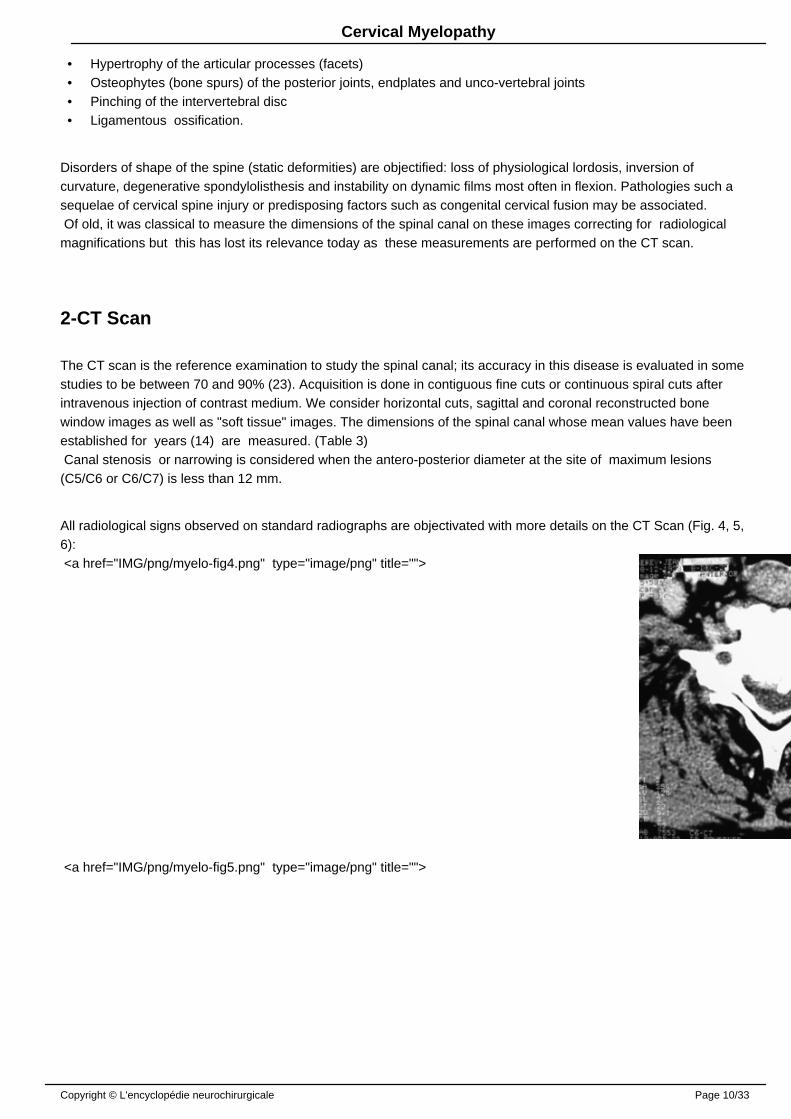
The CT scan is the reference examination to study the spinal canal; its accuracy in this disease is evaluated in somestudies to be between 70 and 90% (23). Acquisition is done in contiguous fine cuts or continuous spiral cuts afterintravenous injection of contrast medium. We consider horizontal cuts, sagittal and coronal reconstructed bonewindow images as well as "soft tissue" images. The dimensions of the spinal canal whose mean values ��have beenestablished for years (14) are measured. (Table 3) Canal stenosis or narrowing is considered when the antero-posterior diameter at the site of maximum lesions(C5/C6 or C6/C7) is less than 12 mm.
All radiological signs observed on standard radiographs are objectivated with more details on the CT Scan (Fig. 4, 5,6): <a href="IMG/png/myelo-fig4.png" type="image/png" title="">
<a href="IMG/png/myelo-fig5.png" type="image/png" title="">
Copyright © L'encyclopédie neurochirurgicale Page 10/33
Cervical Myelopathy
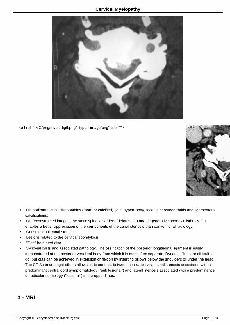
<a href="IMG/png/myelo-fig6.png" type="image/png" title="">
• On horizontal cuts: discopathies ("soft" or calcified), joint hypertrophy, facet joint osteoarthritis and ligamentouscalcifications,
• On reconstructed images: the static spinal disorders (deformities) and degenerative spondylolisthesis. CTenables a better appreciation of the components of the canal stenosis than conventional radiology:
• Constitutional canal stenosis• Lesions related to the cervical spondylosis• "Soft" herniated disc• Synovial cysts and associated pathology. The ossification of the posterior longitudinal ligament is easily
demonstrated at the posterior vertebral body from which it is most often separate. Dynamic films are difficult todo, but cuts can be achieved in extension or flexion by inserting pillows below the shoulders or under the head.The CT Scan amongst others allows us to contrast between central cervical canal stenosis associated with apredominant central cord symptomatology ("sub lesional") and lateral stenosis associated with a predominanceof radicular semiology ("lesional") in the upper limbs.
3 - MRI
Copyright © L'encyclopédie neurochirurgicale Page 11/33
Cervical Myelopathy
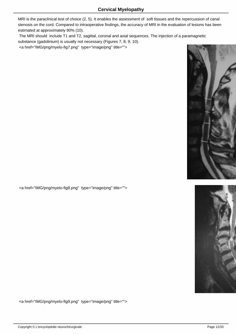
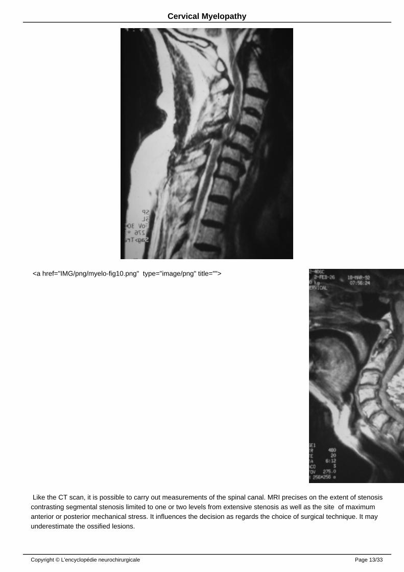
MRI is the paraclinical test of choice (2, 5). It enables the assessment of soft tissues and the repercussion of canalstenosis on the cord. Compared to intraoperative findings, the accuracy of MRI in the evaluation of lesions has beenestimated at approximately 90% (10). The MRI should include T1 and T2, sagittal, coronal and axial sequences. The injection of a paramagneticsubstance (gadolinium) is usually not necessary (Figures 7, 8, 9, 10). <a href="IMG/png/myelo-fig7.png" type="image/png" title="">
<a href="IMG/png/myelo-fig8.png" type="image/png" title="">
<a href="IMG/png/myelo-fig9.png" type="image/png" title="">
Copyright © L'encyclopédie neurochirurgicale Page 12/33
Cervical Myelopathy
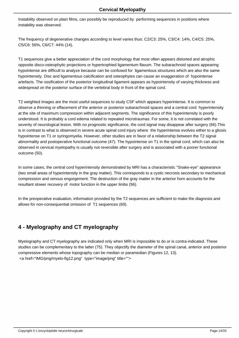
<a href="IMG/png/myelo-fig10.png" type="image/png" title="">
Like the CT scan, it is possible to carry out measurements of the spinal canal. MRI precises on the extent of stenosiscontrasting segmental stenosis limited to one or two levels from extensive stenosis as well as the site of maximumanterior or posterior mechanical stress. It influences the decision as regards the choice of surgical technique. It mayunderestimate the ossified lesions.
Copyright © L'encyclopédie neurochirurgicale Page 13/33
Cervical Myelopathy
Instability observed on plain films, can possibly be reproduced by performing sequences in positions whereinstability was observed.
The frequency of degenerative changes according to level varies thus: C2/C3: 25%, C3/C4: 14%, C4/C5: 25%,C5/C6: 56%, C6/C7: 44% (14).
T1 sequences give a better appreciation of the cord morphology that most often appears distorted and atrophicopposite disco-osteophytic projections or hypertrophied ligamentum flavum. The subarachnoid spaces appearinghypointense are difficult to analyze because can be confused for ligamentous structures which are also the samehypointensity. Disc and ligamentous calcification and osteophytes can cause an exaggeration of hypointenseartefacts. The ossification of the posterior longitudinal ligament appears as hypointensity of varying thickness andwidespread on the posterior surface of the vertebral body in front of the spinal cord.
T2 weighted images are the most useful sequences to study CSF which appears hyperintense. It is common toobserve a thinning or effacement of the anterior or posterior subarachnoid spaces and a central cord hyperintensityat the site of maximum compression within adjacent segments. The significance of this hyperintensity is poorlyunderstood. It is probably a cord edema related to repeated microtraumas. For some, it is not correlated with theseverity of neurological lesion. With no prognostic significance, the cord signal may disappear after surgery (66).Thisis in contrast to what is observed in severe acute spinal cord injury where the hyperintense evolves either to a gliosishypointense on T1 or syringomyelia. However, other studies are in favor of a relationship between the T2 signalabnormality and postoperative functional outcome (47). The hypointense on T1 in the spinal cord, which can also beobserved in cervical myelopathy is usually not reversible after surgery and is associated with a poorer functionaloutcome (50).
In some cases, the central cord hyperintensity demonstrated by MRI has a characteristic "Snake-eye" appearance(two small areas of hyperintensity in the gray matter). This corresponds to a cystic necrosis secondary to mechanicalcompression and venous engorgement. The destruction of the gray matter in the anterior horn accounts for theresultant slower recovery of motor function in the upper limbs (56).
In the preoperative evaluation, information provided by the T2 sequences are sufficient to make the diagnosis andallows for non-consequential omission of T1 sequences (69).
4 - Myelography and CT myelography
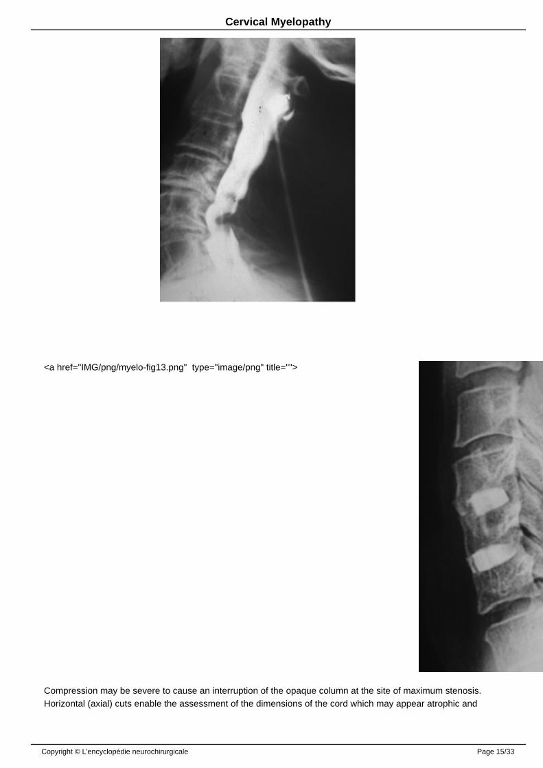
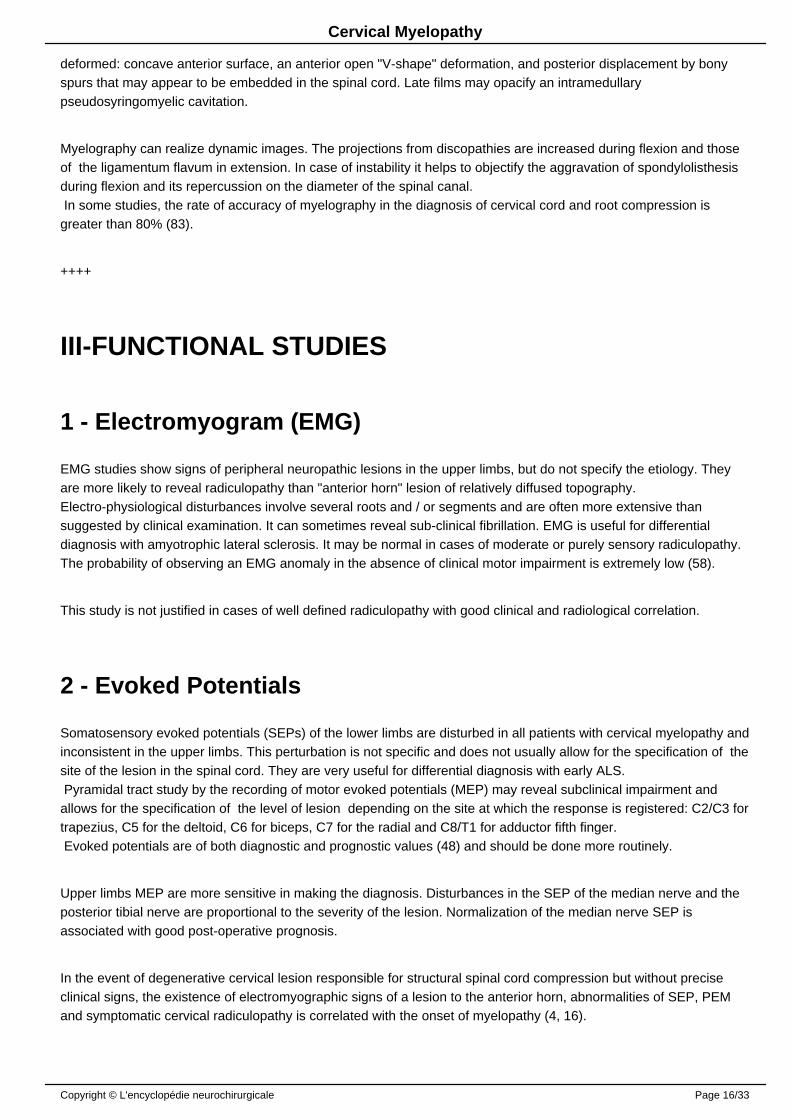
Myelography and CT myelography are indicated only when MRI is impossible to do or is contra-indicated. Thesestudies can be complementary to the latter (75). They objectify the diameter of the spinal canal, anterior and posteriorcompressive elements whose topography can be median or paramedian (Figures 12, 13). <a href="IMG/png/myelo-fig12.png" type="image/png" title="">
Copyright © L'encyclopédie neurochirurgicale Page 14/33
Cervical Myelopathy
<a href="IMG/png/myelo-fig13.png" type="image/png" title="">
Compression may be severe to cause an interruption of the opaque column at the site of maximum stenosis. Horizontal (axial) cuts enable the assessment of the dimensions of the cord which may appear atrophic and
Copyright © L'encyclopédie neurochirurgicale Page 15/33
Cervical Myelopathy
deformed: concave anterior surface, an anterior open "V-shape" deformation, and posterior displacement by bonyspurs that may appear to be embedded in the spinal cord. Late films may opacify an intramedullarypseudosyringomyelic cavitation.
Myelography can realize dynamic images. The projections from discopathies are increased during flexion and thoseof the ligamentum flavum in extension. In case of instability it helps to objectify the aggravation of spondylolisthesisduring flexion and its repercussion on the diameter of the spinal canal. In some studies, the rate of accuracy of myelography in the diagnosis of cervical cord and root compression isgreater than 80% (83).
++++
III-FUNCTIONAL STUDIES
1 - Electromyogram (EMG)
EMG studies show signs of peripheral neuropathic lesions in the upper limbs, but do not specify the etiology. Theyare more likely to reveal radiculopathy than "anterior horn" lesion of relatively diffused topography.Electro-physiological disturbances involve several roots and / or segments and are often more extensive thansuggested by clinical examination. It can sometimes reveal sub-clinical fibrillation. EMG is useful for differentialdiagnosis with amyotrophic lateral sclerosis. It may be normal in cases of moderate or purely sensory radiculopathy.The probability of observing an EMG anomaly in the absence of clinical motor impairment is extremely low (58).
This study is not justified in cases of well defined radiculopathy with good clinical and radiological correlation.
2 - Evoked Potentials
Somatosensory evoked potentials (SEPs) of the lower limbs are disturbed in all patients with cervical myelopathy andinconsistent in the upper limbs. This perturbation is not specific and does not usually allow for the specification of thesite of the lesion in the spinal cord. They are very useful for differential diagnosis with early ALS. Pyramidal tract study by the recording of motor evoked potentials (MEP) may reveal subclinical impairment andallows for the specification of the level of lesion depending on the site at which the response is registered: C2/C3 fortrapezius, C5 for the deltoid, C6 for biceps, C7 for the radial and C8/T1 for adductor fifth finger. Evoked potentials are of both diagnostic and prognostic values (48) and should be done more routinely.
Upper limbs MEP are more sensitive in making the diagnosis. Disturbances in the SEP of the median nerve and theposterior tibial nerve are proportional to the severity of the lesion. Normalization of the median nerve SEP isassociated with good post-operative prognosis.
In the event of degenerative cervical lesion responsible for structural spinal cord compression but without preciseclinical signs, the existence of electromyographic signs of a lesion to the anterior horn, abnormalities of SEP, PEMand symptomatic cervical radiculopathy is correlated with the onset of myelopathy (4, 16).
Copyright © L'encyclopédie neurochirurgicale Page 16/33
Cervical Myelopathy
++++
TREATMENT
I MEDICAL TREATMENT
Medical treatment in cervical spondylotic myelopathy can only be symptomatic. It can however yield transientimprovement in early or slightly advanced forms or when there is a contra-indication for surgery (14). Anepidemiological study on cervical radiculopathy showed that 75% of patients improved on conservative treatment andonly 25% of the patients were operated upon (65). After a follow up of 6 years , 90% of patients remained relieved.Suggestive factors of poor response to conservative treatment may include advanced age, duration of symptoms,severity of myelopathy and canal stenosis (25). However, data from the literature regarding the effectiveness of these various measures in the medical therapy of cervical spondylotic myelopathy, and their position relative to surgery in influencing the evolution of the disease is stillinsufficient (55). Medical treatment may include:
• Class I or II analgesics• Muscle relaxants to alleviate cervical pain (reactive muscle spasm of paravertebral muscles and trapezius) and
radiculalgias.• Non-steriodal anti-inflammatory drugs which can diminish edema from joint lesions and eventually that of the
nerve root sheath.• Steroids have not proven their efficacy in this indication (55). They can nevertheless be prescribed during
exacerbations.• Tricyclic antidepressants and anticonvulsants, depending on the symptomatology of the patient's pain
(neuropathic pain).• Kinesitherapy therapy focused on the para-vertebral muscles, upper limbs motor deficits and gait abnormalities,
combined with physiotherapy.• Heat therapy do not seem useful in the "global" management of the patient during medical visits.• Cervical immobilization may be proposed when there is clinical or radiological instability bearing in mind that
prolonged immobilization favors joint stiffness and para-vertebral muscle atrophy thus aggravating instability. Itcan be temporary and should be associated with rehabilitation of neck muscles. Finally, it is not advisable to beengaged in occupations subjecting the spine to repetitive microtraumas and / or severe injuries. ++++
II-SURGICAL TREATMENT
The choice of the surgical technique is guided by the preoperative analysis of clinical and radiological findings, whichin tend will determine the approach used. An anterior approach is performed in 85% of the cases by French-speakingEuropean neurosurgeons; a posterior approach is performed in 15% of the cases while the use of a combinedanterior-posterior approach is exceptional (12).
1. Anterior Cervical Decompression (see: 71)
Described in the 1950s in the United States by Smith and Robinson (82) and in Europe by Dereymaker Mulier (22),
Copyright © L'encyclopédie neurochirurgicale Page 17/33
Cervical Myelopathy
this technique became popular from the 1970s through the work of Cloward (17) and Verbiest (90) to become aroutine procedure in most cases. To the extended discectomies proposed by these authors have been addedtechniques median or anterolateral corpectomy.
The cervical spine is approached through an anterolateral route. The skin incision is horizontal (transverse) in a neckcrease (Langer's line) for a one or two level decompression and an oblique incision along the anterior border of thesterno- cleido-mastoid mucle for a more extensive decompression. After opening of the subcutaneous tissue and theplatysma, the middle layer of the cervical fascia is incised along the anterior edge of the sterno- cleido-mastoid. Thespine is approached in the space between the vascular sheath that is laterally retracted and the visceral sheath(pretracheal sheath) which is retracted medially. Usually, no vessels or muscles are sacrificed however it maysometimes be necessary to bind the superior thyroid artery and / or the omohyoid muscle. For right sided approachesto the lower cervical spine retraction should be moderate as not to cause injury by stretching the recurrent nervewhose course approximates the bisector of the angle formed by the inferior thyroid artery and tracheo-esophagealaxis. For this reason, some authors prefer a left sided route where the left recurrent laryngeal nerve is deep and lessexposed.
1.1. Discectomy with or without grafting (see: 71)
After radiological confirmation of the level, the anterior longitudinal ligament is incised using a scalpel and the disc ispartially excised using curettes and fine rongeurs. At this stage, the operating microscope is introduced. After placingan interbody spreader, removal of the disc is done, leaving the cartilaginous end plates in place. If a sequestratedfragment has been expelled into the extradural space through a breach of the posterior longitudinal ligament, thelatter may be partially resected to access the entire fragment expelled.
Resection of osteophytes is done if need arises with appropriate size rongeurs with the help of a diamond drill undervisualization with the operating microscope and image intensifier. We can concomitantly perform via the same routean uncusectomy by removing the posterior 5 mm of the uncus. Resection is done from inside to outside with a 5 mmdrill and with the aid of micro-curettes. Preservation of the anterior two thirds of the uncus prevents injury to thevertebral artery and postoperative instability.
At this stage of the procedure according to some authors, an intraoperative image guided injection of contrastmaterial can be performed in situ (epidurography) to assess the quality of disco-osteophytic resection and achieve animmediate appropriate correction in case of insufficient decompression (35).
This type of "wide" discectomy can be done in stages, usually one to three. A complementary interbody graft can beused. The latter aims at preventing narrowing of the disc space that can lead to closure of intervertebral foramina aswell as an eventual post-operative kyphosis. However, an analysis of the literature shows no significant differencebetween clinical series with or without grafting (27).
The "best" graft (ie, one that provides the best fusion rate) is the autologous tricortical iliac bone graft but themorbidity associated with graft harvesting (80) often leads to the use of allografts, xenografts or interbody cages filledwith bone substitutes (Figure 16). Some authors are accustomed to performing a systematic complementary anteriorosteosynthesis combined with interbody graft. This technique seems to provide a better restoration of disc height andlordosis at the operated level, and a higher fusion rate. Here again, comparative studies have failed to demonstratesignificant clinical impact for patients treated with interbody graft plus osteosynthesis versus bone graft only (84). Incase of instability, which can be observed in cases of very advanced degenerative disease, complementary anteriorfixation may be associated with interbody graft. The practice of French-speaking European neurosurgeons is extremely variable and depends more on habit,personal beliefs, school and tradition than on a significant difference in results (13): about 35% do not insert a graft,
Copyright © L'encyclopédie neurochirurgicale Page 18/33
Cervical Myelopathy
26% bone graft without osteosynthesis, 17% place the graft in a cage to improve the mechanical properties and 15%combine a graft with osteosynthesis.
1.2. Cloward's Technique (see: 17-18)
Once the discectomy and scraping off of the osteophytes is done, the width of the endplate is measured using adepth gauge. A strand of 12 to 18 mm in diameter selected according to the patient's morphology most often 14 or 16mm-is endowed with an abutment adjusted to the width of the endplate.This enables the drilling of a circular holewith a diameter corresponding to the straddling on the intervertebral space, starting from the adjacent endplates. Thedrilling of this hole enables the complete and safe removal of the posterior edges of the vertebral endplates seat ofosteophytic proliferation. A trephine with a slightly greater diameter permits the trimming of the iliac graft whoseouter diameter is equal to the inner diameter of the inter-vertebral hole, thus the graft perfectly fits into the drilledhole. However, and this being the main limitation of this technique, the thickness of the iliac crest, where the graft istaken, is often less than the width of the inter-vertebral space, thus the graft is often "short ".
1.3. Median Corpectomy (see: 73)
The approach is classic until the attainment of the anterior border of the concerned vertebral body (74).
Two techniques are possible: (i) if the discs are still present, the classical interbody approach should be done with resection discs at the end of the area to be decompressed (ii) if the ventral surface of the anterior column has beendeformed by osteophytic degenerations which are sometimes asymmetrical, identification of the limits of the cervicalfurrow is more difficult. In the latter case, the inter-vertebral discs are generally completely collapsed making thetrans-discal route impossible. We therefore first of all do a median trans-corporeal approach with a cutting drill, thenwith a diamond drill until we abut the posterior longitudinal ligament.
Corpectomy is initiated with the gouge forceps and then with a mechanical drill within a width of 10 mm until theposterior cortex is abutted and removed with 1 to 2 mm thick rongeurs. Upon contact with the dura mater, thecorpectomy is widened to 12 or 14 mm. To obtain a satisfactory cord decompression, all the disco-osteophytic barsanterior to spinal cord that can cause injury should be resected. Bone resection should also include the endplates ofadjacent vertebrae at the extremities of bone trough (gutter). Similarly, the posterior longitudinal ligament should beresected whenever possible but its adherence to the dura may make this procedure difficult.
At the end of the procedure, re-expansion of the dural sheath and the spinal cord appears as much as after a cervicallaminectomy. The bone trough has the shape of a dovetail, broader at the rear- abutting the spinal cord-than infront.From either side, the lateral portion of the vertebral bodies-at the uncus is respected (preserved) to maintain spinalstability. The vertical extension of the corpectomy is tailored according to the preoperative radiologic findings,essentially MRI.
For most authors, the bony trough is filled with iliac crest or fibular autograft (possibly associated with osteosynthesis)which prevents the risk of postoperative kyphosis. Just as for discectomy, surgical practices are variable: 18% ofEuropeans neurosurgeons do not use grafts, 16% use an embedded bone graft without osteosynthesis and 66% usean embedded graft associated with osteosynthesis. Grafting does not seem necessary if precautions are taken topreserve the uncus (uncinate process) and if active rehabilitation of the muscles of the neck is done (88) (Figure 14).
<a href="IMG/png/myelo-fig14.png" type="image/png" title="">
Copyright © L'encyclopédie neurochirurgicale Page 19/33
Cervical Myelopathy
2. Posterior cervical ( cord) decompression
These are the oldest performed interventions. The patient is placed in the prone position, head slightly flexed, rarelyin lateral decubitus position or sitting position. The lateral decubitus position allows for simultaneous anterior andposterior procedures. The skin incision is marked in the midline and the spine is reached at on the white line(ligamentum nuchae) separating the neck muscles retracted laterally.
2.1. Laminectomy (see: 17)
Laminectomy is the oldest technique performed. Initially, the spinous processes of the vertebrae are cut at theirbases at their junction with the laminae. Bone resection brings out "en bloc" all the concerned spinous processes andthe interspinous ligament. Laminectomy itself is performed by gradually removal of the laminae using fine bonerongeurs or even a mechanical drill. It is carried as far laterally as possible, even up to the inner part of the joints ofwhich the lateral 2/3 should be preserved to avoid creating instability. The ligamentum flavum are usually fragmentedalong with the bone resection. This phase should be conducted with great caution given the narrowness of the spinalcanal and the disappearance of the posterior epidural space caused by the pathology and worsened by the operativeposition.
Laminectomy may cause long-term kyphosis. The latter appears to be the result of insufficiency of neck musclesrather than bone and ligament instability induced by surgery (70). It can be prevented by appropriate earlyrehabilitation. Repeated progression of the disease is sometimes observed after laminectomy and has beenattributed to the formation of a compressive epidural fibrosis (post-laminectomy membrane) whose pathogenic rolehowever remains questionable (46).
2.2. Interventions preserving the posterior arch
To prevent postoperative kyphosis (86) and the formation of post-laminectomy membrane alternative technicalsolutions are possible.
Copyright © L'encyclopédie neurochirurgicale Page 20/33
Cervical Myelopathy
2.2.1. The laminoplasty (see 39, 67)
Proposed as from 1970 by Japanese neurosurgeons, they are more difficult to carry out and do not appear moredangerous. Several technical variants have been described. (i) The "open door" technique is the most frequentlyperformed. It consist of initially creating two paravertebral troughs at the junction of the lamina and facet whilepreserving the spinous processes and interspinous ligament, with the aid of a drill or a fine rongeurs at the side of thelarger lesion over the entire length of the lower cervical spine from C3 to C7 at least to exceed all lesions, including, ifnecessary C2 and the first two thoracic vertebrae.
In principle, the ligamentum flavum is respected and the instrument does not penetrate into the vertebral canal, thuspreventing root or spinal cord injury. On the other hand, on the contralateral side in the corresponding location onlythe outer cortical bone is drilled creating a trough to allow for "hinge-opening" of the lamina. The whole structurecomposed of the spinous processes, the interspinous ligament and the lamina is moved backwards by rotation (as ifa door is opened "open door laminoplasty"). It is recommended to "open the door" for a distance at least equal to thediameter of the spinal canal, ie at least 12mm. The bony block moved backwards is fixed away from the dura matterby attaching it to the paraspinal muscles or better still by interposition of a spacer (spacer), consisting mainly ofhydroxyapatite bone substitute, autograft taken from the spinous processes, in bioabsorbable materials of lactic acidpolymer filled with cancellous bone or allograft. If the yellow ligament is not adherent to the laminae and stays inplace, it should be resected secondarily (91).
(ii) The "double door" technique consists of opening the cervical canal in the midline. By the means of a high speedmechanical drill the spinous processes and the laminae are divided in the mid line to their point of contact with theligamentum flavum, and the thinning of the lamina to their insertion at the facet joints. The opening of the vertebralcanal is performed by introducing a spacer between each half of the spinous process, as in opening the portal of adouble door. It is recommended to introduce spacers of bone substitute between the two halves of the spinous tokeep them apart.
2.2.2. Laminectomy with preservation the spinous processes
Described by C. Large (31) in the lumbar spine, it is a unilateral para-vertebral route where the spinous processesare cut at their base via a trough at the junction between the spinous process and the lamina. The tissue bulkconsisting of the spinous processes and the interspinous ligament is retracted beyond the midline exposing thelaminae. Laminectomy is then conducted in a conventional manner. During closure of muscles, the interspinousligament and spinous process fit back into their natural position on the midline.
2.2.3. Skip laminectomy ("Alternate" Laminectomy) (see 79, 81)
It concerns only alternate laminae: for example to achieve decompression from C3 to C7, only the laminae of C4 andC6 are resected and decompression is performed by resection of the ventral surface of adjacent laminae left in place.In addition, the interspinous ligament is preserved: the laminae to be resected are targeted by sectioning of thespinous process in the midline using a high speed drill and each portion of the spinous process is retracted laterallyto preserve the insertion of the supraspinatus muscle.
2.3. Posterior osteosynthesis
Due to the risk of kyphosis, some authors have proposed the routine performance of a complementary posteriorosteosynthesis. This seems to be reserved for rare cases where surgical decompression requires resection of thefacets and there is preoperative instability. The best technique is that of screw osteosynthesis of the facet associated
Copyright © L'encyclopédie neurochirurgicale Page 21/33
Cervical Myelopathy
with a posterolateral graft.
3. Combined Interventions (see 44)
In case of severe stenosis, a double approach simultaneously or in two successive staged operations can beperformed. In this case, the risk of postoperative instability is relatively high, and decompressive surgery must beaccompanied by grafting and anterior osteosynthesis with screwed plates into vertebral bodies.
4 -Lateral spinal decompression
Described by George (15), this approach may become an alternative to standards anterior approaches.According toits author, this lateral approach could be indicated where anterior compression is predominant and the spine isstraight or kyphotic without instability and without bilateral radiculopathy. With this approach, after a direct control of the vertebral artery, the lateral portion of the intervertebral discs,vertebral bodies, the posterior longitudinal ligament and ipsilateral unco-vertebral joint can be resected if necessarydepending on the extent of the lesions. The advantages of this technique are wide exposure, the lack of need for graft and / or osteosynthesis, nocontra-indication in case of kyphosis without instability and the the possibility of reoperation after a prior standardanterior cervical approach.
5 - Indications
Early and simple forms in the elderly (JOA Score> 12) may justify trial of medical treatment with regular clinicalfollow-up (54).
Surgical treatment should not be delayed in young patients and when faced with any progressive form. It has beenshown that the length of symptoms has more influences on the outcome than age, severity of illness, number oflevels operated and/or pre-operative functional score (40).
Imaging allows for the assessment of the lesions:
• Anteroposterior and transverse diameter of the spinal canal,• Overall appearance or segmental nature of the stenosis,• Number of levels affected• Predominant site of the compression: anterior or posterior• Medial or lateral compression,• Potential instability associated with stenosis assessed on dynamic films taken with caution• State of the spinal cord: atrophic, intramedullary hyperintensity (cord signal). Evoked potentials (somatosensory
and motor) (48, 60, 68) could possibly help in the choice of surgical approach in case of difficulty in identifyingthe responsible clinical and radiological elements causing the compression: anterior or posterior. The approach,anterior, posterior or combined, the extent of decompression and the indication for additional osteosynthesis willbe chosen based on the results of these tests (93) bearing in mind that the anterior approach is preferred interms of its simplicity and efficiency.
Copyright © L'encyclopédie neurochirurgicale Page 22/33

Cervical Myelopathy
5-1 Indications of anterior approaches
The best indications for the anterior approach when there is myelopathy or myeloradiculopathy are represented by:(i) the presence of clinical signs predominantly in the upper limbs, from root lesion (myelo-radiculopathy) or centralcord syndrome (brachial diparesis); (ii) the predominance of anterior compression elements (discs, osteophytes) overposterior factors (laminae, ligaments, facets), (iii) the existence of a preoperative cervical kyphosis.
5-1-1 Indications of discectomy:
Simple discectomy at one, two or three levels can be used if the lesions are limited to the disc levels responsible forsegmental stenosis with the compressive elements close to the midline. Similarly, the anterior approach can be usedas a complement to a posterior route if there is a persistence of anterior compression or as treatment for anevolving post-laminectomy kyphosis. The use of complementary interbody graft is more a matter linked to the schoolsof thought than the quality of long-term clinico-radiologic results (27). In general, the graft diminishes the severity ofpost-operative neck pain and frequency of late kyphosis, without being able to remove them completely.Complementary osteosynthesis is only exceptionally indicated in patients with preoperative instability; correctlyperformed isolated discectomy does not destabilize the spine.
5-1-2 Indications for median corpectomy
In affections involving three levels and / or in case of associated global stenosis a corpectomy can be discussedinstead of the preferred laminectomy by some because in 75% of the cases the compression factors are anterior.
Additional graft is not necessary if the bony trough respects the uncinate processes (88). Reconstruction with bonegraft and an eventual osteosynthesis is necessary when instability is certain (resection of one of the lateral columns)or when there is a tendency for instability, especially when associated with laminectomy. The grafts are generallyautograft from the iliac crest, and sometimes from the tibia, allografts from graft bank, rarely xenograft or bonesubstitutes. Rare cases of instability with formation of kyphosis have been observed after simple corpectomy withoutgrafting. It is therefore necessary to respect a rigorous technique and no hesitation to perform complementarygrafting if in doubt of future stability.
5-2 Indications for posterior approaches
These approaches are preferred to anterior approaches when: (i) Clinically, patients show isolated or predominantsigns of myelopathy, with a preponderance of posterior column affection over pyramidal signs (ii) anatomically,patients have extensive constitutional stenosis (rare situation) and / or predominantly posterior lesions: hypertrophiclaminae, yellow ligament hypertrophy, hypertrophy of the facets.
"Standard" laminectomy is the easiest procedure to perform. The laminoplasty is preferred by Japanese and NorthAmerican authors. The procedure is longer, more difficult and its superiority in terms of performance compared tostandard laminectomy has not been demonstrated (57). Osteosynthesis is only associated in case of obviouspreoperatively instability.
5-3 Indications for combined approaches
In our experience, the indications of mixed(combined) approaches are rare and are limited to severe stenosis withequivalent anterior and posterior lesions. They are sometimes indicated with incomplete results from an initial
Copyright © L'encyclopédie neurochirurgicale Page 23/33
Cervical Myelopathy
anterior or posterior decompression, or in case of recurrence of clinical signs after a period of improvement (44). Adouble approach can destabilize the cervical spine, thus the second operation should be accompanied by anadditional fixation (44). If a double approach is done systematically, osteosynthesis with interbody graft should bedone via an anterior approach: this is where it has the best biomechanical properties.
6 - Results of treatment
Langston t. HoLLy, M.D.,1 PauL g. Matz, M.D.,2 PauL a. anDerson, M.D., Clinical prognostic indicators of surgicaloutcome in cervical spondylotic myelopathy. J Neurosurg Spine 2009;11;112-118
In the English literature, the effectiveness of treatment is assessed by a modification (expressed in percentage) of theJOA score (more accurate than the Nurick classification) according to the following formula: (postoperative score -preoperative score) / (17 - preoperative score) × 100.
6-1 Results of medical treatment
Rigorously followed up medical treatment can stabilize the disease for several years when the affection is in its mildand slowly progressive form (95). Clinical follow-up of patients with mild forms (average JOA score = 14) andmyelopathy from "soft disc" shows that 60% can be stabilized or improved on medical treatment consisting of: spinalimmobilization and reduced physical activity. In contrast, about 40% will worsen and should be operated. Theincriminated lesions may regress on MRI in approximately 50% of cases (51). The JOA score improvement mayreach 80% in initially relatively benign forms.
6-2 The results of surgical treatment
Clinical improvement depends more on the quality of the decompression than on the choice of surgical approachwhen it is adapted to the type and topography of the lesions (93).
There are a few more complications related to surgery in patients over 70 years because of associated pathologies(34).
According to the JOA classification, clinical improvement for all techniques is on average 55% (range 20-80%).The quality of functional outcome deteriorates with time and is only more than 45% after 6 years of progression. Thisdeterioration is related to the continued progression of degenerative lesions and spinal deformities, as well as theonset of associated diseases in older patients: for example; hip osteoarthritis, lumbar canal stenosis.
It is possible to identify many prognostic factors (41, 77, 94):
• Age is not determining (94) when the general condition, the degree of disability and associated pathologies aretaken into account: same degree of improvement was observed before and after 65 years but only in youngersubjects can complete recovery be observed. The aim moreover is not the same; young patients want to resumetheir full activities while older patients just want to gain sufficient autonomy.
• The duration of symptoms is one of the most important prognostic factors: better results are obtained if theduration of the disease is short, especially in patients over 65 years, which could correspond to symptoms offunctional cord affection before the installation of irreversible anatomical lesions (43, 94).
• The severity of preoperative disability seems paradoxically a less important factor (94).
Copyright © L'encyclopédie neurochirurgicale Page 24/33
Cervical Myelopathy
• The antero-posterior diameter of the cervical spinal canal and the surface area at its narrowest portion seems adetermining prognostic factors regardless of age (43).
• Intramedullary T2 hyperintensity (cord signal) on MRI may influence prognosis (47), but it is quite controversialand inconsistently reported in the literature (43, 77, 94).
• Preoperative instability observed mainly in older subjects does not seem to influence the prognosis (41).
7 - Complications of surgical treatment (see 8, 30)
Degenerative cervical spine surgery is currently well mastered by specialized surgeons (11). General complications common to all surgical procedures are rare approximately 1%. This type of surgery shouldnot be feared regardless of the patient's age when functional disability justifies it. There are few contra-indications.
Specific complications related to the anterior approach can sometimes be observed, they are directly related to thesurgeon's experience. The most classical and most common is that recurrent laryngeal nerve palsy which may beobserved in 0.2 to 16.7% of the cases (29, 87), especially in the vicinity of the lower cervical spine (C6/C7 andC7/T1) via right approach. It is prevented by minimizing the retraction of tracheo-oesophageal axis or systematicallyusing the left side approach that is less easy for a right handed surgeon. It is usually reversible. Pharyngeal oresophageal trauma may be observed but are exceptional, estimated to occur between 0.02 and 1.49% (20). Injuriesof the great vessels are rarely observed (8). The occurrence of a postoperative hematoma is documented in 1-11%of cases in some observations (29). This complication in the form of a neck mass, is responsible for dysphagia andputs the patient at risk of respiratory distress.
One can observe aggravation in the neurologic status regardless of the approach. A common complication ofextended decompression (but not often reported in the literature and in hospitalization summary) is paresis of the fifthand / or sixth cervical root, observed in 1-15% of cases (28, 89). Its physiologic mechanism is unclear. It is possiblethat it occurs during the installation of the patient on the operating table from a lesion of the radicular artery, but alsofrom the particular fragility of this root stretched when the cord moves after decompression (89). It is usuallyregressive. Aggravation of clinical manifestations occur in 0.2 to 5% of the cases depending on the series (28, 29).These are the consequences of a spinal cord injury that may occur during the installation of the patient (for exampleexcessive head flexion during a posterior approach) or during completion of the bone resection which requires theintroduction of fine instruments (curettes, rongeurs or mechanical drills ...) in a much narrowed spinal canal. Vascularphenomena can sometimes be the cause of this type of complication. The most dangerous procedure is probably theresection of bony and disc calcifications, on the anterior aspect of the dura mater which we must not try to remove atall cost, thus limiting the procedure to decompression.
The evolution of this type of complication is unpredictable and depends on the severity of the neurological syndromefound during recovery from anesthesia. Some may be permanently quadriplegia.
Mechanical complications related to the surgery are the most important to know as they compel the surgeon to adaptthe technique to the patient's semiology and spinal lesions:
• Interbody grafts can be displaced in about 2% of the cases (29). This is often the result of a misconfiguration ofautografts or inappropriate choice of bone substitutes. Some surgeons advise wearing a cervical collar forseveral weeks. It has not been shown that this reduces the frequency of the incident.
Displacements of material for osteosynthesis used in complementing an anterior approach may be observed. Thiscan cause destabilization of the spine and / or a wound of the posterior part of the esophagus. Although difficult to
Copyright © L'encyclopédie neurochirurgicale Page 25/33

Cervical Myelopathy
accurately estimate, the frequency of this complication is nevertheless exceptional, particularly because of thewidespread use of systems including interlocking screws. In addition, the screw profile has been improved to thepoint that all "loosening" became very rare or occurs simply as a result of a technical foul.
• Pain occurring as a consequence of graft harvesting from the donor site are observed in 20% of the cases. Thisrelatively high figure discourages many surgeons from using autografts in degenerative disease, although it is byfar the best graft (80).
• A post-operative kyphosis is frequently observed after an anterior approach as in the posterior route. Therealization of a graft and / or osteosynthesis reduces this risk (84) but does not completely eliminate it whileincreasing the burden of the operative procedures. This kyphosis seems more related to the inadequacy of neckmuscles (70) which should be subjected to postoperative physiotherapy according to the extent of thedecompression. Prolonged wearing of a collar could lead atrophy of paraspinal muscles and increase the risk ofkyphosis. Finally, the preservation of the uncinate process during anterior approaches and anterior half of thejoints during posterior approaches decreases the frequency of these complications. It has been shown thatinterbody graft is not absolutely necessary after a median corpectomy (88), which thus reduces the risk ofcomplications and the cost of the intervention. Laminoplasty for many authors (86), decreases the risk ofkyphosis compared to laminectomy, but this was not shown in the meta-analysis conducted by Ratliff andCooper in 2003 (67).
• After a laminectomy, a fibrotic scar (post-laminectomy membrane) develops that could be at the origin of thelate deterioration of postoperative outcome (67). This is one of the reasons that contributed to the developmentof laminoplasty. In fact, the pathogenic role of this fibrous scar is questionable (46).
• The fusion of one or more cervical segments leads to early degeneration of adjacent segments which could bethe source of a new pathology in 3% of the patients each year and in 10 years, 15 to 20% of the patients couldrequire further surgery (37). In our experience, this figure is much lower and it has been shown that if theselesions are frequent, they are often asymptomatic (45). There is no evidence till date (for lack of sufficient clinicalexperience) that the use of cervical disc prostheses reduces this risk significantly (9).
• All cervical spine procedures (with or without fusion) lead to a restricted range of neck movement. This usuallyremains mildly debilitating and well compensated for by adjacent segments, especially as these is most often inelderly patients with decreased activity and diffuse degenerative lesions of the entire spine. Despite the long listof potential complications each of which taken in isolation (beside donor site painful sequelae) is relativelyunusual, surgical treatment should not be delayed in a patient with neurological signs related to cervicalmyelopathy. ++++
BIBLIOGRAPHY
(1) Aboulker J, Metzger J, David M et al. Les myélopathies cervicales d'origine rachidienne. Neurochirurgie1965;11:89-1198.
(2) Al-Mefty O, Harkey LH, Middleton TH et al. Myelopathic cervical spondylotic lesions demonstrated by magneticresonance imaging. J Neurosurg 1988;68:217-22.4/C
(3) Asano T, Tsuzuki N. Surgical management of ossification of the posterior longitudinal ligament. In Schmidek &Sweet, Operative neurosurgical techniques, W.B. Saunders Company, Philadelphia, 4e ed 2000;2003-2015.
(4)Bednarik J, Kadanka Z, Dusek L et al. Presymptomatic spondylotic cervical cord compression. Spine2004;29(20):2260-9.2/B
(5) Bell GR, Ross JS. Diagnosis of nerve root compression : Myelography, computed tomography and MRI. Orthop
Copyright © L'encyclopédie neurochirurgicale Page 26/33

Cervical Myelopathy
Clin North Am 1992;23:405-19.
(6) Benzel EC, Lancon J, Kesterson L et al. Cervical laminectomy and dentate ligament section for cervicalspondylotic myelopathy. J Spinal Disorder 1991;4(3):286-95.3/C
(7) Bernhardt M, Hynes RA, Blume HW et al. Cervical spondylotic myelopathy [Current concepts review]. J BoneJoint Surg 1993;75A :119-26.
(8) Boakye M, Patil CG, Santarelli J et al. Cervical spondylotic myelopathy: complications and outcomes after spinalfusion. Neurosurgery 2008;62:455-61.4/C
(9) Botelho RV, Moraes OJ, Fernandes GA et al. A systematic review of randomized trials on the effect of cervicaldisc arthroplasty on reducing adjacent-level degeneration. Neurosurg Focus 2010;28:E5.2/B
(10) Brown BM, Schwartz RH, Franck E et al. Preoperative evaluation of cervical radiculopathy and myelopathy bysurface-coil MR imaging. AJR Am J Roentgenol 1988;151:1205-12.4/C
(11) Brunon J, Fuentes JM. Chirurgie antérieure et antéro-latérale du rachis cervical inférieur (vingt cinq ans après H.Verbiest). Première partie : les bases techniques. Neurochirurgie 1996, 42 : 105-122. Deuxième partie : indications,résultats et complications. Neurochirurgie 1996;42:229-48.
(12) Brunon J. Traitement chirurgical des myélopathies cervicales. Rachis,1997;9:275-280.
(13) Brunon J, Born JD. Chirurgie antérieure et latérale du rachis cervical dégénératif. Place de la greffe et del'ostéosynthèse. Analyse de la pratique des neurochirurgiens européens francophones. Neurochirurgie2000;46:54-58.4/C
(14) Brunon J, Nuti C, Duthel R et al. Myélopathies cervicales. EMC (Elsevier SAS, Paris), Neurologie,2005;17-660-A-10.
(15) Chibbaro S, Mirone G, Bresson D et al. Cervical spine lateral approach for myeloradiculopathy: technique andpitfalls. Surg Neurol 2009;72:318-24.4/C
(16) Chistyakov AV, Soustiel JF, Hafner H et al. The value of motor and somatosensory evoked potentials inevaluation of cervical myelopathy in the presence of peripheral neuropathy. Spine 2004;29(12):E239-47.3/C
(17) Cloward RB. The anterior approach for removal of ruptured cervical disks. J Neurosurg 1958;15:602-17.
(18) Cloward RB. The anterior approach for removal of ruptured cervical disks. 1958. J Neurosurg Spine2007;6:496-511.
(19) Collias JC, Roberts MP. Posterior surgical approaches for cervical disk herniation and spondylotic myelopathy.In Schmidek & Sweet eds. Operative Neurosurgical Techniques: indications, methods, and results, 4th edition, Vol 2,Massachusetts: Marion; 2000;p. 2016-28.
Copyright © L'encyclopédie neurochirurgicale Page 27/33
Cervical Myelopathy
(20) Dakwar E, Uribe JS, Padhya TA et al. Management of delayed esophageal perforations after anterior cervicalspine surgery. J Neurosurg Spine 2009;11:320-5.4/C
(21) Davidson RI, Dunn EJ, Metzmaker JN. The shoulder abduction test in the diagnosis of radicular pain in cervicalextradural compressive monoradiculopathies. Spine 1981;6:441-6.4/C
(22) Dereymaker A, Mulier J. Nouvelle cure chirurgicale des discopathies cervicales. La méniscectomie par voiecentrale suivie d'arthrodèse par greffe intercorporéale. Neurochirurgie 1956;2:233-236.
(23) Dorwart RH, LaMasters DL. Applications of computed tomography scanning of the cervical spine. Orthop ClinNorth Am 1985;16:381-93.
(24) Ebersold MJ, Pare MC, Quast LM. Surgical treatment for cervical spondylitic myelopathy. J Neurosurgery1995;82:745-51.4/C
(25) Edwars CC, Riew KD, Anderson PA et al. Cervical myelopathy: current diagnosis and treatment strategies. TheSpine Journal 2003;3:68-81.2/B
(26) Emery SE. Cervical spondylotic myelopathy : Diagnosis and treatment. J Am Acad Orthop Surg2001;9:376-88.4/C
(27) Fehlings MG, Arvin B. Surgical management of cervical degenerative disease: the evidence related toindications, impact, and outcome. J Neurosurg Spine 2009;11:97-100.1/A
(28) Fehlings MG, Smith JS, Kopjar B et al. Perioperative and delayed complications associated with the surgicaltreatment of cervical spondylotic myelopathy based on 302 patients from the AOSpine North America CervicalSpondylotic Myelopathy Study. J Neurosurg Spine 2012;16:425-32.2/B
(29) Fountas KN, Kapsalaki EZ, Nikolakakos LG et al. Anterior cervical discectomy and fusion associatedcomplications. Spine 2007 ;32 :2310-17.4/C
(30) Gok B, Sciubba DM, McLoughlin GS et al. Revision surgery for cervical spondylotic myelopathy: surgical resultsand outcome. Neurosurgery 2008;63:292-8.2/B
(31) Gros C, Frerebeau P, Privat JM, et al. Conservative lumbar laminectomies: techniques and results.Neurochirurgie 1983;29:207-209.4/C
(32) Harkey HL, Al-Mefty O, Marawi I et al. Experimental chronic compressive myelopathy: effects of decompression.J Neurosurg 1995;83:336-341.4/C
(33) Harrop JS, Hanna A, Silva MT et al. Neurological manifestations of cervical spondylosis : an overview of signs,symptoms and pathophysiology. Neurosurgery 2007;60(1- Suppl 1):S14-20.2/B
(34) Hasegawa K, Homma T, Chiba Y et al. Effects of surgical treatment for cervical spondylotic myelopathy inpatients > 70 years of age: a retrospective comparative study. J Spinal Disord Tech 2002;15:458-60.4/C
Copyright © L'encyclopédie neurochirurgicale Page 28/33
Cervical Myelopathy
(35) Hejazi N, Witzmann A, Hassler W. Intraoperative cervical epidurography: a simple modality for assessing theadequacy of decompression during anterior cervical procedures. Technical note. J Neurosurg 2003;98 (Suppl1):96-9.
(36) Herkowitz H. Cervical laminoplasty: its role in the treatment of cervical radiculopathy. J Spinal Disord1988;1:179-88.3/C
(37) Hilibrand AS, Carlson GD, Palumbo MA et al. Radiculopathy and myelopathy at segments adjacents to the siteof a previous anterior cervical arthrodesis. J Bone Joint Surg 1999;81:519-28.4/C
(38) Hirabayashi K, Miyakawa J, Satomi K et al. Operative results and postoperative progression of ossificationamong patients with ossification of cervical posterior longitudinal ligament. Spine 1981;6:354-64.4/C
(39) Hirabayashi S, Yamada H, Motosuneya et al. Comparison of enlargement of the spinal canal after cervicallaminoplasty: open-door type and double-door type. Eur Spine J 2010;19:1690-94.3/C
(40) Holly LT, Matz PG, Anderson PA, et al. Clinical prognostic indicators of surgical outcomes in cervical spondyloticmyelopathy. J Neurosur Spine 2009;11:112-118.2/B
(41) Kawakami M, Tamaki T, Ando M et al. Preoperative instability does not influence the clinical outcome of patientswith cervical spondylotic myelopathy treated with expansive laminoplasty. J Spinal Disord Techn 2002;15:277-83.4/C
(42) Kim P, Haisa T, Kawamoto T et al. Delayed myelopathy induced by chronic compression in the rat spinal cord.Ann Neurol 2004;55:503-511.4/C
(43) Kohno K, Kumon Y, Oka Y et al. Evaluation of prognostic factors following expansive laminoplasty for cervicalspinal stenotic myelopathy. Surg Neurol 1997;48:237-45.4/C
(44) Konya D, Ozgen S, Gercek A, et al. Outcomes for combined anterior and posterior surgical approaches forpatients with multisegmental cervical spondylotic myelopathy. J Clin Neurosci 2009;16:404-9.4/C
(45) Kulkarni V, Rajshekhar V, Raghuram L. Accelerated spondylotic changes adjacent to the fused segmentfollowing central cervical corpectomy : magnetic resonance imaging study evidence. J Neurosurg 2004;100 (suppl1):2-6.4/C
(46) Larocca H, Macnab I. The laminectomy membrane. Studies in its evolution, characteristics, effects, andprophylaxis in dogs. J Bone Joint Surg Br 1974;44:771-778.4/C
(47) Li F, Chen Z, Zhang F et al.A meta-analysis showing that high signal intensity on T2-weighted MRI is associatedwith poor prognosis for patients with cervical spondylotic myelopathy. J Clin Neurosci 2011;18(12):1592-5.2/B
(48) Lyu RK, Tang LM, Chen CJ et al. The use of evoked potentials for clinical correlation and surgical outcome incervical spondylotic myelopathy with intramedullary high signal intensity on MRI. J Neurol Neurosurg Psychiatry2004;75(2):256-261.2/B
Copyright © L'encyclopédie neurochirurgicale Page 29/33
Cervical Myelopathy
(49) Manelfe C. Imagerie du rachis et de la moelle. Scanner, IRM, ultra sons. Vigot édit. Paris 1989;801p.4/C
(50) Mastronardi L, Elsawaf A, Roperto R et al. Prognostic relevance of the postoperative evolution of intramedullaryspinal cord changes in signal intensity on magnetic resonance imaging after anterior decompression for cervicalspondylotic myelopathy.J Neurosurg Spine 2007;7(6):615-22.2/B
(51) Matsumoto M, Chiba K, Ishikawa M et al. Relationships between outcomes of conservative treatment andmagnetic resonance imaging findings in patients with mild cervical myelopathy caused dy soft disc herniation. Spine2001;26:1492-1598.4/C
(52) Matsunaga S, Sakou T, Taketomi E, Komiya S. Clinical course of patients with ossification of the posteriorlongitudinal ligament: a minimum 10-year cohort study. J Neurosurg 2004;100 (suppl 3):245-248.2/B
(53)Matz PG, Anderson PA, Holly LT et al. The natural history of cervical spondylotic myelopathy. J Neurosurg Spine2009;11(2):104-11.2/B
(54) Matz PG, Holly LT, Mummaneni PV. Anterior cervical surgery for the treatment of cervical degenerativemyelopathy. J Neurosurg Spine 2009;11:170-3.2/B
(55) Mazanec D, Reddy A. Medical management of cervical spondylosis. Neurosurgery 2007;60(1- Suppl1):S43-S50.3/C
(56)Mizuno J, Nakagawa H, Inoue T et al. Clinicopathological study of "snake-eye appearance" in compressivemyelopathy of the cervical spinal cord.J Neurosurg 2003;99(2 Suppl):162-8.4/C
(57) Mummaneni PV, Kaiser MG, Matz PG et al. Cervical surgical techniques for the treatment of cervical spondyloticmyelopathy. J Neurosurg Spine 2009;11:130-41.3/C
(58) Nardin RA, Patel MR, Gudas TF et al. Electromyography and magnetic resonance imaging in the evaluation ofradiculopathy. Muscle Nerve 1999;22:151-55.4/C
(59) Nguyen HV, Ludwig SC, Silber J et al. Rheumatoid arthritis of the cervical spine. The Spine Journal2004;4:329-34.4/C
(60) Nové-Josserand A, André-Obadia N, Mauguière F. Cervical spondylotic myelopathy: motor and somatosensoryevoked potentials, clinical and radiological correlation. Rev Neurol 2002;158:1191-97.4/C
(61) Nurick S. The pathogenesis of the spinal cord disorder associated with cervical spondylosis. Brain1972;95:87-100.3/C
(62) Ono K, Ota H, Toda K. Cervical myelopathy secondary to multiple spondylotic protrusions. Clinicopathologicstudy. Spine 1977;2:109-125.4/C
(63) Parker F, Comoy J, Carlier R et al. Myélopathies cervicales. Encycl Med Chir Neurologie 1993;17-660-A-10,14p.
Copyright © L'encyclopédie neurochirurgicale Page 30/33
Cervical Myelopathy
(64) Pavlov H, Torg JS, Robie R. Cervical spinal stenosis : determination with vertebral body ratio method. Radiology1987;164:171-175.4/C
(65) Radhakrishnan K, Litchy WJ, O'Fallon MW et al. Epidemiology of cervical radiculopathy : A population basedstudy from Rochester, Minnesota, 1976 through 1990. Brain 1994;117:325-35.4/C
(66) Ratliff J, Voorhies R. Increased MRI signal intensity in association with myelopathy and cervical instability:casereport and review of the literature Surg Neurol 2000;53(1):8-13.4/C
(67) Ratliff JK, Cooper PR. Cervical laminoplasty: a critical review. J. Neurosurg 2003;(Spine 3),98:230-8.4/C
(68) Restuccia D, Di Lazzaro V, Valeriani M et al. The role of upper limb somatosensory evoked potentials in themanagement of cervical spondylotic myelopathy: preliminary data. Electroencephalogr Clin Neurophysiol1994;92:502-9.4/C
(69) Ryan AG, Morrissey BM, Newcombe RG et al. Are T1 weighted images helpful in MRI of cervical radiculopathy?Br J Radiol 2004;77:189-196.
(70) Sakaura H, Hosono N, Mukai Y et al. Preservation of the nuchal ligament plays an important role in preventingunfavorable radiologic changes after laminoplasty. J Spinal Disord Tech 2008;21:338-43.2/B
(71) Schmidek HH. Anterior cervical diskectomy and fusion in cervical spondylosis. In Schmidek & Sweet eds.Operative Neurosurgical Techniques: indications, methods, and results, 4th edition, Vol 2, Massachusetts:Marion;2000;p. 1970-78.
(72) Schneider RC. The syndrome of acute central cervical spinal cord injury. J Neurosurgery 1955;12:95-112.
(73) Seifert V. Anterior approaches in multisegmental cervical spondylosis. In Schmidek & Sweet eds. OperativeNeurosurgical Techniques: indications, methods, and results, 4th edition, Vol 2, Massachusetts: Marion;2000;p.1987-2002.
(74) Senegas J, Guerin J, Vital JM. Décompression médullaire étendue par voie antérieure dans le traitement desmyélopathies par cervicarthrose. Rev Chir Orthoped 1985;71:291-300.2/B
(75) Shafaie FF, Wippold FJ 2nd, Gado M et al. Comparison of computed tomography myelography and magneticresonance imaging in the evaluation of cervical spondylotic myelopathy and radiculopathy. Spine1999;24(17):1781-5.4/C
(76) Shedid D, Benzel EC. Cervical spondylosis anatomy : pathophysiology and biomechanics. Neurosurgery2007;60(1- Suppl 1):S1-7.4/C
(77) Shin JJ, Jin BH, Kim KS et al. Intramedullary high signal intensity and neurological status as prognostic factorsin cervical spondylotic myelopathy. Acta Neurochir (Wien) 2010;152:1687-94.2/B
(78) Shingyouchi Y, Nagahama A, Niida M. Ligamentous ossification of the cervical spine in the late middle-aged
Copyright © L'encyclopédie neurochirurgicale Page 31/33
Cervical Myelopathy
Japanese men. Its relation to body mass index and glucose metabolism. Spine 1996;21:2474-8.4/C
(79) Shiraishi T, Fukuda K, Yato Y et al. Results of skip laminectomy. Minimum 2 year follow-up study compared withopen-door laminoplasty. Spine 2003;24:2667-72.2/B
(80) Silber JS, Anderson DG, Daffner SD et al. Donor site morbidity after anterior iliac crest bone harvest for singlelevel anterior cervical discectomy and fusion. Spine 2003;28:134-39.4/C
(81) Sivaraman A, Bhadra AK, Altaf F, et al. Skip laminectomy and laminoplasty for cervical spondylotic myelopathy:a prospective study of clinical and radiologic outcomes. J Spinal Disord Tech 2010;23:96-100.2/B
(82) Smith G, Robinson RA. The treatment of certain cervical spine disorders by anterior removal of intervertebraldisc and interbody fusion. J. Bone Joint Surg 1958;40 A:607-624.2/B
(83) Sobel DF, Barkovich AJ, Munderloh SH. Metrizamide myelography and post-myelographic computedtomography : comparative adequacy in the cervical spine. Am J Neuroradiol 1984;5:385-90.3/C
(84) Song KJ, Taghavi CE, Lee KB, Song JH, Eun JP. The efficacy of plate construct augmenation versus cagealone in anterior cervical fusion. Spine 2009;34:2886-2892.4/C
(85) Spinos P, Matzaroglou C, Partheni M et al. Surgical management of cervical spondylarthropathy in hemodialysispatients. The Open Orthopaedics Journal 2010;4:39-43.4/C
(86) Subramaniam V, Chamberlain RH, Theodore N, et al. Biomechanical effects of laminoplasty versuslaminectomy: stenosis and stability. Spine 2009;34:573-578.
(87) Tervonen H, Niemela M, Lauri ER et al. Dysphonia and dysphagia after anterior cervical decompression. JNeurosurg Spine 2007;7:124-30.4/C
(88) Tourneux H, Nuti C, Fotso MJ, Dumas B, Duthel R, Brunon J. Evaluation of the clinical and radiological results ofcervical longitudinal median corpectomy without graft. Neurochirurgie 2009;55:1-7.3/C
(89) Tsuzuki N, Abe R, Saiki K et al. Extra dural tethering effect as one mechanism of radiculopathy complicatingposterior decompression of the cervical spinal cord. Spine 1996;21:203-11.3/C
(90) Verbiest H. La chirurgie antérieure et latérale du rachis cervical. Neurochirurgie 1970;16, suppl N°2,1-212.
(91) Wang MY, Green BA. Open-door cervical expansile laminoplasty. Neurosurgery 2004;54:119-124.
(92) White AA 3rd, Panjabi MM. Biomechanical considerations in the surgical management of cervical spondyloticmyelopathy. Spine 1988;13 :856-60.
(93) Witwer BP, Trost GR. Cervical spondylosis: ventral or dorsal surgery. Neurosurgery 2007;60(Suppl1):S130-6.3/C
Copyright © L'encyclopédie neurochirurgicale Page 32/33
Cervical Myelopathy
(94) Yamazaki T, Yanaka K, Sato H et al. Cervical spondylotic myelopathy : surgical results and factors affectingoutcome with special reference to age differences. Neurosurgery 2003;52:122-26.4/C
(95) Yoshimata H, Nagata K, Goto H et al. Conservative treatment for spondylotic myelopathy: prediction oftreatment effects by multivariate analysis. The Spine Journal 2001;1:269-73.3/C
Copyright © L'encyclopédie neurochirurgicale Page 33/33