Embed Size (px)
Citation preview
1
Cervical Spine Stenosis
Donnie BellBIDMC Radiology Core Clerkship
October 2005
Donnie Bell, Jr. HMS IIIGillian Lieberman, MD
2
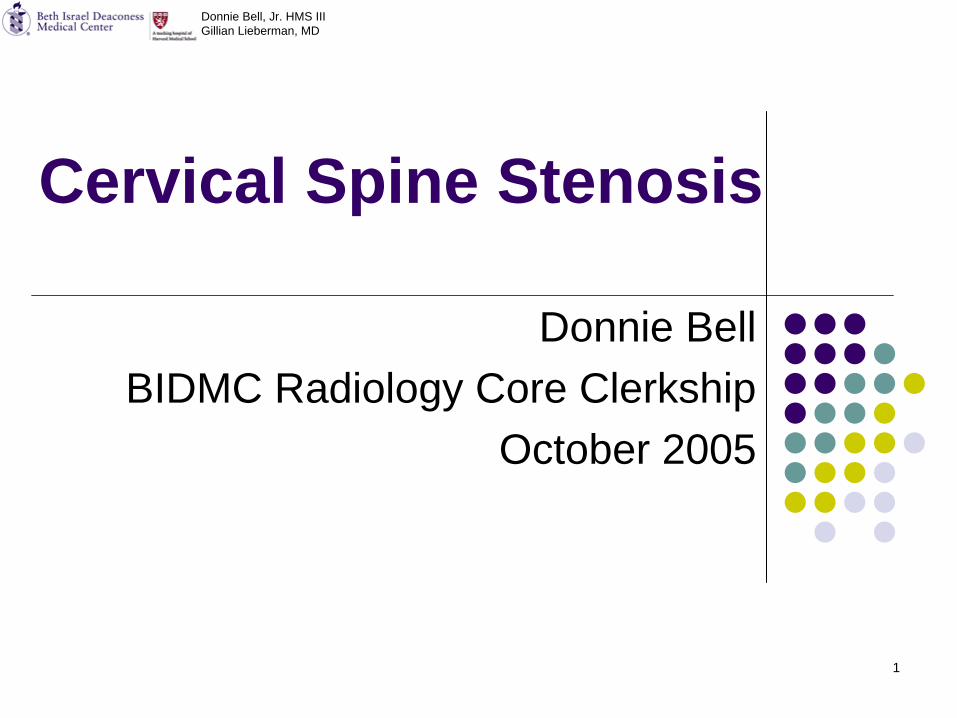
Our PatientMC is a 50 yo Asian female s/p MVA 9.28.05
Low speed rear end collision while stoppedNo LOC or head trauma
Declined AmbulanceThe next day presented to PCP c/o
HA, myalgiasL neck/shoulder painL thumb and 4/5 digit toe painMild numbness of distal L 3/4/5 digit hand
PMHCervical spine sprain 2001
C-spine at that time revealed degenerative changes in lower cervical spine, band-like calcific density C5/6, and loss of normal lordosisTreated with NSAIDs, Flexeril, and PT
Social Hx:Non-contributory
Donnie Bell, Jr. HMS IIIGillian Lieberman, MD October 2005
3
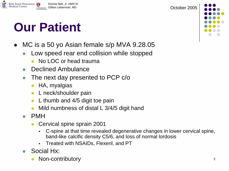
Our Patient cont’dPhysical Exam
Neck: Mild L tendernessROM limited to 45° b/l
Back: No spinous process tenderness+ tenderness to palpation along the edge of the scapula and upper trapezius
Left Ext:Mild numbness on dorsal aspect of 3/4/5 digitsTenderness @ thenar eminence, lateral elbow, 4/5 toes? Faint weakness of upper/lower ext.
AssessmentAcute Whiplash Injury s/p MVA
w/ C8 involvementPlan
Start motrin 400 mg TID, flexeril 10 mg QD, soft cervical collar when upright, PT, accupunctureReturn if sx worsen or sx do not resolve
Donnie Bell, Jr. HMS IIIGillian Lieberman, MD October 2005
4
Follow-up 11.1.05CC:
Persistent neck “tightness”Paresthesias of L upper extremity worse in the morning and with certain positions and movementsPain in left knee assoc. w/ “tightness” in posterior thigh and calf
PEFlexion, extension, and bilateral rotation causes neck discomfort.
A/PCervical Radiculitis w/ C8 involvementGiven duration and interval change in sx:
MRI
Donnie Bell, Jr. HMS IIIGillian Lieberman, MD October 2005
5
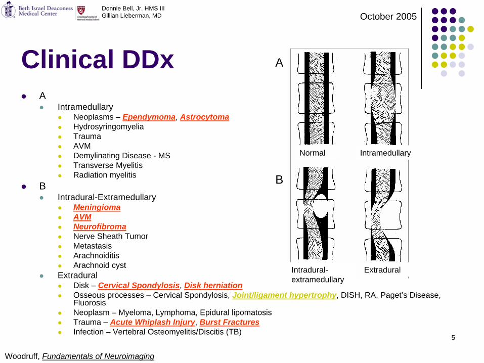
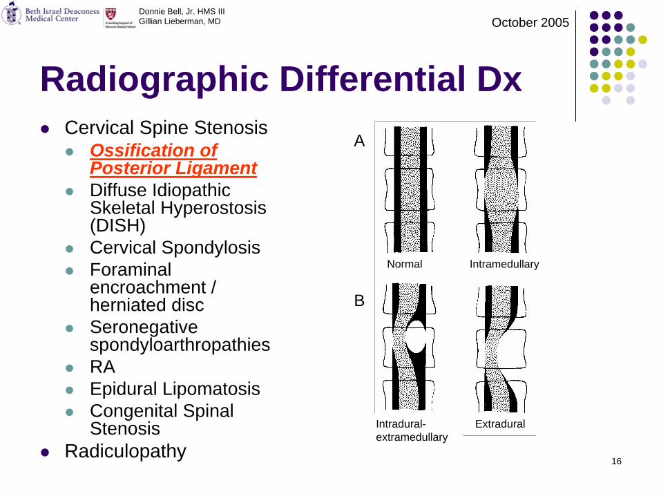
Clinical DDxA
IntramedullaryNeoplasms – Ependymoma, AstrocytomaHydrosyringomyeliaTraumaAVMDemylinating Disease - MSTransverse MyelitisRadiation myelitis
BIntradural-Extramedullary
MeningiomaAVMNeurofibromaNerve Sheath TumorMetastasisArachnoiditisArachnoid cyst
ExtraduralDisk – Cervical Spondylosis, Disk herniationOsseous processes – Cervical Spondylosis, Joint/ligament hypertrophy, DISH, RA, Paget’s Disease, FluorosisNeoplasm – Myeloma, Lymphoma, Epidural lipomatosisTrauma – Acute Whiplash Injury, Burst FracturesInfection – Vertebral Osteomyelitis/Discitis (TB)
Normal
Intradural- extramedullary
A
B
Intramedullary
Extradural
Donnie Bell, Jr. HMS IIIGillian Lieberman, MD October 2005
Woodruff, Fundamentals of Neuroimaging
6
Cervical Spine Anatomy
Let’s quickly review the anatomy of the regionbefore reviewing our patient’s imaging
7
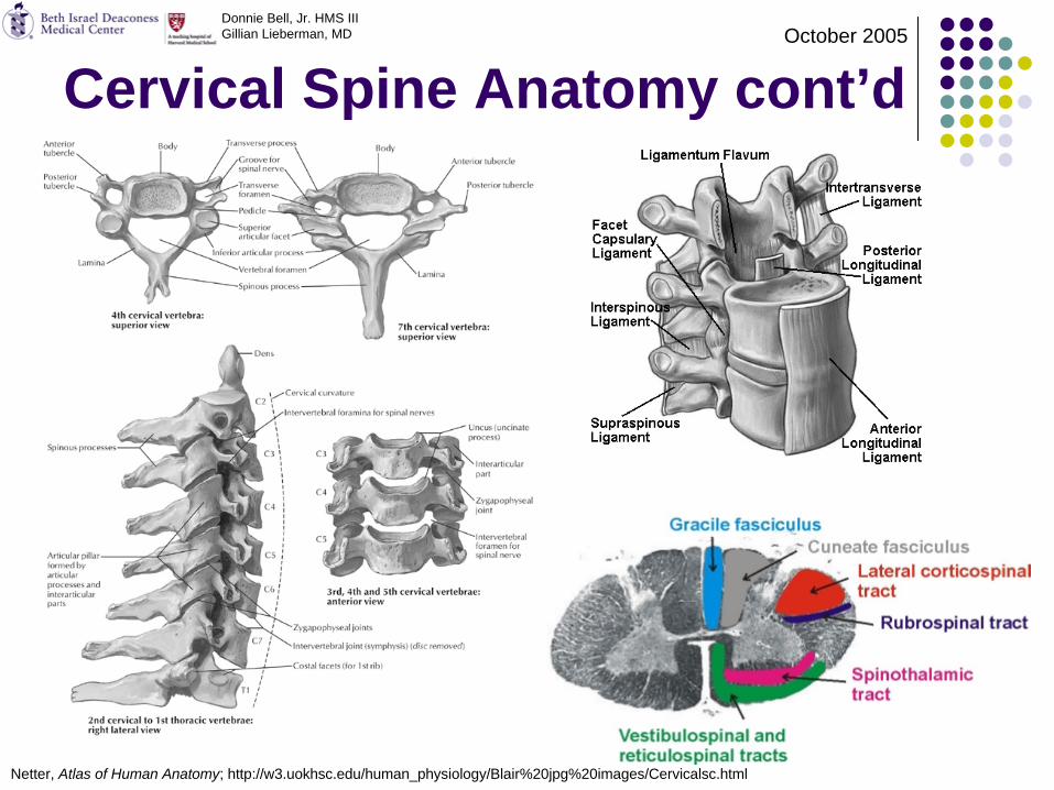
Cervical Spine Anatomy cont’dDonnie Bell, Jr. HMS IIIGillian Lieberman, MD October 2005
Netter, Atlas of Human Anatomy; http://w3.uokhsc.edu/human_physiology/Blair%20jpg%20images/Cervicalsc.html
8
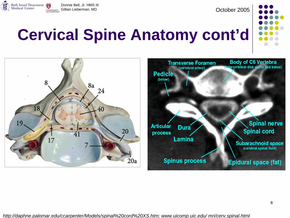
Cervical Spine Anatomy cont’d
Donnie Bell, Jr. HMS IIIGillian Lieberman, MD October 2005
http://daphne.palomar.edu/ccarpenter/Models/spinal%20cord%20XS.htm; www.uicomp.uic.edu/ mri/cerv.spinal.html
9
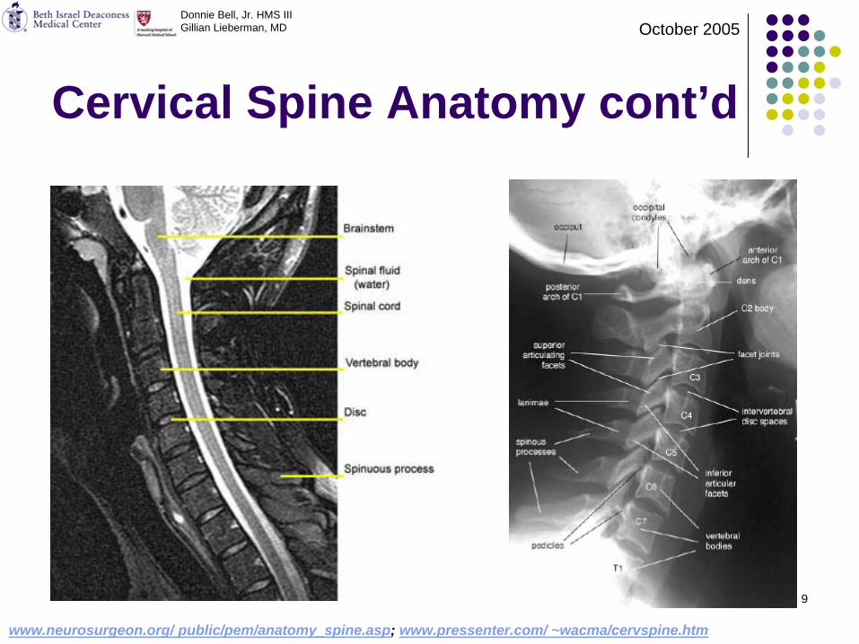
Cervical Spine Anatomy cont’d
Donnie Bell, Jr. HMS IIIGillian Lieberman, MD October 2005
www.neurosurgeon.org/ public/pem/anatomy_spine.asp; www.pressenter.com/ ~wacma/cervspine.htm
10
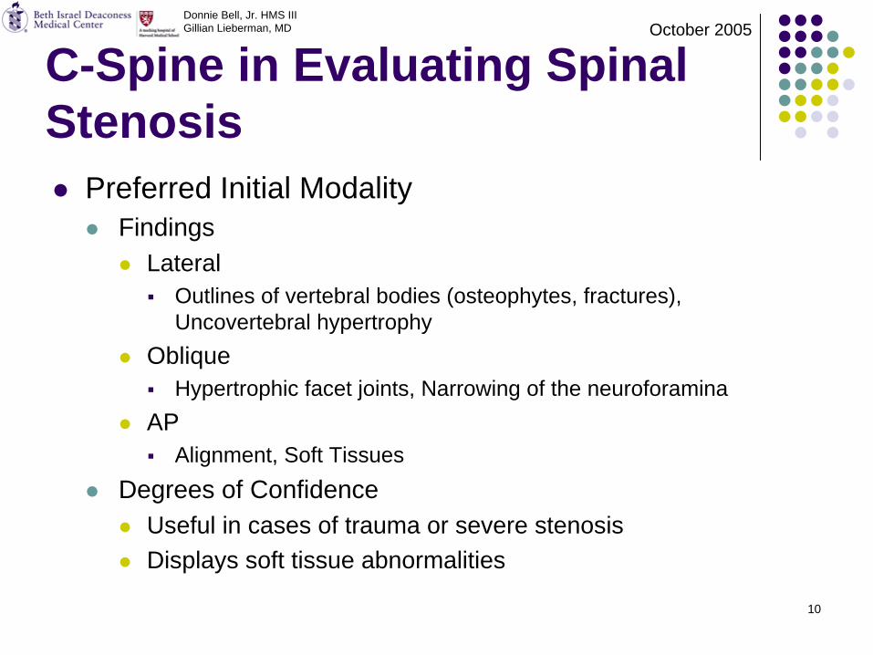
C-Spine in Evaluating Spinal Stenosis
Preferred Initial ModalityFindings
LateralOutlines of vertebral bodies (osteophytes, fractures), Uncovertebral hypertrophy
ObliqueHypertrophic facet joints, Narrowing of the neuroforamina
APAlignment, Soft Tissues
Degrees of ConfidenceUseful in cases of trauma or severe stenosisDisplays soft tissue abnormalities
Donnie Bell, Jr. HMS IIIGillian Lieberman, MD October 2005
11
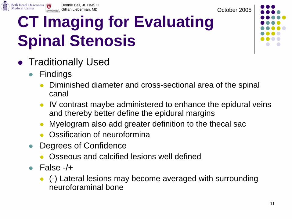
CT Imaging for Evaluating Spinal Stenosis
Traditionally UsedFindings
Diminished diameter and cross-sectional area of the spinal canalIV contrast maybe administered to enhance the epidural veins and thereby better define the epidural marginsMyelogram also add greater definition to the thecal sacOssification of neuroformina
Degrees of ConfidenceOsseous and calcified lesions well defined
False -/+(-) Lateral lesions may become averaged with surrounding neuroforaminal bone
Donnie Bell, Jr. HMS IIIGillian Lieberman, MD October 2005
12
MRI for Evaluating Spinal Stenosis
Preferred ModalityFindings
Osteophytic and calcific lesions appear dark (T1/T2-SE)Vertebral endplates show increased signal intensity post contrast in the setting of inflammation or chronic degenerative changes (T1)Cord enhancement with edema or myelomalaciaSpinal infectionPrimary/Secondary tumor
Degrees of ConfidenceSagittal spinal canal measurements are particularly useful
False +/-(-) Stenosis overestimation and false dorsal stenosis seen as a consequence of gradient-echo and CSF pulsation respectively
Donnie Bell, Jr. HMS IIIGillian Lieberman, MD October 2005
13
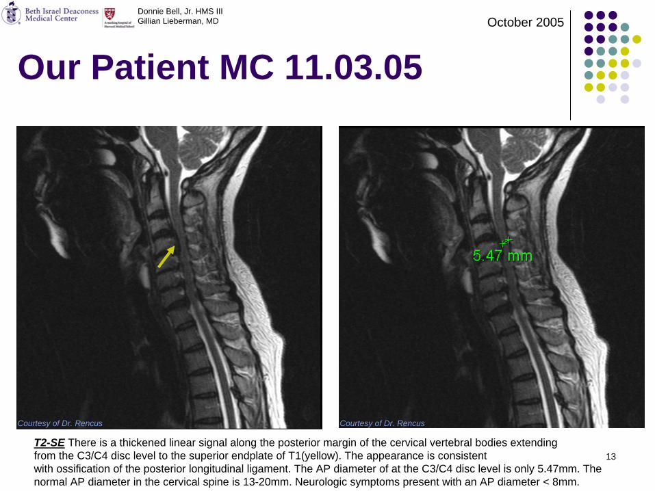
Our Patient MC 11.03.05
Donnie Bell, Jr. HMS IIIGillian Lieberman, MD October 2005
Courtesy of Dr. Rencus Courtesy of Dr. Rencus
T2-SE There is a thickened linear signal along the posterior margin of the cervical vertebral bodies extending from the C3/C4 disc level to the superior endplate of T1(yellow). The appearance is consistentwith ossification of the posterior longitudinal ligament. The AP diameter of at the C3/C4 disc level is only 5.47mm. The normal AP diameter in the cervical spine is 13-20mm. Neurologic symptoms present with an AP diameter < 8mm.
14
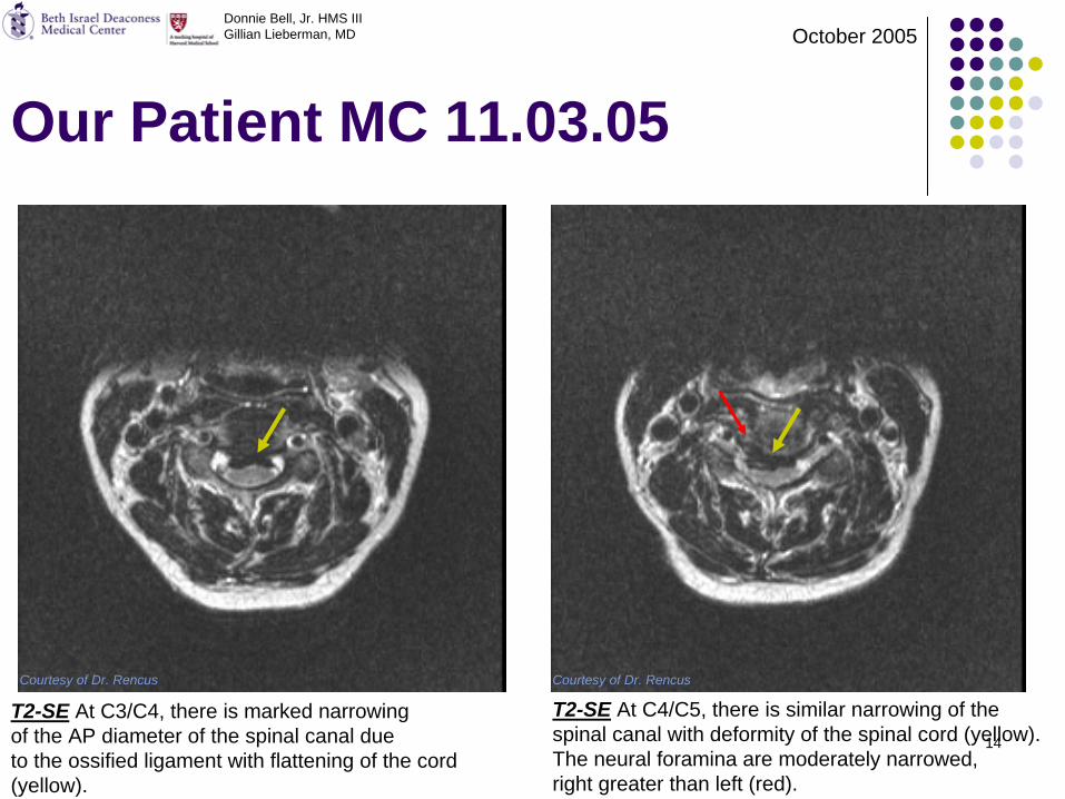
Our Patient MC 11.03.05
Donnie Bell, Jr. HMS IIIGillian Lieberman, MD October 2005
Courtesy of Dr. Rencus Courtesy of Dr. Rencus
T2-SE At C4/C5, there is similar narrowing of thespinal canal with deformity of the spinal cord (yellow). The neural foramina are moderately narrowed,right greater than left (red).
T2-SE At C3/C4, there is marked narrowingof the AP diameter of the spinal canal due to the ossified ligament with flattening of the cord (yellow).
15
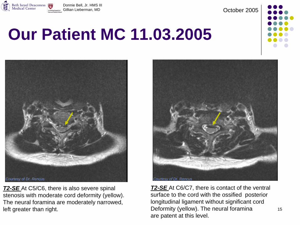
Our Patient MC 11.03.2005
Donnie Bell, Jr. HMS IIIGillian Lieberman, MD October 2005
Courtesy of Dr. Rencus Courtesy of Dr. Rencus
T2-SE At C6/C7, there is contact of the ventralsurface to the cord with the ossified posteriorlongitudinal ligament without significant cordDeformity (yellow). The neural foraminaare patent at this level.
T2-SE At C5/C6, there is also severe spinalstenosis with moderate cord deformity (yellow). The neural foramina are moderately narrowed,left greater than right.
16
Radiographic Differential DxCervical Spine Stenosis
Ossification of Posterior LigamentDiffuse Idiopathic Skeletal Hyperostosis (DISH)Cervical SpondylosisForaminalencroachment / herniated discSeronegativespondyloarthropathiesRAEpidural LipomatosisCongenital Spinal Stenosis
Radiculopathy
Normal
Intradural- extramedullary
A
B
Intramedullary
Extradural
Donnie Bell, Jr. HMS IIIGillian Lieberman, MD October 2005
17
Diagnosis: Ossification of Posterior Longitudinal Ligament
EpidemiologyHighest incidence in Japanese populations
~1.7% of Japanese patients with cervical spine disorders (increasing awareness of OPLL in other populations)2:1 male:female ratioAssociated with and similar to DISH
Clinical PresentationDiagnosis typically established in 5th-7th decadesCord Signs:
Dominant sensorimotor disturbances in lower ext. 16%Dominant sensorimotor disturbances in upper ext. 56%
Cervico-brachialgia – pain in the neck, shoulder, and arm 28%Symptoms typically appear when PLL is 30% of the saggital canal lengthPredisposed to myelopathy precipitated by trauma
PathophysiologyHyperplasia of cartilage cells with eventual endochondral ossification of the posterior longitudinal ligamentMost Common in C3-5Anterior vertebral osteophytes
Radiologic FindingsLow signal intensity between the vertebral body and dural sac on T1 and T2 spin echo (SE) MRI images; calcific densities on CTSpinal Canal AP diameter of <12 mm associated w/ onset of neurologic symptoms (nml 13-20mm)Cord edema/myelomalacia as a result of compression, microtrauma, and ischemia
Donnie Bell, Jr. HMS IIIGillian Lieberman, MD October 2005
Singh et al
18
Molecular Biology of OPLL
OPLL mapped to Npp gene using the ttwmouse model
NppEncodes nucleoside pyrophosphatase
Regulates soft-tissue calcification and bone mineralization by producing inorganic pyrophosphate – an inhibitor of calcificationNon-sense mutation (gly568 to ter) leads to production of a truncated protein thought to be responsible for the accelerated bone formation
http://www.ncbi.nlm.nih.gov/entrez/dispomim.cgi?id=602475
Donnie Bell, Jr. HMS IIIGillian Lieberman, MD October 2005
19
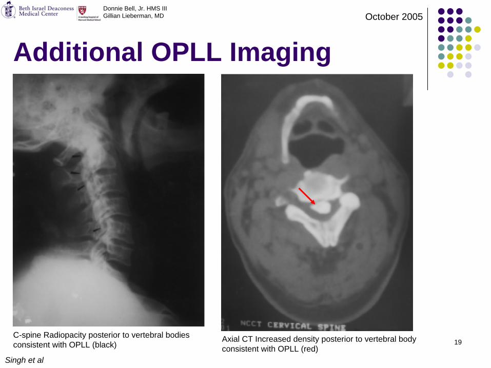
Additional OPLL Imaging
Donnie Bell, Jr. HMS IIIGillian Lieberman, MD October 2005
Singh et al
C-spine Radiopacity posterior to vertebral bodies consistent with OPLL (black)
Axial CT Increased density posterior to vertebral body consistent with OPLL (red)
20
Cervical Spine Stenosis
Now let’s review the common causes of cervical spine stenosis and their radiolographic
features
21
Cervical Spine StenosisEpidemiology
Cervical spondylosis myelopathy (CSM) is the most common cause in adults > 5595% of adults >65 have spondylositic changes and 20% develop myelopathyFrequent in high contact sports athletes
Clinical PresentationPredisposed to myelopathy as result of minor traumaInitial sx:
Loss of hand dexterity/mild proximal lower extremity weakness+/- neck, arm pain
Progression:Progresses in up to 1/3 of patients (initial onset, period of stability, and deterioration to myelopathy)+ Babinski, Hoffman sign, clonus, ataxia, hyperreflexiaSpastic quadriparesis +/- Pain
+/- RadiculopathyPathophysiology
CSM: Cord compression due to mechanical compression and degenerative instabilityAging -> intervertebral discs degeneration…1. Collapse and spur formation -> compression (C5-7)2. Joint instability with antero/retrolisthesis -> compression (C3-5) (often accompanied by
ligamentum flavum hypertrophy)Other Common Etiologies
Disk herniation, AVM, neoplasms, RA, DISH, TB spondylitis, Paget’s Disease, Metastatic Disease
Donnie Bell, Jr. HMS IIIGillian Lieberman, MD October 2005
Adams et al
22
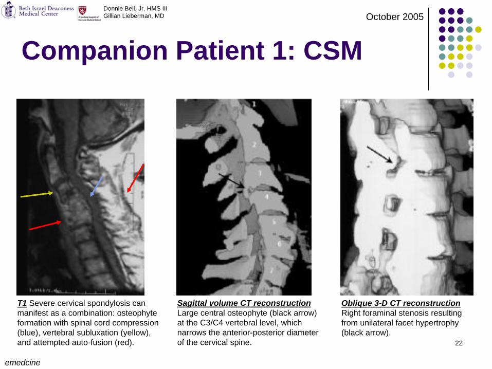
Companion Patient 1: CSM
Donnie Bell, Jr. HMS IIIGillian Lieberman, MD October 2005
emedcine
T1 Severe cervical spondylosis can manifest as a combination: osteophyte formation with spinal cord compression (blue), vertebral subluxation (yellow), and attempted auto-fusion (red).
Sagittal volume CT reconstruction Large central osteophyte (black arrow) at the C3/C4 vertebral level, which narrows the anterior-posterior diameter of the cervical spine.
Oblique 3-D CT reconstruction Right foraminal stenosis resulting from unilateral facet hypertrophy (black arrow).
23
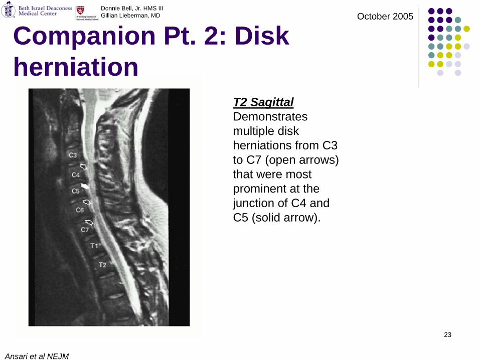
Companion Pt. 2: Disk herniation
Ansari et al NEJM
Donnie Bell, Jr. HMS IIIGillian Lieberman, MD October 2005
T2 Sagittal Demonstrates multiple disk herniations from C3 to C7 (open arrows) that were most prominent at the junction of C4 and C5 (solid arrow).
24
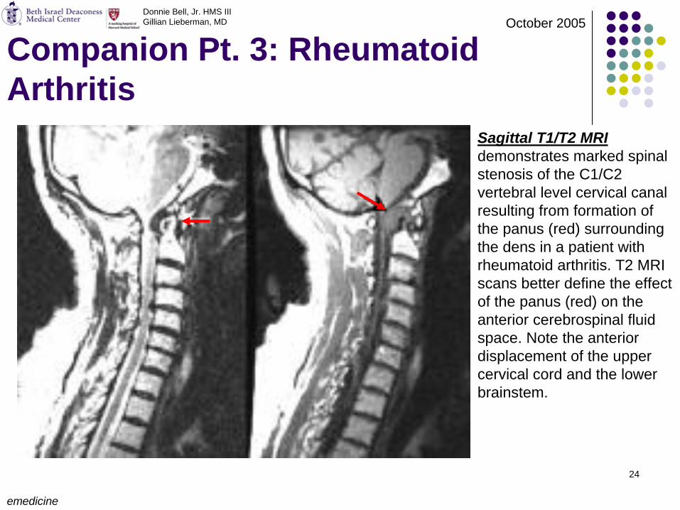
Companion Pt. 3: Rheumatoid Arthritis
Donnie Bell, Jr. HMS IIIGillian Lieberman, MD October 2005
emedicine
Sagittal T1/T2 MRI demonstrates marked spinal stenosis of the C1/C2 vertebral level cervical canal resulting from formation of the panus (red) surrounding the dens in a patient with rheumatoid arthritis. T2 MRI scans better define the effect of the panus (red) on the anterior cerebrospinal fluid space. Note the anterior displacement of the upper cervical cord and the lower brainstem.
25
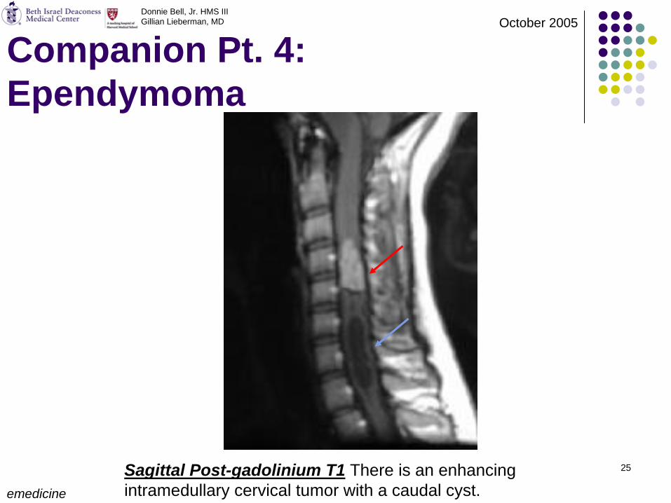
Companion Pt. 4: Ependymoma
emedicine
Donnie Bell, Jr. HMS IIIGillian Lieberman, MD October 2005
Sagittal Post-gadolinium T1 There is an enhancing intramedullary cervical tumor with a caudal cyst.
26
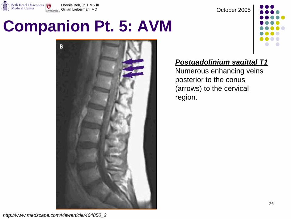
Companion Pt. 5: AVM
http://www.medscape.com/viewarticle/464850_2
Donnie Bell, Jr. HMS IIIGillian Lieberman, MD October 2005
Postgadolinium sagittal T1 Numerous enhancing veins posterior to the conus (arrows) to the cervical region.
27
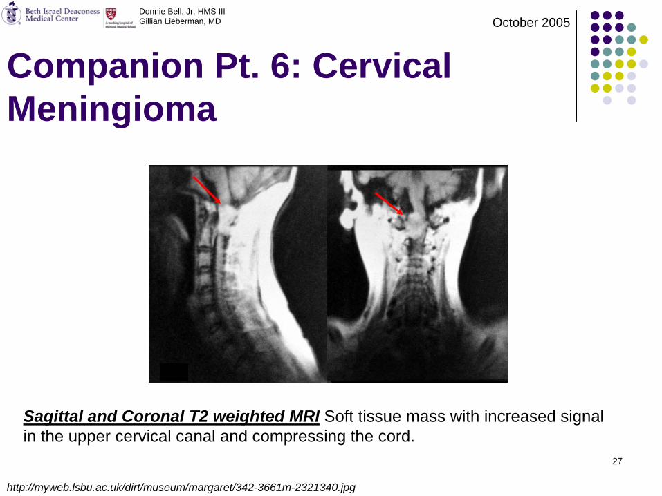
Companion Pt. 6: Cervical Meningioma
http://myweb.lsbu.ac.uk/dirt/museum/margaret/342-3661m-2321340.jpg
Donnie Bell, Jr. HMS IIIGillian Lieberman, MD October 2005
Sagittal and Coronal T2 weighted MRI Soft tissue mass with increased signal in the upper cervical canal and compressing the cord.
28
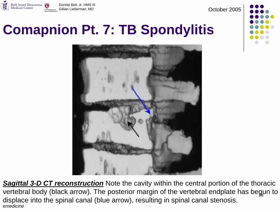
Comapnion Pt. 7: TB Spondylitis
Donnie Bell, Jr. HMS IIIGillian Lieberman, MD October 2005
emedicine
Sagittal 3-D CT reconstruction Note the cavity within the central portion of the thoracic vertebral body (black arrow). The posterior margin of the vertebral endplate has begun to displace into the spinal canal (blue arrow), resulting in spinal canal stenosis.
29
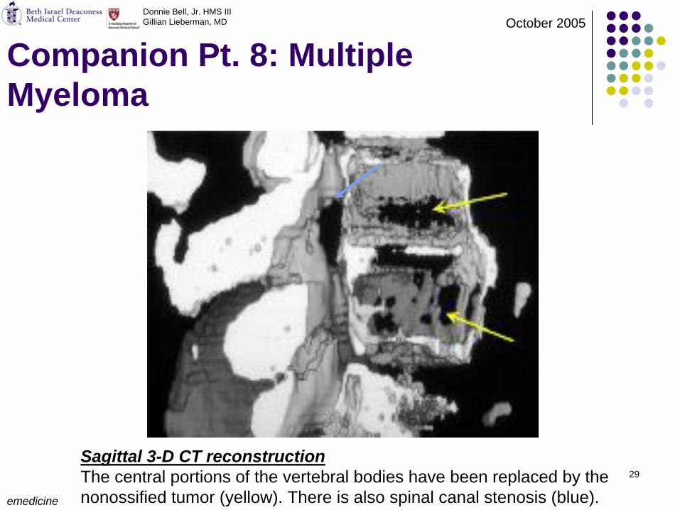
Companion Pt. 8: Multiple Myeloma
Donnie Bell, Jr. HMS IIIGillian Lieberman, MD October 2005
emedicine
Sagittal 3-D CT reconstruction The central portions of the vertebral bodies have been replaced by the nonossified tumor (yellow). There is also spinal canal stenosis (blue).
30
SummaryCervical Spine Stenosis
Adults > 5040% have clinical sx (eg neck crepitus/pain restricted ROM)Remaining asymptomatic individuals have radiographic evidence of cervical spine stenosisMost common cause CSM
ImagingMRI is preferred modality due to its superior soft tissue depictions (spinal cord and surrounding soft tissues)CT and CT myelography also helpful particularly with osseous abnormalities
Patient Presentation and surveyed common causes of Cervical Spine Stenosis
Donnie Bell, Jr. HMS IIIGillian Lieberman, MD October 2005
31
ReferencesWoodruff, W. Fundamentals of Neuroimaging. WB Saunders, Philadelphia, 1993.Netter, FH. Atlas of Human Anatomy, 3rd ed., ICON Learning Systems, Teterboro, New Jersey, 2003.http://w3.uokhsc.edu/human_physiology/Blair%20jpg%20images/Cervicalsc.htmlhttp://daphne.palomar.edu/ccarpenter/Models/spinal%20cord%20XS.htmwww.uicomp.uic.edu/ mri/cerv.spinal.htmlwww.neurosurgeon.org/ public/pem/anatomy_spine.aspwww.pressenter.com/ ~wacma/cervspine.htmSingh et al. Ossification of the Posterior Longitudinal Ligament, MJAFI, vol. 60, No. 3, 2004.Adams et al. Principles of Neurology 6th ed, McGraw-Hill, New York, 1997.http://www.emedicine.com/radio/topic644.htmAnsari A., Rockswold G. N Engl J Med 1998; 338:1358, May 7, 1998http://www.medscape.com/viewarticle/464850_2http://myweb.lsbu.ac.uk/dirt/museum/margaret/342-3661m-2321340.jpg
Donnie Bell, Jr. HMS IIIGillian Lieberman, MD October 2005
32
Acknowledgements Tal Rencus, MD Neuroradiology FellowPeter Rosal, MD 4th yr ResidentGillian Lieberman, MD Clerkship Director Pamela Lepkowski, Clerkship CoordinatorLarry Barbaras, WebmasterFuture MDs of Class of ’06 and ‘07
Donnie Bell, Jr. HMS IIIGillian Lieberman, MD October 2005