Embed Size (px)
Citation preview

Ch. 5: Integumentary System
Integumentary System Functions
• Protection– chemical: acidic skin secretions, melanin, DNA
– physical: keratinized cells
– biological: dendritic cells in epidermis, macrophages in dermis
• Temperature regulation– perspiration, skin blood flow
• Cutaneous sensation– hairs, and see chapter 13
• Metabolism– synthesizes vitamin D precursor (in presence of sunlight)
• Blood reservoir– up to 5% of total blood volume
Skin (Integument)
• Consists of two (or three) major regions
– Epidermis – outermost superficial region
– Dermis – middle region
– Hypodermis (superficial fascia) – a layer of loose connective tissue below the dermis. Most authorities consider it to be below and not a part of the skin; some authorities consider it the third and deepest layer of the skin.
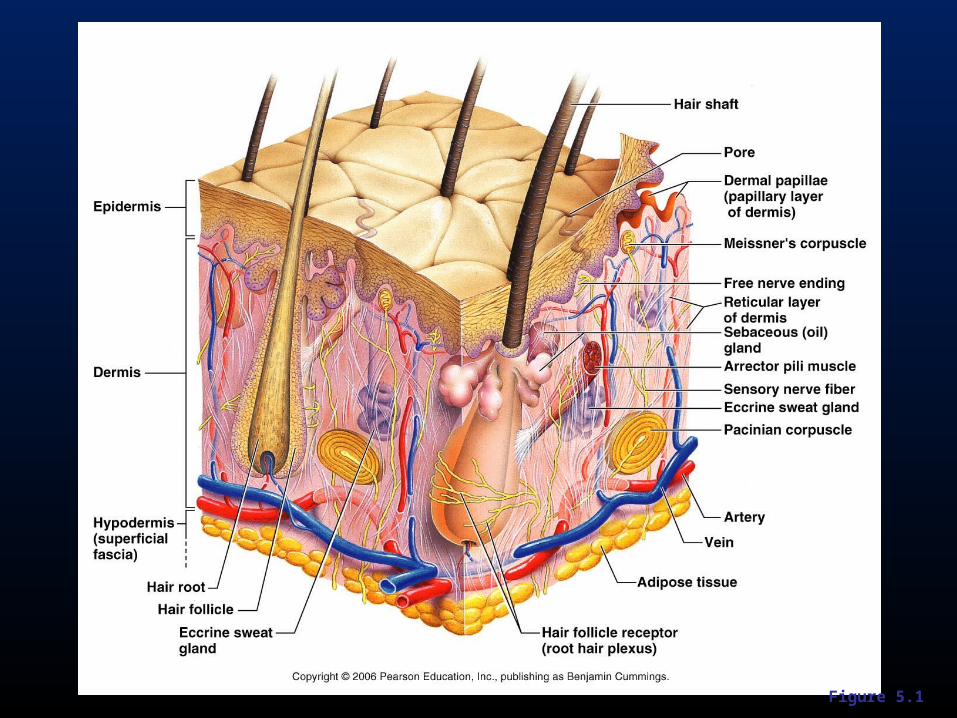
Figure 5.1
Epidermis
• Composed of keratinized stratified squamous epithelium, consisting of four distinct cell types and four or five layers
• Cell types include keratinocytes, melanocytes, Merkel cells, and Langerhans’ cells
• Outer portion of the skin is exposed to the external environment and functions in protection

Figure 5.1
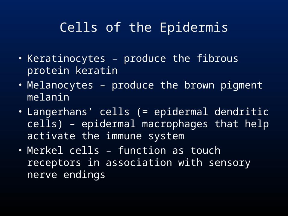
Cells of the Epidermis
• Keratinocytes – produce the fibrous protein keratin
• Melanocytes – produce the brown pigment melanin
• Langerhans’ cells (= epidermal dendritic cells) – epidermal macrophages that help activate the immune system
• Merkel cells – function as touch receptors in association with sensory nerve endings
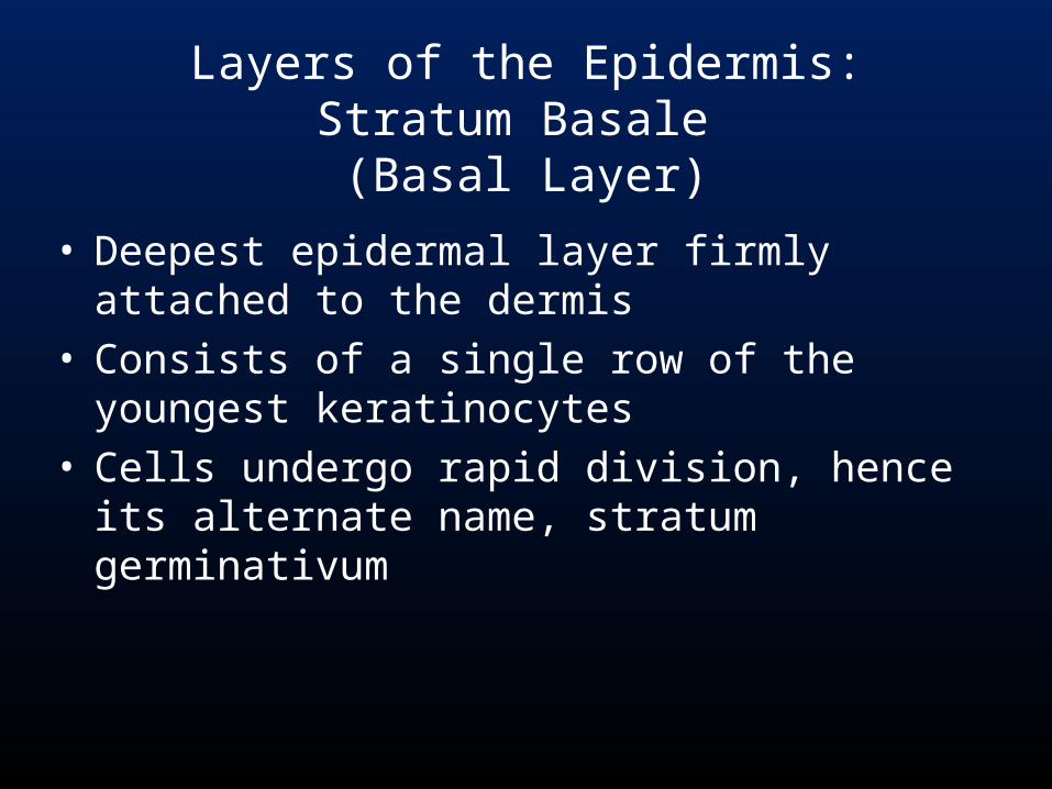
Layers of the Epidermis:Stratum Basale
(Basal Layer)
• Deepest epidermal layer firmly attached to the dermis
• Consists of a single row of the youngest keratinocytes
• Cells undergo rapid division, hence its alternate name, stratum germinativum

Figure 5.2
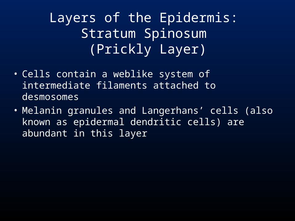
• Cells contain a weblike system of intermediate filaments attached to desmosomes
• Melanin granules and Langerhans’ cells (also known as epidermal dendritic cells) are abundant in this layer
Layers of the Epidermis: Stratum Spinosum
(Prickly Layer)
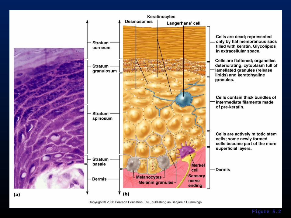
Figure 5.2
• Thin; three to five cell layers in which drastic changes in keratinocyte appearance occurs
• Keratohyaline and lamellated granules accumulate in the cells of this layer
Layers of the Epidermis: Stratum Granulosum
(Granular Layer)
• Thin, transparent band superficial to the stratum granulosum
• Consists of a few rows of flat, dead keratinocytes• Present only in thick skin
Layers of the Epidermis:Stratum Lucidum
(Clear Layer)

Figure 5.2
• Outermost layer of keratinized cells• Accounts for three quarters of the epidermal
thickness• Functions include:
– Waterproofing– Protection from abrasion and penetration– Rendering the body relatively insensitive to
biological, chemical, and physical assaults
Layers of the Epidermis:Stratum Corneum
(Horny Layer)
Dermis
• Second major skin region containing strong, flexible connective tissue
• Cell types include fibroblasts, macrophages, and occasionally mast cells and white blood cells
• Composed of two layers – papillary and reticular

Figure 5.1
Layers of the Dermis: Papillary Layer
• Papillary layer– Areolar connective tissue with collagen
and elastic fibers– Its superior surface contains peglike
projections called dermal papillae– Dermal papillae contain capillary loops,
Meissner’s corpuscles, and free nerve endings
Layers of the Dermis:Reticular Layer
• Reticular layer– Accounts for approximately 80% of the
thickness of the skin– Collagen fibers in this layer add strength
and resiliency to the skin– Elastin fibers provide stretch-recoil
properties
Hypodermis
• Subcutaneous layer deep to the skin• Composed of adipose and areolar
connective tissue• Functions: energy storage; cushion
underlying tissues from external forces
Skin Color
• Three pigments contribute to skin color– Melanin – yellow to reddish-brown to black pigment,
responsible for dark skin colors• Freckles and pigmented moles – result from local
accumulations of melanin– Carotene – yellow to orange pigment, most obvious in
the palms and soles of the feet– Hemoglobin – reddish pigment responsible for the
pinkish hue of the skin
Sweat Glands
• Different types prevent overheating of the body; secrete cerumen and milk– Eccrine sweat glands – found in palms,
soles of the feet, and forehead– Apocrine sweat glands – found in axillary
and anogenital areas– Ceruminous glands – modified apocrine
glands in external ear canal that secrete cerumen
– Mammary glands – specialized sweat glands that secrete milk
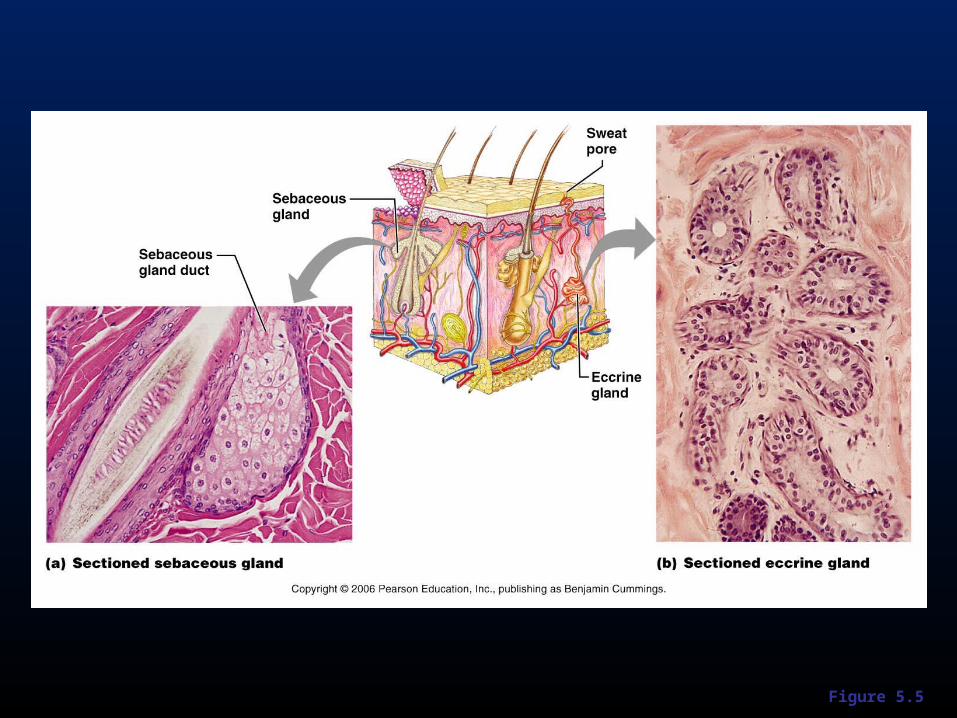
Figure 5.5
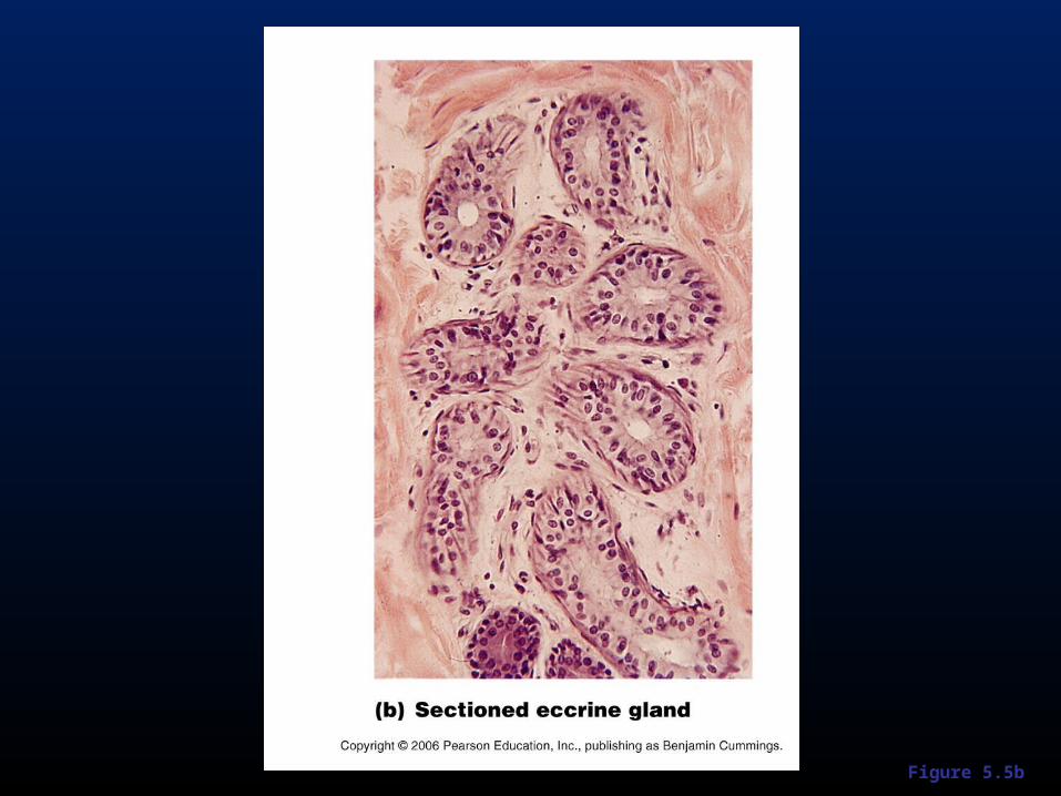
Figure 5.5b
Sebaceous Glands
• Simple alveolar glands found all over the body• Soften skin when stimulated by hormones• Secrete an oily secretion called sebum
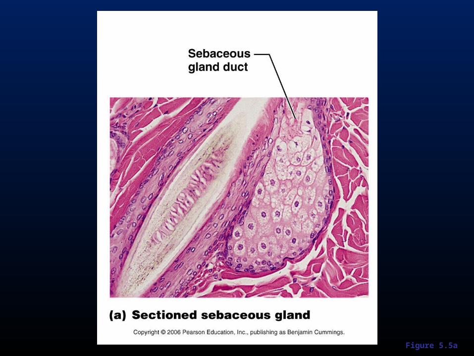
Figure 5.5a
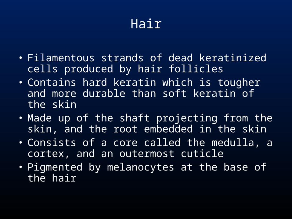
Hair
• Filamentous strands of dead keratinized cells produced by hair follicles
• Contains hard keratin which is tougher and more durable than soft keratin of the skin
• Made up of the shaft projecting from the skin, and the root embedded in the skin
• Consists of a core called the medulla, a cortex, and an outermost cuticle
• Pigmented by melanocytes at the base of the hair
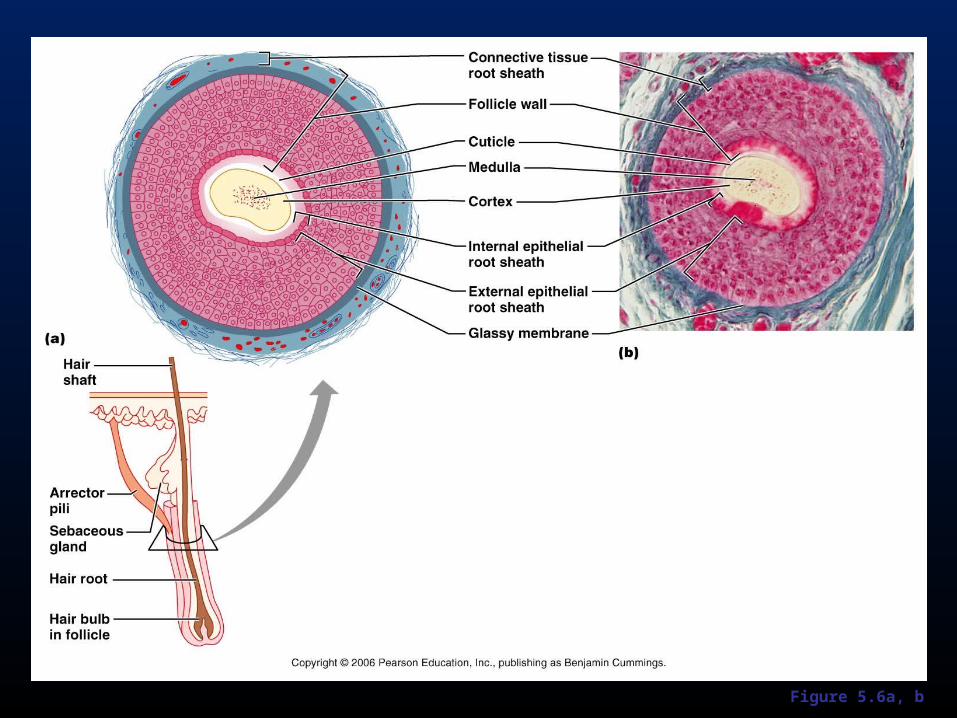
Figure 5.6a, b
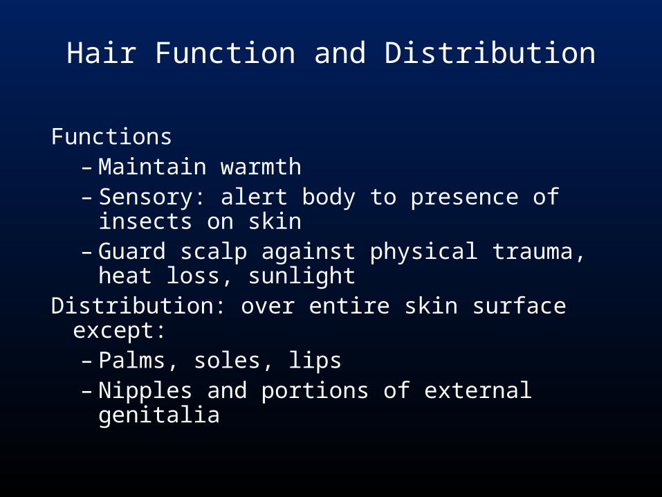
Hair Function and Distribution
Functions– Maintain warmth– Sensory: alert body to presence of insects
on skin – Guard scalp against physical trauma, heat
loss, sunlightDistribution: over entire skin surface except:
– Palms, soles, lips– Nipples and portions of external genitalia
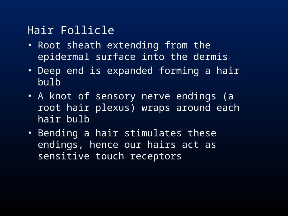
Hair Follicle• Root sheath extending from the epidermal surface
into the dermis• Deep end is expanded forming a hair bulb• A knot of sensory nerve endings (a root hair plexus)
wraps around each hair bulb• Bending a hair stimulates these endings, hence our
hairs act as sensitive touch receptors
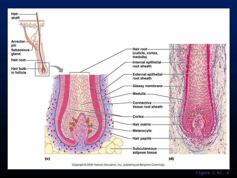
Figure 5.6c, d
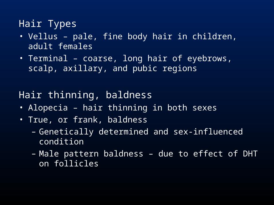
Hair Types• Vellus – pale, fine body hair in children, adult females • Terminal – coarse, long hair of eyebrows, scalp, axillary,
and pubic regions
Hair thinning, baldness• Alopecia – hair thinning in both sexes• True, or frank, baldness
– Genetically determined and sex-influenced condition – Male pattern baldness – due to effect of DHT on
follicles
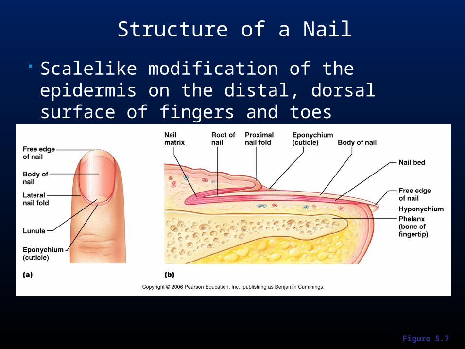
Figure 5.7
Structure of a Nail
Scalelike modification of the epidermis on the distal, dorsal surface of fingers and toes
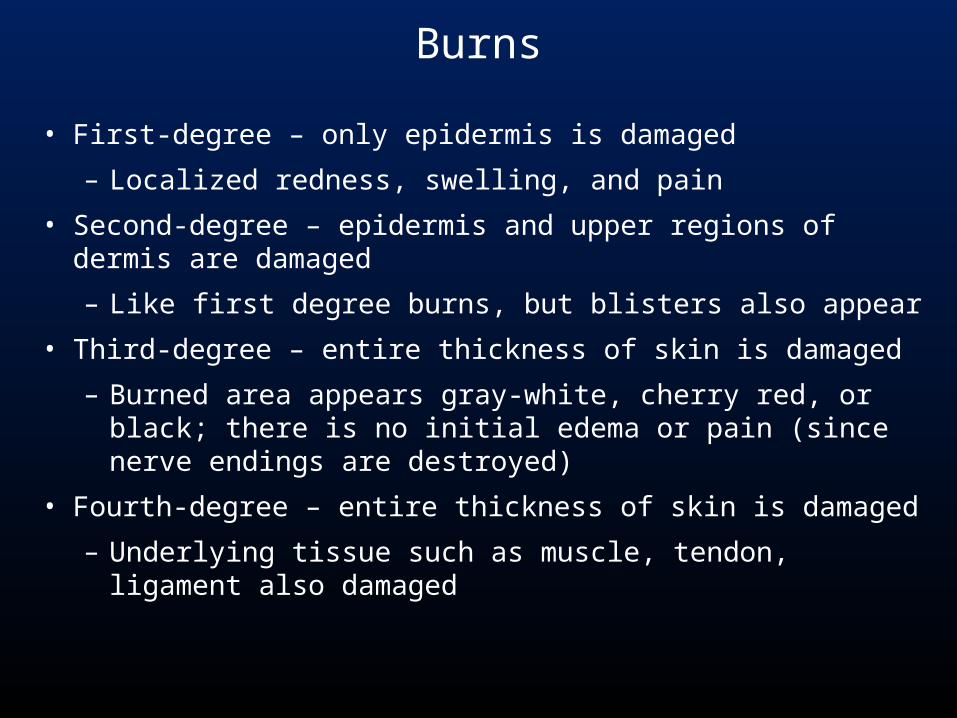
Burns
• First-degree – only epidermis is damaged
– Localized redness, swelling, and pain
• Second-degree – epidermis and upper regions of dermis are damaged
– Like first degree burns, but blisters also appear
• Third-degree – entire thickness of skin is damaged
– Burned area appears gray-white, cherry red, or black; there is no initial edema or pain (since nerve endings are destroyed)
• Fourth-degree – entire thickness of skin is damaged
– Underlying tissue such as muscle, tendon, ligament also damaged
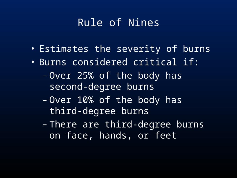
Rule of Nines
• Estimates the severity of burns• Burns considered critical if:
– Over 25% of the body has second-degree burns
– Over 10% of the body has third-degree burns
– There are third-degree burns on face, hands, or feet
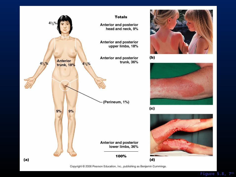
Figure 5.8, 7th ed.