Embed Size (px)
Citation preview
University of Groningen
Pathophysiology of thoracic irradiationGhobadi, Ghazaleh
IMPORTANT NOTE: You are advised to consult the publisher's version (publisher's PDF) if you wish to cite fromit. Please check the document version below.
Document VersionPublisher's PDF, also known as Version of record
Publication date:2013
Link to publication in University of Groningen/UMCG research database
Citation for published version (APA):Ghobadi, G. (2013). Pathophysiology of thoracic irradiation Groningen: s.n.
CopyrightOther than for strictly personal use, it is not permitted to download or to forward/distribute the text or part of it without the consent of theauthor(s) and/or copyright holder(s), unless the work is under an open content license (like Creative Commons).
Take-down policyIf you believe that this document breaches copyright please contact us providing details, and we will remove access to the work immediatelyand investigate your claim.
Downloaded from the University of Groningen/UMCG research database (Pure): http://www.rug.nl/research/portal. For technical reasons thenumber of authors shown on this cover page is limited to 10 maximum.
Download date: 18-02-2018
CHAPTER 2
Lung irradiation induces pulmonary vascular remodeling resembling pulmonary arterial
hypertension
G. Ghobadi1,2, B. Bartelds3, S. J. van der Veen1,2, M.G. Dickinson3, S.
Brandenburg4, R.M.F. Berger3, J. A. Langendijk1, R. P. Coppes1,2*, P. van
Luijk1*
1Department of Radiation Oncology, 2Department of Cell Biology, Section of Radiation and Stress Cell Biology,
3Center for Congenital Heart Disease, Pediatric Cardiology, Beatrix Children Hospital, University Medical
Center Groningen, 4Kernfysisch Versneller Instituut, University of Groningen, Groningen, The Netherlands.
*These authors contributed equally
Thorax. 2012 Apr;67(4):334-41.

35
Lung irradiation induces pulmonary hypertension
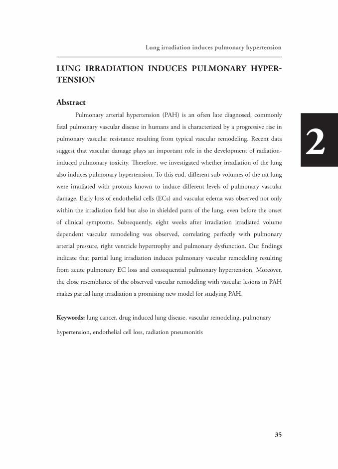
LUNG IRRADIATION INDUCES PULMONARY HYPER-TENSION
AbstractPulmonary arterial hypertension (PAH) is an often late diagnosed, commonly
fatal pulmonary vascular disease in humans and is characterized by a progressive rise in
pulmonary vascular resistance resulting from typical vascular remodeling. Recent data
suggest that vascular damage plays an important role in the development of radiation-
induced pulmonary toxicity. Therefore, we investigated whether irradiation of the lung
also induces pulmonary hypertension. To this end, different sub-volumes of the rat lung
were irradiated with protons known to induce different levels of pulmonary vascular
damage. Early loss of endothelial cells (ECs) and vascular edema was observed not only
within the irradiation field but also in shielded parts of the lung, even before the onset
of clinical symptoms. Subsequently, eight weeks after irradiation irradiated volume
dependent vascular remodeling was observed, correlating perfectly with pulmonary
arterial pressure, right ventricle hypertrophy and pulmonary dysfunction. Our findings
indicate that partial lung irradiation induces pulmonary vascular remodeling resulting
from acute pulmonary EC loss and consequential pulmonary hypertension. Moreover,
the close resemblance of the observed vascular remodeling with vascular lesions in PAH
makes partial lung irradiation a promising new model for studying PAH.
Keywords: lung cancer, drug induced lung disease, vascular remodeling, pulmonary
hypertension, endothelial cell loss, radiation pneumonitis
2
36
Chapter 2
1. INTRODUCTIONPulmonary arterial hypertension (PAH) is a severe and progressive form of pulmo-
nary hypertension that leads to right heart failure and premature death, without a cure
available1. PAH is characterized by typical angioproliferative lesions, such as neointimal
lesions, leading to increased pulmonary vascular resistance2.The elevated pulmonary
vascular resistance leads to high right ventricle systolic pressure and subsequent right
ventricular hypertrophy (RVH) ultimately resulting in heart failure 3,4. The pathogen-
esis of the increased pulmonary vascular resistance is due to combination of sustained
vasoconstriction, arterial wall remodeling and thrombosis5. Although the typical histopa-
thology of the angioproliferative lesions is well described, the mechanisms are so far
unknown. A variety of animal models has been developed to study the mechanism
of development and maintenance of pulmonary hypertension. However, few of these
models fully describe human PAH6. Interestingly, we recently showed in a preclinical
model that also thoracic irradiation leads to vascular damage, being the predominant
histological change after irradiation of large volumes with a relatively low dose7. Our
findings agreed with what was suggested from clinical8 and preclinical studies9 indicating
that treatment of large lung volumes even to a low dose is an important risk factor in the
development of pulmonary toxicity. Moreover, whole-thoracic-irradiation with a sub-
lethal dose was shown to induce an increased pulmonary vascular resistance as well as a
decrease in pulmonary arterial distensibility and vascular density early after radiation10.
These features seem similar to that what is observed in patients with pulmonary hyperten-
sion11. Interestingly, individuals already suffering from pre-existing pulmonary vascular
disease, manifesting as sub-clinical increases in pulmonary artery pressure, are known to
have an increased risk for radiation-induced pulmonary toxicity12. However, the type,
evolution and consequences of pulmonary vascular remodeling after radiation are largely
unknown. In the present study, we hypothesize that vascular damage after irradiation
of the lung may develop into PAH. Using high-precision proton radiation beams, this
hypothesis was tested by inducing different levels of vascular damage by irradiating small,
intermediate and large volumes of rat lungs with graded radiation doses, resulting in an
equal risk of inducing pulmonary dysfunction13. Since vascular damage plays a central
37
Lung irradiation induces pulmonary hypertension
role in radiation-induced pulmonary dysfunction as well as pulmonary hypertension, we
investigated the commonalities between radiation-induced vascular remodeling and that
of previously described PAH models.
2. MATERIAL AND METHODS 2.1 Animals
Adult male albino wistar rats (n=78, 270gr-320gr, 8-9 weeks old) of the Hsd/
Cpb:WU strain bred in a specific pathogen free colony (Harlan-CPB, Rijswijk, The
Netherlands) were used in the experiments. They were housed five to a cage under a
12 h light - 12 h dark cycle and fed rodent chow (RMH-B, Hope Farms, Woerden,
The Netherlands) and water ad libitum. The experiments were performed in agreement
with the Netherlands Experiments on Animals Act (1977) and the European Convention
for the Protection of Vertebrate Animals Used for Experimental Purposes (Strasbourg,
18.III.1986).
2.2 Irradiation technique
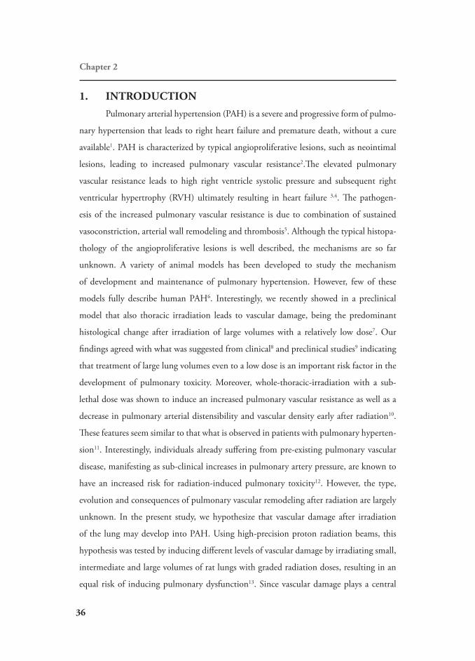
To induce different levels of vascular damage, 33%, 50%, 75% or 100% of the rats’
lungs were irradiated to 28, 20/22, 17 and 13 Gy (single fraction) respectively (Figure.1).
Figure.1: Overview of applied irradiation ports
This was done with 150 MeV protons from the cyclotron at the Kernfysisch
Versneller Instituut, using the shoot-through technique as published previously13-16.
This results in very sharp lateral field edges (20–80% isodose distance: 1 mm)16 thus
sparing the shielded part of the lung very effectively. The irradiation ports (Figure.1) were
designed using computed tomography scans of animals of the same age and weight17.
2
38
Chapter 2
2.3 Histopathology
For morphological analysis, tissue samples were taken from both in- and out-of-
field of the left lung at a distance of at least 3 mm from the field edge and compared with
non-irradiated controls (n=3). The radiation response of the pulmonary vasculature was
assessed by evaluating the morphology before (2 weeks post-irradiation) and at the peak
of pulmonary dysfunction (8 weeks post-irradiation)17 in 3 rats per group. Pulmonary
sections (5-μm thick) were stained with Haematoxylin and Eosin, rat endothelial cells
specific marker (HIS52) or Verhoeff’s elastica stain. Since 70% of the pulmonary vascular
bed consists of radiosensitive microvasculature18,19 and damage of these can be expected
to have the largest impact on lung function, small intra-acinar vessels (<50 μm) were
selected for morphological analysis. For morphometric analysis of the vascular dimen-
sions, lung sections with Verhoeff’s elastica stain were used according to van Albada et
al.20. In short, 40 randomly chosen pulmonary intra-acinar vessels <50 μm were assessed
at 400x magnification using an image analysis system (CZ KS400; Imaging Associates,
Bicester, UK). Two different vascular areas were defined: outer vessel area and luminal
area. The outer vessel area was defined as the area within the external elastic lamina.
The area within the internal elastic lamina was defined as the luminal area. Areas were
transformed into diameters and subsequently wall thickness was defined by subtracting
the external diameter from the luminal diameter. Occlusion was then calculated in these
pulmonary vessels accordingly, as (outer vessel diameter- luminal diameter) / (outer vessel
diameter). Vessels were excluded from the measurement if they were too elliptical (ratio
of the largest and smallest diameter exceeded 2), had an incomplete circular shape, were
collapsed along more than one quarter of the vessel wall or were located adjacent to an
airway.
2.4 Hemodynamics
To assess the effect of the early radiation response of the lung vasculature on
pulmonary/cardiac hemodynamics, right ventricle and pulmonary artery pressure (RVP
and PAP) were directly measured at the peak of pulmonary dysfunction, 8 weeks post-
lung irradiation, according to Rabinovitch et al.21. In short, the rats (n=4 per group) first
were anesthetized (isoflurane) and ventilated with room air. A fluid-filled catheter with
39
Lung irradiation induces pulmonary hypertension
the pressure transducer was induced via right external jugular vein into right ventricle(RV)
cavity and guided to the pulmonary artery (PA) under pressure waveform monitoring to
record the RVP and PAP. After completion of the hemodynamic measurement, the thorax
was opened and heart and lungs were excised. The heart was divided into atria, ventricles
and septum and weighted separately. Right ventricle hypertrophy (RVH) was assessed by
measuring the ratio of the weights of RV to the combination of left ventricle (LV) and
intraventricular septum (IVS). The measurements were compared with non-irradiated
controls (n=3).
2.5 Breathing rate (BR) assay
In pre-clinical studies, BR is considered as a surrogate for pulmonary function17,22.
BR was measured before and every two weeks after the irradiations up to week 10 (n=24
per group), as previously described17. The increase of the BR at this time, relative to
the mean BR in weeks 0–2 after irradiation, was used as an indicator of early pulmo-
nary dysfunction. The BR of non-irradiated controls was available in our database from
previous experiments (n=9).
2.6 Statistics
To test whether differences exist between groups in pressure measurements and
RVH assessment, the Post hoc-Bonferroni test was performed using one-way ANOVA.
For morphology quantification, the linear mixed model and F-test was specifically used to
test differences between quantified parameters when comparing all groups at once. In this
test randomly chosen vessels per rat per group were considered as randomly repeated vari-
ables. The nominal level of significance was 0.05. In order to determine to what extent the
changes in BR is explained by changes in PAP, the Pearson’s product-moment correlation
coefficient of these 2 parameters was used. A correlation was considered significant if the
hypothesis of no correlation was rejected at p<0.05. Breathing rate change has significant
amount of variation due to random errors in the measurement. Therefore when using all
individual data points for correlation, in order to answer our main question, the correla-
tion coefficient has to be corrected for attenuation due to the random errors of BR meas-
urement according to the following formula 23.
2
40
Chapter 2
BR
BRmPAPcorrected yreliabilit
rr ,=
The reliability is inversely related to random errors and is the consistency of a set
of measurements24. In order to assess the random error in the BR measurement, 4 random
groups of rats with random dose distributions were chosen to resemble the variation in
the irradiated population. Their BR measurements during the whole follow up post-irra-
diation were duplicated which was carried out by the same person. The reliability is then
estimated as the concordance correlation coefficient between these two administrations of
the same measure 24 and was 0.77 25.
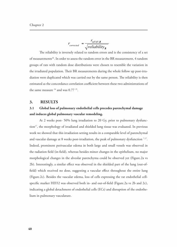
3. RESULTS3.1 Global loss of pulmonary endothelial cells precedes parenchymal damage
and induces global pulmonary vascular remodeling.
At 2 weeks post- 50% lung irradiation to 20 Gy, prior to pulmonary dysfunc-
tion17, the morphology of irradiated and shielded lung tissue was evaluated. In previous
work we showed that this irradiation setting results in a comparable level of parenchymal
and vascular damage at 8 weeks post-irradiation, the peak of pulmonary dysfunction 7,17.
Indeed, prominent perivascular edema in both large and small vessels was observed in
the radiation field (in-field), whereas besides minor changes in the epithelium, no major
morphological changes in the alveolar parenchyma could be observed yet (Figure.2a vs
2b). Interestingly, a similar effect was observed in the shielded part of the lung (out-of-
field) which received no dose, suggesting a vascular effect throughout the entire lung
(Figure.2c). Besides the vascular edema, loss of cells expressing the rat endothelial cell-
specific marker HIS52 was observed both in- and out-of-field (Figure.2a vs 2b and 2c),
indicating a global detachment of endothelial cells (ECs) and disruption of the endothe-
lium in pulmonary vasculature.
41
Lung irradiation induces pulmonary hypertension
Figure.2: Global loss of pulmonary endothelial cells (ECs) precedes paren-chymal damage. The effect of lung irradiation on lung tissue and ECs was assessed by evaluation of the morphology of lung tissue by H&E and immunohistochemical staining of rat ECs specific marker (HIS52) respectively. Lung samples were taken from both in- and out-of-field 2 weeks post-50% lung irradiation to 20 Gy. Nuclei are stained with DAPI (blue) and HIS52 positive are stained with brown. Panel a shows a large and a small vessel in non-irradiated lung tissue with fine lung paren-chyma. A close view of a small vessel in non-irradiated lung tissue shows an intact endothelial layer; the integrated lining of ECs is shown in a higher magnification. Perivascular edema (both in-field and out-of-field, b and c respectively; pointed by arrows) and EC detachment (leading to a disrupted endothelial layer) are the predominant damage. The results are representative sections from 3 irradiated rat lungs.
Since it is well known that changes in the integrity of ECs lead to increased perme-
ability and leakage in irradiated tissues26, the early response of pulmonary vasculature to
lung irradiation may be initiated from the global lung EC loss.
2
42
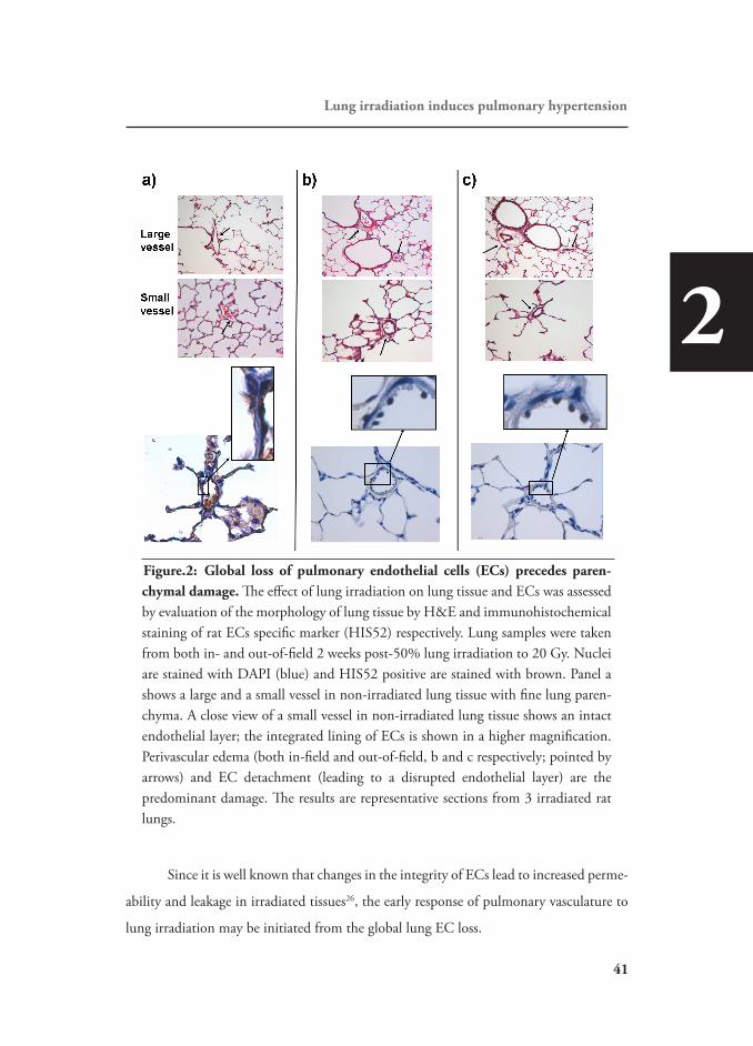
Chapter 2
Figure.3: Early radiation response of lung vasculature leads to global pulmo-nary vascular remodeling. (a) Anatomy of non-irradiated control pulmonary intra-acinar vessel (<50 μm). Pulmonary vascular remodeling was assessed by immunohistochemical staining (Verhoeff’s elastica stain) of pulmonary intra-acinar vessels 8 weeks post-irradiation of 33, 50 and 75% rats’ lungs to 28, 22 and 17 Gy respectively. (b) Lung irradiation induces vascular remodeling such as musculariza-tion, adventitia thickening and neointima formation. (c) Vascular remodeling were present both in- and out-of- field showing a global pulmonary vascular response in all irradiated volumes. L= luminal diameter, O= outer vessel diameter, SMCs= smooth muscle cells. Magnification 400x.
43
Lung irradiation induces pulmonary hypertension
EC injury is a hallmark in initiating/mediating structural changes in pulmonary
vasculature 27. Therefore, the morphology of the lung vasculature was investigated at the
peak of pulmonary dysfunction17. Indeed lung irradiation-induced structural changes
in the vasculature such as muscularization of the media layer, thickening of the adven-
titia as well as more advanced lesions, including neointimal lesions and obliteration of
small pulmonary vessels (Figure.3b), indicative of advanced remodeling of all layers. As
expected, due to the global nature of vascular response, these features were also present
out-of-field. In order to check whether the vascular response occurs at lower doses, the
morphology of 100% to a low dose of 13 Gy irradiated lungs, were evaluated. In previous
work 7,17 we showed that this irradiation setting has the lowest applicable dose which
produces a pronounced pulmonary dysfunction. Indeed prior to pulmonary dysfunc-
tion, again predominant perivascular edema was observed with no sign of parenchymal
damage (Supplementary figure.1a). At the peak of pulmonary dysfunction again severe
vascular remodeling was observed without major parenchymal remodeling (Supplemen-
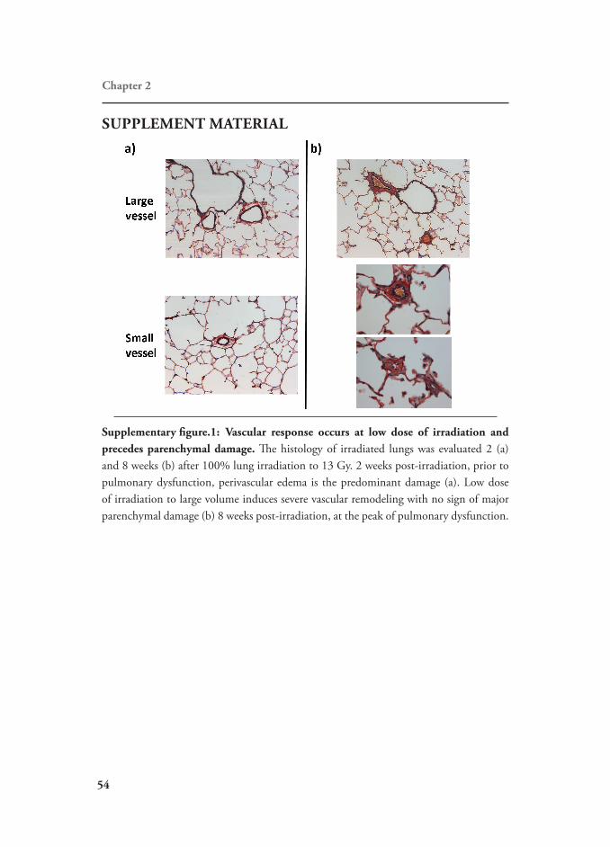
tary figure.1b). This may indicate that the tolerance dose for vascular response is lower
than parenchymal response.
3.2 Global response of pulmonary vasculature is irradiated volume dependent.
Development of pulmonary dysfunction depends on the irradiated lung
volume7,17,28. Therefore, the extent to which pulmonary dysfunction is determined by
pulmonary vascular remodeling was assessed by morphological quantification of vascular
damage, induced to different levels by irradiating different lung volumes (Figure.3c)
Strikingly irradiated-volume dependent increases were observed in absolute wall thick-
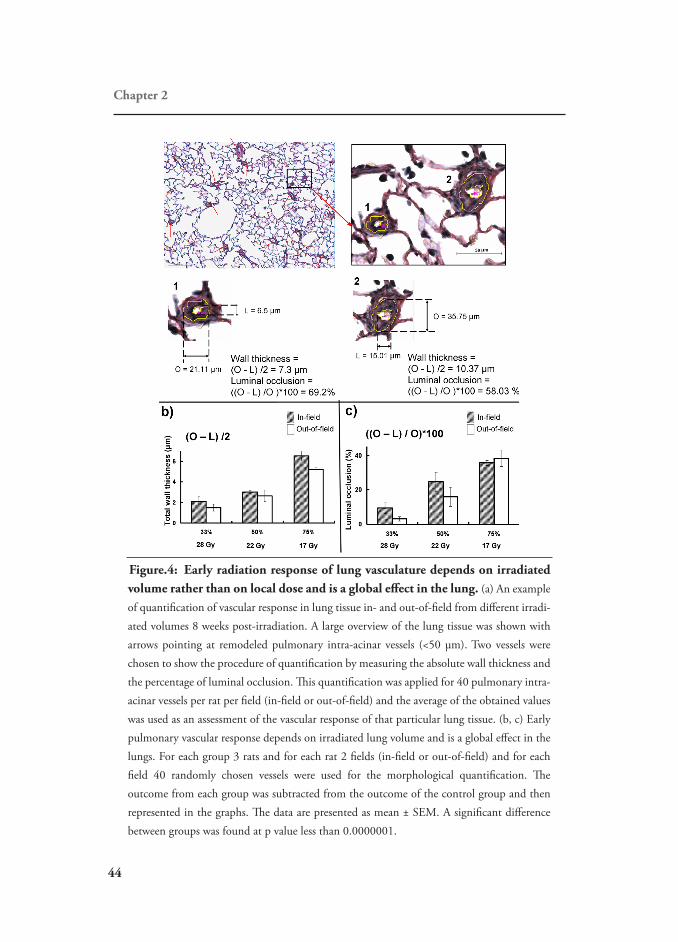
ness (Figure.4b, p<0.05) and the percentage of luminal occlusion (Figure.4c, p<0.05).
Vascular remodeling was more pronounced with an increase in irradiated volume despite
the lower radiation dose. Moreover, the out-of-field response increased as the irradiated
volume increased (Figure.4b and c, p<0.05).This indicates a global pulmonary vascular
response rather than a response confined only to the area where the radiation dose is
deposited.
2
44
Chapter 2
Figure.4: Early radiation response of lung vasculature depends on irradiated volume rather than on local dose and is a global effect in the lung. (a) An example of quantification of vascular response in lung tissue in- and out-of-field from different irradi-ated volumes 8 weeks post-irradiation. A large overview of the lung tissue was shown with arrows pointing at remodeled pulmonary intra-acinar vessels (<50 μm). Two vessels were chosen to show the procedure of quantification by measuring the absolute wall thickness and the percentage of luminal occlusion. This quantification was applied for 40 pulmonary intra-acinar vessels per rat per field (in-field or out-of-field) and the average of the obtained values was used as an assessment of the vascular response of that particular lung tissue. (b, c) Early pulmonary vascular response depends on irradiated lung volume and is a global effect in the lungs. For each group 3 rats and for each rat 2 fields (in-field or out-of-field) and for each field 40 randomly chosen vessels were used for the morphological quantification. The outcome from each group was subtracted from the outcome of the control group and then represented in the graphs. The data are presented as mean ± SEM. A significant difference between groups was found at p value less than 0.0000001.
45
Lung irradiation induces pulmonary hypertension
3.3 Global pulmonary vasculature response determines pulmonary dysfunction.
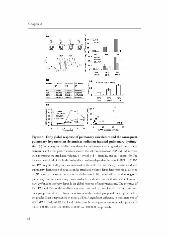
Global changes in vascular bed increases the pulmonary vascular resistance and
induces elevated RVP and PAP, features that are known as pulmonary hypertension29.
Indeed lung irradiation was found to induce pulmonary hypertension by showing an
irradiated-volume dependent increase in all components of RVP and PAP, despite the
decreasing dose (Figure.5a). Moreover, as can be expected with an increase in pulmo-
nary vascular resistance and PAP, the increased RV workload led to an irradiated-volume
dependent RVH and a decrease in IVS weight (Figure.5b). These are generally consid-
ered as characteristics of the severity of pulmonary hypertension29. Therefore the results
showed that rats irradiated to the largest lung volume (75%) even to a relatively low
dose (17 Gy) suffer from severe pulmonary hypertension. In addition, as a measure of
pulmonary function, the breathing rate (BR) of the irradiated animals increased in a
similar manner to all other measured parameters, indicating that the development of
pulmonary dysfunction is related to pulmonary hypertension (Figure.5c). To determine
to what extent the global pulmonary vascular response and hence pulmonary hyperten-
sion is responsible for pulmonary dysfunction, the increase in BR and mPAP were corre-
lated using all individual data(Figure.5c). Remarkably, a strong correlation of these two
parameters (Figure.5c, r=0.9) was found. Taken together our results indicate that the
global pulmonary vascular response plays a major role in the development of early radia-
tion-induced pulmonary dysfunction.
4. DISCUSSIONIn preclinical studies radiation of the lungs at large volumes leading to pulmo-
nary dysfunction seems to be caused by vascular damage. Since vascular remodeling is a
hallmark of PAH, we investigated whether irradiation could induce pulmonary hyperten-
sion with the related pulmonary vascular changes. We show the proof of principle that
irradiation of the lung leads to pulmonary vascular remodeling with subsequent pulmo-
nary hypertension and RVH. The pulmonary vascular remodeling induced by irradia-
tion showed striking similarities with the characteristic histopathology of PAH, included
endothelial cell damage, vascular cell proliferation and neointimal lesions 30.
2
46
Chapter 2
Figure.5: Early global response of pulmonary vasculature and the consequent pulmonary hypertension determines radiation-induced pulmonary dysfunc-tion. (a) Pulmonary and cardiac hemodynamics measurement with right-sided cardiac cath-eterization at 8 weeks post-irradiation showed that all components of RVP and PAP increase with increasing the irradiated volume. s = systolic, d = diastolic, and m = mean. (b) The increased workload of RV leaded to irradiated volume dependent increase in RVH. LV, RV, and IVS weights of all groups are indicated in the table. (c) Indeed early radiation-induced pulmonary dysfunction showed a similar irradiated volume dependent response as assessed by BR increase. The strong correlation of the increase in BR and mPAP as a readout of global pulmonary vascular remodeling (r corrected = 0.9) indicates that the development of pulmo-nary dysfunction strongly depends on global response of lung vasculature. The increases of RVP, PAP and RVH of the irradiated rats were compared to control levels. The outcome from each group was subtracted from the outcome of the control group and then represented in the graphs. Data is represented in mean ± SEM. A significant difference in measurements of sRVP, sPAP, dPAP, mPAP, RVH and BR-increase between groups was found with p values of 0.004, 0.0004, 0.0001, 0.00005, 0.00006 and 0.000003 respectively.
47
Lung irradiation induces pulmonary hypertension
The first sign of vascular changes such as EC loss and perivascular edema were
observed both within the irradiated and shielded parts of the lung before any apparent
parenchymal or BR changes. Subsequent to global pulmonary vascular remodeling accom-
panied by PAP elevation, RVH was observed indicative of pulmonary hypertension. This
cascade of events was shown to play a major role in the development of BR increase as a
surrogate measure for pulmonary dysfunction. BR is a nonspecific measure for pulmo-
nary function. It is only part of the lung function (which is not easily measurable in rats)
and changes can be induced by several factors such as reduced diffusion capacity due to
parenchymal damage. However, the very strong correlation between pulmonary hyper-
tension and BR in our study suggests that in our model the reduced ventilatory efficiency
as a consequence of reduced perfusion is responsible for the increases in BR, as frequently
observed in patients with PAH 31. Hence in this model of PAH in rats, BR may seem to
be a sufficient surrogate measure of pulmonary function.
The acute EC loss and perivascular edema found in the current study may result
from ECs detaching from the basal lamina32, loss of endothelium integrity due to EC
retraction and increased permeability to low molecular weight solutes26 shown to occur
hours to days after radiation of pulmonary ECs. Although not yet studied in lungs, the
importance of acute EC loss in the development of radiation-induced normal tissue
damage has been established for other tissues such as intestine, spinal cord and rectum33-
35. In patients treated for rectal cancer with preoperative radiotherapy the radiation injury
to normal tissue correlated strongly with the radiation-induced vascular damage as quan-
tified by vascular wall thickness33. In the same study33, it was also demonstrated that
irradiated ECs induce migration and proliferation of vascular smooth muscle cells, an
important feature of vascular remodeling in PAH 36. It therefore seems that ECs may be
the main target of radiation in many organs and may have a pivotal role in the develop-
ment of early radiation-induced vascular changes in the lung. However, the current study
shows for the first time that this phenomenon can induce pulmonary hypertension and
function failure. Indeed in many pulmonary vascular diseases, such as PAH, EC dysfunc-
tion is considered to play an initiating role in the pathogenesis of vascular remodeling27.
The features of vascular remodeling in our model show striking similarities with those
2
48
Chapter 2
observed in other experimental pulmonary hypertension models using the same strain
of rats and age20 or Sprague-Dawley with almost the same age and weight (300-350
gr) 37,38. This supports the hypothesis that EC loss also plays an initiating role in the
pathogenesis of the early radiation-induced vascular changes in the lung. Moreover, in
other animal models observations in line with ours have been published after thoracic
irradiation, such as acute vascular remodeling in August rats39 and increased mean pulmo-
nary artery pressure in Sprague-Dawley rats40, dogs41 and sheep42,43. Therefore, our find-
ings open a new way of studying the radiation model of pulmonary hypertension for a
better understanding of EC dysfunction and degeneration of vasculature in pulmonary
vascular disease leading to PAH, a disease in which the inducing mechanisms are so far
not completely understood 44,45.
One of the most striking observations in this model of radiation-induced pulmo-
nary vascular damage is that the histomorphology shows the development of neointimal
lesions, which are considered hallmarks of PAH6,20,46-49. Treatment of patients with PAH,
in the international classification type I PAH, still have a poor prognosis despite improve-
ment in therapeutic regimens1, hence it is referred to as the “irreversible” form of PAH 46,47. Irreversibility of pulmonary hypertensive disease has been specifically associated with
the presence of these neointimal lesions 50. Although many PAH models have shown to
develop medial hypertrophy, so far, only few models have been described that consistently
develop neointima lesions 29,37,49,51. In the current era, neointimal models are needed to
adequately study the pathobiology of PAH and the effects of new treatment strategies.
The occurrence of actual neointimal lesions in this radiation-induced pulmonary hyper-
tension may offer such a novel model. A unique feature of the radiation-induced model
is the irradiated lung volume dependence of the induction of damage52 and consequent
changes in PAP (present work), which facilitates well-controlled induction to pre-deter-
mined levels of PAH to study different stages of disease development in a controlled
manner.
Radiation-induced EC loss (e.g. due to apoptosis) and the consequent disrup-
tion of endothelial lining with increased permeability and perivascular edema shown in
this model may decrease the blood flow in the irradiated vasculature. As a compensa-
49
Lung irradiation induces pulmonary hypertension
tory effect, the pressure, blood flow and thereby the shear stress would increase in the
vasculature in the non-irradiated part of the lungs. This increase of the shear stress may
then lead to changes in function and structure of ECs in the non-irradiated vasculature.
Indeed, changes in blood flow and shear stress are known to specifically regulate vascular
remodeling by altering EC and smooth muscle cells apoptosis and proliferation rates53,54.
Increased shear stress induced by increased pulmonary blood flow in patients and animal
models with large congenital cardiac shunts is known to lead to pulmonary endothelial
dysfunction and progressive vascular remodeling and, thus, flow-associated PAH 30,44.
This may be a mechanism by which out-of-field EC loss and whole lung vascular remod-
eling develops. It is expected that irradiation of larger lung volumes even to low dose
(high enough to induce EC apoptosis) induces a more pronounced increase in blood flow
and shear stress in non-irradiated vasculatures leading to an enhanced out-of-field effect.
The unique behavior of our radiation-induced pulmonary hypertension model enables
inducing different levels of vascular response in different parts of the lung and hence may
provide opportunities to study the mechanisms involved in progressive vascular remode-
ling in flow-associated PAH. So far, treatment for PAH has focused on the use of epopros-
terenol, phosphodieestares inhibitors and endothelin-recepetor antagonists with limited
success 1. Recently, the use of antiproliferative agents as imatinib, a platelet derived growth
factor inhibitor, was suggested to be a new class of drugs leading to improved outcome
of patients with PAH 55. Alternatively, cell-based therapies, as the use of bone-marrow
derived endothelial progenitor cells have recently been introduced in experimental PAH
and was shown to have a positive effect on pulmonary vascular hemodynamics 27,56,57. For
all putative new treatment strategies, however, it is of paramount importance that they
show improvements in experimental models mimicking the unique histomorphology of
PAH i.e. with neointimal lesions.
5. CONCLUSIONLung irradiation was shown to induce pulmonary vascular degeneration and
pulmonary hypertension possibly initiated from EC loss. As the histopathology of these
lesions closely resembles vascular remodeling in PAH, including neointimal lesions,
2
50
Chapter 2
partial lung irradiation may be a promising new model to study and develop strategies for
the prevention and treatment of pulmonary arterial hypertension.
51
References
References(1) Humbert M, Sitbon O, Chaouat A, Bertocchi M, Habib G, Gressin V, et al. Survival in patients with idiopathic, familial, and anorexigen-associated pulmonary arterial hypertension in the modern management era. Circulation 2010 Jul 13;122(2):156-163. (2) Wagenvoort CA. The pathology of primary pulmonary hypertension. J Pathol 1970 Aug;101(4):Pi. (3) van Wolferen SA, Marcus JT, Boonstra A, Marques KM, Bronzwaer JG, Spreeuwenberg MD, et al. Prog-nostic value of right ventricular mass, volume, and function in idiopathic pulmonary arterial hypertension. Eur Heart J 2007 May;28(10):1250-1257. (4) Champion HC, Michelakis ED, Hassoun PM. Comprehensive invasive and noninvasive approach to the right ventricle-pulmonary circulation unit: state of the art and clinical and research implications. Circulation 2009 Sep 15;120(11):992-1007. (5) Chan SY, Loscalzo J. Pathogenic mechanisms of pulmonary arterial hypertension. J Mol Cell Cardiol 2008 Jan;44(1):14-30. (6) Stenmark KR, Meyrick B, Galie N, Mooi WJ, McMurtry IF. Animal models of pulmonary arterial hyper-tension: the hope for etiological discovery and pharmacological cure. Am J Physiol Lung Cell Mol Physiol 2009 Dec;297(6):L1013-32. (7) Novakova-Jiresova A, van Luijk P., van Goor H., Kampinga HH, Coppes RP. Changes in expression of injury after irradiation of increasing volumes in rat lung. Int J Radiat Oncol Biol Phys 2007 04/01;67(5):1510-1518. (8) Schallenkamp JM, Miller RC, Brinkmann DH, Foote T, Garces YI. Incidence of radiation pneumonitis after thoracic irradiation: Dose-volume correlates. Int J Radiat Oncol Biol Phys 2007 Feb 1;67(2):410-416. (9) Semenenko VA, Molthen RC, Li C, Morrow NV, Li R, Ghosh SN, et al. Irradiation of varying volumes of rat lung to same mean lung dose: a little to a lot or a lot to a little? Int J Radiat Oncol Biol Phys 2008 Jul 1;71(3):838-847. (10) Ghosh SN, Wu Q, Mader M, Fish BL, Moulder JE, Jacobs ER, et al. Vascular injury after whole thoracic x-ray irradiation in the rat. Int J Radiat Oncol Biol Phys 2009 May 1;74(1):192-199. (11) Berger RM, Cromme-Dijkhuis AH, Hop WC, Kruit MN, Hess J. Pulmonary arterial wall distensibility assessed by intravascular ultrasound in children with congenital heart disease: an indicator for pulmonary vascular disease? Chest 2002 Aug;122(2):549-557. (12) Murayama S, Akamine T, Sakai S, Oshiro Y, Kakinohana Y, Soeda H, et al. Risk factor of radiation pneu-monitis: assessment with velocity-encoded cine magnetic resonance imaging of pulmonary artery. J Comput Assist Tomogr 2004 Mar-Apr;28(2):204-208. (13) van Luijk P., Faber H, Meertens H, Schippers JM, Langendijk JA, Brandenburg S, et al. The impact of heart irradiation on dose-volume effects in the rat lung. Int J Radiat Oncol Biol Phys 2007 10/01;69(2):552-559. (14) van Luijk P., Novakova-Jiresova A, Faber H, Schippers JM, Kampinga HH, Meertens H, et al. Radiation damage to the heart enhances early radiation-induced lung function loss. Cancer Res 2005 08/01;65(15):6509-6511. (15) van Luijk P., Bijl HP, Coppes RP, van der Kogel AJ, Konings AW, Pikkemaat JA, et al. Techniques for precision irradiation of the lateral half of the rat cervical spinal cord using 150 MeV protons. Phys Med Biol 2001 11;46(11):2857-2871. (16) van Luijk P., van t,V, Zelle HD, Schippers JM. Collimator scatter and 2D dosimetry in small proton beams. Phys Med Biol 2001 03;46(3):653-670. (17) Novakova-Jiresova A, van Luijk P., van Goor H., Kampinga HH, Coppes RP. Pulmonary radiation injury: identification of risk factors associated with regional hypersensitivity. Cancer Res 2005 05/01;65(9):3568-3576.
2
52
Chapter 2
(18) O’Connor MM, Mayberg MR. Effects of radiation on cerebral vasculature: a review. Neurosurgery 2000 Jan;46(1):138-49; discussion 150-1. (19) Effros RM, Parker JC. Pulmonary vascular heterogeneity and the Starling hypothesis. Microvasc Res 2009 Jun;78(1):71-77. (20) van Albada ME, Schoemaker RG, Kemna MS, Cromme-Dijkhuis AH, van Veghel R, Berger RM. The role of increased pulmonary blood flow in pulmonary arterial hypertension. Eur Respir J 2005 Sep;26(3):487-493. (21) Rabinovitch M, Gamble W, Nadas AS, Miettinen OS, Reid L. Rat pulmonary circulation after chronic hypoxia: hemodynamic and structural features. Am J Physiol 1979 Jun;236(6):H818-27. (22) Ward HE, Kemsley L, Davies L, Holecek M, Berend N. The pulmonary response to sublethal thoracic irradiation in the rat. Radiat Res 1993 10;136(1):15-21. (23) Spearman C. The proof and measurement of association between two things. Int J Epidemiol 2010 Oct;39(5):1137-1150. (24) Bland JM, Altman DG. Statistical methods for assessing agreement between two methods of clinical meas-urement. Lancet 1986 Feb 8;1(8476):307-310. (25) Ghobadi G, Hogeweg LE, Faber H, Tukker WG, Schippers JM, Brandenburg S, et al. Quantifying local radiation-induced lung damage from computed tomography. Int J Radiat Oncol Biol Phys 2010 Feb 1;76(2):548-556. (26) Waters CM, Taylor JM, Molteni A, Ward WF. Dose-response effects of radiation on the permeability of endothelial cells in culture. Radiat Res 1996 Sep;146(3):321-328. (27) Jurasz P, Courtman D, Babaie S, Stewart DJ. Role of apoptosis in pulmonary hypertension: from experi-mental models to clinical trials. Pharmacol Ther 2010 Apr;126(1):1-8. (28) Rodrigues G, Lock M, D’Souza D, Yu E, Van DJ. Prediction of radiation pneumonitis by dose - volume histogram parameters in lung cancer--a systematic review. Radiother Oncol 2004 05;71(2):127-138. (29) van Albada ME, du Marchie Sarvaas GJ, Koster J, Houwertjes MC, Berger RM, Schoemaker RG. Effects of erythropoietin on advanced pulmonary vascular remodelling. Eur Respir J 2008 Jan;31(1):126-134. (30) Dickinson MG, Bartelds B, Molema G, Borgdorff MA, Boersma B, Takens J, et al. Egr-1 expression during neointimal development in flow-associated pulmonary hypertension. Am J Pathol 2011 Nov;179(5):2199-2209. (31) Sun XG, Hansen JE, Oudiz RJ, Wasserman K. Exercise pathophysiology in patients with primary pulmo-nary hypertension. Circulation 2001 Jul 24;104(4):429-435. (32) Maisin JR, Reyners H, de Reyners EG. Changes in the ultrastructure and the permeability of the capillaries after irradiation. Bibl Anat 1977;(15 Pt 1)(15 Pt 1):311-314. (33) Milliat F, Francois A, Isoir M, Deutsch E, Tamarat R, Tarlet G, et al. Influence of endothelial cells on vascular smooth muscle cells phenotype after irradiation: implication in radiation-induced vascular damages. Am J Pathol 2006 Oct;169(4):1484-1495. (34) Paris F, Fuks Z, Kang A, Capodieci P, Juan G, Ehleiter D, et al. Endothelial apoptosis as the primary lesion initiating intestinal radiation damage in mice. Science 2001 Jul 13;293(5528):293-297. (35) Li YQ, Chen P, Haimovitz-Friedman A, Reilly RM, Wong CS. Endothelial apoptosis initiates acute blood-brain barrier disruption after ionizing radiation. Cancer Res 2003 Sep 15;63(18):5950-5956. (36) Morrell NW, Adnot S, Archer SL, Dupuis J, Jones PL, MacLean MR, et al. Cellular and molecular basis of pulmonary arterial hypertension. J Am Coll Cardiol 2009 Jun 30;54(1 Suppl):S20-31. (37) Okada K, Tanaka Y, Bernstein M, Zhang W, Patterson GA, Botney MD. Pulmonary hemodynamics modify the rat pulmonary artery response to injury. A neointimal model of pulmonary hypertension. Am J Pathol 1997 Oct;151(4):1019-1025. (38) Abe K, Toba M, Alzoubi A, Ito M, Fagan KA, Cool CD, et al. Formation of plexiform lesions in experi-
53
References
mental severe pulmonary arterial hypertension. Circulation 2010 Jun 29;121(25):2747-2754. (39) Down JD. The nature and relevance of late lung pathology following localised irradiation of the thorax in mice and rats. Br J Cancer Suppl 1986;7:330-332. (40) Ward WF, Lin PJ, Wong PS, Behnia R, Jalali N. Radiation pneumonitis in rats and its modification by the angiotensin-converting enzyme inhibitor captopril evaluated by high-resolution computed tomography. Radiat Res 1993 Jul;135(1):81-87. (41) Gillette SM, Powers BE, Orton EC, Gillette EL. Early radiation response of the canine heart and lung. Radiat Res 1991 Jan;125(1):34-40. (42) Loyd JE, Bolds JM, Sheller JR, Duke SS, Gillette AW, Malcolm AW, et al. Acute effects of thoracic irradia-tion on lung function and structure in awake sheep. J Appl Physiol 1987 Jan;62(1):208-218. (43) Guerry-Force ML, Perkett EA, Brigham KL, Meyrick B. Early structural changes in sheep lung following thoracic irradiation. Radiat Res 1988 Apr;114(1):138-153. (44) van Albada ME, Bartelds B, Wijnberg H, Mohaupt S, Dickinson MG, Schoemaker RG, et al. Gene expres-sion profile in flow-associated pulmonary arterial hypertension with neointimal lesions. Am J Physiol Lung Cell Mol Physiol 2010 Apr;298(4):L483-91. (45) Rabinovitch M. Pathobiology of pulmonary hypertension. Annu Rev Pathol 2007;2:369-399. (46) Sakao S, Tatsumi K, Voelkel NF. Reversible or irreversible remodeling in pulmonary arterial hypertension. Am J Respir Cell Mol Biol 2010 Dec;43(6):629-634. (47) Wagenvoort CA, Mooi WJ. Biopsy pathology of the pulmonary vasculature. In: Neville A, Walker F, Gott-lieb LS, editors. Plexogenic arteriopathy London: Chapman and Hall Medical; 1989. p. 56. (48) Taraseviciene-Stewart L, Kasahara Y, Alger L, Hirth P, Mc Mahon G, Waltenberger J, et al. Inhibition of the VEGF receptor 2 combined with chronic hypoxia causes cell death-dependent pulmonary endothelial cell proliferation and severe pulmonary hypertension. FASEB J 2001 Feb;15(2):427-438. (49) White RJ, Meoli DF, Swarthout RF, Kallop DY, Galaria II, Harvey JL, et al. Plexiform-like lesions and increased tissue factor expression in a rat model of severe pulmonary arterial hypertension. Am J Physiol Lung Cell Mol Physiol 2007 Sep;293(3):L583-90. (50) Wagenvoort CA. Open lung biopsies in congenital heart disease for evaluation of pulmonary vascular disease. Predictive value with regard to corrective operability. Histopathology 1985 Apr;9(4):417-436. (51) Ivy DD, McMurtry IF, Colvin K, Imamura M, Oka M, Lee DS, et al. Development of occlusive neointimal lesions in distal pulmonary arteries of endothelin B receptor-deficient rats: a new model of severe pulmonary arterial hypertension. Circulation 2005 Jun 7;111(22):2988-2996. (52) Coppes RP, Muijs CT, Faber H, Gross S, Schippers JM, Brandenburg S, et al. Volume-Dependent Expres-sion of In-Field and Out-of-Field Effects in the Proton-Irradiated Rat Lung. Int J Radiat Oncol Biol Phys 2011 May 21:269. (53) Macario DK, Entersz I, Abboud JP, Nackman GB. Inhibition of apoptosis prevents shear-induced detach-ment of endothelial cells. J Surg Res 2008 Jun 15;147(2):282-289. (54) Ueno H, Kanellakis P, Agrotis A, Bobik A. Blood flow regulates the development of vascular hypertrophy, smooth muscle cell proliferation, and endothelial cell nitric oxide synthase in hypertension. Hypertension 2000 Jul;36(1):89-96. (55) Schermuly RT, Dony E, Ghofrani HA, Pullamsetti S, Savai R, Roth M, et al. Reversal of experimental pulmonary hypertension by PDGF inhibition. J Clin Invest 2005 Oct;115(10):2811-2821. (56) Diller GP, Thum T, Wilkins MR, Wharton J. Endothelial progenitor cells in pulmonary arterial hyperten-sion. Trends Cardiovasc Med 2010 Jan;20(1):22-29. (57) Zhao YD, Courtman DW, Deng Y, Kugathasan L, Zhang Q, Stewart DJ. Rescue of monocrotaline-induced pulmonary arterial hypertension using bone marrow-derived endothelial-like progenitor cells: efficacy of combined cell and eNOS gene therapy in established disease. Circ Res 2005 Mar 4;96(4):442-450.
2
54
Chapter 2
SUPPLEMENT MATERIAL
Supplementary figure.1: Vascular response occurs at low dose of irradiation and precedes parenchymal damage. The histology of irradiated lungs was evaluated 2 (a) and 8 weeks (b) after 100% lung irradiation to 13 Gy. 2 weeks post-irradiation, prior to pulmonary dysfunction, perivascular edema is the predominant damage (a). Low dose of irradiation to large volume induces severe vascular remodeling with no sign of major parenchymal damage (b) 8 weeks post-irradiation, at the peak of pulmonary dysfunction.