Embed Size (px)
Citation preview
Chapter 5:
Aerobic Respiration
and the Mitochondrion
Mitochondrial outer membrane
• ~50% lipid by weight• Contains many enzymes involved in diverse
activities: epinephrine oxidation, tryptophan degradation, fatty acid elongation, etc.
• Porin channel is surrounded by a barrel of β strands
• If porin channels wide open, outer membrane is freely permeable to molecules like ATP, NAD & coenzyme A
Porins
• Molecules up to ~5,000 daltons to penetrate
• The intermembrane space & cytoplasm are basically continuous with respect to ATP, NAD, CoA, etc.
Mitochondrial inner membrane
• Very high protein/lipid ratio
(3:1 by weight; ~1 protein/every 15 phospholipids)
• >100 different polypeptides; devoid of cholesterol• Rich in the unusual phospholipid cardiolipin• Both the presence of cardiolipin & the absence of
cholesterol are characteristic of bacterial plasma membranes
Mitochondrial inner membrane
• Ca2+-ATPase
• Electron transport chain
• ATP synthase
Mitochondrial matrix
• Enzymes• Ribosomes • Circular double-stranded DNAs (encode inner
membrane proteins; nuclear DNA codes for some, too)
• Humans mitochondrial DNA encodes– 13 mitochondrial polypeptides– rRNAs and 22 tRNAs that are used in protein
synthesis within the organelle
The role of anaerobic and aerobic metabolism in exercise
• Muscle cells contain a store of creatine phosphate (CrP )
• CrP + ADP Cr + ATP• Human skeletal muscles consist of fast-
twitch fibers and slow-twitch fibers
Fast-twitch fibers
• Contract very rapidly; 15 – 40 msec
• Nearly devoid of mitochondria
• Unable to make much ATP by aerobic respiration
Slow-twitch fibers
• Contract more slowly; 40 – 100 msec
• Have large numbers of mitochondria
Aerobic exercise
• Energy source– Initially by glucose stored as glycogen in
muscles– After a few minutes the muscles depend
increasingly on free fatty acids released into blood from adipose (fat) tissue
• The longer the exercise period, the greater the dependency on fatty acids
Direct evidence for rotation of γ subunit relative to αβ subunits
• Prepared a genetically engineered version of working part of ATP synthase (3α, 3β & a γ [α3β3γ])
• Fixed polypeptide complex to glass coverslip by its head & attached short, fluorescently labeled actin filament to γ subunit end jutting into medium
• Add ATP & rotation seen (like propellor) • Powered by energy released as ATPs were bound
& catalyzed by β subunit catalytic sites
The mechanism by which H+ movement drives c ring rotation
• Each a subunit has 2 half-channels that are physically separated (offset) from one another
• One half-channel leads from intermembrane (cytosolic) space into the middle of the a subunit; the other leads from the middle of the a subunit into the matrix
• Each proton moves from the intermembrane space through the half-channel & binds to a negatively charged Asp residue situated at the surface of the adjoining c subunit
The mechanism by which H+ movement drives c ring rotation
• Binding of H+ to Asp carboxyl group generates a major conformational change in the c subunit that causes the subunit to rotate ~30° in a counterclockwise direction
• This movement of the recently protonated c subunit brings the adjoining ring subunit (protonated at an earlier step) into alignment with the second a subunit half-channel
The mechanism by which H+ movement drives c ring rotation
• The Asp releases its associated proton, which diffuses into the matrix
• After proton dissociation, the c subunit then returns to its original conformation & is ready to accept another proton from the intermembrane space & repeat the cycle
Peroxisomes
• Found in 1954 & called microbody• Simple membrane-bound vesicles with 0.1 - 1.0
µm diameter• Often have dense, crystalline core of an oxidative
enzyme(s) & consequently granular appearance• Multifunctional organelles containing >50
enzymes involved in diverse activities like:– Oxidation of very long chain fatty acids
(VLCFAs); whose chains typically contain 24 – 26 C
Peroxisomes
• Synthesis of plasmalogens– Abnormalities in plasmalogen synthesis can
lead to severe neurological dysfunction
• Luciferase– which generates light emitted by fireflies, is
also a peroxisomal enzyme
Peroxisomes
• Named peroxisomes since they are the site of synthesis & degradation of H2O2
• H2O2 is produced by a number of peroxisomal enzymes– Urate oxidase, glycolate oxidase & amino acid
oxidases that utilize molecular oxygen to oxidize their respective substrates
• Catalase (at high concentration in peroxisomes) rapidly breaks down H2O2 generated in these reactions
Peroxisomes
• Form by splitting from preexisting organelles
• Import preformed proteins from cytosol
• Do similar kinds of oxidative metabolism in mitochondria– Alanine/glyoxylate aminotransferase, is seen in
the mitochondria of some mammals (cats, dogs) & peroxisomes of others (rabbits, humans)
Glyoxysomes
• A specialized type of peroxisome found only in plants
• Contain some of same enzymes (catalase, fatty acid oxidase), but others as well
• Plant seedlings rely on stored fatty acids to provide energy & material to form new plant
• Glyoxylate cycle
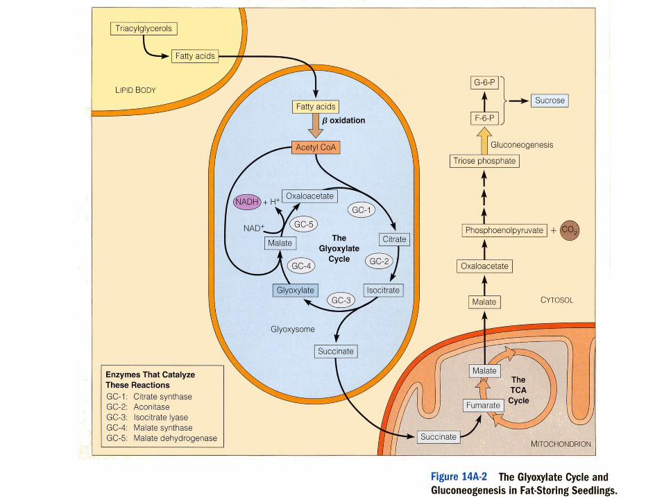
Glyoxysomes
• A primary metabolic activity in these germinating seedlings is the conversion of stored fatty acids to carbohydrate
• Stored fatty acid disassembly produces acetyl CoA & it condenses with oxaloacetate to form citrate
• Citrate is then converted to glucose by a series of glyoxylate cycle enzymes found in glyoxysomes
Diseases result from abnormal mitochondrial or peroxisomal function
• Muscle & nerve tissues tend to be most seriously impacted in these disorders since they have the highest demand for ATP
• Depending on protein(s) affected, conditions vary in severity from diseases that lead to death during infancy to disorders that produce seizures ( 中風驟發 ), blindness, deafness and/or strokelike episodes
• Sometimes conditions are mild & characterized by intolerance to exercise or nonmotile sperm
Abnormal mitochondria
• Closer examination of mitochondria reveals large numbers of abnormal inclusions
• A number of common neurological diseases with adult onset (like Parkinson's disease) might be a consequence of degenerative changes in mitochondrial function
• The first such disease-causing mutation was reported in 1995 – The mutation occurred in gene encoding the
flavoprotein subunit of the TCA enzyme succinate dehydrogenase
Mitochondrial disorder inheritance contrasts in several ways with nuclear
gene Mendelian inheritance
• Mitochondria in cells of human embryo are derived exclusively from mitochondria present in the egg at the time of conception without any contribution from the fertilizing sperm
• Mitochondrial disorders are inherited maternally• Mitochondria in cell can contain mixture of
normal (wild-type) & mutant mtDNA (heteroplasmy)
mtDNA Mutation
• Nuclear DNA is protected from damage by a variety of DNA repair systems which are generally lacking in mitochondria
• mtDNA may also be subjected to high levels of mutagenic oxygen radicals
• mtDNA experiences >10 times the mutation rate of nuclear DNA
Abnormal peroxisomes
• Zellweger syndrome (ZS) is a rare inherited disease characterized by a variety of neurological, visual & liver abnormalities leading to death during early infancy
• Sidney Goldfischer et al. (1973) – reported that liver & renal cells from these patients lacked peroxisomes
• Later studies showed that peroxisomes were not entirely absent from the cells of these individuals
Zellweger syndrome (ZS)
• Peroxisomes were present as empty membranous ghosts (organelles lacking the enzymes normally found in peroxisomes)
• These individuals can make peroxisomal enzymes but the enzymes fail to be imported into peroxisomes & stay largely in cytosol where they are unable to carry out their normal functions
• Mutations in at least 11 different genes– Encoding proteins involved in uptake of
peroxisomal enzymes from cytosol
Adrenoleukodystrophy (ALD), subject of the movie Lorenzo's Oil
• Absence of a single peroxisomal enzyme • A defect in a membrane protein that transports
very-long-chain-fatty-acids (VLCFAs) into the peroxisomes where they are normally metabolized
• In the absence of this protein, VLCFAs accumulate in brain & destroy myelin sheaths that insulate nerve cells
• Boys with the disease are typically unaffected until midchildhood, when symptoms of adrenal insufficiency & neurological dysfunction begin
Adrenoleukodystrophy (ALD)
• A diet rich in certain fatty acids is able to retard the progress of the disease
• A number of ALD patients have been successfully treated by bone marrow transplantation, which provides normal cells capable of metabolizing VLCFAs
• Administration of drugs (e.g., lovastatin) that may lower VLCFA levels
• Clinical studies employing gene therapy are also being planned
Chapter 8-1:
Cytoplasmic Membrane Systems:
Structure, Function, and
Membrane Trafficking
Endomembrane system
• Plasma membrane, vesicles, vacuoles, ER, Golgi apparatus, nuclear membrane, lysosome– Have distinct structures & functions but
together form an endomembrane system– Dynamic, integrated network– Materials are shuttled (transport vesicles)
between the endomembrane system
Transport vesicles in endomembrane system
• Transport vesicles form by budding from donor compartment
• Transport vesicles move in directed manner, often pulled by motor proteins operating on tracks formed by microtubules & microfilaments of the cytoskeleton
• When they reach their destination, they fuse with acceptor compartment
Transport in endomembrane system
• Endocytic pathway
• Exocytotic pathway
– Secretory pathway
Biosynthetic (secretory) pathway
• Synthesis in ER (protein) or Golgi (lipid, carbohydrate)
• Many materials made in ER (proteins) & Golgi (complex polysaccharides) fated for secretion from cell
• Two types of secretory activity– Constitutive secretion– Regulated secretion
Constitutive secretion
• Synthesis & secretion into extracellular space occurs in continual, unregulated manner
• Form extracellular matrix & plasma membrane
Regulated secretion
• Secretory materials stored in large, densely packed, membrane-bound secretory granules in cell periphery
• Secreted after correct stimulus – Endocrine cells release hormones – Pancreatic acinar cells release digestive
enzymes – Nerve cells release neurotransmitters
Proteins targeting
• Through sorting signals located on proteins & receptors in transport vesicle walls that recognize them
• Salivary gland cell protein trafficking– Salivary mucus proteins (made in ER)
specifically targeted to secretory granules• Lysosome enzymes (also made in ER) specifically
sent to lysosome • Sorting signals are encoded in protein amino acid
sequence or in attached oligosaccharides
Approaches to the study of cytomembranes
• EM micrographs give detailed view of cell cytoplasm, but little insight into functions of the structures
• Insights gained from autoradiography– Detect location of radioactively labeled
materials in cell • Insights from pulse-chase trials
Pulse-chase trials
• Expose to hot amino acids briefly (pulse)
• Wash to remove excess isotope from tissue • Transferred tissue to medium with
unlabeled amino acids (chase), which lasts for varying time periods
• See wave of radioactivity moving through cell, discern pathway sequence
Use of green fluorescent protein (GFP) reveals the movement of proteins within
a living cell
• GFP is small protein from certain jellyfish that emits a green fluorescent light
• GFP gene fused to DNA encoding protein to be studied
• Introduce the chimeric DNA into cells• Chimeric DNA expresses chimeric protein
(GFP fused to the protein to be studied)
Use of green fluorescent protein (GFP) reveals the movement of proteins within
a living cell
• Usually, GFP stuck to end of a protein has little or no effect on its movement or function & protein under study has no effect on fluorescence of attached GFP
Example: infect a mammalian cell with vesicular stomatitis virus (VSV) strain in which a viral gene
(VSVG) is fused to GFP gene
• Cell begins to make massive amounts of VSVG protein in RER
• VSVG then goes to Golgi complex & eventually to the plasma membrane of the infected cell where they are incorporated into viral envelopes
• Can see relatively synchronous wave of protein movement (green fluorescence) soon after infection
Infect a mammalian cell with vesicular stomatitis virus (VSV) strain in which a viral
gene (VSVG) is fused to GFP gene
• Synchrony is enhanced by use of virus with mutant VSVG protein that cannot leave ER of infected cells grown at elevated temperature (40°C).
• The green fluorescence is restricted to the ER.
• When temperature is lowered to 32°C, the fluorescent GFP-VSVG protein that had accumulated in ER moves synchronously to Golgi complex for various processing events & then to membrane
• Temperature-sensitive mutants– Permissive temperature
Mutants function normally – Restrictive temperatures
Mutants function abnormally
Cell fractionation
• Homogenization
• Organelles fractionation by centrifugation