Embed Size (px)
DESCRIPTION
Chapter 69 Management of Patients With Musculoskeletal Trauma. Injuries of the Musculoskeletal System. Contusion: soft tissue injury produced by blunt force with bleeding into soft tissue Pain, swelling, and discoloration: ecchymosis - PowerPoint PPT Presentation
Citation preview
Chapter 69
Management of Patients With Musculoskeletal
Trauma
1
Injuries of the Musculoskeletal System
Contusion: soft tissue injury produced by blunt force with bleeding into soft tissue
Pain, swelling, and discoloration: ecchymosis Strain: Pulled muscle-injury to the
musculotendinous unit (Excessive stretching of a ligament)
Pain, edema, muscle spasm, ecchymosis, and loss of function are on a continuum graded 1st , 2nd, and 3rd degree
2
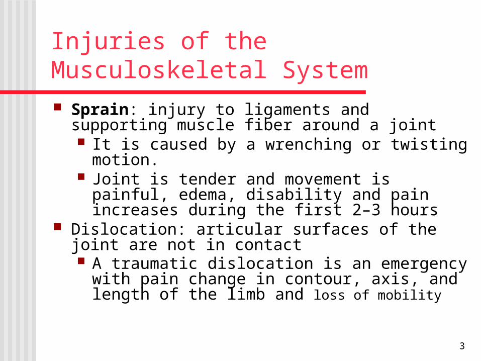
Injuries of the Musculoskeletal System Sprain: injury to ligaments and supporting muscle fiber
around a joint It is caused by a wrenching or twisting motion. Joint is tender and movement is painful, edema,
disability and pain increases during the first 2–3 hours
Dislocation: articular surfaces of the joint are not in contact A traumatic dislocation is an emergency with pain
change in contour, axis, and length of the limb and loss of mobility
3
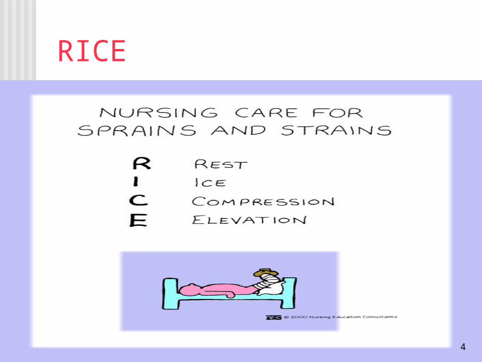
RICE
Rest Ice Compression Elevation
4
Common Sports-Related Injuries
Contusions, strains, sprains and dislocations
Tendonitis: inflammation of a tendon by overuse
Meniscal injuries of the knee occur with excessive rotational stress
Traumatic fractures Stress fractures
5
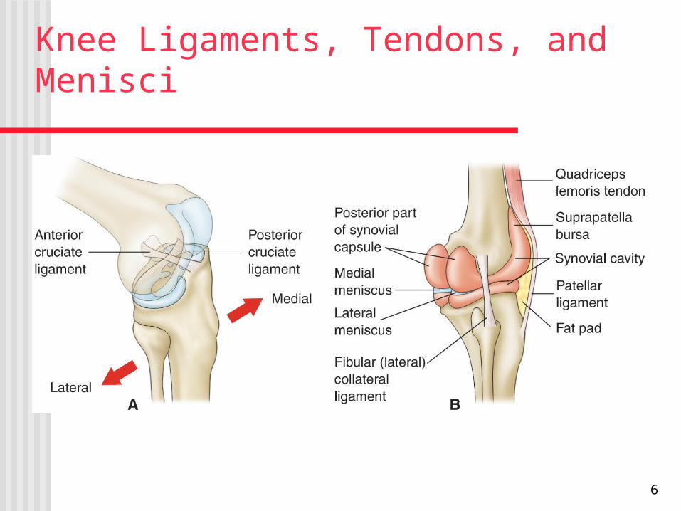
Knee Ligaments, Tendons, and Menisci
6
Prevention of Sports-Related Injuries
Use of proper equipment; running shoes for runners, wrist guards for skaters, etc.
Effective training and conditioning specific for the person and the sport
Stretching prior to engaging in a sport or exercise has been recommended but may not prevent injury
Changes in activity and stresses should occur gradually Time to “cool down” Tune in to the body; be aware of limits and capabilities Modify activities to minimize injury and promote healing
7
Occupational-Related Injuries
Common injuries include strains, sprains, contusions, fractures, back injuries, tendonitis, and amputations.
Prevention measures may include personnel training, proper use of equipment, availability of safety and other types of equipment (patient lifting equipment, back belts), correct use of body mechanics, and institutional policies.
8
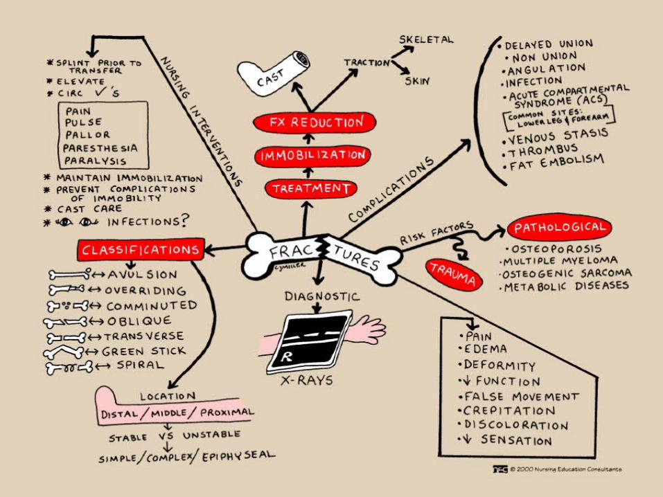
Fractures Break in the continuity of bone Causes:
Direct blow Crushing force (compression) Sudden twisting motions (torsion) Severe muscle contraction Disease (pathologic fracture)
9
Types of Fractures
Complete Incomplete Closed or simple Open or compound/complex
Grade I Grade II Grade III
10
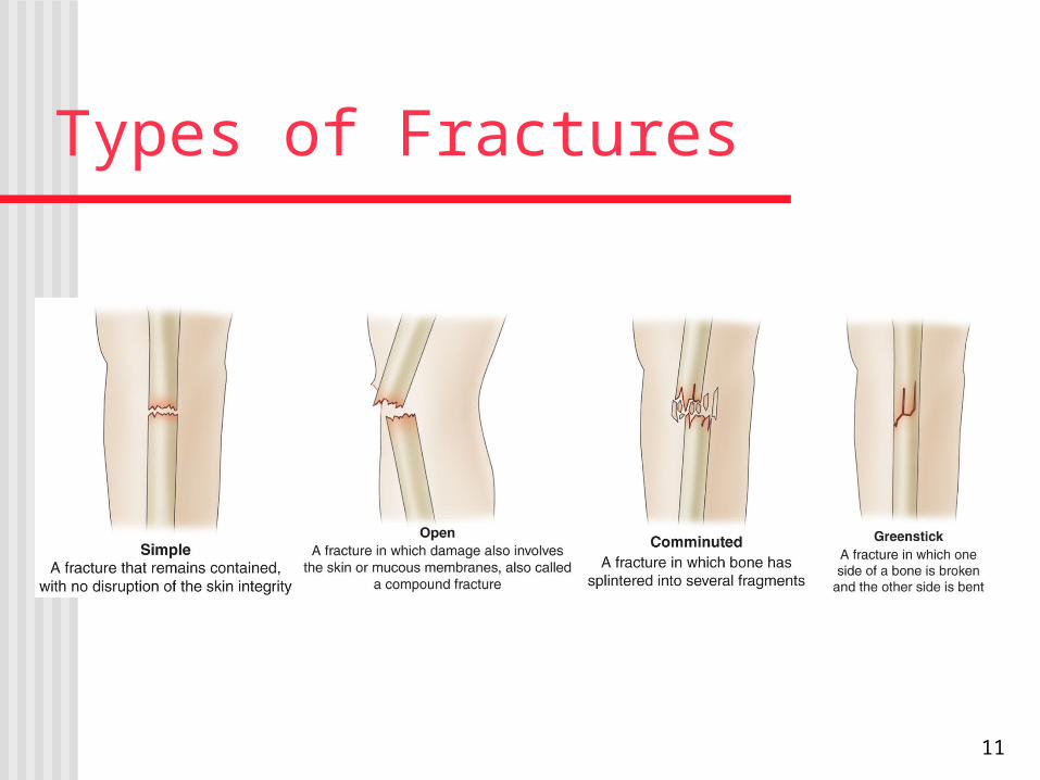
Types of Fractures
11
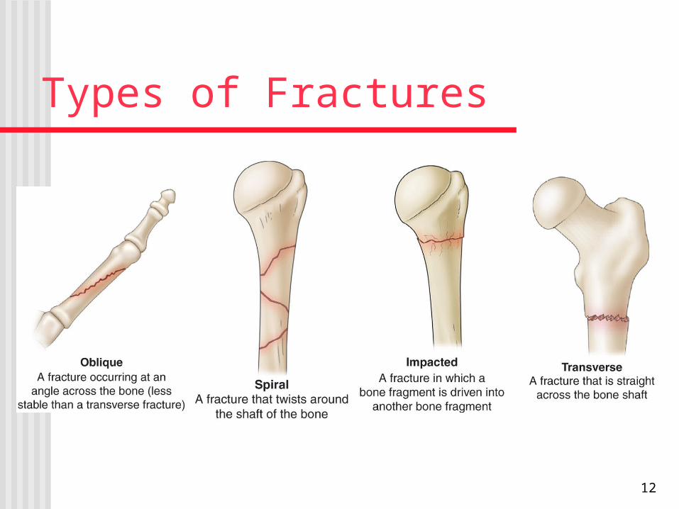
Types of Fractures
12
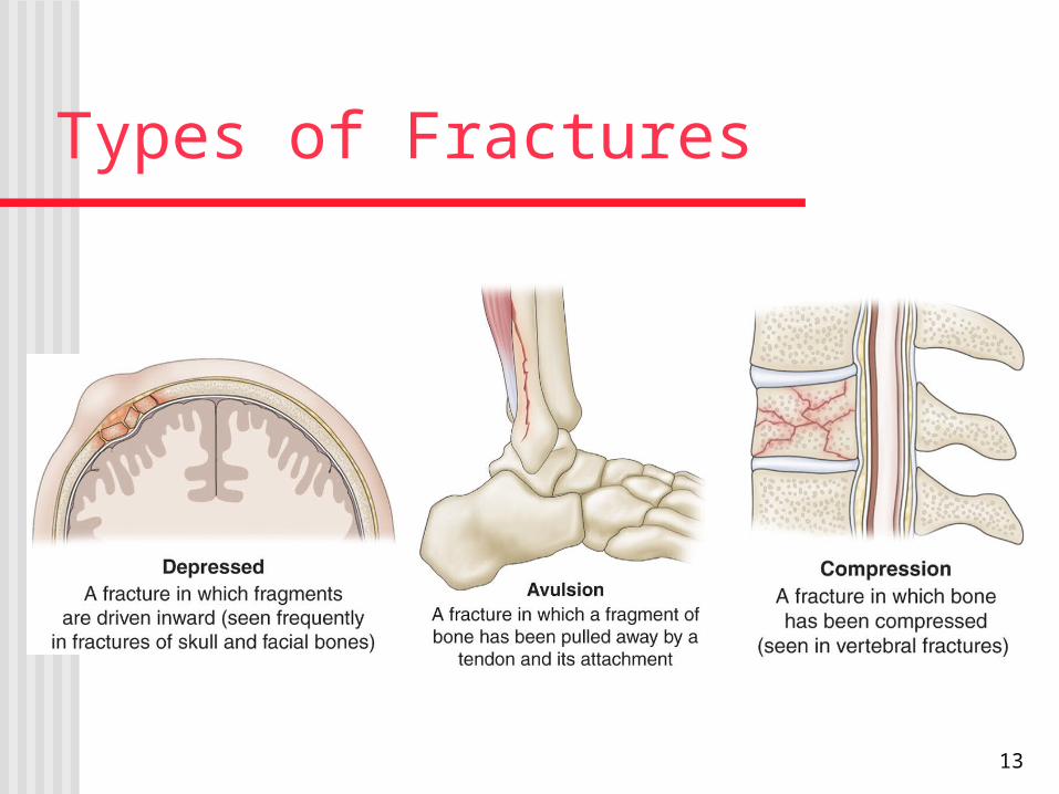
Types of Fractures
13
Manifestations of Fracture Pain Loss of function Deformity Shortening of the extremity Crepitus Local swelling and discoloration Diagnosis by symptoms and x-ray Patient usually reports an injury to the area
14

Emergency Management Immobilize the body part Splinting: joints distal and proximal to the
suspected fracture site must be supported and immobilized
Assess neurovascular status before and after splinting
Open fracture: cover with sterile dressing to prevent contamination
Do not attempt to reduce the fracture
15
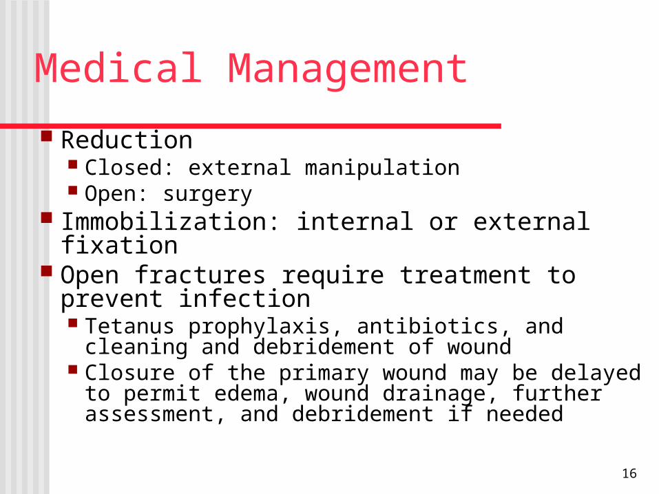
Medical Management Reduction
Closed: external manipulation Open: surgery
Immobilization: internal or external fixation Open fractures require treatment to prevent
infection Tetanus prophylaxis, antibiotics, and cleaning and
debridement of wound Closure of the primary wound may be delayed to
permit edema, wound drainage, further assessment, and debridement if needed
16
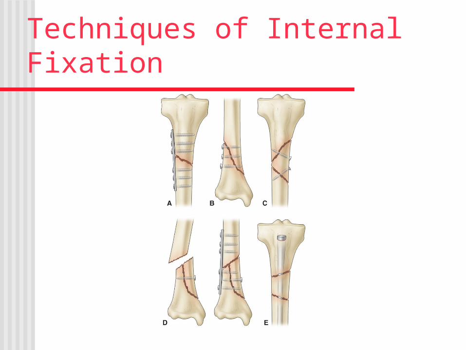
Techniques of Internal Fixation
17
Factors That Enhance Fracture Healing
Immobilization of fracture fragments Maximum bone fragment contact Sufficient blood supply Proper nutrition Exercise: weight bearing for long bones Hormones: growth hormone, thyroid,
calcitonin, vitamin D, anabolic steroids
18
Factors That Inhibit Fracture Healing Extensive local trauma Bone loss Inadequate immobilization Space or tissue between bone fragments Infection Local malignancy Metabolic bone disease (as Paget's disease) Avascular necrosis Intra-articular fracture (synovial fluid contains fibrolysins, which
lyse the initial clot and retard clot formation) Age (elderly persons heal more slowly) Corticosteroids (inhibit the repair rate)
19
QuestionIs the following statement True or False?
Testing for crepitus can produce further tissue damage and should be avoided.
20
AnswerTrue
Testing for crepitus can produce further tissue damage and should be avoided.
21
Techniques of Internal Fixation
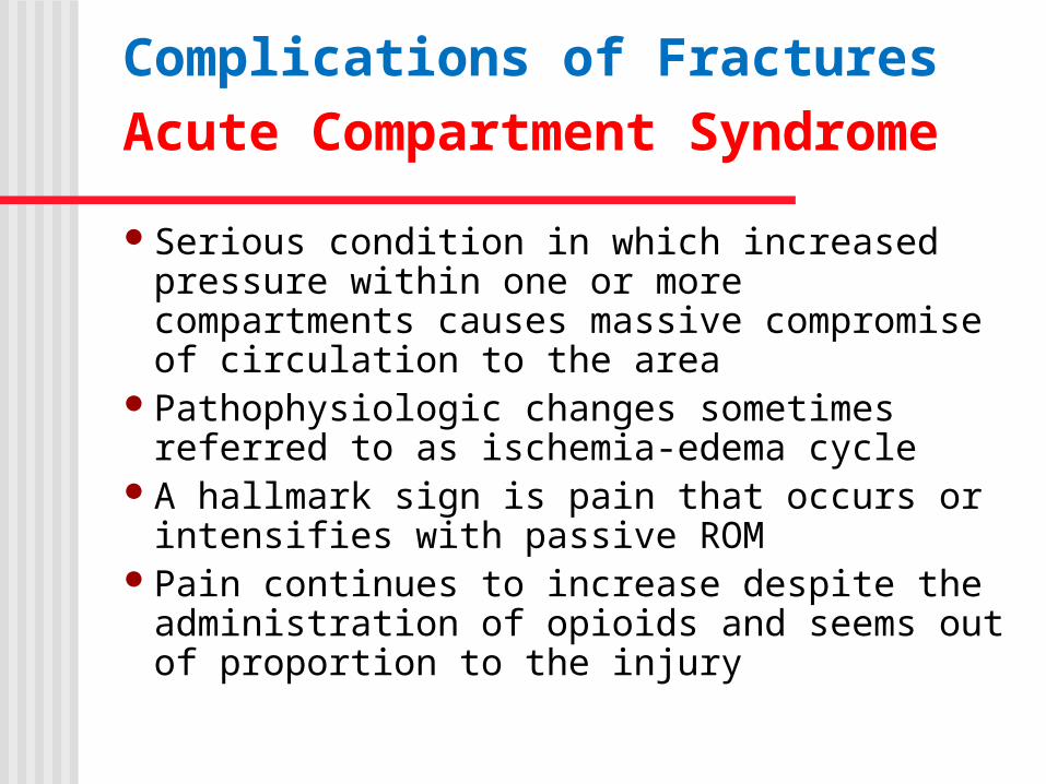
Complications of FracturesAcute Compartment Syndrome Serious condition in which increased pressure
within one or more compartments causes massive compromise of circulation to the area
Pathophysiologic changes sometimes referred to as ischemia-edema cycle
A hallmark sign is pain that occurs or intensifies with passive ROM
Pain continues to increase despite the administration of opioids and seems out of proportion to the injury
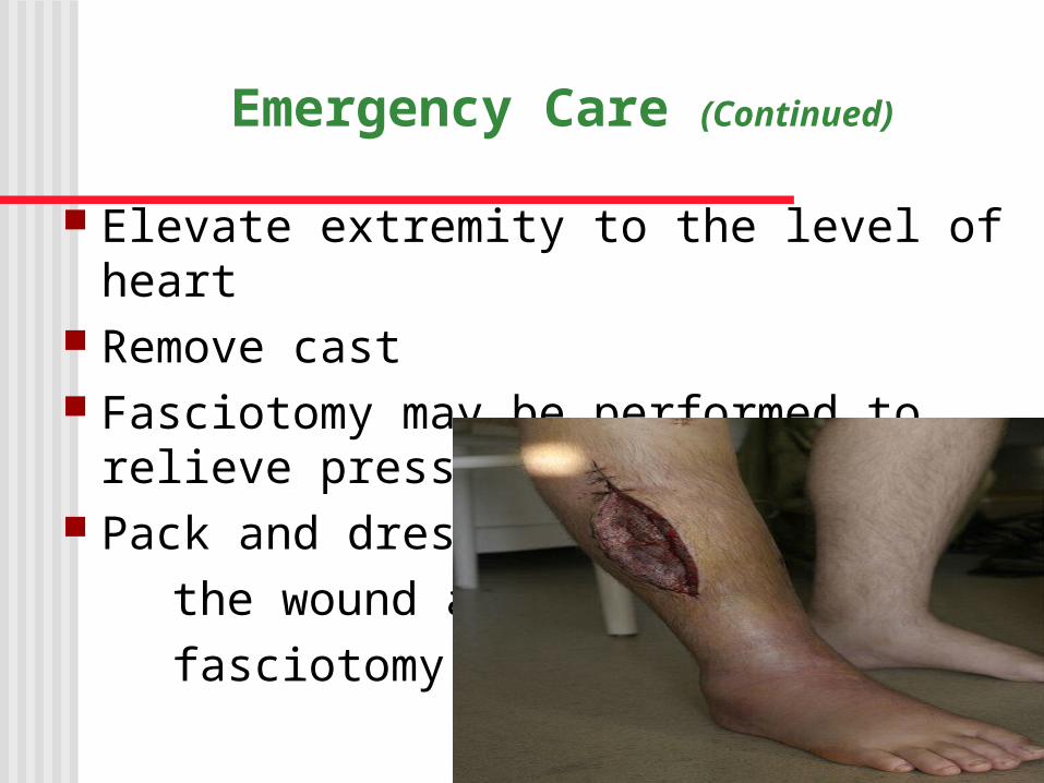
Emergency Care (Continued)
Elevate extremity to the level of heart Remove cast Fasciotomy may be performed to relieve
pressure. Pack and dress the wound after fasciotomy.
Other Complications of Fractures
ShockFat embolism syndrome: serious
complication resulting from a fracture; fat globules are released from yellow bone marrow into bloodstream
Venous thromboembolism Infection Ischemic necrosis delayed union, nonunion, and malunion
Possible Results of Acute Compartment Syndrome
Infection Motor weakness Volkmann’s contractures: (a deformity of the
hand, fingers, and wrist caused by a lack of blood flow
(ischemia) to the muscles of the forearm)
Musculoskeletal Complications (continued)
Muscle Atrophy, loss of muscle strength range of motion, pressure ulcers, and other problems associated with immobility
Embolism/Pneumonia/ARDS TREATMENT – hydration, albumin, corticosteroids
Constipation/Anorexia UTI DVT
QuestionIs the following statement True or False?
Avascular necrosis is prolongation of expected healing time for a fracture.
28
AnswerFalse
Avascular necrosis is death of tissue secondary to poor perfusion and hypoxemia. Delayed union is prolongation of expected healing time for a fracture.
29
Rehabilitation Related to Specific Fractures
Clavicle Use of claviclar strap (“figure 8”) or sling Exercises Limitation of activities Do not elevate arm above shoulder for approximately
6 weeks Humeral neck and shaft fractures
Slings and bracing Activity limitations and pendulum exercises
30
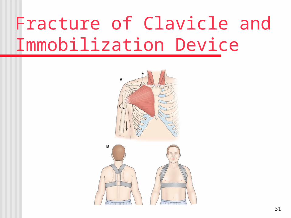
Fracture of Clavicle and Immobilization Device
31
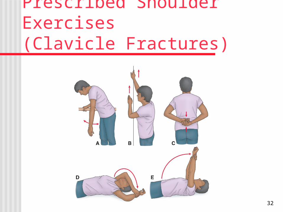
Prescribed Shoulder Exercises (Clavicle Fractures)
32
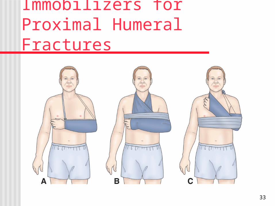
Immobilizers for Proximal Humeral Fractures
33
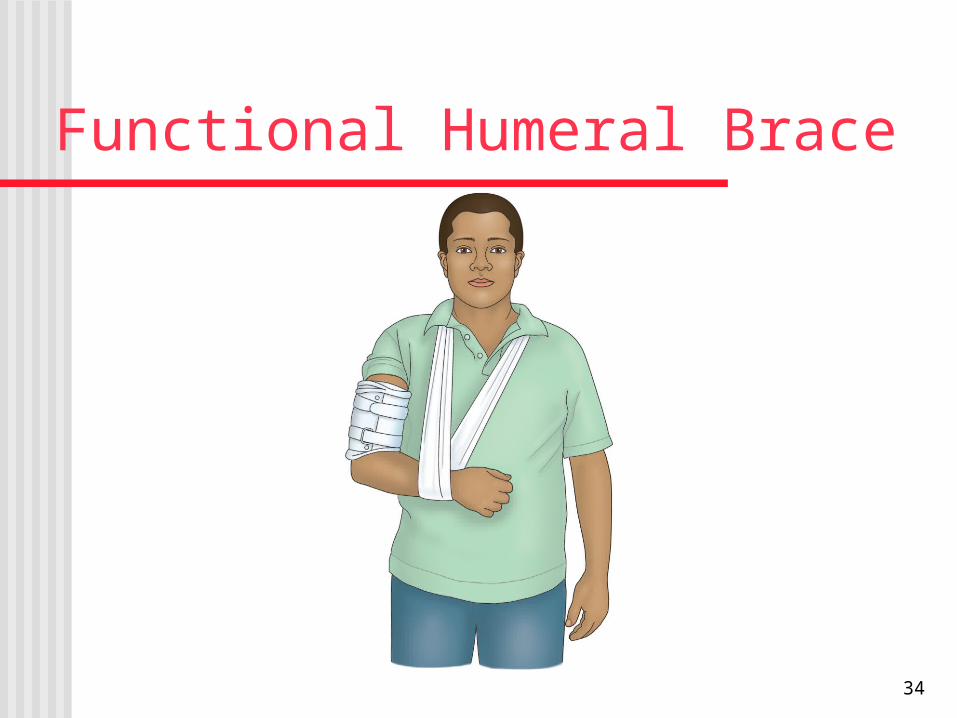
Functional Humeral Brace
34
Rehabilitation Related to Specific Fractures Elbow fractures
Monitor regularly for neurovascular compromise and signs of compartment syndrome
Potential for Volkmann's contracture Active exercises and ROM are encouraged to
prevent limitation of joint movement after immobilization and healing (4–6 weeks for nondisplaced, casted) or after internal fixation (about 1 week)
35
Volkmann's Contracture Observe the distal part of the extremity for swelling, skin color,
nail bed capillary refill, and temperature. Compare affected and unaffected hands.
Assess radial pulse. Assess for paresthesia in the hand, which may indicate nerve
injury or impending ischemia. Evaluate the patient's ability to move the fingers. Explore the intensity and character of the pain. Report indications of diminished nerve function or diminished
circulatory perfusion promptly before irreparable damage occurs; fasciotomy may become necessary.
36
Fractures of the Pelvis
Result from falls or accidents Associated internal damage is the chief concern
in fracture management of pelvic fractures Management depends upon type and extent of
fracture and associated injuries. Stable fractures are treated with a few days bed
rest and symptom management. Early mobilization reduces problems related to
immobility.
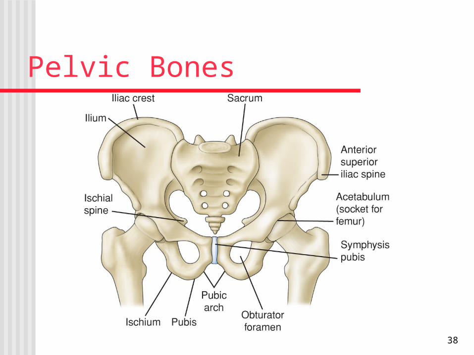
Pelvic Bones
38
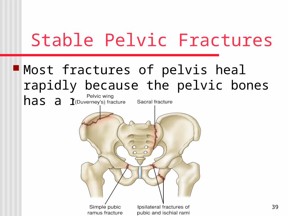
Stable Pelvic Fractures Most fractures of pelvis heal rapidly because
the pelvic bones has a rich blood supply
39
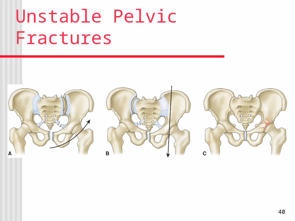
Unstable Pelvic Fractures
40

Hip fracture Most common among elderly (due to falls and
osteoporosis) Fracture can intracapsular or extracapsular Surgery is usually done to reduce and fixate the
fracture. Care is similar to that of a patient undergoing
other orthopedic surgery or hip replacement surgery.
41
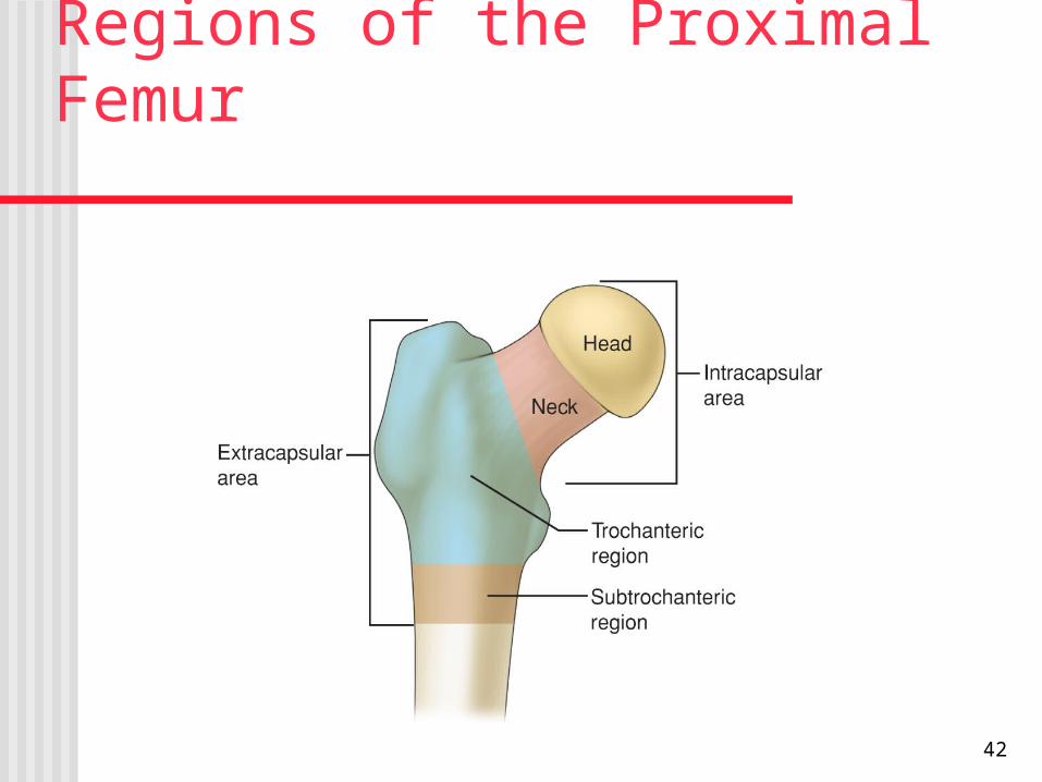
Regions of the Proximal Femur
42
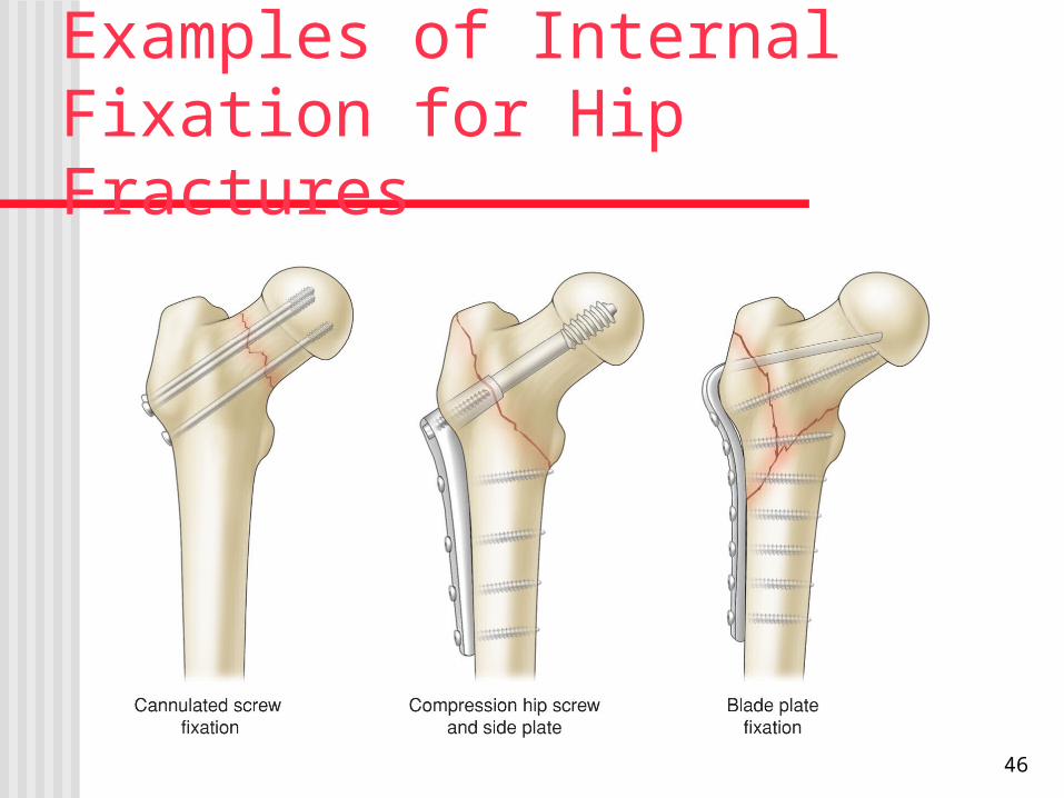
Examples of Internal Fixation for Hip Fractures
46
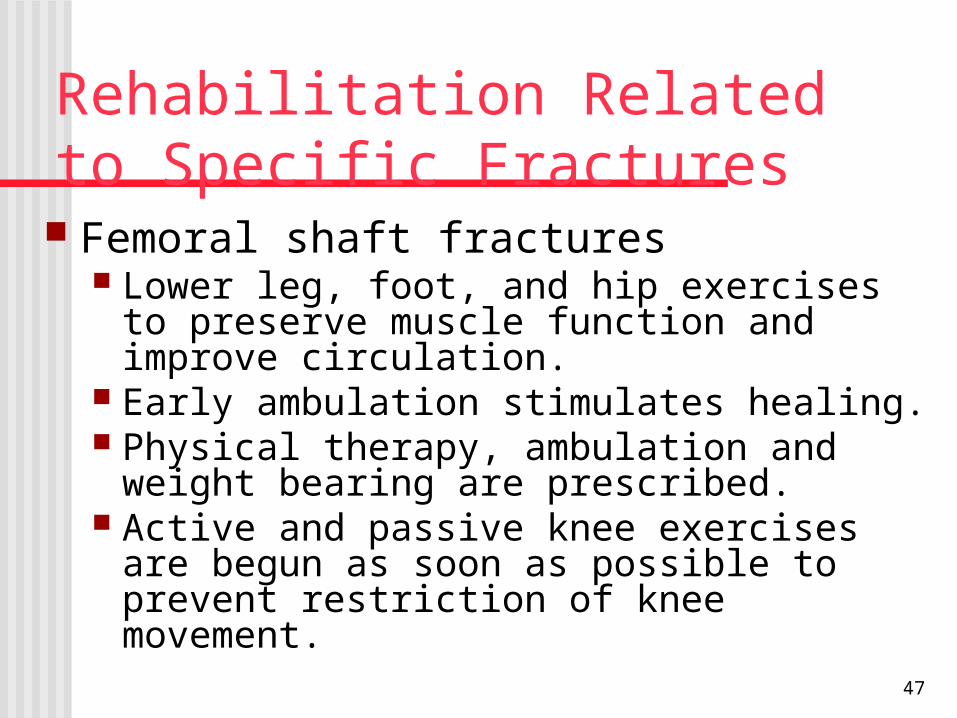
Rehabilitation Related to Specific Fractures Femoral shaft fractures
Lower leg, foot, and hip exercises to preserve muscle function and improve circulation.
Early ambulation stimulates healing. Physical therapy, ambulation and weight bearing
are prescribed. Active and passive knee exercises are begun as
soon as possible to prevent restriction of knee movement.
47
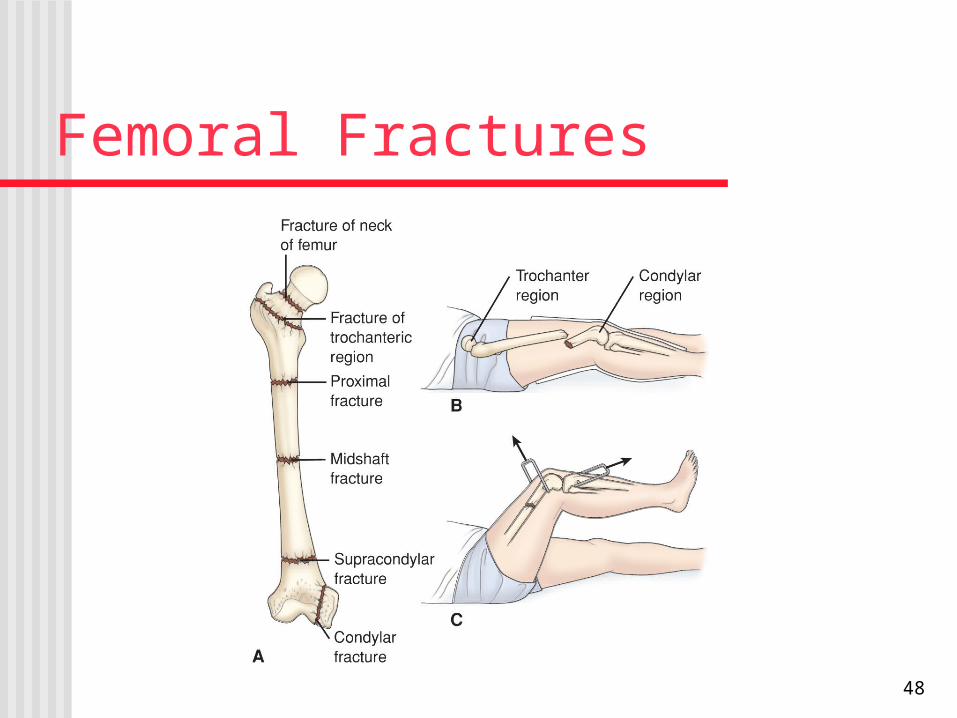
Femoral Fractures
48
Nursing Process: The Care of the Patient with Fracture of the Hip—Assessment
Health history and presence of concomitant problems Pain VS, respiratory status, LOC, and signs and symptoms
of shock Affected extremity including frequent neurovascular
assessment Bowel and bladder elimination; bowel sounds, I&O Skin condition Anxiety and coping
49
Nursing Process: The Care of the Patient with Fracture of the Hip—Diagnoses
Acute pain Impaired physical mobility Impaired skin integrity Risk for impaired urinary elimination Risk for ineffective coping Risk for disturbed thought processes
50
Collaborative Problems/Potential Complications
Hemorrhage Peripheral neurovascular dysfunction DVT Pulmonary complications Pressure ulcers
51
Nursing Process: The Care of the Patient with Fracture of the Hip—Planning
Major goals may include relief of pain; achievement of a pain-free, functional, and stable hip; healed wound; maintenance of normal urinary elimination pattern; use of effective coping mechanisms; remains oriented and participates in decision-making; and absence of complications.
52
Relief of Pain
Administer analgesics as prescribed Use of Buck’s traction as prescribed Handle extremity gently Support extremity with pillows and when
moving Positioning for comfort Frequent position changes Alternative pain relief methods
53
Prompting Physical Mobility
Maintain neutral position of hip Use trochanter rolls Maintain abduction of hip Isometric, quad-setting, and gluteal-setting
exercises Use of trapeze Use of ambulatory aids Consultation with physical therapy
54
Interventions
Use aseptic technique with dressing changes Avoid/minimize use of indwelling catheters Supporting coping
Provide and reinforce information Encourage patient to express concerns Support coping mechanisms Encourage patient to participate in decision
making and planning
55
Interventions
Orient patient to & stabilize the environment Provide for patient safety Encourage participation in self-care Encourage coughing and deep breathing exercises Ensure adequate hydration Apply hose / crib bandage as prescribed Encourage ankle exercises Patient and family teaching
56
Rehabilitation of Patients with Amputation Amputation may be congenital, traumatic, or due
to conditions such as progressive peripheral vascular disease, infection, or malignant tumor.
Amputation is used to relieve symptoms, improve function, and save the person's life.
The health care team needs to communicate a positive attitude to facilitate acceptance and participation in rehabilitation.
57
Amputations
Surgical amputation Traumatic amputation Levels of amputation Complications of amputations:
hemorrhage, infection, phantom limb pain, problems associated with immobility, neuroma (a growth or tumour of nerve tissue), flexion contracture
Amputation
Nursing Management relieving pain minimizing altered sensory
perception promoting wound healing enhancing body image self-care
QuestionIs the following statement True or False?
Phantom limb pain is perceived in the amputated limb.
60
AnswerTrue
Phantom limb pain is perceived in the amputated limb.
61
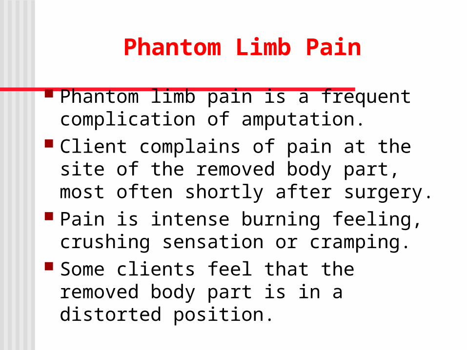
Phantom Limb Pain
Phantom limb pain is a frequent complication of amputation.
Client complains of pain at the site of the removed body part, most often shortly after surgery.
Pain is intense burning feeling, crushing sensation or cramping.
Some clients feel that the removed body part is in a distorted position.
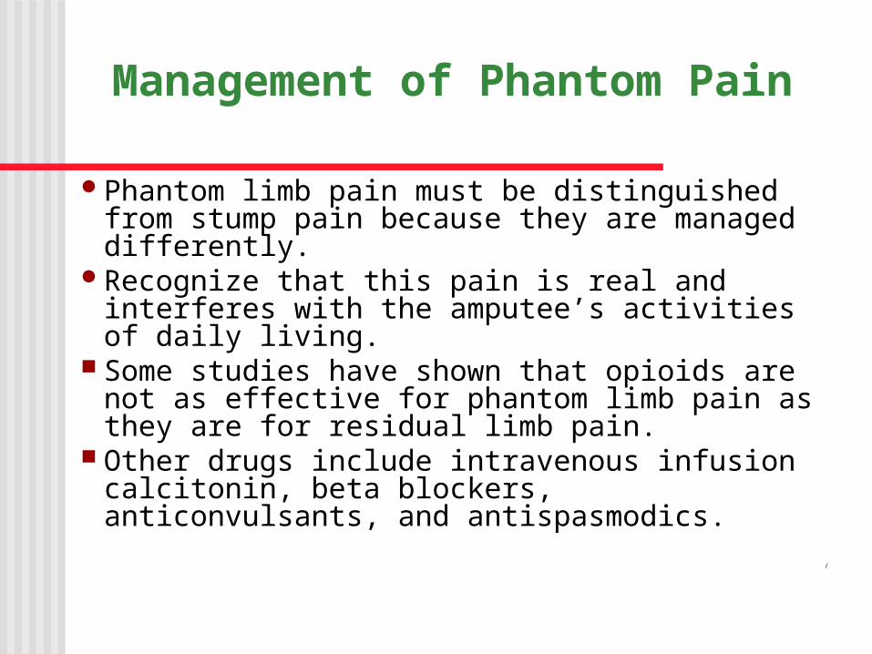
Management of Phantom Pain
Phantom limb pain must be distinguished from stump pain because they are managed differently.
Recognize that this pain is real and interferes with the amputee’s activities of daily living.
Some studies have shown that opioids are not as effective for phantom limb pain as they are for residual limb pain.
Other drugs include intravenous infusion calcitonin, beta blockers, anticonvulsants, and antispasmodics.
(
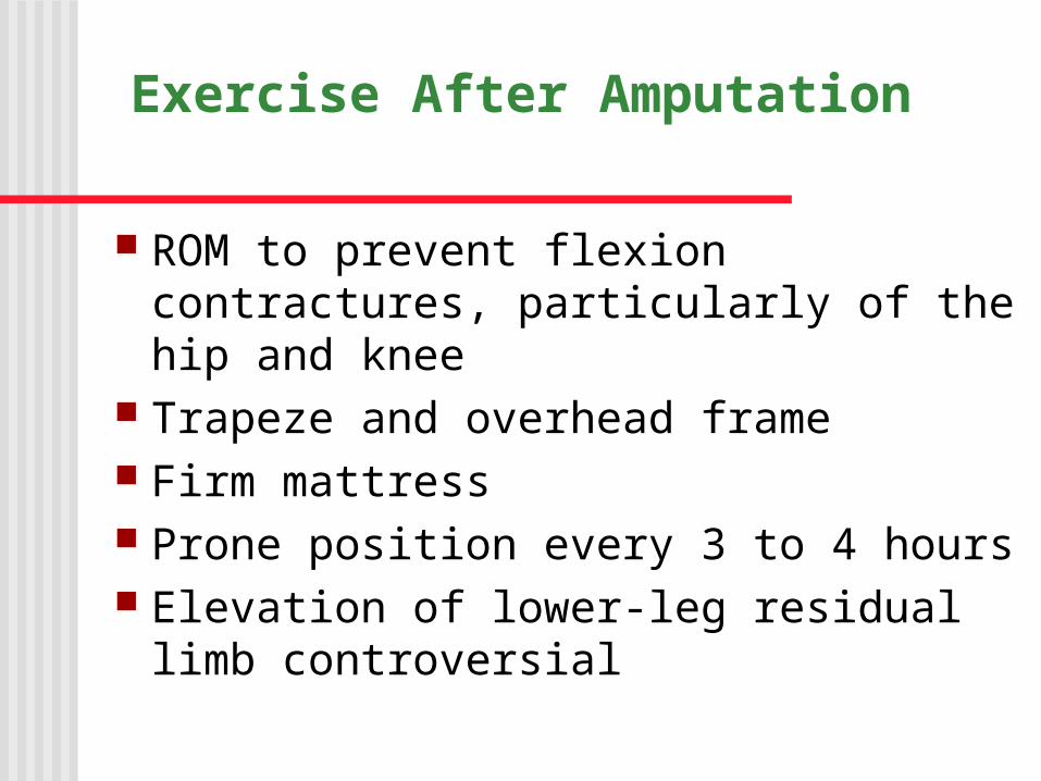
Exercise After Amputation
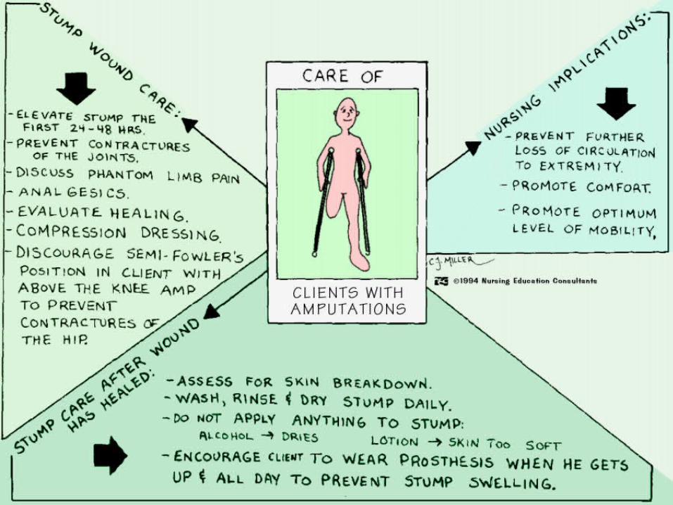
ROM to prevent flexion contractures, particularly of the hip and knee
Trapeze and overhead frame Firm mattress Prone position every 3 to 4 hours Elevation of lower-leg residual limb
controversial
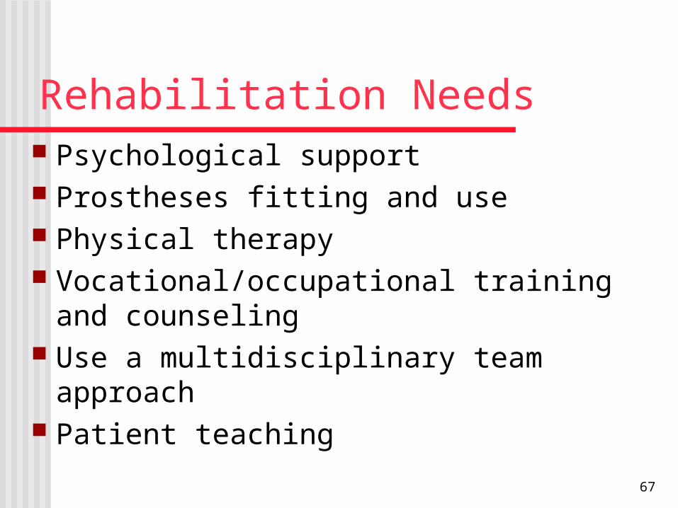
Rehabilitation Needs Psychological support Prostheses fitting and use Physical therapy Vocational/occupational training and
counseling Use a multidisciplinary team approach Patient teaching
67
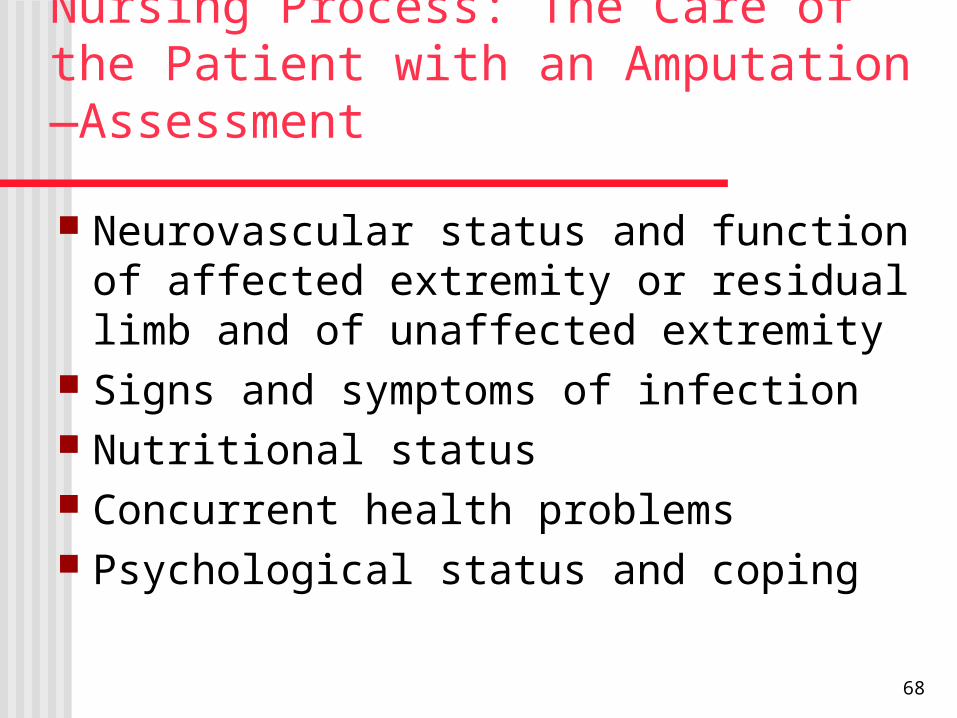
Nursing Process: The Care of the Patient with an Amputation—Assessment
Neurovascular status and function of affected extremity or residual limb and of unaffected extremity
Signs and symptoms of infection Nutritional status Concurrent health problems Psychological status and coping
68
Nursing Process: The Care of the Patient with an Amputation—Diagnoses
Acute pain Risk for disturbed sensory perception Disturbed body image Ineffective coping Risk for anticipatory or dysfunctional grieving Self-care deficit Impaired physical mobility
69
Collaborative Problems/Potential Complications
Postoperative hemorrhage Infection Skin breakdown
70
Nursing Process: The Care of the Patient with an Amputation—Planning
Major goals may include: relief of pain, absence of altered sensory perceptions, wound healing, acceptance of altered body image, resolution of grieving processes, restoration of physical mobility, and absence of complications.
71
Interventions
Relief of pain Administer analgesic or other medications as
prescribed Changing position Putting a light sand bag on residual limb Alternative methods of pain relief- distraction,
TENS unitNote: Pain may be an expression of grief and
altered body image Promoting wound healing
Handle limb gently Residual limb shaping
72
Resolving Grief and Enhancing Body Image Encourage communication and expression of
feelings Create an accepting, supportive atmosphere Provide support and listen Encourage patient to look at, feel, and care for the
residual limb Help patient set realistic goals Help patient resume self-care & independence Referral to counselors and support groups
73
Achieving Physical Mobility
Proper positioning of limb; avoid abduction, external rotation and flexion
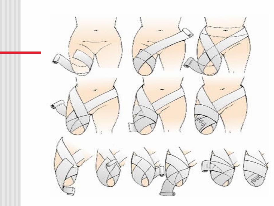
Turn frequently; prone positioning if possible Use of assistive devices ROM exercises Muscle strengthening exercises “Preprosthetic care”; proper bandaging,
massage, and “toughening” of the residual limb
74