Embed Size (px)
Citation preview

CONSTRUCTION AND EXPRESSION OF HUMAN
ADIPONECTIN IN PROKARYOTIC AND EUKARYOTIC
EXPRESSION SYSTEMS AND THE STUDY OF ITS
EFFECT ON SELECTED BLOOD PARAMETERS AND
EXPRESSION OF RELATED GENES
HUSSIN ALWAN ROTHAN AL-MAAMURI
FACULTY OF SCIENCE
UNIVERSITY OF MALAYA
KUALA LUMPUR
2011
CONSTRUCTION AND EXPRESSION OF HUMAN
ADIPONECTIN IN PROKARYOTIC AND EUKARYOTIC
EXPRESSION SYSTEMS AND THE STUDY OF ITS EFFECT
ON SELECTED BLOOD PARAMETERS AND EXPRESSION
OF RELATED GENES
HUSSIN ALWAN ROTHAN AL-MAAMURI
THESIS SUBMITTED IN FULFILMENT OF
THE REQUIREMENT FOR THE DEGREE
OF DOCTOR OF PHILOSOPHY
INSTITUTE OF BIOLOGICAL SCIENCES
FACULTY OF SCIENCE
UNIVERSITY OF MALAYA
KUALA LUMPUR
2011
Abstract
i
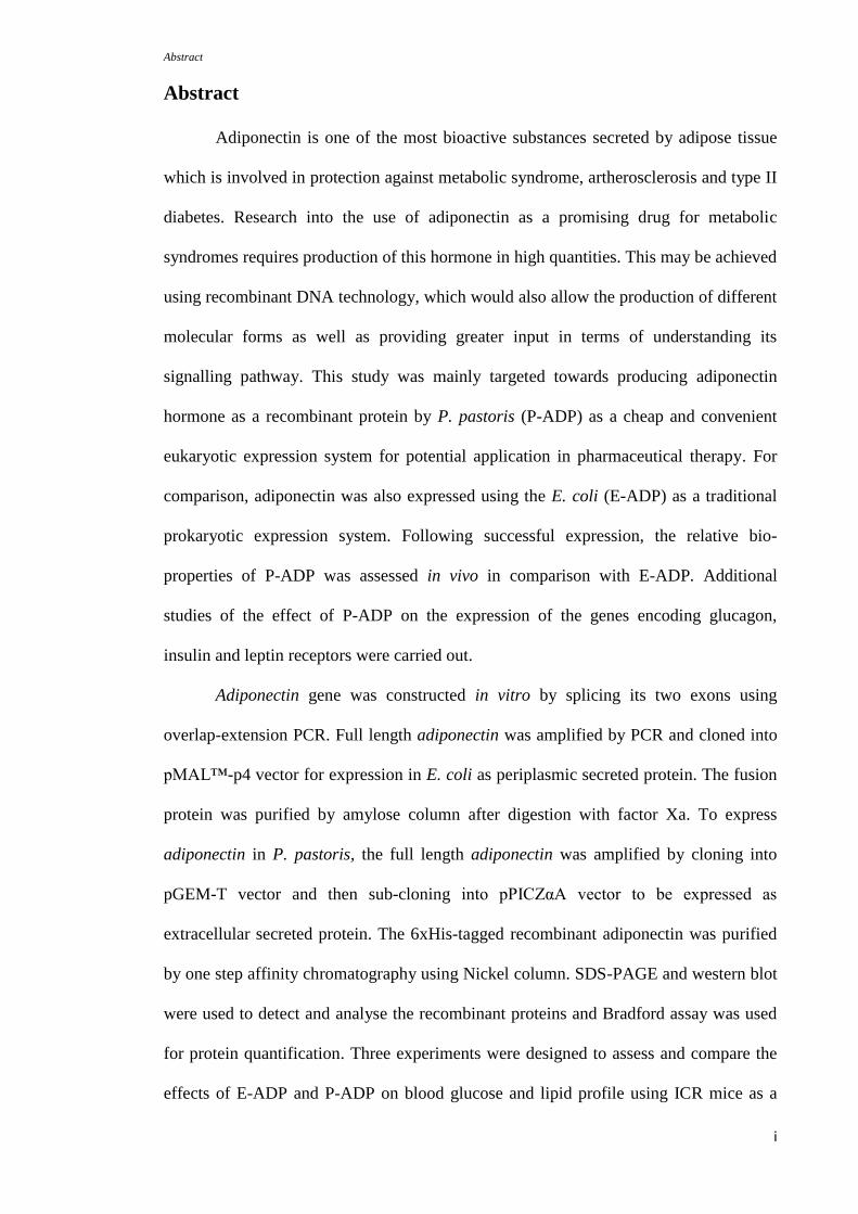
Abstract
Adiponectin is one of the most bioactive substances secreted by adipose tissue
which is involved in protection against metabolic syndrome, artherosclerosis and type II
diabetes. Research into the use of adiponectin as a promising drug for metabolic
syndromes requires production of this hormone in high quantities. This may be achieved
using recombinant DNA technology, which would also allow the production of different
molecular forms as well as providing greater input in terms of understanding its
signalling pathway. This study was mainly targeted towards producing adiponectin
hormone as a recombinant protein by P. pastoris (P-ADP) as a cheap and convenient
eukaryotic expression system for potential application in pharmaceutical therapy. For
comparison, adiponectin was also expressed using the E. coli (E-ADP) as a traditional
prokaryotic expression system. Following successful expression, the relative bio-
properties of P-ADP was assessed in vivo in comparison with E-ADP. Additional
studies of the effect of P-ADP on the expression of the genes encoding glucagon,
insulin and leptin receptors were carried out.
Adiponectin gene was constructed in vitro by splicing its two exons using
overlap-extension PCR. Full length adiponectin was amplified by PCR and cloned into
pMAL™-p4 vector for expression in E. coli as periplasmic secreted protein. The fusion
protein was purified by amylose column after digestion with factor Xa. To express
adiponectin in P. pastoris, the full length adiponectin was amplified by cloning into
pGEM-T vector and then sub-cloning into pPICZαA vector to be expressed as
extracellular secreted protein. The 6xHis-tagged recombinant adiponectin was purified
by one step affinity chromatography using Nickel column. SDS-PAGE and western blot
were used to detect and analyse the recombinant proteins and Bradford assay was used
for protein quantification. Three experiments were designed to assess and compare the
effects of E-ADP and P-ADP on blood glucose and lipid profile using ICR mice as a
Abstract
ii
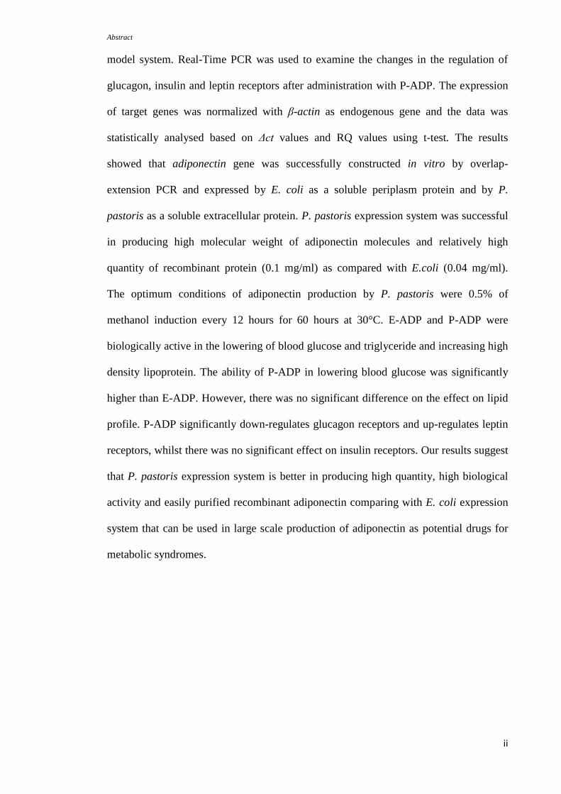
model system. Real-Time PCR was used to examine the changes in the regulation of
glucagon, insulin and leptin receptors after administration with P-ADP. The expression
of target genes was normalized with β-actin as endogenous gene and the data was
statistically analysed based on Δct values and RQ values using t-test. The results
showed that adiponectin gene was successfully constructed in vitro by overlap-
extension PCR and expressed by E. coli as a soluble periplasm protein and by P.
pastoris as a soluble extracellular protein. P. pastoris expression system was successful
in producing high molecular weight of adiponectin molecules and relatively high
quantity of recombinant protein (0.1 mg/ml) as compared with E.coli (0.04 mg/ml).
The optimum conditions of adiponectin production by P. pastoris were 0.5% of
methanol induction every 12 hours for 60 hours at 30°C. E-ADP and P-ADP were
biologically active in the lowering of blood glucose and triglyceride and increasing high
density lipoprotein. The ability of P-ADP in lowering blood glucose was significantly
higher than E-ADP. However, there was no significant difference on the effect on lipid
profile. P-ADP significantly down-regulates glucagon receptors and up-regulates leptin
receptors, whilst there was no significant effect on insulin receptors. Our results suggest
that P. pastoris expression system is better in producing high quantity, high biological
activity and easily purified recombinant adiponectin comparing with E. coli expression
system that can be used in large scale production of adiponectin as potential drugs for
metabolic syndromes.

iii
ACKNOWLEDGEMENTS
This project was supported through UMRG Research University grant RG022/09B10
and short term grant PS232/2008C, awarded to the author and his supervisor by the
University Malaya and Ministry of Higher Education, Malaysia. The author wishes to
thank these funding bodies for enabling this project to be completed. The author also
wishes to acknowledge Faculty of Science, University of Malaya for access and use of
all relevant facilities and many thanks to Center for Biotechnology Research in
Agriculture (CEBAR) for use of equipments and facilities. The main person who guided
me through all the steps in this work was my supervisor Associate Professor Dr.
Zulqarnain Bin Mohamed, who gave ideas, time, support, and encouraged me to do the
best; he deserves the highest respect and thanks. Many thanks and appreciation to Miss
Teh Ser Huy. She provided so much gracious help with the research. Much appreciation
is also expressed to Associate Professor Dr. Mahmood Ameen Abdulla who provided
help, and research associates for bioassay experiments. I would really like to thank all
my wonderful lab mates. Personally, I would like to thank my wife Rehab for her
patience and support, and also to my brothers and sisters.
Contents
iv
Contents Page
Abstract
Acknowledgements
contents
i
iii
iv
List of figures xi
List of tables xvi
Abbreviations xvii
Chapter 1: Introduction
1.1. Adiponectin 1
1.1.1. Adiponectin gene characteristics 6
1.1.2. Adiponectin protein characteristics 7
1.1.3. Glycosylation of adiponectin protein 11
1.1.4. Regulation of adiponectin gene expression 13
1.1.5. Adiponectin single nucleotide polymorphisms (SNPs) 17
1.1.6. Adiponectin biological activity 20
1.1.6.1. Anti-diabetic properties of adiponectin 22
1.1.6.2. Anti-atherogenic, anti-inflammatory and
cardioprotective properties of adiponectin
27
1.2. Expression of proteins in prokaryotic and eukaryotic expression
systems.
30
1.2.1. Expression in Escherichia coli 31
1.2.1.1. The features of E. coli expression system 32
1.2.1.2. Formation of inclusion bodies and protein
folding
34
Contents
v
1.2.2. Pichia pastoris expression system 38
1.2.2.1. Advantages of using P. pastoris as an
expression system
39
1.2.2.2. Pichia pastoris promoters 41
1.3. Objectives of study 42
Chapter 2: Materials and Methods
2.1. Construction of human adiponectin by overlap-extension PCR 44
2.1.1. Primers design 44
2.1.1.1. Primer design for expression of adiponectin in
P. pastoris
44
2.1.1.2. Primer design for expression of adiponectin in
E. coli
45
2.1.2. PCR reaction 47
2.2. Agarose gel electrophoresis 48
2.3. DNA extraction by gel extraction kit 48
2.4. Adiponectin expression by E. coli 49
2.4.1. Preparation of Luria-Bertani (LB) broth and agar media 49
2.4.2. Competent cells preparation of TB1 E. coli strain 50
2.4.3. Cloning of adiponectin into pMAL™ vector 50
2.4.4. Transformation into E. coli 53
2.4.5. Colony screening PCR 53
2.4.6. Small scale expression of adiponetin by E. coli 54
2.4.7.
Native sodium dodecyl sulfate poly-acrylamide gel
electrophoresis (SDS-PAGE) and SDS-PAGE without
54
Contents
vi
denaturing condition.
2.4.8. Western blot 56
2.4.9. Scale up expression of adiponectin by E. coli 57
2.4.10. Protein purification by amylose resin column 58
2.4.11. Cleavage, denaturing and re-purification of recombinant
protein
58
2.4.12. Bradford assay 59
2.5. Adiponectin expression by Pichia pastoris 59
2.5.1. Cloning of adiponectin into pGEM-T vector 60
2.5.1.1. Ligation reaction 60
2.5.1.2. Transformation 60
2.5.1.3. Colony selection 61
2.5.1.4. Plasmid isolation 61
2.5.1.5. Restriction enzyme digestion of pGEMT-
adiponectin recombinant plasmid
63
2.5.2. Clone of adiponectin in pPICZαA vector 63
2.5.2.1. Preparation of Low Salt Luria-Bertani (LSLB)
broth and agar media
64
2.5.2.2. Preparation of Top10F E. coli competent cells 64
2.5.2.3. Ligation reaction 64
2.5.2.4. Transformation 65
2.5.2.5. Colony selection 65
2.5.2.6. Plasmid isolation 65
2.5.2.7. Linearization of the recombinant plasmid 66
2.5.3. Cloning into Pichia pastoris 66
Contents
vii
2.5.3.1. Preparation of P. pastoris broth and agar
media
66
2.5.3.2. Preparation of P. pastoris competent cells 67
2.5.3.3. Transformation 67
2.5.3.4. Selection of positive colonies 68
2.5.3.4.1. Colony selection via PCR 68
2.5.3.4.2. Colonies capacity of the Zeocin™
resistance
69
2.5.3.4.3. Differentiation between Mut+ and
MutS phenotypes
69
2.5.3.5. Preparation of expression media 69
2.5.3.6. Protein expression and optimization 70
2.5.3.7. Protein purification 71
2.5.3.8. Scale-up expression of recombinant
adiponectin
71
2.5.4. Bioactivity tests 72
2.5.4.1. Effect of recombinant adiponectin produced by
P. pastoris and E. coli on blood glucose and
lipids.
73
2.5.4.2. High and low doses effect of recombinant
adiponectin on blood glucose and lipid profile.
73
2.5.4.3. Oral glucose tolerance test 74
2.5.5. Assessment of genes expression 74
2.5.5.1. Total RNA extraction 74
2.5.5.2. DNase treatment 75
Contents
viii
2.5.5.3. cDNA preparation 76
2.5.5.4. Real-Time PCR 78
2.5.4.4.1. Primers design and validation 81
2.5.4.4.2. Data analysis of Real-Time PCR 84
Chapter 3: Results
3.1. In vitro construction of adiponectin 86
3.2. Expression of adiponectin gene in E. coli 91
3.2.1. Restriction enzyme digestion 93
3.2.2. Selection of recombinant E. coli colonies 95
3.2.3. Sequencing analysis 98
3.2.4. Recombinant adiponectin analysis 100
3.2.5. Scale up expression of adiponectin by E. coli 103
3.2.6. Recombinant adiponectin purification 103
3.3. Expression of adiponectin by Pichia pastoris 108
3.3.1. Cloning of adiponectin in pGEM-T vector 110
3.3.2. EcoRI and NotI digestion 114
3.3.3. Cloning of adiponectin in pPICZαA 118
3.3.4. Sequencing data 121
3.3.5. Transformation into Pichia pastoris 124
3.3.6. Colony selection 126
3.3.7. Selection of Mut+ phenotype with high insertion copy
number colonies
129
3.3.8. Small scale expression of adiponectin 131
3.3.9. Optimization of adiponectin expression 134
Contents
ix
3.3.10. Analysis of recombinant adiponectin protein produced by
E. coli and P. pastoris
140
3.4. Biological activity assessment of recombinant adiponectin 142
3.4.1. Comparison of adiponectin bioactivity produced by P.
pastoris and E. coli
142
3.4.2. Effect of the high and low doses of P-ADP on blood
glucose and lipid profile
145
3.4.3. Oral glucose tolerant test 148
3.5. Effect of P-ADP administration on the expression of glucagon,
insulin and leptin receptors
150
3.5.1. Total RNA extraction and DNase treatment 150
3.5.2. RT- PCR 152
3.5.2.1. Expression of glucagon receptors 152
3.5.2.2. Expression of insulin receptors 157
3.5.2.3. Expression of leptin receptors 162
Chapter 4: Discussion
4.1. In vitro gene construction 168
4.2. Expressions of adiponectin in prokaryotic and eukaryotic expression
systems
172
4.2.1. Expression of adiponectin by E. coli 173
4.2.1.1. Adiponectin protein solubility 175
4.2.1.2. Adiponectin protein purification 177
4.2.2. Adiponectin expression by Pichia pastoris 178
4.2.2.1. Production of adiponectin as extracellular 179
Contents
x
secreted protein
4.2.2.2. Optimization of protein expression 180
4.2.2.3. Protein solubility 182
4.2.3. Comparison between adiponectin expression in E. coli and
P. pastoris
182
4.3. Adiponectin bioactivity 184
4.3.1. Effects of E-ADP and P-ADP on blood glucose 185
4.3.2. P-ADP potential in lowering blood glucose 187
4.3.3. Effects of E-ADP and P-ADP on lipid profile 189
4.4. Effect of P-ADP administration on related gene expression 191
4.4.1. Effect of P-ADP administration on the expression of
glucagon receptors
192
4.4.2. Effect of P-ADP administration on the expression of insulin
receptors
193
4.4.3. Effect of P-ADP administration on the expression of leptin
receptors
193
4.5. Summary and conclusion 195
References 198
Appendices
Appendix A Coference abstracts and presentations
Appendix B Awards received

Contents
xi
List of figures
xi
List of figures Page
Figure 1.1 Summary of the diversity of cell-signalling responses to
adipokines
2
Figure 1.2 Research interest in adiponectin, a protein secreted by adipose
tissue
4
Figure 1.3 The relationships between plasma adiponectin levels, obesity
and insulin resistance
5
Figure 1.4 Structure and adiponectin domains 8
Figure 1.5 Multimer formation of adiponectin 10
Figure 1.6 Structural characteristics of adiponectin protein. 12
Figure 1.7 Regulation of adiponectin synthesis and function. 14
Figure 1.8 PPAR agonists thiazolidinediones (TZDs) ameliorate insulin
resistance and diabetes by both adiponectin-dependent and -
independent pathways.
16
Figure 1.9
Figure 1.10
Figure 1.11
Figure 1.12
Figure 1.13
Figure 1.14
Figure 1.15
Figure 2.1
Figure 2.2
Diagram of polymorphic variants of the adiponectin gene.
Main target organs and bio-activities of adiponectin.
The hypothesis of adiponectin effect on metabolic syndromes.
Proposed structure of adiponectin receptors.
Signal transduction by adiponectin receptors.
Adiponectin functions as anti-atherogenic factor.
The pathway of protein folding and secretion in E. coli
The design of RT-PCR experiment.
Melt curves of each gene of interest and an endogenous gene.
19
21
23
26
26
29
37
80
83
Figure 3.1 In vitro construction of ADPs. 88
Figure 3.2 In vitro construction of ADPws. 89
Figure 3.3 Amplification of ADPs and ADPws using different primers for 90
List of figures
xii
cloning into P.pastoris and E.coli.
Figure 3.4 The map of pMAL™-p4x and pMAL™-c4x vectors explaining
enzymes restriction sites.
92
Figure 3.5 Restriction enzyme digestion for cloning into E. coli 94
Figure 3.6 Agarose gel electrophoresis of colony screening ADPs. 96
Figure 3.7 Agarose gel electrophoresis of colony screening for ADPws. 97
Figure 3.8 Sequencing data of ADPs. 99
Figure 3.9 SDS-PAGE analysis of adiponectin expression before and after
induction with IPTG.
101
Figure 3.10 Western blot analysis of adiponectin expression before and after
induction with IPTG.
102
Figure 3.11 Purification by amylose resin column. 104
Figure 3.12 SDS-PAGE for optimization of Factor Xa digestion. 106
Figure 3.13 SDS-PAGE for second purification by hydroxyapatite column
and amylose resin column.
107
Figure 3.14 The map of pPICZα A plasmid that was used to clone the gene
of interest into P. pastoris.
109
Figure 3.15 PGEMT-vector map. 111
Figure 3.16 Agarose gel electrophoresis of colony selection by PCR using
M13 forward and reverse primers to detect existence of ADPws.
112
Figure 3.17 Agarose gel electrophoresis of colony selection by PCR using
M13 forward and reverse primers to confirm existence of ADPs.
113
Figure 3.18 Agarose gel electrophoresis of adiponectin-pGEMT
recombinant plasmid.
115
Figure 3.19 Agarose gel electrophoresis of restriction enzyme digestion to 115
List of figures
xiii
generate ADPs with two sticky ends.
Figure 3.20 Agarose gel electrophoresis of restriction enzyme digestion to
generate ADPws with two sticky ends.
116
Figure 3.21 Agarose gel electrophoresis of pPICZαA plasmid digestion with
EcoRI and NotI.
117
Figure 3.22 Colony screening PCR using α-factor forward primer and
3’AOX1 reverse primer to detect ADPws.
119
Figure 3.23 Colony screening PCR using α-factor forward primer and
3’AOX1 reverse primer to detect ADPs.
120
Figure 3.24 Sequencing data of ADPws after cloning in pPICZαA. 122
Figure 3.25 The sequence of ADPs after cloning in pPICZαA. 123
Figure 3.26 Recombinant plasmid linearization with SacI restriction enzyme. 125
Figure 3.27 Colony PCR screening using α-factor forward primer and
3’AOX1 reverse primer to detect ADPws into yeast colonies.
127
Figure 3.28 Colony screening PCR using α-factor forward primer and
3’AOX1 reveres primer to detect ADPs into yeast colonies.
128
Figure 3.29 Multi insertion of expression cassette into yeast cells. 130
Figure 3.30 SDS- PAGE analysis of the supernatant collected from cultures
inoculated with yeast colonies carrying ADPws.
132
Figure 3.31 SDS- PAGE analysis of the supernatant collected from cultures
inoculated with yeast colonies carrying ADPs.
133
Figure 3.32 The growth rate of yeast during time course expression. 135
Figure 3.33 SDS-PAGE analysis of the time course for adding methanol and
culture harvesting every 12 hours after methanol induction.
136
Figure 3.34 Western blot analysis of the time course for adding methanol
and culture harvesting every 12 hours after induction.
137
List of figures
xiv
Figure 3.35 SDS-PAGE without denaturing condition showed less oligomers
for E-ADP compared with P-ADP.
141
Figure 3.36 Effect of P-ADP and E-ADP on the levels of blood glucose. 143
Figure 3.37 Effect of P-ADP and E-ADP on lipid profile. 144
Figure 3.38 Effect of the high and low doses of P-ADP on blood glucose
levels.
146
Figure 3.39 Effect of the high and low doses of P-ADP on blood lipid
profile.
147
Figure 3.40 Oral glucose tolerance test. 149
Figure 3.41 Total RNA extraction by TRIzol™ method. 151
Figure 3.42 Total RNA after DNase treatment. 151
Figure 3.43 Expression of glucagon receptors: The comparative ΔCt value of
the biological groups that contain control or treatment groups.
153
Figure 3.44 Expression of glucagon receptors: The mean of biological
group’s ΔCt value for liver, abdominal fat tissue and kidney for
treatment and control groups.
154
Figure 3.45 Expression of glucagon receptors: Relative quantification
differences between treatment and control groups.
156
Figure 3.46 Expression of insulin receptors: The comparative ΔCt value of
the biological groups that contain control or treatment groups.
158
Figure 3.47 Expression of insulin receptors: The mean of biological group’s
ΔCt value for liver, abdominal fat tissue, skeletal muscle and
kidney for treatment and control groups.
159
Figure 3.48 Expression of insulin receptors: Relative quantification
differences between treatment and control groups.
161
Figure 3.49 Expression of leptin receptors: The comparative ΔCt value of the 163
List of figures
xv
biological groups that contain control or treatment groups.
Figure 3.50 Expression of leptin receptors: The mean of biological group’s
ΔCt value for liver, abdominal fat tissue and kidney for
treatment and control groups.
164
Figure 3.51 Expression of leptin receptors: Relative quantification
differences between treatment and control groups.
166
Figure 4.1 In vitro construction of adiponectin gene by overlap-extension
PCR
170
List of tables
xvi
List of tables Page
Table 2.1 Sequences of primers used to express adiponectin in P. pastoris. 46
Table 2.2 Sequences of primers used to express adiponectin in E. coli. 46
Table 2.3 The components of the ligation mix. 52
Table 2.4 The components of 2X RT master mix. 77
Table 2.5 Thermal cycle conditions of RT reaction. 77
Table 2.6 The components of RT-PCR reaction 79
Table 2.7
Table 2.8
Table 3.1
Table 3.2
Table 3.3
Table 3.4
Thermal cycle conditions of RT-PCR reaction
Forward and reverse primers for each gene of interest and an
endogenous gene.
The differences were observed in the optical density, total cells
mass and protein concentration at different time points after
methanol induction.
The results of real time PCR and the results of statistical analysis
for mean ΔCt values and relative quantification values (RQ)
using t-test statistical analysis.
The results of real time PCR and the results of statistical analysis
for mean ΔCt values and relative quantification values (RQ)
using t-test statistical analysis.
The results of real time PCR and the results of statistical analysis
for mean ΔCt values and relative quantification values (RQ)
using t-test statistical analysis.
79
82
139
155
160
165
Abbreviation
xvii
Abbreviations
3T3-L1
Cell line derived from 3T3 cells used in biological research
on adipose tissue.
C degree Celsius
g microgram
g/ml microgram per millilitre
l microlitre
M micro Molar
3T3-F442A preadipocyte cell lines
ADPs adiponectin DNA fragment with signal peptide sequence
ADPws adiponectin DNA fragment without signal peptide
sequence
AMPK 5' adenosine monophosphate-activated protein kinase
AOX Alcohol oxidase
APS
TEMED
ammonium persulfate
N, N, N', N'-tetramethylethylenediamine
Arg Arginine
BMGY Buffered Glycerol-complex Medium
BMMY Buffered Methanol-complex Medium
bp base pair
BSA bovine serum albumin
BW Body weight
CaCl2 calcium chloride
cDNA complementary DNA
Abbreviation
xviii
CDS coding sequence
CHO Chinese hamster ovary
CHOL total cholesterol
Ct Cycle time
CV coefficient of variation
Cys cysteine
DEPC Diethylpyrocarbonate
DNA deoxyribonucleic acid
dNTPs Deoxyribonucleoside triphosphate
e.g for example
E-ADP recombinant adiponectin expressed in E. coli
EDTA ethylenediaminetetraacetic acid
ERp44 endoplasmic reticulum folding assistant protein
et al. et alii (and other people)
EtBr ethidium bromide
FLD1 formaldehyde dehydrogenase gene
g/L gram per litre
Glu Glutamic acid
Gly Glycine
HDL high density lipoprotein
His Histidine
HMW High molecular weight
hrs hours
i.e. that is
IGF-1 Insulin like growth factor 1
Ile Isoleucine

Abbreviation
xix
IPTG isopropyl-beta-dthiogalactopyranoside
kb kilo bases
kDa kilo dalton
KOAc potassium acetate
lbs/sq.in.
LB
pounds per square inch
Luria-Bertani broth
LDL low-density lipoproteins
LMW Low molecular weight
LSLB low salt Luria-Bertani broth
M molar
MBP maltose binding protein
mg/L milligram per litre
mg/ml milligram per millilitre
Mg2+
magnesium ion
min minute
mM milli Molar
mRNA messenger RNA
Mut- methanol utilization minus
Mut+ methanol utilization plus
MutS methanol utilization slow
NaCl sodium chloride
NaOH sodium hydroxide
ng nanogram
Ni nickel
nm nanometer
O2 oxygen
Abbreviation
xx
OD600 optical density at 600nm
P-ADP recombinant adiponectin expressed in P. pastoris
PAGE polyacrylamide gel electrophoresis
PAOX1 promoter from the alcohol oxidase 1 gene
PBS phosphate buffered saline
PCR polymerase chain reaction
Pfu Pyrococcus furiosus
PPAR peroxisome proliferator-activated receptor
Pro proline
RNA ribonucleic acid
RNase A ribonuclease A
rpm revolutions per minute
RQ relative quantification
RT reverse transcription
RT-PCR Real-Time PCR
s second
sdH2O sterile distilled water
SDS sodium dodecyl sulfate
SEM standard error means
Ser Serine
SNPs single nucleotide polymorphisms
Stdev standard deviation
Taq Thermus aquaticus
TBE Tris Borate EDTA
TBS Tris buffered saline
TBST Tris buffered saline-Tween 20
Abbreviation
xxi
TCA trichloroacetic acid
TE Tris-EDTA
TG triglyceride
Tm Melting temperature
TNF-α Tumor necrosis factor-alpha
TZDs thiazolidinediones
U unit
UV ultraviolet
V volume
X-Gal bromo-chloro-indolyl-galactopyranoside
YPD Yeast Extract Peptone Dextrose