Embed Size (px)
Citation preview
J Periodoni Res 1995: 30: 245-251Printed in Denmark . All rights reserved
Copyright © Munksgaard 1995
JOURNAL OFPERIODONTAL RESEARCH
tSSN 0022-3484
Characterisation ofblack-pigmented anaerobesisolated from diseased andhealthy periodontal sites
R.Teanpaisan\ CW. I.Douglas\T.RWalsh^Departments of, ^Oral Pathology, ^RestorativeDentistry, University of Sheffield, England
Teanpaisan R, Douglas CWI, Walsh TE: Characterisation of black-pigmentedanaerobes isolated from diseased and healthy periodontal sites. J Periodont Res1995: 30: 245-25 f. © Munksgaard, 1995
Prevotella intermedia has recently been re-defined and a new species, Prevotellanigrescens has been proposed. However, there is little data available on the inci-dence of these new species in periodontal health or disease. Black-pigmentedanaerobes isolated from diseased and healthy subgingival sites were identified byserotyping, SDS-PAGE and physiological tests. In adult periodontitis subjects,64% of active sites, 35.7% of inactive sites and 38.5% of healthy sites yieldedblack-pigmented anaerobes. Of these, Porphyromonas gingivaiis was found in11% of active and 5% of healthy sites in diseased patients, Prevotella intermediain 15.5% of active and 20.5% of healthy sites, Prevotella nigrescens in 31.TA ofactive and 11.5% of healthy sites and Prevotella denticola in 3% of active and 1%of healthy sites. In healthy subjects, 50%) of sites yielded black-pigmented an-aerobes. P. gingivaiis was not found in healthy subjects but P. intermedia wasfound in 18% and P. nigrescens in 31%) of sites. SDS-PAGE proved to be auseful method for routinely differentiating P. intermedia and P. nigrescens andtwo sub-types of the latter species were detected on the basis of band pattern.Only one P. nigrescens sub-type was found in any given individual and one type,typified by ATCC 25261, was more commonly found in deep pockets. However,overall both P. nigrescens and P. intermedia as species were just as frequentlyfound at healthy sites as diseased sites. Thus, these species, in contrast to P.gingivaiis. appear to be common commensals but they may act as opportunisticpathogens.
C. W. I. Douglas, Department of Oral Pathology,School of Clinical Dentistry, University ofSheffield, Sheffield S10 2TA, England
Key words: Prevotella intermedia, Prevotellanigrescens; Porphyromonas gingivaiis;black-pigmented anaerobes
Accepted for publication October 25, 1994
Certain oral anaerobic gram-negative bacilli thatproduce a black/brown pigment on blood agarhave been found to be associated with destructiveforms of inflammatory periodontal disease. Ofthese, Porphyromonas gingivaiis has been associ-ated with advancing lesions (1) and is the mostvirulent species in a variety of in vivo and in vitroassays (2). Prevotella intermedia has been fre-quently recovered from subgingival plaque in pa-tients suffering from adult periodontitis (3, 4)acute necrotizing gingivitis (5) and pregnancy gin-givitis (6) and thus is thought to have pathogenicsignificance in these conditions. However, it is alsofound in healthy subjects (7, 8).
Several studies have shown Prevotella intermediato be heterogeneous (9-12). Gmur & Guggenheim(10) have described five serotypes, one of which(serotype 1) has been noted to be more frequentlyassociated with deep periodontal pockets than theothers (9). More recently, Shah & Gharbia (13)identified two DNA homology groups amongstrains designated as P. intermedia and proposeda new species, Prevotella nigrescens, for homologygroup 2 strains. The latter species includes sero-type II and III strains, whereas P. intermedia con-tains only serotype I strains. In the light of theserecent taxonomic changes we aimed to determinethe relative incidence of P. intermedia and P. nigre-
246 Teanpaisan et al.
scens in healthy subjects and subjects with chronicadult periodontitis and to compare these with theincidence of other black-pigmented species.
Material and methodsPatients, sampling and culture
A total of 58 subjects were involved in the study,46 had a clinical diagnosis of adult periodontitisand 12 were periodontally healthy adult volun-teers. In the diseased subjects, sites were categor-ised as 1) active disease, when they had probingdepths of 5 mm or greater, bleeding on probingand presence of either erythema or suppuration, 2)inactive disease, when they had probing depths of5 mm or greater but no bleeding on probing orsigns of erythema or pus, or 3) healthy, when theyhad probing depths of 2 mm or less, no bleedingon probing and no erythema or suppuration.
Samples of subgingival plaque were collected on2 sterile paper points, which were placed directlyinto Fastidious Anaerobe broth (100 /.A: Lab M) ineppendorf tubes and plated within 1 h onto Fas-tidious Anaerobe agar (Lab M) supplemented with0.5% yeast extract and 8% horse blood (Oxoid).This non-selective medium has been reported to bea superior medium for the recovery of oral an-aerobes (14). Cultures were incubated in anaerobicjars under an atmosphere of 10% CO^, 10% H2,80% N. at 37°C for 7-14 days.
From each specimen, up to 10 black-pigmentingcolonies were picked and identified to species levelusing the methods described below. Some coloniesvaried in the rate at which they pigmented or intheir physical consistency but representatives of allcolony variants were picked and speciated. In ad-dition to the clinical isolates, 7 reference strainswere employed in the study as shown in Table 1.
Identification of bacterial strains
Black-pigmented colonies that fluoresced in ultra-violet light were tentatively identified as F. interme-dia using tests for trypsin, a-glucosidase, indoleproduction and hydrolysis of aesculin (15). Allputative clinical isolates of F. gingivalis and F. in-termedia were further identified using the API
Table 1. Sources of reference strains
Strain
ATCC 25611ATCC 25261NCTC 9338MH7MH15MHllMH20
Provided as
P. intermedia serogroup IP. intermedia serogroup IIP. intermediaP. intermediaP. intermediaP. nigrescensP. nigreseens
Source
DA DevineDA DevineA. EleyHN ShahHN ShahHN ShahHN Shah
Rapid 32 A system (BioMerieux Ltd). Tests forglycyl-prolidase activity and lipase activity wereperformed as described by Shah & Gharbia(13) using glycyl-prolyl-7-amido-4-methylcou-marin, glycyl-prolyl-p-nitroanilide and 4-methyl-umbelliferyl elaidate as substrates (Sigma) respec-tively. The reference strains were also checked bythe above schemes.
Serotyping
Organisms identified as F. intermedia by the abovecriteria were grown in Brain Heart Infusion broth(Oxoid) supplemented with vitamin K (1 mg/L)and haemin (5 mg/L) under anaerobic conditionsfor 1-2 days at 37°C and after centrifugation werestored as pellets at — 20°C until required. For usecells were washed and re-suspended in phosphate-buffered saline and aliquots (5 fiX) of each suspen-sion were air dried onto microscope slides and fix-ed in methanol. Serogrouping was performedusing monoclonal antibodies 37BI6.1, 39BI1.1.2and 40BI3.2.2, kindly supplied by Dr R. Gmur,University of Zurich. These were used diluted 1:2in PBS contaning 0.5% BSA and 0.05% Tween 20and followed by fiuorescein isothiocyanate-conju-gated goat anti-rat immunoglobulin (Sigma) as de-scribed previously (10). Forphyromonas gingivalisW50 was included in each test as a negative con-trol.
SDS-PAGE
Organisms were grown for 1-2 days on plates, har-vested, washed and resuspended in distilled water.The protein content of aliquots of suspensions wasdetermined by the method of Lowry (16) aftersolubilizing cells with 0.6 M NaOH at 100°C for10 min and neutralizing. Equal volumes of cell sus-pensions and SDS sample buffer (17) were boiledand then centrifuged. Samples of the supernatantscontaining 25 //g protein were electrophoresed on12% polyacrylamide gels. Gels were fixed and thenstained in Coomassie blue in 7% acetic acid/meth-anol.
ResultsRecovery of organisms from diseased and healthy subjects
Of the 224 sites sampled in diseased subjects, 90were categorised as active, 56 as inactive and 78 ashealthy. In addition, 60 sites in healthy subjectswere sampled. The sampling technique employedwas similar to that of other workers and has beenreported (18) to be superior to curettage for therecovery of black-pigmented anaerobes from sub-gingival sites. Samples were scored as positive if a

Black-pigmented anaerobes in health and disase 247
single black-pigmented colony was present, al-though in practice all samples yielded several colo-nies and diseased sites (active and inactive) alwaysyielded much higher levels of black-pigmented or-ganisms than healthy sites. No attempt was madein this study to enumerate the black-pigmented an-aerobes recovered from samples because we weremore concerned with the distribution of species inrelation to health and disease and in the geneticrelatedness of isolates from diseased and healthysites. This latter aspect is the subject of a separatereport.
A total of 138 sites (48.2%) yielded black-pig-mented anaerobes, which comprised 64% of activesites, 35.7% of inactive sites and 38.5%) of healthysites in diseased subjects and 50% of sites inhealthy subjects. The recovery of the variousblack-pigmented species from diseased and healthysites is summarised in Table 2. It was notable thatP. gingivaiis was isolated most frequently from ac-tive sites but was present at fewer sites than was P.nigrescens or P. intermedia. However, P. gingivaiiswas not isolated at all from healthy subjects. P.nigrescens was the most commonly recovered spe-cies and it was found with similar frequency inboth diseased and healthy sites. P. intermedia wasalso found with similar frequency in diseased andhealthy sites and, although not quatitated, the or-ganism was obviously present at much higher levelsin diseased sites. A mixture of black-pigmented an-aerobes was found in only 6/224 sites in diseasedsubjects and all except one of these were from sitesdesignated as active (data not shown).
Differentiation between P. intermedia and P. nigrescens
Physiological tests - Eighty nine sites harbouredstrains resembling P. intermedia. For all thesestrains, the identification obtained using the pre-liminary screening tests was confirmed by the APIsystem; however, no differentiation between P. in-termedia and P. nigrescens was possible usingeither scheme. A few strains, including the P. inter-media reference strains MH7 & MH15, showedonly a weak positive reaction for indole produc-
tion. Attempts to separate the two species by testsfor glycyl-prolidase and lipase using fluorescentsubstrates was unsuccessful, partly due to the reac-tions being very weak.
Serotyping - Three reference strains were suppliedto us as P. intermedia (MH 7, MH 15 and NCTC9338) and one strain as P. intermedia serogroup I(ATCC 25611). All except NCTC 9338 were recog-nised by monoclonal antibodies (MAb) 37, 39 and40 and were, therefore, consistent with serotype Iof Gmur & Guggenheim (10), which has since beenclassified as P. intermedia (12).
Two strains were supplied as P. nigrescens (MH11 and MH 20) and one strain as P. intermediaserogroup II (ATCC 25261). These strains, as wellas NCTC 9338, all bound MAb 37 and one(ATCC 25261) also bound MAb 39 but nonebound MAb 40. These strains were, therefore, con-sistent with serotype II or III of Gmur & Gug-genheim (10), which are currently classified as P.nigrescens (13).
Of the 89 clinical isolates from diseased subjectsidentified initially as P. intermedia by physiologicaltests, 30 strains bound the same monoclonal anti-bodies as the P. intermedia reference strains, 16strains bound MAbs 37 and 39 and 43 strainsbound MAb 37 only. The latter two groups weretherefore similar to the P. nigrescens referencestrains. There was no correlation between the reac-tivity of strains with the monoclonals and the clin-ical status of the sites from which they were de-rived.
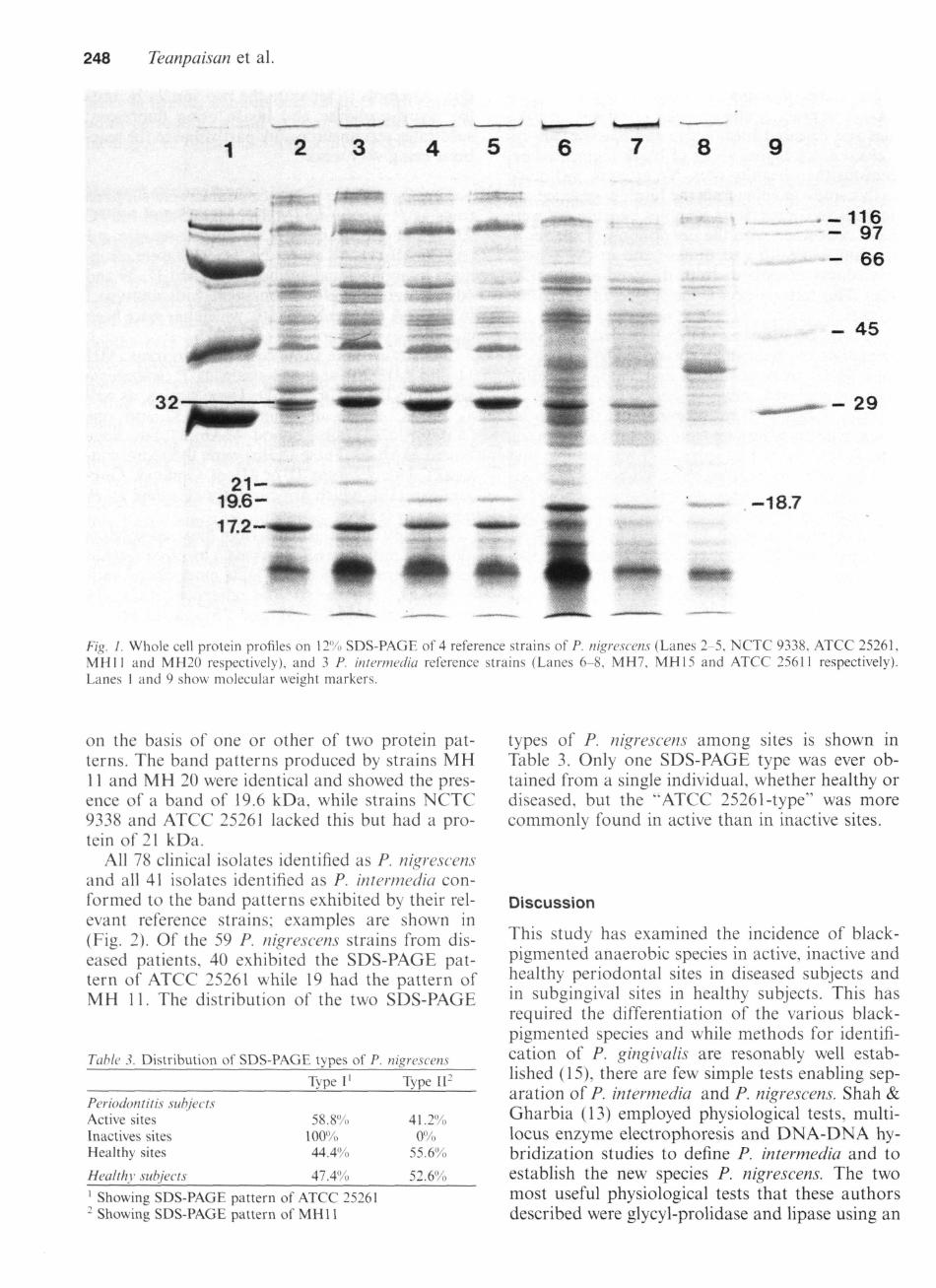
SDS-PAGE - The whole cell protein profiles ofthree P. intermedia and four P. nigrescens referenceor laboratory strains are shown in Fig. 1. Clearand reproducible differences between the profilesof the two species could be seen. All three P. inter-media strains exhibited one relatively heavily stain-ing band of Mr 18.7 kDa, while two strains alsoshowed a strong band of 32 kDa. Two heavilystaining bands with Mr of 32 kDa and 17.2 kDawere seen in all the P. nigrescens reference strains.The P. nigrescens strains could be divided further
Table 2. Incidence of black-pigmented anaerobes in diseased and healthy sites
P. gingivaiis P. intermedia P.nigrescens P. denticola
Periodonlal patientsTotal sitesActive sitesInactive sitesHealthy sites
Healthy subjectsTotal sites
46224905678
1260
8.5%'11%8.9%5%
0%
13.4%15.5%0%
20.5%
18.3%
26.3%37.7%18.6%n.5%
31.6%
1.8%3.3%0%L3%
0%1 0•() sites harbouring each species
248 Teanpaisan et al.
8
jAm.'. ifi. .,
KPPr
-116- 97- 66
- 45
- 29
2 1 -19.6-17.2-
-18.7
Eig. 1. Whole cell protein profiles on 12% SDS-PAGE of 4 reference strains of P. nigrescens (Lanes 2-5, NCTC 9338. ATCC 25261,MHII and MH20 respectively), and 3 P. intermedia reference strains (Lanes 6-8, MH7, MH15 and ATCC 25611 respectively).Lanes I and 9 show molecular weight markers.
on the basis of one or other of two protein pat-terns. The band patterns produced by strains MH11 and MH 20 were identical and showed the pres-ence of a band of 19.6 kDa, while strains NCTC9338 and ATCC 25261 lacked this but had a pro-tein of 21 kDa.
All 78 clinical isolates identified as P. nigrescensand all 41 isolates identified as P. intertnedia con-formed to the band patterns exhibited by their rel-evant reference strains; examples are shown in(Fig. 2). Of the 59 P. nigrescens strains from dis-eased patients, 40 exhibited the SDS-PAGE pat-tern of ATCC 25261 while 19 had the pattern ofMH 11. The distribution of the two SDS-PAGE
Table 3. Distribution of SDS-PAGE types of P. nigrescens
Periodontitis subjectsActive sitesInactives sitesHealthy sites
Healthy subjects
Type I'
58,8%)100%44.4%
47,4'/o
Type 11-
41,2%,0%
55,6%
52,6%
' Showing SDS-PAGE pattern of ATCC 25261- Showing SDS-PAGE pattern of MHl 1
types of p. nigrescens among sites is shown inTable 3. Only one SDS-PAGE type was ever ob-tained from a single individual, whether healthy ordiseased, but the "ATCC 25261-type" was morecommonly found in active than in inactive sites.
Discussion
This study has examined the incidence of black-pigmented anaerobic species in active, inactive andhealthy periodontal sites in diseased subjects andin subgingival sites in healthy subjects. This hasrequired the differentiation of the various black-pigmented species and while methods for identifi-cation of P. gingivalis are resonably well estab-lished (15)., there are few simple tests enabling sep-aration of P. intermedia and P. nigrescens. Shah &Gharbia (13) employed physiological tests, multi-locus enzyme electrophoresis and DNA-DNA hy-bridization studies to define P. intermedia and toestablish the new species P. nigrescens. The twomost useful physiological tests that these authorsdescribed were glycyl-prolidase and lipase using an

Black-pigmented anaerobes in health and disase 249
8
66-
4 5 -
* * > •
29-
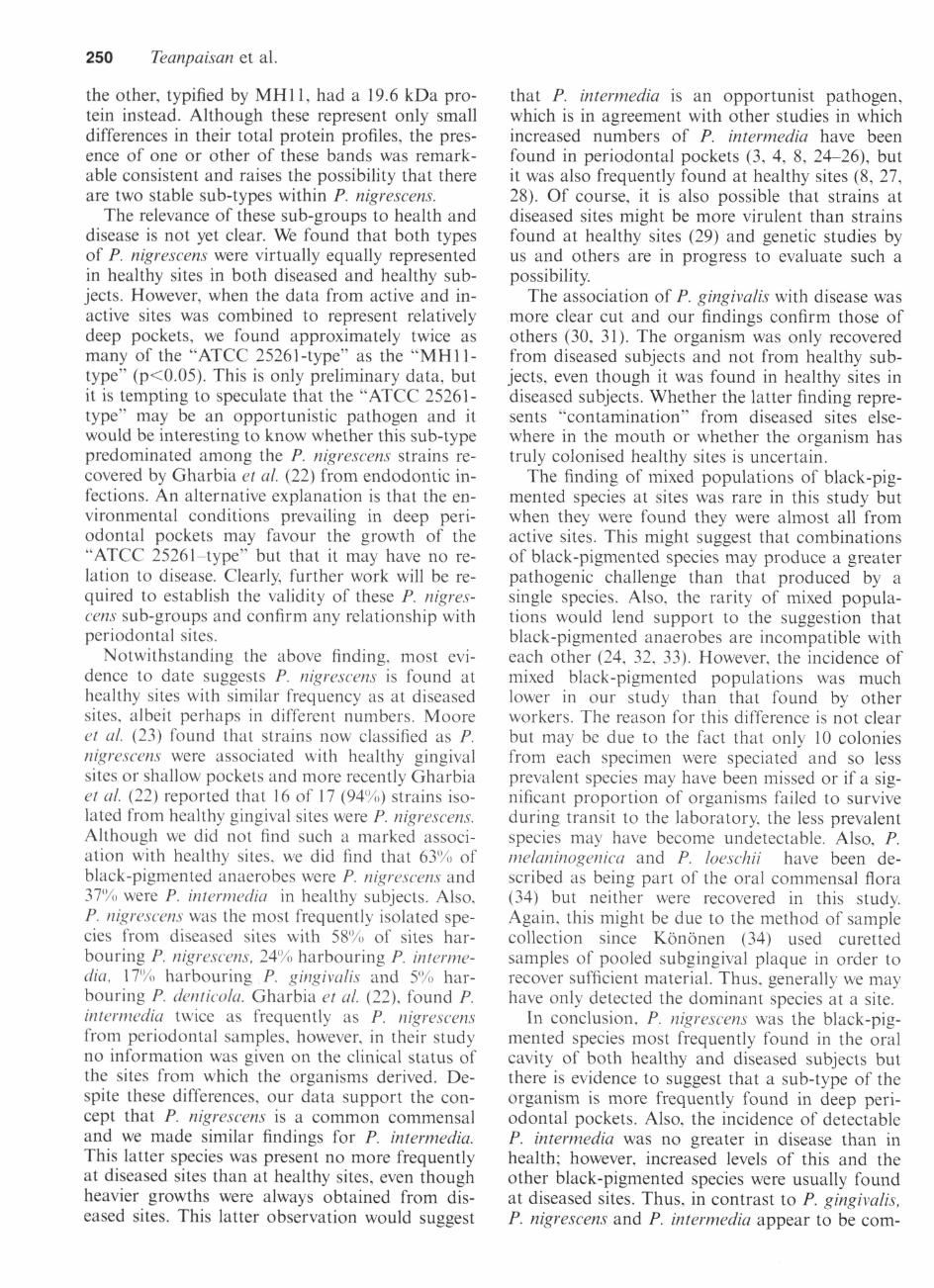
/g. 2. Whole cell protein profiles on \2% SDS-PAGE of representative clinical strains of P. intermedia (Lanes 3-5). a P. intermediareference strain (MH15. Lane 2), representative clinical strains of P. nigrescens (Lanes 6, 7) and reference strains MH20 (Lane 8)and NCTC 9338 (Lane 9). Lane 1 shows molecular weight markers.
elaidate substrate. However, in our hands, thesetests did not reliably differentiate between eitherthe reference strains of the two species or a numberof clinical isolates identified by the other methodsemployed here. The reason for this variance in re-sults is not clear but may be related to the sourcefrom which the enzyme substrates were obtained.The type strains for P. nigrescens is ATTCC 33563,which was not available to us when these studieswere performed but MHl 1 and MH20 were kindlysupplied to us as representatives of P. nigrescensby Shah who originally described the species (13).
In contrast to biochemical tests, serotyping andSDS-PAGE proved useful for differentiating P. in-termedia from P. nigrescens. Of these, serotypingwas the most rapid but the monoclonal antibodiesrequired are not generally available. However,SDS-PAGE is a technique available in most lab-oratories and has been previously used to charac-terise numerous oral species. In most previousstudies, the taxonomy of the P. intermedia grouphad not been clarified but variation in protein pro-files was noted (19, 20). Kinder et al. (21) showedthat P. intermedia ATCC 25611 produced several
major bands, particularly ones with molecularweights of approximately 21.5, 29, 32 and 47 kDa.We also observed protein bands of similar sizes inextracts of ATCC 25611 but a protein of 18.7 kDaproved to be the most useful as a consistentcharacteristic of P. intermedia. Gharbia et al. (22)observed a major band of 31 kDa in P. nigrescensstrains but not in P. intermedia. While we observeda 32 kDa band in P. nigrescens, which is probablythe same as that observed by Gharbia et al. (22),we isolated several P. intermedia strains which hada major band of the same size, making this an un-reliable marker for P. nigrescens. However, we didfind that a protein of 17.2 kDa was present in allstrains of P. nigrescens and this was not seen inany P. intermedia strains. Thus, P. intermedia wascharacterised by an 18.7 kDa band with variablepresence of a 32 kDa band, whereas P. nigrescenswas characterised by both a 17.2 kDa and a 32kDa protein. In addition, all our P. nigrescens clin-ical isolates and reference strains could be sub-di-vided into two groups on the basis of their SDS-PAGE profiles. One was typified by ATCC 25261and showed the presence of a 21 kDa protein and
250 Teanpaisan et al.
the other, typified by MHl 1, had a 19.6 kDa pro-tein instead. Although these represent only smalldifferences in their total protein profiles, the pres-ence of one or other of these bands was remark-able consistent and raises the possibility that thereare two stable sub-types within P. nigrescens.
The relevance of these sub-groups to health anddisease is not yet clear. We found that both typesof P. nigrescens were virtually equally representedin healthy sites in both diseased and healthy sub-jects. However, when the data from active and in-active sites was combined to represent relativelydeep pockets, we found approximately twice asmany of the "ATCC 25261-type" as the "MHll -type" (p<0.05). This is only preliminary data, butit is tempting to speculate that the "ATCC 25261-type" may be an opportunistic pathogen and itwould be interesting to know whether this sub-typepredominated among the P. nigrescens strains re-covered by Gharbia et al. (22) from endodontic in-fections. An alternative explanation is that the en-vironmental conditions prevailing in deep peri-odontal pockets may favour the growth of the"ATCC 25261-type" but that it may have no re-lation to disease. Clearly, further work will be re-quired to establish the validity of these P. nigres-cens sub-groups and confirm any relationship withperiodontal sites.
Notwithstanding the above finding, most evi-dence to date suggests P. nigrescens is found athealthy sites with similar frequency as at diseasedsites, albeit perhaps in different numbers. Mooreet al. (23) found that strains now classified as P.nigrescens were associated with healthy gingivalsites or shallow pockets and more recently Gharbiaet al. (22) reported that 16 of 17 (94%) strains iso-lated from healthy gingival sites were P. nigrescens.Although we did not find such a marked associ-ation with healthy sites, we did find that 63'Mi ofblack-pigmented anaerobes were P. nigrescens and37% were P. intermedia in healthy subjects. Also,P. nigrescens was the most frequently isolated spe-cies from diseased sites with 5%% of sites har-bouring P. nigrescens, 24% harbouring P. interme-dia, 17'/t) harbouring P. gingivaiis and 5% har-bouring P. denticola. Gharbia et al. (22), found P.intermedia twice as frequently as P. nigrescensfrom periodontal samples, however, in their studyno information was given on the clinical status ofthe sites from which the organisms derived. De-spite these differences, our data support the con-cept that P. nigrescens is a common commensaland we made similar findings for P. intermedia.This latter species was present no more frequentlyat diseased sites than at healthy sites, even thoughheavier growths were always obtained from dis-eased sites. This latter observation would suggest
that P. intermedia is an opportunist pathogen,which is in agreement with other studies in whichincreased numbers of P. intermedia have beenfound in periodontal pockets (3, 4, 8, 24-26), butit was also frequently found at healthy sites (8, 27,28). Of course, it is also possible that strains atdiseased sites might be more virulent than strainsfound at healthy sites (29) and genetic studies byus and others are in progress to evaluate such apossibility.
The association of P. gingivaiis with disease wasmore clear cut and our findings confirm those ofothers (30, 31). The organism was only recoveredfrom diseased subjects and not from healthy sub-jects, even though it was found in healthy sites indiseased subjects. Whether the latter finding repre-sents "contamination" from diseased sites else-where in the mouth or whether the organism hastruly colonised healthy sites is uncertain.
The finding of mixed populations of black-pig-mented species at sites was rare in this study butwhen they were found they were almost all fromactive sites. This might suggest that combinationsof black-pigmented species may produce a greaterpathogenic challenge than that produced by asingle species. Also, the rarity of mixed popula-tions would lend support to the suggestion thatblack-pigmented anaerobes are incompatible witheach other (24, 32, 33). However, the incidence ofmixed black-pigmented populations was muchlower in our study than that found by otherworkers. The reason for this difference is not clearbut may be due to the fact that only 10 coloniesfrom each specimen were speciated and so lessprevalent species may have been missed or if a sig-nificant proportion of organisms failed to surviveduring transit to the laboratory, the less prevalentspecies may have become undetectable. Also, P.melaninogenica and P. loeschii have been de-scribed as being part of the oral commensal flora(34) but neither were recovered in this study.Again, this might be due to the method of samplecollection since Kononen (34) used curettedsamples of pooled subgingival plaque in order torecover sufficient material. Thus, generally we mayhave only detected the dominant species at a site.
In conclusion, P. nigrescens was the black-pig-mented species most frequently found in the oralcavity of both healthy and diseased subjects butthere is evidence to suggest that a sub-type of theorganism is more frequently found in deep peri-odontal pockets. Also, the incidence of detectableP. intermedia was no greater in disease than inhealth; however, increased levels of this and theother black-pigmented species were usually foundat diseased sites. Thus, in contrast to P. gingivaiis,P. nigrescens and P. intermedia appear to be com-
Black-pigmented anaerobes in health and disase 251
mon commensals but they make act as opportun-istic pathogens.
Acknowledgements
We are grateful to H.N. Shah, Institute of DentalSurgery, London, A. Eley, University of Sheffield,and DA, Devine, University of Bradford for pro-vision of the reference strains and to R, Gmur,University of Zurich for provision of the mono-clonal antibodies. This work was partly supportedby a studentship to RT from the British Council.
References
1, Slots J, Emrich LJ, Genco RJ, Rosling BG, Relationshipbetween some subgingival bacteria and periodontal pocketdepth and gain or loss of periodontai attachment aftertreatrnent of adult periodontitis, J Clin Periodontol 1985;12: 540-552,
2, Sundqvist G, Pathogenicity and virulence of black-pig-mented Gram-negative anaerobes, FFMS Immunol MedMicrobiol 1993; 6: 125-138,
3, Slots J, Subgingival microflora and periodontal disease, JClin Periodontol 1979; 6: 351-382,
4, Zambon JJ, Reynolds HS, Slots J, Black-pigmented Baeter-oides spp, in the human oral cavity. Infect Immun 1981; 32:198-203,
5, Loesche WJ, Syed SA, Laughon BE, Stoll J, The bacter-iology of acute necrotizing ulcerative gingivitis, J Peri-odontol 1982; 53: 223-230,
6, Kornman KS, Loesche WJ, The subgingival microfloraduring pregnancy, J Periodontal Res 1980; 15: 111-122,
7, van Steenbergen TJM, van Winkelhoff AJ, de Graaf J,Black-pigmented anaerobic rods: classificatton and role inperiodontal disease. In: Hamada S, Holt SC, McGhee JR,eds. Periodontal disease: pathogens and host immune re-sponses. Tokyo: Quintessence, 1991: 41-52.
8, Okuda K, Fukumoto Y, Takazoe L Enumeration of culti-vable black-pigmented Baeteroides spp, in human subgin-gival dental plaque and fecal samples. Oral Microbiol Im-munol 1988; 3: 28-31,
9, Dahlen GM, Wikstrom S, Renvert S, Gmur R, Gug-genheim B, Biochemical and serological characterizationof Baeteroides intertnedius strains isolated from the deepperiodontal pocket, J Clin Microbiol 1990; 28: 2269-2274,
10, Gmur R, Guggenheim B, Antigenic heterogeneity ofBaeteroides intermedius as recognized by monoclonal anti-bodies. Infect Immun 1983; 42: 459-470.
11, Nakazawa F, Zambon JJ, Reynolds HS, Genco RJ, Sero-logical studies on oral Baeteroides intermedius. Infect Im-mun 1988:56: 1647-1651,
12, Swindlehurst CA, Shah HN, Parr CW Williams RAD, So-dium dodecyl sulphate-polyacrylamide gel electrophoresisof polypeptides from Baeteroides melaninogenicus. J ApplBacteriol 1977; 43: 319-324,
13, Shah HN, Garbia SE, Biochemical and chemical studieson strains designated Prevotella intermedia and proposal ofa new pigmented species, Prevotella nigrescens. Int J SystBacteriol 1992; 42: 542-546,
14, Heiginbottom M, Fitzgerald TC, Wade WG, Comparisonof solid media for cultivation of anaerobes, J Clin Pathol1990; 43: 253-256,
15, Laughon BE, Syed SA, Loesche WJ, Rapid identiflcation
of Baeteroides gingivalis. J Clin Microbiol 1982; 15: 345-346,
16, Lowry OH, Rosebrough NJ, Farr AL, Randall RJ, Proteinmeasurement with Folin phenol reagent, J Biol Chem 1951;193: 262-275,
17, Laemmli UK, Cleavage of structural proteins during theassembly of the head of bacteriophage T4. Nature 1970;227: 680-685,
18, Kiel RA, Lang NP, Effect of subgingival sampling tech-nique on periodontal microbiological culturing, J Dent Res1983:62: 247,
19, Swindlehurst CA, Shah HN, Parr CW, WiUiams RAD. So-dium dodecyl sulphate-polyacrylamide gel electrophoresisof polypeptides from Baeteroides melaninogenicus. J ApplBacteriol 1977; 43: 319-324,
20, Johnson JL, Holdeman LV, Baeteroides intermedius comb,nov, and descriptions of Baeteroides corporis sp. nov. andBaeteroides levii sp. nov, Int J Syst Bacteriol 1983; 33: 15-25.
21, Kinder SA, Kornman KS, Holt SC, Characterization ofselected gram-negative oral microorganisms by SDS-PAGE, Oral Microbiol Immunol 1989; 4: 52-56,
22, Gharbia SE, Haapasalo M, Shah HN, et al. Characteriza-tion of Prevotella intermedia and Prevotella nigrescens iso-lates from periodontic and endodontic infections, J Peri-odontol 1994; 65: 56-61,
23, Moore WEC, Moore LVH, Ranney RR, Smibert RM,Burmeister JA, Schenkein HA, The microflora of peri-odontal sites showing active destructive progression, J ClinPeriodontol 1991; 18: 729-739,
24, Loesche WJ, Syed SA, Schmidt E, Morrison EC, Bacterialproflles of subgingival plaques in periodontitis../ PeriodontRes 1985; 56: 447-456,
25, Slots J, Emrich LJ, Genco RJ, Rosling BG, Relationshipbetween some subgingival bacteria and periodontal pocketdepth and gain or loss of periodontal attachment aftertreatment of adult periodontitis, J Clin Periodontol 1985;12: 540-552,
26, Savitt E, Socransky SS, Distribution of certain subgingivalmicrobial species in selected periodontal conditions,,/ Peri-odont Res 1984; 19: 111-123,
27, Mombelli A, McNab H, Lang NP, Black-pigmentingGram-negative bacteria in periodontal disease, L Topo-graphic distribution in the human dentition, J PeriodontRes 1991: 26: 301-307,
28, Van Oosten MAC, Mombelli A, Gusberti FA, Lang NPBlack-pigmented baeteroides species and spirochetes in thesubgingival microbiota of prepubertal school children, JPeriodont Res 1988; 23: 199-203,
29, Socransky SS, Haffajee AD, The bacterial aetiology of de-structive periodontal disease: current concepts, J Peri-odontol 1992; 63: 322-331.
30, Dzink JL, Socransky SS, Haffajee AD. The predominantcultivable microbiota of active and inactive lesions of de-structive periodontal diseases, J Clin Periodontol 1988; 15:316-323,
31, Kojima T, Yasui S, Ishikawa I, Distribution of Porphyro-monas gingivalis in adult periodontitis patients, J Peri-odontol 1993; 64: 1231-1237.
32, van Winkelhoff AJ, van der Velden U, de Graaf J. Mi-crobial succession in recolonizing deep periodontal pocketsafter a single course of supra- and subgingival debride-ment, J Clin Periodontol 1988; 15: 116-122,
33, Torkko H, Asikainen S, Occurrence of Porphyromonas gin-givalis with Prevotella intermedia in periodontal samples,FEMS Immunol Med Microbiol 1993; 6: 195-198,
34, Kononen E, Pigmented prevotella species in the peri-odontally healthy oral cavity, FFMS Immunol Med Micro-biol 1993; 6: 201-206,