Embed Size (px)
Citation preview
Characterization of a Drosophila Alzheimer’s DiseaseModel: Pharmacological Rescue of Cognitive DefectsRanjita Chakraborty1., Vidya Vepuri1,2., Siddhita D. Mhatre1, Brie E. Paddock1, Sean Miller1, Sarah J.
Michelson1, Radha Delvadia1, Arkit Desai1, Marianna Vinokur1, David J. Melicharek1, Suruchi Utreja1,
Preeti Khandelwal1, Sara Ansaloni1, Lee E. Goldstein3, Robert D. Moir4, Jeremy C. Lee5, Loni P. Tabb6,
Aleister J. Saunders1,7,8*, Daniel R. Marenda1,8*
1 Department of Biology, Drexel University, Philadelphia, Pennsylvania, United States of America, 2 Department of Biological Sciences, University of the Sciences in
Philadelphia, Philadelphia, Pennsylvania, United States of America, 3 Department of Psychiatry, Boston University, Boston, Massachusetts, United States of America,
4 Genetics and Aging Research Unit, MIND, Massachusetts General Hospital, Harvard Medical School, Boston, Massachusetts, United States of America, 5 Department of
Molecular, Cell, and Developmental Biology, University of California Santa Cruz, Santa Cruz, California, United States of America, 6 Department of Epidemiology and
Biostatistics, Drexel University, Philadelphia, Pennsylvania, United States of America, 7 Department of Biochemistry and Molecular Biology, Drexel University College of
Medicine, Philadelphia, Pennsylvania, United States of America, 8 Department of Neurobiology and Anatomy, Drexel University College of Medicine, Philadelphia,
Pennsylvania, United States of America
Abstract
Transgenic models of Alzheimer’s disease (AD) have made significant contributions to our understanding of ADpathogenesis, and are useful tools in the development of potential therapeutics. The fruit fly, Drosophila melanogaster,provides a genetically tractable, powerful system to study the biochemical, genetic, environmental, and behavioral aspectsof complex human diseases, including AD. In an effort to model AD, we over-expressed human APP and BACE genes in theDrosophila central nervous system. Biochemical, neuroanatomical, and behavioral analyses indicate that these flies exhibitaspects of clinical AD neuropathology and symptomology. These include the generation of Ab40 and Ab42, the presence ofamyloid aggregates, dramatic neuroanatomical changes, defects in motor reflex behavior, and defects in memory. Inaddition, these flies exhibit external morphological abnormalities. Treatment with a c-secretase inhibitor suppressed thesephenotypes. Further, all of these phenotypes are present within the first few days of adult fly life. Taken together these datademonstrate that this transgenic AD model can serve as a powerful tool for the identification of AD therapeuticinterventions.
Citation: Chakraborty R, Vepuri V, Mhatre SD, Paddock BE, Miller S, et al. (2011) Characterization of a Drosophila Alzheimer’s Disease Model: PharmacologicalRescue of Cognitive Defects. PLoS ONE 6(6): e20799. doi:10.1371/journal.pone.0020799
Editor: Mel B. Feany, Brigham and Women’s Hospital, Harvard Medical School, United States of America
Received September 21, 2010; Accepted May 13, 2011; Published June 6, 2011
Copyright: � 2011 Chakraborty et al. This is an open-access article distributed under the terms of the Creative Commons Attribution License, which permitsunrestricted use, distribution, and reproduction in any medium, provided the original author and source are credited.
Funding: This work is supported by grants from Drexel University’s Human Cognition Enhancement Program (to DRM), Cure Alzheimer’s Fund InvestigatorAward and National Institutes of Health (NIH) grant 1R01AI081990-01A1 (to RDM), NIH R21RR026074 (to DRM), and NIH R01NS057295 (to AJS). The funders had norole in study design, data collection and analysis, decision to publish, or preparation of the manuscript.
Competing Interests: The authors have declared that no competing interests exist.
* E-mail: [email protected] (AJS); [email protected] (DRM)
. These authors contributed equally to this work.
Introduction
Alzheimer’s disease (AD) is a progressive neurodegenerative
disorder and is the most common cause of dementia in the
developed world [1]. The pathological features of AD include the
presence of amyloid plaques, neurofibrillary tangles, and loss of
neurons, primarily in the cerebral cortex and hippocampus [2].
Amyloid plaques are extracellular deposits mainly composed of a
small peptide (,4 kD) called b-amyloid (Ab), surrounded by
dystrophic neurites, reactive microglia and astrocytes [3]. Several
lines of evidence support the amyloid hypothesis of AD, according
to which Ab plays the central role in initiating the AD pathogenic
cascade [4].
Ab peptides are generated by proteolytic processing of the b-
amyloid precursor protein (APP) through sequential proteolysis by
b- and c-secretases in the amyloidogenic processing pathways [5].
This pathway is initiated when APP undergoes proteolytic
cleavage by b-secretase, encoded by the BACE gene. This cleavage
produces a soluble extracellular/lumenal fragment of APP (sAPPb)
and a membrane spanning C-terminal fragment (bCTF/C99).
The c-secretase complex then cleaves bCTF to produce Abpeptides and the APP intracellular domain (AICD) [5]. Abpeptides of a variety of lengths are produced but Ab40 and Ab42
are the major isoforms produced in the central nervous system
(CNS). Compared to Ab40, Ab42 is more prone to oligomerization
and has been shown to be more neurotoxic [6].
APP also undergoes an alternative proteolytic processing
pathway termed the non-amyloidogenic pathway. In this pathway,
a-secretase initially cleaves APP, rather than b-secretase, to
produce a soluble extracellular/lumenal fragment of APP (sAPPa)
and a membrane spanning C-terminal fragment (aCTF/C83).
Again, the c-secretase complex then cleaves aCTF to produce the
P3 peptide and AICD [5].
APP proteolysis is an important step towards development of
AD. Therefore, it is important to identify genes and pharmaceu-
ticals that modulate APP metabolism and Ab production and
PLoS ONE | www.plosone.org 1 June 2011 | Volume 6 | Issue 6 | e20799
clearance. Developing in vivo disease models has proven crucial to
illuminating disease mechanisms, since in vitro studies do not
always represent the natural physiological complexity of the tissue
and/or organism. In particular, the fruit fly, Drosophila melanogaster,
has been tremendously important and influential in furthering our
understanding of the mechanisms of many forms of neurodegen-
erative diseases, including AD [7,8,9,10,11].
Drosophila endogenously express orthologues to the human APP
[12], a-secretase [13,14], and c-secretase [15,16,17,18]. Recently,
a functional Drosophila homolog of the BACE (b-secretase) family
of proteins has also been identified [19]. Though the Drosophila
homolog to human APP, Appl, does not contain significant
sequence similarity within the Ab region of human APP [12],
there is recent evidence suggesting that the fly Appl protein is also
capable of generating neurotoxic Ab-like fragments when the fly
Appl and fly b-secretase proteins are overexpressed in Drosophila
tissues [19]. These features position the fly as an attractive model
to further study the evolutionarily conserved functions of these
endogenous proteins.
Even though flies express orthologues of APP and secretase
proteins, other Drosophila models of AD have been generated that
express the human genes to gain insight into mechanism of disease
and to illuminate potential therapeutic approaches. Many of these
Drosophila AD models express the toxic human Ab42 to study its
effects on a molecular and behavioral level [9,20,21,22,23,24].
These models have been useful in further dissecting the basic
mechanisms behind human disease phenotypes such as amyloid
deposits, learning and memory deficiences, and premature death.
This method of expressing wild-type Ab and disease associated Absequence variants is useful for modulating the disease phenotype
after disease progression has begun. Fewer reports have been
published that rely on human APP proteolytic processing in the
Drosophila CNS to generate Ab oligomers [11], even though it has
been shown that the endogenous fly secretases can process the
human form of APP [7,8].
The targeted expression of human AD genes in the fly has been
used previously, with a focus on expression in the retina, wing, and
the nervous system [7,8,11]. Here we express the human APP and
BACE genes within the developing nervous system of Drosophila.
This results in a model that displays very similar pathology to
human Alzheimer’s patients, including accumulation of Ab-
containing puncta in their brains, decreased dendritic and axonal
fields in areas of the brain important for learning and memory,
and memory deficits. A significant advantage of the model we
describe is that these neuropathologies and memory defects are
evident within days. We demonstrate that all of these phenotypes
can be pharmacologically suppressed by the c-secretase inhibitor
L-685,458, illustrating the efficacy of this model for the rapid
testing of small molecules for therapeutic intervention in vivo.
Results
Expression of the human APP gene alone or in combination
with the human b-secretase (BACE) gene exclusively in the
developing fly nervous system was accomplished using the
GAL4/UAS system [25]. Specifically, we utilized elav-GAL4,
which drives protein expression throughout the fly CNS [26].
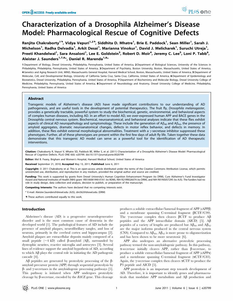
Using Western blot analysis, full-length APP is detected in the
brains of flies expressing either APP or APP/BACE under control
of the elav promoter, but APP is not detected in control flies lacking
the APP transgene (Figure 1), as expected in the absence of the
elav-GAL4 driver. In brain tissue from elav; APP; BACE heterozy-
gous flies, detection of BACE (Figure S1D) andb CTFs are
evidence of BACE expression and b-secretase activity (Figure 1,
red arrows in third lane) respectively. b-secretase activity is not
evident in elav; APP heterozygous flies, since only the a-secretase
generated aCTFwas detected (Figure 1, red arrow in second lane).
To determine if the bCTFs were further processed by c-
secretase we measured Ablevels by ELISA and by Western blot.
AbELISA results demonstrate that elav; APP; BACE heterozygous
flies produce signficantly higher levels of Ab40 and Ab42 than those
not expressing human APP or BACE (Table 1). When elav; APP;
BACE heterozygous flies are raised on food containing 100 nM L-
685,458, a c-secretase transition state inhibitor, Ablevels are
undetectable (Table 1). This indicates that c-secretase activity is
inhibited successfully in these flies, as is the subsequent production
of Ab. This result was confirmed by Western blot analysis of elav;
APP; BACE fly heads, which demonstrate decreased Ab levels in
the L-685,458 treated flies compared to the DMSO (vehicle) raised
elav; APP; BACE heterozygous flies (Figure S1C and S1F). The
bCTF is the substrate for c-secretase cleavage in APP amyloido-
genic processing. Inhibition of c-secretase activity should result in
increased CTF levels. Consistent with this, we observed increased
bCTF levels in the elav; APP; BACE heterozygous flies raised on L-
685,458 containing food compared to those raised on DMSO
(Figures S1B and S1F), as well as a modest increase in full length
APP levels in flies raised on L-685,458 (Figures S1A and S1F).
Treatment with either DMSO or L-685, 458 did not alter
expression of BACE (Figures S1E and S1F). Therefore, CNS
expression of human APP and BACE recapitulates APP
amyloidogenic processing observed in vitro and in rodent transgenic
AD models.
In this model APP and BACE are expressed continuously
during fly development. Upon adult eclosion, we observed two
Figure 1. Transgene expression and proteolytic processing.Western blot analysis of human APP and fly b-actin detected in fly headlysates of: 1) elav; +; + heterozygous flies 2) elav; APP; + heterozygousflies, and 3) elav; APP; BACE heterozygous flies. APP-FL (full length APP,,110 kD) and APP-CTFs (C terminal fragments, ,10–12 kD) weredetected using A8717 (Sigma). Red arrows indicate a-CTF (lane 2) and b-CTFs (lane 3). A fly b-actin specific antibody was utilized (Abcam).doi:10.1371/journal.pone.0020799.g001
Table 1. Ab levels.
Genotype Treatment Ab40 (pg/mL) Ab42 (pg/mL)
elav; +; + – 36614 ,5
elav; PP; BACE DMSO 11068 114622
elav; APP; BACE L-685,458 ,5 ,5
Ab levels detected in fly head lysates (genotypes indicated) by ELISA.doi:10.1371/journal.pone.0020799.t001
Drosophila AD Model
PLoS ONE | www.plosone.org 2 June 2011 | Volume 6 | Issue 6 | e20799
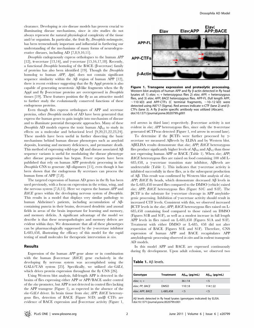
distinct morphological abnormalities in these flies: crumpled
wings, and the presence of melanotic masses on both the abdomen
and proboscis (arrows in Figure 2B). Crumpled wings were
observed in 61% of all elav; APP; BACE heterozygous flies, while
necrotic tissue was observed in 26% of the same genotype
(Figure 2C). These phenotypes are observed in flies expressing
human APP alone but at an approximately tenfold reduced
penetrance compared to flies expressing both human APP and
human BACE (Figure 2C) consistent with the idea that the
phenotypes are dependent upon the expression of human BACE
and amyloidogenic APP processing. More evidence to support this
idea is that elav; APP; BACE heterozygous flies raised on food
containing L-685,458 display significantly fewer crumpled wings
(p = 0.001) or necrotic tissue (p = 0.001; Figure 2C). Specifically, L-
685,458 treatment reduced the occurrence of crumpled wings to
17% from 52% observed in elav; APP; BACE flies treated with
vehicle (DMSO). Additionally, L-685,458 treatment reduced the
presence of melanotic masses to 3% from 16%.
We next compared the longevity of flies expressing human APP
and human BACE to those flies only expressing human APP or
human BACE alone. We created both survival and hazard plots
for each genotype for analysis (Figure S2). We calculated the
median survival time as the time when the survivor function equals
50%. As some flies were censored during the experiment (those
that flew away or died of unnatural causes), median survivorship
reflects a more reliable metric than the mean survival time.
The median survival time for elav; APP; BACE heterozygous flies
was 32 days, compared to 42 days for elav; + heterozygous flies,
and 56 days for +; APP; BACE heterozygous flies (Figure S2A). We
found that until day 45, elav; APP; BACE heterozygous flies
consistently had a lower probability of survival (Figure S2A). We
found a statistical difference in survival between elav; APP; BACE
heterozygous flies and controls (p,0.0001), suggesting that these
flies displayed decreased survival, but this effect was limited to only
young adults. While we found that there was no significant
difference in the probability of survival between elav; BACE; +heterozygous flies and controls (p = 0.1207) (Figure S2D), we did
find a significant difference in the probability of survival between
elav; APP; + heterozygous flies and controls (p,0.0001) (Figure
S2C). The median survival time of elav; APP heterozygous flies was
6 days, while the median survival time for the +; APP heterozygous
and elav; + heterozygous flies was 38 and 45, respectively (Figure
S2C). Again, the effect on survival was limited to only young
adults. Finally, we compared the survival time for elav; APP; BACE
heterozygous flies fed on DMSO and L-685, 458, and found no
significant difference in the probability of survival (p = 0.5038).
Taken together, these results suggest that while there is an effect on
survival in the elav; APP; BACE heterozygous genotype in young
adult flies, this effect does not require either BACE or c-secretase
function, and therefore most likely does not represent an effect of
human Ab accumulation in these flies.
We next compared the gross anatomical features of whole
brains from elav-CD8; +; + heterozygous flies and elav-CD8; APP;
BACE heterozygous flies (Figures S3A and S3B, respectively). We
co-expressed a membrane tagged form of GFP (CD8-GFP) in the
CNS of flies with the indicated transgenes to fluorescently visualize
whole brain anatomy. elav-CD8; APP; BACE heterozygous flies
displayed evidence of significant neuroanatomical changes com-
pared to elav-CD8; +; + heterozygous flies and elav-CD8; APP; +heterozygous flies (Figures S3A and S3B). A number of specific
brain structures/regions are altered including the the mushroom
body (Figure 3A), the antennal lobes, and the optic lobes (Figure
S3). The mushroom bodies are axonal bundles involved in
learning and memory behavior in multiple experimental para-
digms [27,28,29,30,31]. These axons extend from a population of
neurons that consist of three distinct groups, which give rise to a
final adult structure consisting of 5 projections to the a, a9, b, b9,
and c lobes (Figure 3A) [32]. These structures are significantly
smaller in elav-CD8; APP; BACE heterozygous flies compared to
controls (Figure 3A). In multiple cases axons from the mushroom
body extending to the a and a9 lobes are either significantly
shorter than controls, and/or missing completely (Figure 3A).
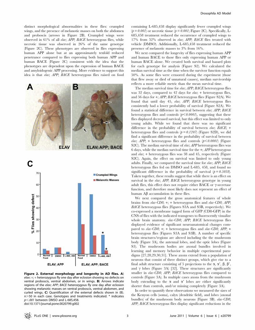
In order to quantify these observations we measured the sizes of
the Kenyon cells (soma), calyx (dendritic field), and lobes (axonal
bundles) of the mushroom body neurons (Figure 3B). elav-CD8;
APP; BACE heterozygous flies display significant reductions in the
Figure 2. External morphology and longevity in AD flies. A)elav; +; + heterozygous fly one day after eclosion showing no defects onventral proboscis, ventral abdomen, or in wings. B) Arrows indicateregions of the elav; APP; BACE heterozygous fly one day after eclosionshowing melanotic masses on ventral proboscis, ventral abdomen, andcurled wings. C) Quantification of the external defects shown in (B).n.50 in each case. Genotypes and treatments indicated. * indicatesp,.001 between DMSO and L-685,458.doi:10.1371/journal.pone.0020799.g002
Drosophila AD Model
PLoS ONE | www.plosone.org 3 June 2011 | Volume 6 | Issue 6 | e20799
sizes of the calyx (p = 0.005) and the lobes (p = 0.005) compared to
elav-CD8; APP; + heterozygous flies, elav-CD8; BACE; + heterozy-
gous flies, or elav; + heterozygous control flies (Figures 3B, C).
However, elav-CD8; APP; BACE heterozygous flies do not display a
significant (p = 0.099) decrease in Kenyon cell soma size compared
to either elav; + heterozygous flies, or elav; APP; + heterozygous flies
(Figure 3C). elav-CD8; APP; BACE heterozygous flies do show a
significant difference in size compared to elav; BACE; +heterozygous flies (p = 0.018). However, Kenyon cell fields from
elav; BACE; + heterozygous flies do not show a significant different
between either elav; APP; + heterozygous flies or Elav; +heterozygous flies themselves (p = 0.393 and 0.989 respectively).
Culturing elav-CD8; APP; BACE heterozygous flies on food
containing the common drug vehicle DMSO resulted in a
decrease in Kenyon cell size as compared to the same genotype
cultured on medium containing no DMSO, indicating that
DMSO has a negative effect on these neurons in this genetic
background. This is consistent with previous literature suggesting
that DMSO has deleterious effects in flies as well as other systems
[33,34,35]. However, when elav-CD8; APP; BACE heterozygous
flies were raised on food containing L-685,458 dissolved in DMSO
we observe a significant increase in the size of the Kenyon cell
soma (p = 0.03) and calyx (p = 0.03) compared to elav-CD8; APP;
BACE heterozygous flies raised on DMSO alone (Figure 3D).
However, a significant increase in the axonal fields upon L-
685,458 treatment was not observed (p = 0.14; Figure 3D). In
conclusion, elav-CD8; APP; BACE heterozygous flies have reduced
dendritic and axonal fields in structures involved in learning and
memory. Further, these reductions require the expression of
human APP and BACE, as well as the proper function of the c-
secretase complex, consistent with the idea that these neuroana-
tomical phenotypes are dependent upon amyloidogenic APP
processing.
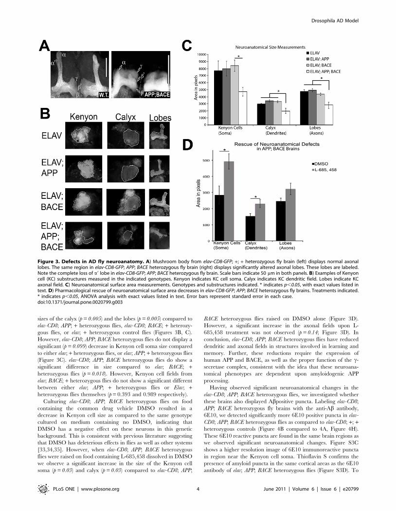
Having observed significant neuroanatomical changes in the
elav-CD8; APP; BACE heterozygous flies, we investigated whether
these brains also displayed Abpositive puncta. Labeling elav-CD8;
APP; BACE heterozygous fly brains with the anti-Ab antibody,
6E10, we detected significantly more 6E10 positive puncta in elav-
CD8; APP; BACE heterozygous flies as compared to elav-CD8; +; +heterozygous controls (Figure 4B compared to 4A, Figure 4H).
These 6E10 reactive puncta are found in the same brain regions as
we observed significant neuroanatomical changes. Figure S3C
shows a higher resolution image of 6E10 immunoreactive puncta
in region near the Kenyon cell soma. Thioflavin S confirms the
presence of amyloid puncta in the same cortical areas as the 6E10
antibody of elav; APP; BACE heterozygous flies (Figure S3D). To
Figure 3. Defects in AD fly neuroanatomy. A) Mushroom body from elav-CD8-GFP; +; + heterozygous fly brain (left) displays normal axonallobes. The same region in elav-CD8-GFP; APP; BACE heterozygous fly brain (right) displays significantly altered axonal lobes. These lobes are labeled.Note the complete loss of a9 lobe in elav-CD8-GFP; APP; BACE heterozygous fly brain. Scale bars indicate 50 mm in both panels. B) Examples of Kenyoncell (KC) substructures measured in the indicated genotypes. Kenyon indicates KC cell soma. Calyx indicates KC dendritic field. Lobes indicate KCaxonal field. C) Neuroanatomical surface area measurements. Genotypes and substructures indicated. * indicates p,0.05, with exact values listed intext. D) Pharmacological rescue of neuroanatomical surface area decreases in elav-CD8-GFP; APP; BACE heterozygous fly brains. Treatments indicated.* indicates p,0.05, ANOVA analysis with exact values listed in text. Error bars represent standard error in each case.doi:10.1371/journal.pone.0020799.g003
Drosophila AD Model
PLoS ONE | www.plosone.org 4 June 2011 | Volume 6 | Issue 6 | e20799
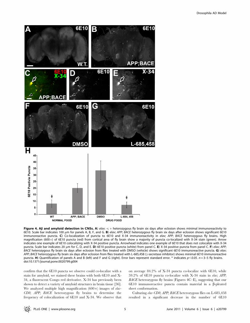
confirm that the 6E10 puncta we observe could co-localize with a
stain for amyloid, we stained these brains with both 6E10 and X-
34, a fluorescent Congo red derivative. X-34 has previously been
shown to detect a variety of amyloid structures in brain tissue [36].
We analyzed multiple high magnification (6006) images of elav-
CD8; APP; BACE heterozygous fly brains to determine the
frequency of colocalization of 6E10 and X-34. We observe that
on average 84.2% of X-34 puncta co-localize with 6E10, while
59.2% of 6E10 puncta co-localize with X-34 stain in elav; APP;
BACE heterozygous fly brains (Figures 4C–E), suggesting that our
6E10 immunoreactive puncta contain material in a b-pleated
sheet conformation.
Culturing elav-CD8; APP; BACE heterozygous flies on L-685,458
resulted in a significant decrease in the number of 6E10
Figure 4. Ab and amyloid detection in CNSs. A) elav; +; + heterozygous fly brain six days after eclosion shows minimal immunoreactivity to6E10. Scale bar indicates 100 mm for panels A, B, F, and G. B) elav; APP; BACE heterozygous fly brain six days after eclosion shows significant 6E10immunoreactive puncta. C) Co-localization of puncta to 6E10 and X-34 immunoreactivity in elav; APP; BACE heterozygous fly brains. Highmagnification (6006) of 6E10 puncta (red) from cortical area of fly brain show a majority of puncta co-localized with X-34 stain (green). Arrowindicates one example of 6E10 colocalizing with X-34 positive puncta. Arrowhead indicates one example of 6E10 that does not colocalize with X-34puncta. Scale bar indicates 20 mm for C, D, and E. D) 6E10 positive puncta (white) from panel C. E) X-34 positive puncta from panel C. F) elav; APP;BACE heterozygous fly brain six days after eclosion from flies treated with DMSO (vehicle) shows significant 6E10 immunoreactive puncta. G) elav;APP; BACE heterozygous fly brain six days after eclosion from flies treated with L-685,458 (c-secretase inhibitor) shows minimal 6E10 immunoreactivepuncta. H) Quantification of panels A and B (left) and F and G (right). Error bars represent standard error. * indicates p,0.05. n = 3–5 fly brains.doi:10.1371/journal.pone.0020799.g004
Drosophila AD Model
PLoS ONE | www.plosone.org 5 June 2011 | Volume 6 | Issue 6 | e20799
immunoreactive puncta compared to DMSO treated flies of the
same genotype (Figures 4F, 4G, and 4H). These results further
confirm the presence of Ab positive puncta in the brains of the
elav-CD8; APP; BACE heterozygous flies, and show that presence of
these puncta is dependent on c-secretase activity, as expected.
Having observed marked neuroanatomical and neuropatholog-
ical changes in the elav-CD8; APP; BACE heterozygous flies, we
wanted to determine if CNS function may be compromised. As an
initial test of CNS function, we utilized a simple, yet powerful
behavioral assay, the climbing assay [37]. This well-established
assay has been previously used to assess nervous system
dysfunction in fly models of multiple diseases, including AD [9].
Briefly, flies display a negative geotaxis response when given a
mechanical stimulus. When tapped to the bottom of a vial, flies
normally orient themselves rapidly and begin climbing to the top.
By assaying the fly’s ability to climb to the top of a vial in a set time
period (18 seconds) we are able to compare broad nervous system
function of reflex behavior between flies of different genotypes, or
flies treated with different pharmacologic agents. When cultured
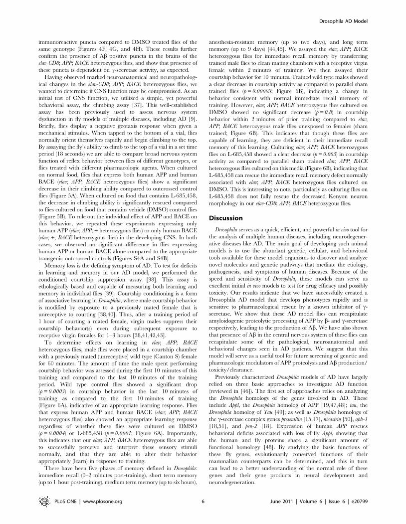
on normal food, flies that express both human APP and human
BACE (elav; APP; BACE heterozygous flies) show a significant
decrease in their climbing ability compared to outcrossed control
flies (Figure 5A). When cultured on food that contains L-685,458,
the decrease in climbing ability is significantly rescued compared
to flies cultured on food that contains vehicle (DMSO) control flies
(Figure 5B). To rule out the individual effect of APP and BACE on
this behavior, we repeated these experiments expressing only
human APP (elav; APP; + heterozygous flies) or only human BACE
(elav; +; BACE heterozygous flies) in the developing CNS. In both
cases, we observed no significant difference in flies expressing
human APP or human BACE alone compared to the appropriate
transgenic outcrossed controls (Figures S4A and S4B).
Memory loss is the defining symptom of AD. To test for deficits
in learning and memory in our AD model, we performed the
conditioned courtship suppression assay [38]. This assay is
ethologically based and capable of measuring both learning and
memory in individual flies [39]. Courtship conditioning is a form
of associative learning in Drosophila, where male courtship behavior
is modified by exposure to a previously mated female that is
unreceptive to courting [38,40]. Thus, after a training period of
1 hour of courting a mated female, virgin males suppress their
courtship behavior(s) even during subsequent exposure to
receptive virgin females for 1–3 hours [38,41,42,43].
To determine effects on learning in elav; APP; BACE
heterozygous flies, male flies were placed in a courtship chamber
with a previously mated (unreceptive) wild type (Canton S) female
for 60 minutes. The amount of time the male spent performing
courtship behavior was assessed during the first 10 minutes of this
training and compared to the last 10 minutes of the training
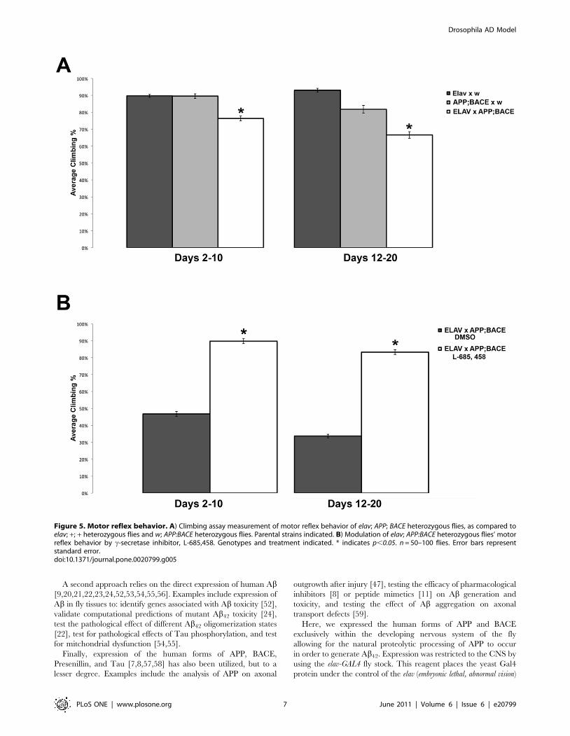
period. Wild type control flies showed a significant drop
(p = 0.0003) in courtship behavior in the last 10 minutes of
training as compared to the first 10 minutes of training
(Figure 6A), indicative of an appropriate learning response. Flies
that express human APP and human BACE (elav; APP; BACE
heterozygous flies) also showed an appropriate learning response
regardless of whether these flies were cultured on DMSO
(p = 0.0004) or L-685,458 (p = 0.0001; Figure 6A). Importantly,
this indicates that our elav; APP; BACE heterozygous flies are able
to successfully perceive and interpret these sensory stimuli
normally, and that they are able to alter their behavior
appropriately (learn) in response to training.
There have been five phases of memory defined in Drosophila:
immediate recall (0–2 minutes post-training), short term memory
(up to 1 hour post-training), medium term memory (up to six hours),
anesthesia-resistant memory (up to two days), and long term
memory (up to 9 days) [44,45]. We assayed the elav; APP; BACE
heterozygous flies for immediate recall memory by transferring
trained male flies to clean mating chambers with a receptive virgin
female within 2 minutes of training. We then assayed their
courtship behavior for 10 minutes. Trained wild type males showed
a clear decrease in courtship activity as compared to parallel sham
trained flies (p = 0.00003; Figure 6B), indicating a change in
behavior consistent with normal immediate recall memory of
training. However, elav; APP; BACE heterozygous flies cultured on
DMSO showed no significant decrease (p = 0.8) in courtship
behavior within 2 minutes of prior training compared to elav;
APP; BACE heterozygous male flies unexposed to females (sham
trained; Figure 6B). This indicates that though these flies are
capable of learning, they are deficient in their immediate recall
memory of this learning. Culturing elav; APP; BACE heterozygous
flies on L-685,458 showed a clear decrease (p = 0.005) in courtship
activity as compared to parallel sham trained elav; APP; BACE
heterozygous flies cultured on this media (Figure 6B), indicating that
L-685,458 can rescue the immediate recall memory defect normally
associated with elav; APP; BACE heterozygous flies cultured on
DMSO. This is interesting to note, particularly as culturing flies on
L-685,458 does not fully rescue the decreased Kenyon neuron
morphology in our elav-CD8; APP; BACE heterozygous flies.
Discussion
Drosophila serves as a quick, efficient, and powerful in vivo tool for
the analysis of multiple human diseases, including neurodegener-
ative diseases like AD. The main goal of developing such animal
models is to use the abundant genetic, cellular, and behavioral
tools available for these model organisms to discover and analyze
novel molecules and genetic pathways that mediate the etiology,
pathogenesis, and symptoms of human diseases. Because of the
speed and sensitivity of Drosophila, these models can serve as
excellent initial in vivo models to test for drug efficacy and possibly
toxicity. Our results indicate that we have successfully created a
Drosophila AD model that develops phenotypes rapidly and is
sensitive to pharmacological rescue by a known inhibitor of c-
secretase. We show that these AD model flies can recapitulate
amyloidogenic proteolytic processing of APP by b- and c-secretase
respectively, leading to the production of Ab. We have also shown
that presence of Ab in the central nervous system of these flies can
recapitulate some of the pathological, neuroanatomical and
behavioral changes seen in AD patients. We suggest that this
model will serve as a useful tool for future screening of genetic and
pharmacologic modulators of APP proteolysis and Ab production/
toxicity/clearance.
Previously characterized Drosophila models of AD have largely
relied on three basic approaches to investigate AD function
(reviewed in [46]). The first set of approaches relies on analyzing
the Drosophila homologs of the genes involved in AD. These
include Appl, the Drosophila homolog of APP [19,47,48]; tau, the
Drosophila homolog of Tau [49]; as well as Drosophila homologs of
the c-secretase complex genes presenillin [15,17], nicastrin [50], aph-1
[18,51], and pen-2 [18]. Expression of human APP rescues
behavioral deficits associated with loss of fly Appl, showing that
the human and fly proteins share a significant amount of
functional homology [48]. By studying the basic functions of
these fly genes, evolutionarily conserved functions of their
mammalian counterparts can be determined, and this in turn
can lead to a better understanding of the normal role of these
genes and their gene products in neural development and
neurodegeneration.
Drosophila AD Model
PLoS ONE | www.plosone.org 6 June 2011 | Volume 6 | Issue 6 | e20799
A second approach relies on the direct expression of human Ab[9,20,21,22,23,24,52,53,54,55,56]. Examples include expression of
Ab in fly tissues to: identify genes associated with Ab toxicity [52],
validate computational predictions of mutant Ab42 toxicity [24],
test the pathological effect of different Ab42 oligomerization states
[22], test for pathological effects of Tau phosphorylation, and test
for mitchondrial dysfunction [54,55].
Finally, expression of the human forms of APP, BACE,
Presenillin, and Tau [7,8,57,58] has also been utilized, but to a
lesser degree. Examples include the analysis of APP on axonal
outgrowth after injury [47], testing the efficacy of pharmacological
inhibitors [8] or peptide mimetics [11] on Ab generation and
toxicity, and testing the effect of Ab aggregation on axonal
transport defects [59].
Here, we expressed the human forms of APP and BACE
exclusively within the developing nervous system of the fly
allowing for the natural proteolytic processing of APP to occur
in order to generate Ab42. Expression was restricted to the CNS by
using the elav-GAL4 fly stock. This reagent places the yeast Gal4
protein under the control of the elav (embryonic lethal, abnormal vision)
Figure 5. Motor reflex behavior. A) Climbing assay measurement of motor reflex behavior of elav; APP; BACE heterozygous flies, as compared toelav; +; + heterozygous flies and w; APP:BACE heterozygous flies. Parental strains indicated. B) Modulation of elav; APP:BACE heterozygous flies’ motorreflex behavior by c-secretase inhibitor, L-685,458. Genotypes and treatment indicated. * indicates p,0.05. n = 50–100 flies. Error bars representstandard error.doi:10.1371/journal.pone.0020799.g005
Drosophila AD Model
PLoS ONE | www.plosone.org 7 June 2011 | Volume 6 | Issue 6 | e20799
genomic enhancer region on the X chromosome [25]. elav encodes
for an RNA binding protein that is expressed in all post-mitotic
neurons [60], and has recently been shown to be expressed in
embryonic glial cells, but not larval or adult glia [61]. Though
other Drosophila models have similarly expressed both of these
human proteins to model AD in the fly, these models have largely
restricted their analysis to the developing retina [8] and wing
tissues [7,8]. In the retina, Greeve and colleagues observe that
expression of human APP resulted in more neurodegeneration
than the co-expression of human APP and human BACE [8].
They postulated that this suprising difference was due to the
claveage of APP by a putative d-secretase when BACE was not
expressed. d-secretase cleaves APP 12 residues N terminal of the b-
secretase site. The long Ab that results from d- and c-secretase
cleavage produces more photoreceptor neurodegeneration. Inter-
estingly, in our study the consequences of human APP and human
BACE co-expression far outweigh those observed by expression of
human APP alone in the CNS. We found no evidence of APP d-
secretase cleavage in our Western blot results (Figure 1 & Figure
S1). This may suggest that d-secretase expression is high in the
retina and lower in the brain.
AD can be caused by increased APP expression levels. In
humans, the APP gene is located on chromosome 21. Patients with
Trisomy 21 (Down’s Syndrome) invariably develop AD [62,63].
Furthermore, APP locus duplications have also been identified in a
small number of patients developing AD early in life [64]. This
suggests that increased APP levels cause AD presumably by
increased Ab levels. Consistent with these clincial findings, we find
that using a strong CNS promoter, daughterless (da-GAL4), to drive
co-expression of human APP and human BACE results in pupal
death (data not shown). Because of this effect, we utilized the elav
promoter to drive transgene expression since it is weaker than the
da promoter. Even with this relatively weak promoter, we observe
strong biochemical, neuroanatomical, and behavioral effects.
Using an even weaker promoter may result in more subtle
phenotypes.
Recently, Sarantseva et al expressed both human APP and
human BACE using the elav-GAL4 driver at 29uC using standard
yeast medium [11]. Consistent with the results presented here,
these authors showed that these flies expressed APP, that this APP
was processed successfully to generate Ab monomers and
oligomers, and that this Ab accumulated in cortical regions of
fly brains [11]. In their model, changes in neuroanatomy are also
observed. Decreased mushroom bodies and antennal lobe sizes,
consistent with the decreased mushroom body stuctures we
observe in our model, are observed in 30 day old flies. These
authors also report a defect in immediate recall learning (also
called immediate recall memory) using an olfactory learning assay
with 1–2 day old flies [11]. This defective immediate recall is
consistent with our observations in 3 day old AD model flies,
which display normal learning during the training period, but
defective immediate recall memory. Because the olfactory learning
task does not allow for testing of learning during training, these
results suggest that in both of our models, immediate recall
memory is defective in young adult flies.
Surprisingly, there are some significant differences between our
two models. The neuroanatomical changes observed by Sarant-
seva et al are not apparent in young adult flies (two days old) but
only in 30 day old flies [11]. This is in stark contrast to significant
neuroanatomical changes we observe in six day old adult flies. The
neuroanatomical changes we observe also occur concurrently with
memory deficits, while this concurrence is not observed by
Sarantseva et al. While both models observe strong neuropatho-
logical changes in the brains of elav; APP; BACE heterozygous flies,
the flies described here also display phenotypes outside of the
brain. Specifically we observe abnormal wing development and
melanotic masses on the abdomen and proboscis, Sarantseva et al
did not report such observations.
What could account for the differences we observe between our
models? The calorie content of the fly food may be one cause. There
are multiple recipes in use for fly media (http://flystocks.bio.
indiana.edu/Fly_Work/media-recipes/media-recipes.htm). Each
differ in the kind and amount of sugar(s) used. We used a standard
Figure 6. Learning and memory behavior. A) Panel denoteslearning ability during the first 10 minutes (white columns) and last10 minutes (grey columns) of the courtship suppression assay trainingphase. Treatments are indicated and WT indicates Canton S. Notenormal learning response of elav; APP; BACE heterozygous flies raisedon either DMSO or L-685, 458. B) Panel denotes immediate recallmemory (0–2 minutes post-training) of trained flies (white columns) ascompared to sham trained flies of matching genotypes and age (greycolumns). elav; APP; BACE heterozygous flies treated with DMSO(vehicle) show no significant difference between trained and shamtrained flies, indicating no immediate recall memory of training. Thismemory defect was rescued by treating flies with c-secretase inhibitor,L-685,458. Error bars represent standard error. * indicates p,0.05. n$18for panels A and B.doi:10.1371/journal.pone.0020799.g006
Drosophila AD Model
PLoS ONE | www.plosone.org 8 June 2011 | Volume 6 | Issue 6 | e20799
medium containing molasses, which has a higher calorie content
than the standard media containing sucrose or dextrose. Consistent
with this, we have observed that reducing calorie intake in another
Drosophila AD model reduces c-secretase cleavage of APP
(Chakraborty et al., in preparation).
Abnormal wing development was previously observed in flies
expressing human APP in wings [7]. Melanotic masses have not
been previously described when human APP or human APP and
human BACE are expressed. These masses are an immune
response in flies due to the localized buildup of hemocytes
(invertebrate phagocytes) in the presence of tissue damage,
necrotic tissue, infection, or altered self components [65].
Melanotic masses are formed due to the activation of the Toll
pathway, which is the major effector of the innate immune
response in flies [66]. The hemocyte response includes cell
aggregation, phagocytosis, encapsulation of material (self or
foreign), and the induction of the melanization cascade [65].
Though these melanotic masses appear in flies that only express
human APP, the frequency of these melanotic masses increases
almost tenfold when human BACE is co-expressed. Further, the
frequency of these masses is significantly decreased when these
flies are fed L-685,458, suggesting that these masses consist of Abor are induced by Ab. This Drosophila immune response is
reminscent of the inflammatory response that is invariably
observed in human AD brain tissue [67], and may be in response
to Ab accumulation or Ab-induced cellular/tissue damage. In
mammals, Ab has been shown to induce the inflammatory
response via activation of Toll-like receptors 4 and 6 (TLR4/6)
[68]. In light of a recent paper that suggests Ab is an
antimicrobial peptide in the innate immune response in humans
[69], an alternative explanation to the role of Ab in melanotic
masses is that Ab is an active participant in the innate immune
response and thereby does not directly ‘‘cause’’ the response.
Regardless of the mechanism of formation of these masses, they
are apparent when flies first eclose from their pupal case, and
may act as a proxy for cerebral Ab accumulation.
In humans, there is a poor correlation between plaque load and
cognitive function [70,71,72]. Therefore in addition to monitoring
a proxy for cerebral Ab accumulation, it is important to have a
quick measure of CNS function. Our AD flies display a rapid
decline in their reflex climbing behavior within the first 10 days
after eclosion (Figure 5). As expected for any Ab dependent
process, this climbing defect can be rescued by treating the flies
with L-685,458.
In each case, we see that there are significant effects of genetic
background on climbing ability for each experimental genotype
(Figure 5, and Figure S4). However, the only valid comparisons
that can be made for these experimental genotypes must be made
between experimental and outcrossed controls of the same groups.
Thus, while w; APP; + heterozygous flies and w; +; BACE
heterozygous flies show similar levels of climbing compared to elav;
APP; BACE heterozygous flies, the fact that the elav; APP; BACE
heterozygous flies contain both UAS:APP and UAS:BACE trans-
genes precludes comparison between these genotypes. The
appropriate comparison must be made between elav; APP; BACE
heterozygous flies and w; APP; BACE heterozygous flies, as it is the
w; APP; BACE heterozygous genetic background that is included
within our elav; APP; BACE heterozygous flies, and not the APP; +heterozygous genetic background alone. Thus, based on these
genetic experiments, we can only conclude that induction of APP
expression via the GAL4/UAS system is not detrimental to the
climbing ability compared to either the elav-GAL4 or UAS:APP
backgrounds alone (Figure S4). This is also similar for BACE
expression alone (Figure S4). However, when we combine the
genetic background of elav-Gal4 with the UAS:APP; UAS:BACE
genetic background, we observe a significant decrease in climbing
ability in the subsequent elav; APP; BACE heterozygous genetic
background compared to either the elav; + heterozygous genetic
background, or the w; APP; BACE heterozygous genetic back-
grounds alone (Figure 5).
Finally, treatment of our AD model flies (elav; APP; BACE
heterozygous flies) with the drug vehicle DMSO has deleterious
effects on these flies, decreasing Kenyon Cell size, decreasing
climbing reflex behavior, and increasing the number of Ab puncta
in fly brains (Figures 3, 4, 5). Previous literature has shown that
DMSO can induce cytotoxicity in transgenic flies lines expressing
hsp70-lacZ at 0.3% of dietary concentration [34]. Though the
concentration of DMSO we used was lower at 0.1%, it is easy to
imagine a scenario where a lower concentration of DMSO could
have a deleterious effect on these flies is the cell’s stress response is
already activated or compromised, as is the case for our AD model
flies (elav; APP; BACE heterozygous flies). Further, while the
previous study examined hatchability, emergence, fecundity,
reproductive performance, and hsp70 expression [34], our study
focuses on neural function, which may be a more sensitive assay,
especially within our model. In mice, DMSO has been shown to
cause apoptosis throughout the central nervous system [33].
Further, cells from AD patients show increased endoplasmic
reticulum calcium stores, a well-defined target of oxidative stress
present in AD. DMSO treatment of these cells exaggerates H2O2
enhancement of this increased calcium storage [35,73]. In each
case analyzed, treatment with L-685, 458 in the presence of
DMSO suppresses the phenotypes associated with our model,
suggesting that though DMSO enhances the pathology in our fly
model, suppression of the gamma secretase complex rescues this
effect, in many cases back to relatively normal levels (ex. Figures 4
& 5). These data are consistent with a requirement for gamma
secretase activity to induce these phenotypes.
In summary, the expression of human APP and human BACE
genes in the Drosophila CNS results in biochemical, neuroana-
tomical, neuropathological, and behavioral changes that are
reminiscent of clinical AD. We observe these changes early in
the life of adult flies, and importantly, these changes are prevented
with c-secretase inhibitor treatment. Taken together, these
measures provide a powerful and quick method to assess AD
progression in our fly model, and may be used for the rapid testing
of small molecules for therapeutic intervention.
Materials and Methods
Western Blot AnalysisFor Western blot analysis, 15–20 fly heads were collected from
respective genotypes and immediately lysed in RIPA buffer
(50 mM Tris, 150 mM NaCl, 1% SDS, 1% NP-40, 0.5%
deoxycholate, pH 8.0) containing a cocktail of protease inhibitors
[Antipain(100 mM), Aprotinin (2 mg/ml), Benzamide (15 mg/
ml), Chymostatin (100 mM), Leupeptin (100 mM), Pepstatin A
(1 mM), PMSF (1 mM), Sodium Metabisulfite(0.1 nM)]. These
lysates were stored at 280uC. As a control for BACE and APP
expression, cell lysates of HEK293 cells were also prepared. The
protein concentration of these fly head lysates was determined
using the BCA Protein Assay Kit (Pierce, Inc.). According to the
protein concentrations, samples for Western Blot were prepared
using the 46 NuPage LDS sample buffer (Invitrogen, Inc.)
containing 0.2% BME (b-Mercaptoethanol, Sigma Aldrich). Equal
amounts of protein were loaded on to each well of NuPAGE 4–
12% Bis Tris Gel. From the gel the proteins were transferred on to
0.25 mm PVDF (Immobilon FL) membrane (Millipore) using a
Drosophila AD Model
PLoS ONE | www.plosone.org 9 June 2011 | Volume 6 | Issue 6 | e20799
semi-dry transfer apparatus. Blots were probed with the indicated
antibodies and the target protein densitometry was normalized to
b-actin densitometry using Odyssey Infrared Imaging system (LI-
COR Biosciences).
AntibodiesAPP C-terminal antibody (A8717; Sigma Aldrich, Inc), BACE
(ab2077, Abcam) monoclonal anti b-Actin (A5441, Sigma Aldrich,
Inc), APP 6E10 (ab10146, Covance), goat anti-Rabbit IR-Dye800
CW (926–3211; LiCor) and/or goat anti-Mouse IR Dye 680 (926–
3200; LiCor) were used as secondary antibodies.
ImmunohistochemistryAdult and larval brains were dissected, fixed and prepared as
described [74]. Adult and larval brains were dissected directly in
fix. Brains were mounted in vectashield (Vector Labs, H-1000). All
fluorescent imaging was done using an Olympus FluoView
FV1000 laser scanning confocal microscope. Secondary antibodies
for immunohistochemistry used were goat anti-mouse TRITC (#115-116-072, 1:150), goat anti-rabbit TRITC (# 111-116-144,
1:250), goat anti-rabbit Cy5 (#111-176-144, 1:1000), goat anti-
mouse Cy5 (# 115-176-072, 1:500). All secondary antibodies were
from Jackson ImmunoResearch. Thioflavin S staining was
performed as described [22]. X-34 staining was performed as
described [36].
Brain structure/puncta measurements and analysisTo measure the size of soma, calyx, and lobes, a membrane
tagged form of GFP (CD8-GFP) was expressed in the nervous
system under UAS control. Serial confocal microscope sections
were obtained at 2006 magnification, and the appropriate brain
regions (Kenyon Cells, Calyx, Lobes) were stacked and pixels
measured using Image J (http://rsbweb.nih.gov/ij/). Pixel mea-
surements were generated using Image J. Five brains were
analyzed for each genotype.
To count 6E10 positive puncta, 5 brains of each genotype were
fully optically sectioned by confocal microscopy. An observer
blinded to genotype scanned through each brain section and
counted the number of 6E10 immunoreactive puncta in each
section. These numbers were then averaged out for each genotype,
and significance was determined by using an unpaired Student’s t-
test.
To count 6E10 and X-34 colocalization, 4 brains of from elav-
CD8; APP; BACE heterozygous flies were imaged at 6006 in areas
near the mushroom body soma (as determined by GFP
fluorescence). The number of 6E10 puncta were counted and
averaged between each brain, as were the number of X-34 puncta.
The number of puncta that were positive for both 6E10 and X-24,
was determined by dividing the total number of co-labeled puncta
by either 6E10 or X-34 positive puncta to derive % colocalization.
Pharmacologic reagents usedc-secretase transition state inhibitor, L-685,458, was purchased
from Sigma Aldrich. 100 nM L-685,458 was used for preparing
food vials for AD model flies. Drug or DMSO was added to water
and mixed to homogeneity prior to preparing food. DMSO
concentration was 0.1% in all cases. Flies were raised on food
containing either drug or DMSO alone for their entire
development and adult life (embryogeneis to death). After
hatching, flies were maintained on DMSO or drug food
containing L685, 458 dissolved in DMSO throughout their entire
lifespan. No external yeast was added to this food at any point
during the analysis.
ELISA AnalysisAb40 and Ab42 levels were determined using commercially
available human Ab specific ELISA kits (BetaMark, Covance,
Dedham, MA) according to the manufacturer’s instructions.
BetaMark ELISA kits are resistant to interference from detergent
and are compatible with tissue extracts containing low levels of
SDS (,0.1%). RIPA buffer homogenates were diluted to a final
SDS concentration of less than 0.1%. Ab standards were assayed
at in homogenization vehicle buffer at the same dilution as test
samples. Lysates from an equal number of fly heads were
compared across treatment or genotype. Lysates from 50 heads
were used for each ELISA well. Two dilutions of each extract were
prepared for assays. A 3:5 dilution sample consisted of: 86 mL
sample+124 mL PBS (0.6 dilution containing 0.06% SDS) further
diluted 1:1 in kit assay buffer (final 0.3 sample dilution containing
0.03% SDS in the well). A 3:25 dilution sample consisted of:
37.2 mL sample+272.8 mL PBS (0.12 dilution containing 0.012%
SDS) further diluted 1:1 in kit assay buffer (final 0.06 dilution
containing 0.006% SDS in the well). Blanks contained BSA
protein in buffer. Fly head were dissected, immediately homog-
enized in RIPA buffer (with protease inhibitors) and then stored at
280uC.
Drosophila Stocks and GeneticsAll crosses and stocks were maintained at 25uC. Normal food
consisted of a standard cornmeal, yeast, molasses recipe as follows:
120 g cornmeal (LabScientific FLY-8009-10), 48 g yeast (LabS-
cientific 8030-5), 9 g agar, 120 ml molasses (LabScientific FLY-
8008-4), 24 ml Tegosept (10% w/v methyl p-hydroxybenzoate in
95% ethanol), and 9.5 ml Propionic Acid) with 840 ml of water.
Drug food was prepared adding the indicated drug to 17 ml of
water and mixing thoroughly. Cornmeal, yeast, agar, molasses,
tegosept, and propionic acid were then added to a final volume of
30 ml, and food was prepared as normal. Flies were cultured on
drug food for their entire lifespan from embryogenesis to death.
Drug food was changed every 3–4 days to ensure fresh exposure to
drug.
The GAL4/UAS system was used for the overexpression of
UAS transgenes in Drosophila as described [25]. BL# refers to
Bloomington Stock Center stock number. Bloomington stocks
P{GawB}elavC155, P{UAS-mCD8::GFP.L}LL4, P{hsFLP}1, w*
(BL#5146) and P{GawB}elavC155 (BL#458) were used to drive
transgene expression and are abbreviated in the text as elav and
elav-CD8, respectively. The P{UAS:APP} and the P{UAS:APP};
P{UAS:BACE} [8] stock, referred to in the text as APP and APP;
BACE respectively, were generous gifts from Rita Reifegerste.
w1118; P{UAS-BACE1.L}2 (BL#29877) referred to in the text as
BACEused to examine the effects of the individual transgene.
Bloomington stock w1118 (BL#3605) was used to generate
outcrossed controls and is referred to as w in the text. All
transgenes are examined in the heterozygous state.
Wild type flies used for controls and training during the learning
and memory assays were Canton S. All other controls are the
appropriate transgenic controls, either lacking the GAL4 driver or
UAS-linked transgene, as indicated.
Behavioral testing and trainingFor all behavioral tests, flies were maintained at 25uC in a 12:12
light:dark cycle at 60% humidity. For longevity studies, flies were
collected between 0–8 hours after eclosion, and were maintained
in vials of 10 or fewer flies for their lifespan. These vials were kept
on their side to minimize flies falling into the food at the bottom of
the vial and perishing due to becoming stuck in the food. Any flies
that died due to these food deaths or that flew away during the
Drosophila AD Model
PLoS ONE | www.plosone.org 10 June 2011 | Volume 6 | Issue 6 | e20799
study were marked as censored from the longevity assay. Flies were
checked every day during the relative light cycle and total number
of living flies were recorded in each vial each day. A total of 50–
140 flies of each genotype were assayed. Each vial maintained and
tested had 10 or fewer flies. Due to certain flies flying away during
the course of the study period and some flies dying in their food,
every fly does not experience the event of interest; therefore, these
flies were deemed censored observations and their observed data
was classified as non-informative censoring. All statistical analyses
for longevity were carried out using the statistical software package
SAS 9.2. The Wilcoxon test of homogeneity (a test statistic that is
often used in survival analysis to compare survival functions,
especially when survivor functions tend to cross each other
throughout points during the follow-up time) was used to
determine statistical significance at the alpha level of 0.05 in all
cases.
For climbing assays, a modified version of Le Bourg and Lints
was used [37]. Flies were collected between 0–8 hours after
eclosion and assayed every two days. Groups of 10 or fewer flies
were maintained in vials kept on their side as above. During the
climbing assay, these flies were transferred to a clean, empty vial
and given 18 seconds to climb 5 cm. The number of flies that
successfully reach the 5 cm line were recorded. Between 50–140
flies of each genotype were assayed for each vial of 10 or fewer
flies. The average climbing success for days 2–10 and 12–20 for
each genotype was binned, and significance was determined
between genotypes by a one-way ANOVA analysis with genotype
as the independent variable.
For courtship behavioral training, virgin male flies of the
appropriate genotype were collected between 0 and 6 hours after
eclosion and transferred to individual food vials. All flies were
maintained at 25uC in a 12:12 light:dark cycle at 60% humidity.
All behavioral tests were performed in a separate room maintained
at 25uC and 60% humidity and illuminated under a constant
130 V white light Kodak Adjustable Safelight Lamp mounted
above the courtship chambers. All behavior was digitally recorded
using a Sony DCR-SR47 Handycam with Carl Zeiss optics.
Subsequent digital video analysis of time spent performing
courtship behavior was quantified using iMovies software (Apple).
The total time that a male performed courtship activity was
measured and scored. The Courtship Index (CI) was calculated as
the total time observed performing courting behavior divided by
the total time assayed, as described [38].
Virgin female wild type (Canton S) flies were collected and kept
in normal food vials in groups of 10. Male flies were aged for 3
days before behavioral training and testing. All tests were
performed during the relative light phase. Mated Cantons S
females used for training were 5 days old, and observed to have
mated with a Canton S male the evening prior to training. Virgin
female Canton S targets used were 4 days old. Male flies were
assigned to random groups the day of training, and assays were set
up and scored blind. Male flies were transferred without anesthesia
to one half of a partitioned mating chambers from Aktogen
(http://www.aktogen.com) that contained a previously mated
Canton S female in the other partitioned half. Males were allowed
to acclimate for 1 minute, then the partition between the male and
female was removed. Male flies were then trained for 60 minutes.
After 60 minutes, male flies were transferred within 2 minutes
without anesthesia to one half of a clean partitioned mating
chamber that contained a virgin Canton S female in the other
partitioned half. The partition was removed and the flies were
recorded for 10 minutes. A total of 18–22 flies were scored for
each genotype, both trained and sham. To determine significance
among the same individuals for the learning phase of this assay, a
two-tailed paired Student’s t-test was performed. To determine
significance among different individuals of the same gentotype a
two-tailed unpaired Student’s t-test was performed.
Statistical AnalysisAll statistical analysis was performed on PASWStatistics version
18.0 with the exception of the survival data described above. To
determine significance between multiple different genotypes, a
one-way ANOVA analysis was performed with Tukey posthoc
analysis. Genotype is the independent variable. To determine
significance between different measures of the same genotype, a
two-tailed paired Student’s t-test was performed. An unpaired
Student’s t-test was performed between 2 groups of different
genotypes. To determine significance in phenotypic frequencies
between different genotypes, a G test of goodness-of-fit was
performed. Significance was determined at the 95% confidence
interval.
Supporting Information
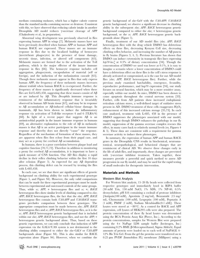
Figure S1 Western blot analysis of elav; APP; BACEheterozygous fly heads. Fly treatments are indicated in figure
for each lane. A) Detection of human APP in elav; APP; BACE
heterozygous fly heads by A8717 anti-APP antibody (Sigma). Lane
1 shows cellular lysates from HEK293 cells stably expressing APP
as a positive control. B) Detection of human CTFs in elav; APP;
BACE heterozygous fly heads by A8717 anti-APP antibody
(Sigma). Note that c-secretase inhibitor, L-685,458 increases
CTF levels. Lane 1 shows cellular lysates from HEK293 cells
stably expressing APP as a positive control. C) Detection of human
Ab by 6E10 (Covance) elav; APP; BACE heterozygous fly head
lysates. D) Detection of BACE (Abcam) in elav; APP; BACE
heterozygote fly head lysates. Lane 1 shows cellular lysates from
HEK293 cells stably expressing APP-Sw as a positive control.
Note no BACE immunoreactivity was observed in elav/w; +; + fly
head lysates, while BACE immunoreactivity was observed in elav;
APP; BACE heterozygousfly head lysates. E) Detection of human
BACE in elav; APP; BACE heterozygous fly heads. Lane 1 shows
cellular lysates from HEK293 cells stably expressing BACE as a
positive control. F) Quantification of panels A, B, C, and E
Western blot signal intensity.
(TIF)
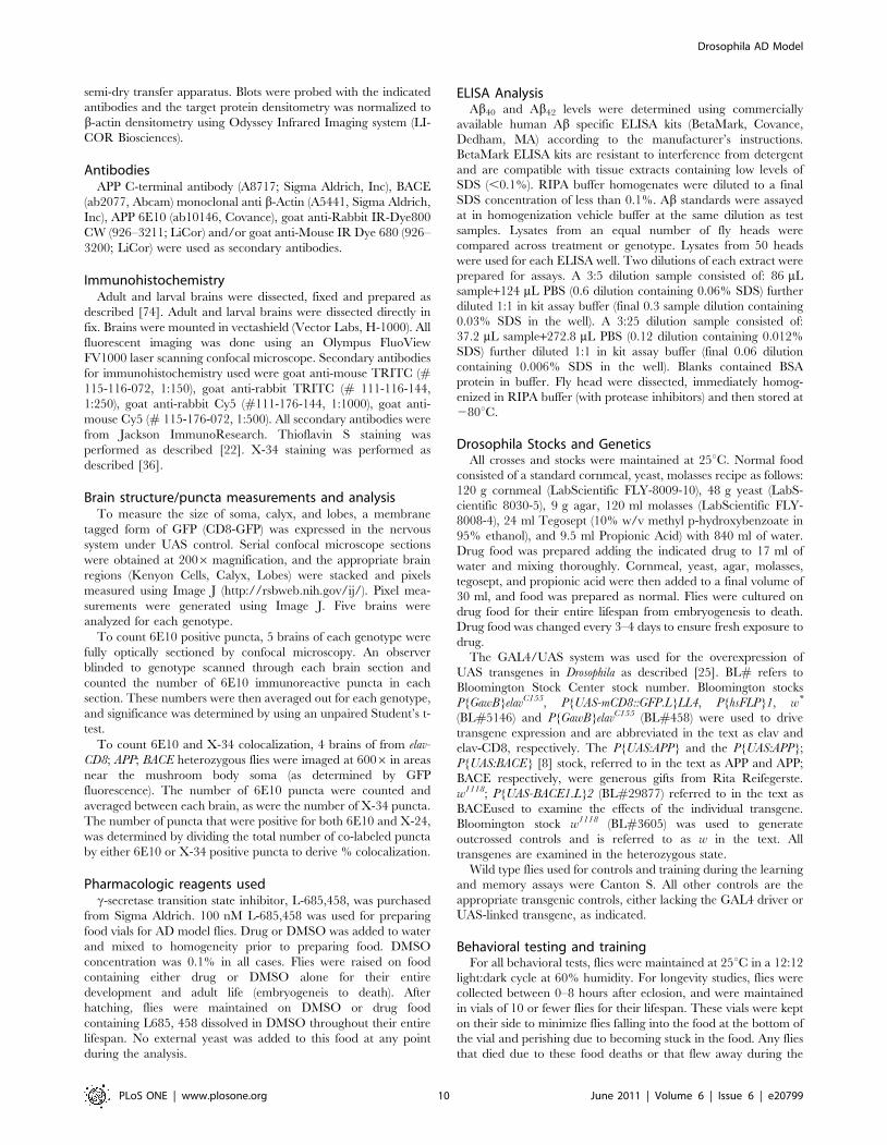
Figure S2 Longevity and mortality analysis. In each
panel, longevity analysis is top chart and mortality chart is
bottom panel. A) Longevity and mortality analysis of flies
expressing human APP and human BACE (elav; APP; BACE
heterozygous flies) compared to genetic background controls that
either lack the driver (w; APP; BACE heterozygous flies) or UAS
transgene (elav; +; + heterozygous flies). B) Longevity and
mortality analysis of flies expressing human APP and human
BACE (elav; APP; BACE heterozygous flies) raised on food
containing DMSO (vehicle) or L-685, 458. C) Longevity and
mortality analysis of flies expressing human APP alone (elav; APP
heterozygous flies) compared to genetic background controls that
either lack the driver (w; APP: + heterozygous flies) or UAS
transgene (elav; +; + heterozygous flies). D) Longevity and
mortality analysis of flies expressing human BACE alone (elav;
BACE; + heterozygous flies) compared to genetic background
controls that either lack the driver (w; BACE; + heterozygous flies)
or UAS transgene (elav; +; + heterozygous flies).
(TIF)
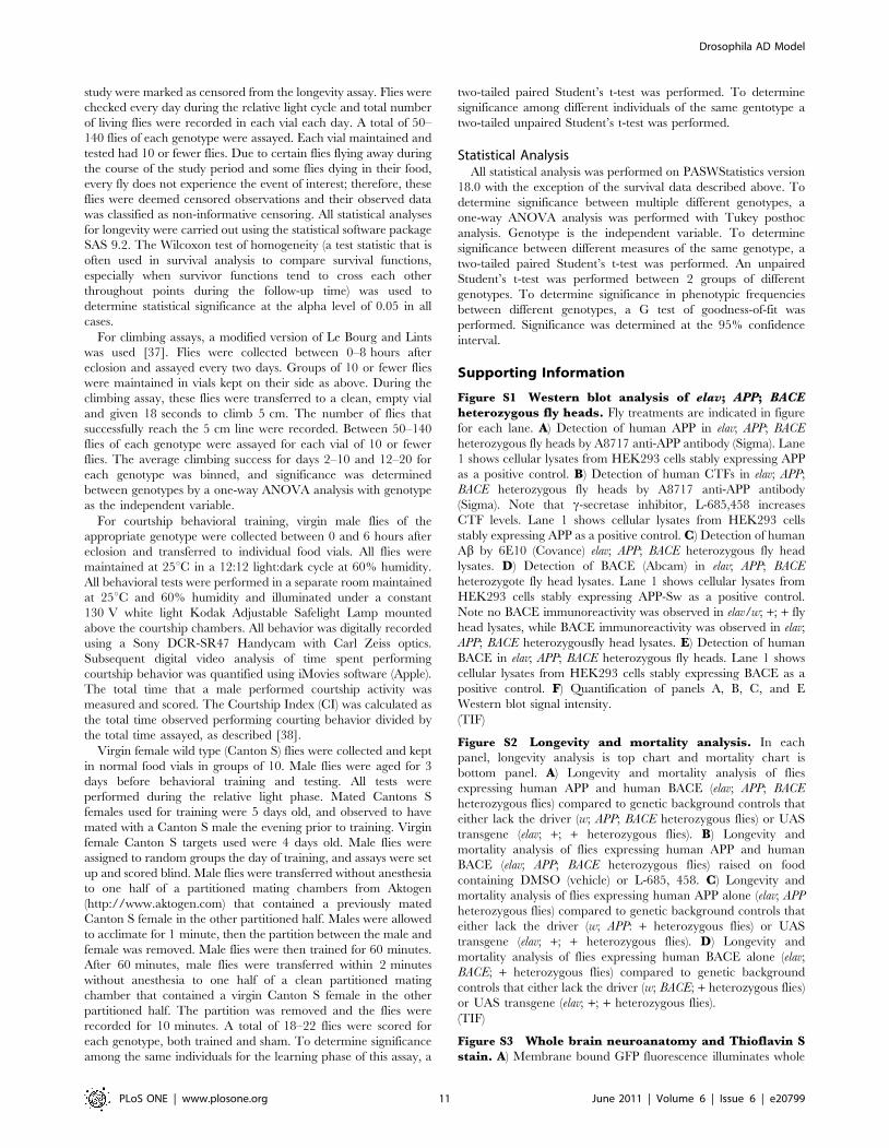
Figure S3 Whole brain neuroanatomy and Thioflavin Sstain. A) Membrane bound GFP fluorescence illuminates whole
Drosophila AD Model
PLoS ONE | www.plosone.org 11 June 2011 | Volume 6 | Issue 6 | e20799
brain morphology in elav-CD8-GFP; +; + heterozygous fly brain
from fly six days after eclosion. B) Dramatic changes in elav-CD8-
GFP; APP; BACE heterozygous brain morphology six days after
eclosion. C) High magnification (6006) of Thioflavin S positive
puncta in cortical region of elav; APP; BACE heterozygous fly brain.
Arrows indicate Thioflavin S positive puncta.
(TIF)
Figure S4 Motor reflex behavior. A) Expression of human
APP alone does not change motor reflex behavior, as measured by
the climbing assay. Parental strains listed. Error bars show
standard error. No significant difference exists between elav; APP;
+ heterozygous flies and elav; +; + heterozygous flies in days 2–10
(ANOVA, p = .601) or in days 12–20 (p = .677). B) Expression of
human BACE alone does not change motor reflex behavior (elav;
BACE; + heterozygous flies). No significant difference was found
between elav; BACE; + heterozygous flies and w; BACE; +
heterozygous flies in days 2–10 (ANOVA, p = .106) or in days
12–20 (p = .066). Error bars represent standard error.
(TIF)
Acknowledgments
We would like to thank Rita Reifegerste for the UAS:APP and UAS:APP;
UAS:BACE flies, the Bloomington Stock Center for various stocks, William
Klunk for X-34, and the Iowa Developmental Studies Hybridoma Bank for
antibodies. We would also like to thank all the members of the Saunders
and Marenda laboratories for their helpful discussions and comments.
Author Contributions
Conceived and designed the experiments: RC VV SDM BEP LEG JCL
AJS DRM. Performed the experiments: RC VV SDM BEP SJM RD AD
SM MV SA SU PJK DJM RDM DRM. Analyzed the data: RC VV SDM
BEP RDM JCL LPT AJS DRM. Wrote the paper: RC VV BEP AJS
DRM.
References
1. Alzheimer Association U (2010) 2010 Alzheimer’s disease facts and figures.
Alzheimers Dement 6: 158–194.
2. Nelson PT, Braak H, Markesbery WR (2009) Neuropathology and cognitiveimpairment in Alzheimer disease: a complex but coherent relationship.
J Neuropathol Exp Neurol 68: 1–14.
3. LaFerla FM, Oddo S (2005) Alzheimer’s disease: Abeta, tau and synaptic
dysfunction. Trends Mol Med 11: 170–176.
4. Hardy J, Selkoe DJ (2002) The amyloid hypothesis of Alzheimer’s disease:
progress and problems on the road to therapeutics. Science 297: 353–356.
5. De Strooper B, Annaert W (2000) Proteolytic processing and cell biological
functions of the amyloid precursor protein. J Cell Sci 113(Pt 11): 1857–1870.
6. Tanzi RE, Bertram L (2005) Twenty Years of the Alzheimer s Disease Amyloid
Hypothesis: A Genetic Perspective 120: 545–555.
7. Fossgreen A, Bruckner B, Czech C, Masters CL, Beyreuther K, et al. (1998)Transgenic Drosophila expressing human amyloid precursor protein show
gamma-secretase activity and a blistered-wing phenotype. Proc Natl Acad
Sci U S A 95: 13703–13708.
8. Greeve I, Kretzschmar D, Tschape JA, Beyn A, Brellinger C, et al. (2004) Age-dependent neurodegeneration and Alzheimer-amyloid plaque formation in
transgenic Drosophila. J Neurosci 24: 3899–3906.
9. Iijima K, Liu HP, Chiang AS, Hearn SA, Konsolaki M, et al. (2004) Dissecting
the pathological effects of human Abeta40 and Abeta42 in Drosophila: a
potential model for Alzheimer’s disease. Proc Natl Acad Sci U S A 101:6623–6628.
10. Jeibmann A, Paulus W (2009) Drosophila melanogaster as a Model Organism of
Brain Diseases. Int J Mol Sci 10: 407–440.
11. Sarantseva S, Timoshenko S, Bolshakova O, Karaseva E, Rodin D, et al. (2009)
Apolipoprotein E-mimetics inhibit neurodegeneration and restore cognitivefunctions in a transgenic Drosophila model of Alzheimer’s disease. PLoS One 4:
e8191.
12. Rosen DR, Martin-Morris L, Luo LQ, White K (1989) A Drosophila gene
encoding a protein resembling the human beta-amyloid protein precursor. Proc
Natl Acad Sci U S A 86: 2478–2482.
13. Allinson TM, Parkin ET, Turner AJ, Hooper NM (2003) ADAMs familymembers as amyloid precursor protein alpha-secretases. J Neurosci Res 74:
342–352.
14. Rooke J, Pan D, Xu T, Rubin GM (1996) KUZ, a conserved metalloprotease-
disintegrin protein with two roles in Drosophila neurogenesis. Nature 273:1227–1231.
15. Hong CS, Koo EH (1997) Isolation and characterization of Drosophilapresenilin homolog. Neuroreport 8: 665–668.
16. Chung HM, Struhl G (2001) Nicastrin is required for Presenilin-mediatedtransmembrane cleavage in Drosophila. Nat Cell Biol 3: 1129–1132.
17. Boulianne GL, Livne-Bar I, Humphreys JM, Liang Y, Lin C, et al. (1997)Cloning and characterization of the Drosophila presenilin homologue.
Neuroreport 8: 1025–1029.
18. Francis R, McGrath G, Zhang J, Ruddy DA, Sym M, et al. (2002) aph-1 and
pen-2 are required for Notch pathway signaling, gamma-secretase cleavage ofbetaAPP, and presenilin protein accumulation. Dev Cell 3: 85–97.
19. Carmine-Simmen K, Proctor T, Tschape J, Poeck B, Triphan T, et al. (2009)
Neurotoxic effects induced by the Drosophila amyloid-beta peptide suggest a
conserved toxic function. Neurobiol Dis 33: 274–281.
20. Finelli A, Kelkar A, Song HJ, Yang H, Konsolaki M (2004) A model for studyingAlzheimer’s Abeta42-induced toxicity in Drosophila melanogaster. Mol Cell
Neurosci 26: 365–375.
21. Crowther DC, Kinghorn KJ, Miranda E, Page R, Curry JA, et al. (2005)
Intraneuronal Abeta, non-amyloid aggregates and neurodegeneration in a
Drosophila model of Alzheimer’s disease. Neuroscience 132: 123–135.
22. Iijima K, Chiang HC, Hearn SA, Hakker I, Gatt A, et al. (2008) Abeta42
mutants with different aggregation profiles induce distinct pathologies in
Drosophila. PLoS One 3: e1703.
23. Iijima-Ando K, Hearn SA, Granger L, Shenton C, Gatt A, et al. (2008)
Overexpression of neprilysin reduces alzheimer amyloid-beta42 (Abeta42)-
induced neuron loss and intraneuronal Abeta42 deposits but causes a reduction
in cAMP-responsive element-binding protein-mediated transcription, age-
dependent axon pathology, and premature death in Drosophila. J Biol Chem
283: 19066–19076.
24. Luheshi LM, Tartaglia GG, Brorsson AC, Pawar AP, Watson IE, et al. (2007)
Systematic in vivo analysis of the intrinsic determinants of amyloid Beta
pathogenicity. PLoS Biol 5: e290.
25. Brand AH, Perrimon N (1993) Targeted gene expression as a means of altering
cell fates and generating dominant phenotypes. Development 118: 401–
415.
26. Yao KM, White K (1994) Neural specificity of elav expression: defining a
Drosophila promoter for directing expression to the nervous system.
J Neurochem 63: 41–51.
27. Heisenberg M, Borst A, Wagner S, Byers D (1985) Drosophila mushroom body
mutants are deficient in olfactory learning. J Neurogenet 2: 1–30.
28. Connolly JB, Roberts IJ, Armstrong JD, Kaiser K, Forte M, et al. (1996)
Associative learning disrupted by impaired Gs signaling in Drosophila
mushroom bodies. Science 274: 2104–2107.
29. McBride SM, Giuliani G, Choi C, Krause P, Correale D, et al. (1999)
Mushroom body ablation impairs short-term memory and long-term memory of
courtship conditioning in Drosophila melanogaster. Neuron 24: 967–977.
30. Dubnau J, Tully T (2001) Functional anatomy: from molecule to memory. Curr
Biol 11: R240–243.
31. Zars T, Fischer M, Schulz R, Heisenberg M (2000) Localization of a short-term
memory in Drosophila. Science 288: 672–675.
32. Lee T, Lee A, Luo L (1999) Development of the Drosophila mushroom bodies:
sequential generation of three distinct types of neurons from a neuroblast.
Development 126: 4065–4076.
33. Hanslick JL, Lau K, Noguchi KK, Olney JW, Zorumski CF, et al. (2009)
Dimethyl sulfoxide (DMSO) produces widespread apoptosis in the developing
central nervous system. Neurobiol Dis 34: 1–10.
34. Nazir A, Mukhopadhyay I, Saxena DK, Chowdhuri DK (2003) Evaluation of
the No Observed Adverse Effect Level of Solvent Dimethyl Sulfoxide in
Drosophila melanogaster. Toxicol Mech Methods 13: 147–152.
35. Gibson GE, Huang HM (2004) Mitochondrial enzymes and endoplasmic
reticulum calcium stores as targets of oxidative stress in neurodegenerative
diseases. J Bioenerg Biomembr 36: 335–340.
36. Ikonomovic MD, Abrahamson EE, Isanski BA, Debnath ML, Mathis CA, et al.
(2006) X-34 labeling of abnormal protein aggregates during the progression of
Alzheimer’s disease. Methods Enzymol 412: 123–144.
37. Le Bourg E, Lints FA (1992) Hypergravity and aging in Drosophila
melanogaster. 6. Spontaneous locomotor activity. Gerontology 38: 71–79.
38. Siegel RW, Hall JC (1979) Conditioned responses in courtship behavior of
normal and mutant Drosophila. Proc Natl Acad Sci U S A 76: 3430–3434.
39. Broughton SJ, Tully T, Greenspan RJ (2003) Conditioning deficits of CaM-
kinase transgenic Drosophila melanogaster in a new excitatory courtship assay.
J Neurogenet 17: 91–102.
40. Siwicki KK, Riccio P, Ladewski L, Marcillac F, Dartevelle L, et al. (2005) The
role of cuticular pheromones in courtship conditioning of Drosophila males.
Learn Mem 12: 636–645.
41. Kane NS, Robichon A, Dickinson JA, Greenspan RJ (1997) Learning without
performance in PKC-deficient Drosophila. Neuron 18: 307–314.
Drosophila AD Model
PLoS ONE | www.plosone.org 12 June 2011 | Volume 6 | Issue 6 | e20799
42. Joiner Ml A, Griffith LC (1997) CaM kinase II and visual input modulate
memory formation in the neuronal circuit controlling courtship conditioning.J Neurosci 17: 9384–9391.
43. Kamyshev NG, Iliadi KG, Bragina JV (1999) Drosophila conditioned courtship:
two ways of testing memory. Learn Mem 6: 1–20.44. McBride SM, Choi CH, Wang Y, Liebelt D, Braunstein E, et al. (2005)
Pharmacological rescue of synaptic plasticity, courtship behavior, and mush-room body defects in a Drosophila model of fragile X syndrome. Neuron 45:
753–764.
45. Greenspan RJ (1995) Flies, genes, learning, and memory. Neuron 15: 747–750.46. Bonner JM, Boulianne GL (2010) Drosophila as a model to study age-related
neurodegenerative disorders: Alzheimer’s disease. Exp Gerontol.47. Leyssen M, Ayaz D, Hebert SS, Reeve S, De Strooper B, et al. (2005) Amyloid
precursor protein promotes post-developmental neurite arborization in theDrosophila brain. EMBO J 24: 2944–2955.
48. Luo L, Tully T, White K (1992) Human amyloid precursor protein ameliorates
behavioral deficit of flies deleted for Appl gene. Neuron 9: 595–605.49. Heidary G, Fortini ME (2001) Identification and characterization of the
Drosophila tau homolog. Mech Dev 108: 171–178.50. Yu G, Nishimura M, Arawaka S, Levitan D, Zhang L, et al. (2000) Nicastrin
modulates presenilin-mediated notch/glp-1 signal transduction and betaAPP
processing. Nature 407: 48–54.51. Littleton JT, Bellen HJ (1994) Genetic and phenotypic analysis of thirteen
essential genes in cytological interval 22F1-2; 23B1-2 reveals novel genesrequired for neural development in Drosophila. Genetics 138: 111–123.
52. Cao W, Song HJ, Gangi T, Kelkar A, Antani I, et al. (2008) Identification ofNovel Genes That Modify Phenotypes Induced by Alzheimer’s {beta}-Amyloid
Overexpression in Drosophila. Genetics 178: 1457–1471.
53. Chiang HC, Iijima K, Hakker I, Zhong Y (2009) Distinctive roles of differentbeta-amyloid 42 aggregates in modulation of synaptic functions. FASEB J 23:
1969–1977.54. Iijima K, Gatt A, Iijima-Ando K (2010) Tau Ser262 phosphorylation is critical
for Abeta42-induced tau toxicity in a transgenic Drosophila model of
Alzheimer’s disease. Hum Mol Genet 19: 2947–2957.55. Iijima-Ando K, Hearn SA, Shenton C, Gatt A, Zhao L, et al. (2009)
Mitochondrial mislocalization underlies Abeta42-induced neuronal dysfunctionin a Drosophila model of Alzheimer’s disease. PLoS One 4: e8310.
56. Iijima-Ando K, Iijima K (2010) Transgenic Drosophila models of Alzheimer’sdisease and tauopathies. Brain Struct Funct 214: 245–262.
57. Folwell J, Cowan CM, Ubhi KK, Shiabh H, Newman TA, et al. (2010) A[beta]
exacerbates the neuronal dysfunction caused by human tau expression in aDrosophila model of Alzheimer’s disease. Experimental Neurology 223:
401–409.58. Wittmann CW, Wszolek MF, Shulman JM, Salvaterra PM, Lewis J, et al. (2001)
Tauopathy in Drosophila: Neurodegeneration Without Neurofibrillary Tangles.
Science 293: 711–714.
59. Stokin GB, Almenar-Queralt A, Gunawardena S, Rodrigues EM, Falzone T,
et al. (2008) Amyloid precursor protein-induced axonopathies are independent
of amyloid-beta peptides. Hum Mol Genet 17: 3474–3486.
60. Robinow S, White K (1988) The locus elav of Drosophila melanogaster is expressed
in neurons at all developmental stages. Dev Biol 126: 294–303.
61. Berger C, Renner S, Luer K, Technau GM (2007) The commonly used marker
ELAV is transiently expressed in neuroblasts and glial cells in the Drosophila
embryonic CNS. Dev Dyn 236: 3562–3568.
62. Rumble B, Retallack R, Hilbich C, Simms G, Multhaup G, et al. (1989)
Amyloid A4 protein and its precursor in Down’s syndrome and Alzheimer’s
disease. N Engl J Med 320: 1446–1452.
63. Podlisny MB, Lee G, Selkoe DJ (1987) Gene dosage of the amyloid beta
precursor protein in Alzheimer’s disease. Science 238: 669–671.
64. Rovelet-Lecrux A, Hannequin D, Raux G, Le Meur N, Laquerriere A, et al.
(2006) APP locus duplication causes autosomal dominant early-onset Alzheimer
disease with cerebral amyloid angiopathy. Nat Genet 38: 24–26.
65. Minakhina S, Steward R (2006) Melanotic mutants in Drosophila: pathways and
phenotypes. Genetics 174: 253–263.
66. Lemaitre B, Meister M, Govind S, Georgel P, Steward R, et al. (1995)
Functional analysis and regulation of nuclear import of dorsal during the
immune response in Drosophila. EMBO J 14: 536–545.
67. McGeer EG, McGeer PL (1998) The importance of inflammatory mechanisms
in Alzheimer disease. Exp Gerontol 33: 371–378.
68. Stewart CR, Stuart LM, Wilkinson K, van Gils JM, Deng J, et al. (2010) CD36
ligands promote sterile inflammation through assembly of a Toll-like receptor 4
and 6 heterodimer. Nat Immunol 11: 155–161.
69. Soscia SJ, Kirby JE, Washicosky KJ, Tucker SM, Ingelsson M, et al. (2010) The
Alzheimer’s disease-associated amyloid beta-protein is an antimicrobial peptide.
PLoS One 5: e9505.
70. Green MS, Kaye JA, Ball MJ (2000) The Oregon brain aging study:
neuropathology accompanying healthy aging in the oldest old. Neurology 54:
105–113.
71. Crystal HA, Dickson DW, Sliwinski MJ, Lipton RB, Grober E, et al. (1993)
Pathological markers associated with normal aging and dementia in the elderly.
Ann Neurol 34: 566–573.
72. Morris JC, Storandt M, McKeel DW, Jr., Rubin EH, Price JL, et al. (1996)
Cerebral amyloid deposition and diffuse plaques in ‘‘normal’’ aging: Evidence
for presymptomatic and very mild Alzheimer’s disease. Neurology 46: 707–719.
73. Gibson GE, Zhang H, Xu H, Park LC, Jeitner TM (2002) Oxidative stress
increases internal calcium stores and reduces a key mitochondrial enzyme.
Biochim Biophys Acta 1586: 177–189.
74. Tio M, Moses K (1997) The Drosophila TGF alpha homolog Spitz acts in
photoreceptor recruitment in the developing retina. Development 124: 343–351.
Drosophila AD Model
PLoS ONE | www.plosone.org 13 June 2011 | Volume 6 | Issue 6 | e20799


![Drosophila Vps13 Is Required for Protein Homeostasis in ... · Results Characterization of Drosophila Vps13 mutant flies ChAc is caused by mutations in the VPS13A gene [3,4], which](https://img.pdfslide.net/doc/110x75/5ccc3ff188c99335448bc7de/drosophila-vps13-is-required-for-protein-homeostasis-in-results-characterization.jpg)