Embed Size (px)
Citation preview
Glycolysis Glycolytic endpoints -‐ depending on which cell and conditions, glucose metabolism results in the production of ethanol, lactate and CO2, H2O via pyruvate
• This is the predominate fate of glucose in mammalian cells • Limited potential energy w/o O2 . Captured as ATP • Pathway is found in all cells of the body • This is primarily a cytosolic pathway
Net reaction: Glucose + 2 ADP +2 Pi + 2 NAD+ <-‐> 2 Pyruvate + 2 ATP + 2 NADH The overall metabolism can be considered to be in two stages. In the first stage there is a cost of energy (in the form of ATP). Glucose is metabolized to two trisoses -‐ 3 phosphoglycerate. In essence the carbohydrate is activated and prepared for energy production by phosphorylation. The second stage results in the partial oxidation to pyruvate and the formation of energy currency (NADH and ATP).
Reactions and Thermodynamics of Glycolysis
There are a few items you need to keep in mind when looking at metabolism.
• The structure of the reactants and products • Where did the carbons come from. Be able to trace the carbons from glucose through each step of glycolysis • What are the important points for each enzyme… regulation, reaction mechanism, isozymes … • The overall control of glycolysis and the fate of the metabolism
– This is more than just memorizing the pathway but you do need to know that too. PATHWAY FIVE TIMES EVERYTHING – that is right! He said EVERYTHING. Darn it!!!!
Reactions of Glycolysis Metabolism Glucokinase / Hexokinase -‐ (GK/HK)
Key glycolytic enzyme -1st step in glycolysis; DG large, negative -Nucleophilic attack from 6’OH on g phosphate requires Mg+2
-Hexokinase (and glucokinase) act to phosphorylate glucose and keep Glc in the cell -1 ATP is consumed -‐ is considered an irreversable step. -Also a key glycolytic step -GK is NOT inhibited by G6P but HK IS
Hexokinase
Glucose-‐6-‐P is common to several metabolic pathways
Glucokinase / Hexokinase -‐ (GK/HK) - Mg+2 is a common cofactor for all kinases. -‐ the metal forms a complex with
the a and b phosphates and allows the g phosphate to be reactive. EDTA is often used as an inhibitor or kinase reactions – why?
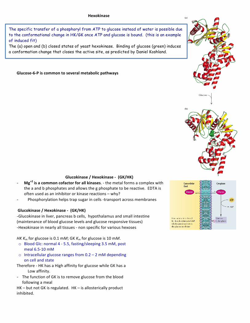
- Phosphorylation helps trap sugar in cells -‐transport across membranes Glucokinase / Hexokinase -‐ (GK/HK) -Glucokinase in liver, pancreas b cells, hypothalamus and small intestine (maintenance of blood glucose levels and glucose responsive tissues) -Hexokinase in nearly all tissues -‐ non specific for various hexoses HK Km for glucose is 0.1 mM; GK Km for glucose is 10 mM. o Blood Glc: normal 4 -‐ 5.5, fasting/sleeping 3.5 mM, post
meal 6.5-‐10 mM o Intracellular glucose ranges from 0.2 – 2 mM depending
on cell and state Therefore -‐ HK has a High affinity for glucose while GK has a
Low affinity. - The function of GK is to remove glucose from the blood
following a meal HK – but not GK is regulated. HK – is allosterically product inhibited.
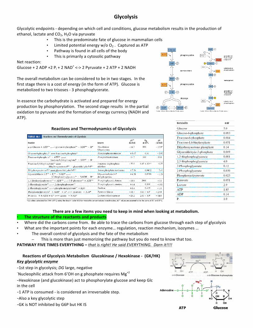
The specific transfer of a phosphoryl from ATP to glucose instead of water is possible due to the conformational change in HK/GK once ATP and glucose is bound. (this is an example of induced fit) The (a) open and (b) closed states of yeast hexokinase. Binding of glucose (green) induces a conformation change that closes the active site, as predicted by Daniel Koshland.
Concept Question Two isozymes bind and react with a common substrate (duh, they are isozymes) but each have a different Km.
– CASE ONE: Both isoforms are found in the same compartment of epithelial cells. Isoform 1 has a higher Km (25 mM substrate) than isoform 2 (5 mM substrate). • Which enzyme will be bind and convert the substrate to product when the substrate concentration is 2.5
mM, 10 mM or 50 mM?
Concept Question II Two isozymes bind and react with a common substrate (duh, they are isozymes) but each have a different Km. Isoform 1 has a higher Km (25 mM) than isoform 2 (5 mM).
– CASE TWO: Each enzyme is found in a different tissue. Enz 1 – tissue 1 and Enz 2 – tissue 2. Both tissues get their supply of substrate from interstitial fluid. Transport of substrate is not a problem or regulated for either tissue.
• Which tissue will be constantly supplied with product when the blood concentration is 5 mM? • If the concentration of substrate in blood spikes to 50 mM, which tissue is responsible for reducing the
blood substrate concentration. • If the preferred bld substrate concentration is 10 mM, which tissue will receive a constant flow of product
and which tissue is a regulator to maintain the blood substrate concentration
Phosphoglucose Isomerase (PGI) -Isomerase reaction -Phosphohexose isomerase = phosphoglucose isomerase -The reaction is a controlled sequence of -‐>open structure/ reaction / close ring -Acid/ base catalyst mechanism -Fructose 6-‐P produced -‐ no energy input
Phosphoglucoisomerase -‐ Glucose-‐6-‐P to Fructose-‐6-‐P Why does this reaction occur?
– next step (phosphorylation at C-‐1) would be tough for hemiacetal -‐OH, but easy for primary -‐OH – isomerization activates C-‐3 for cleavage in aldolase reaction – Ene-‐diol intermediate in this reaction
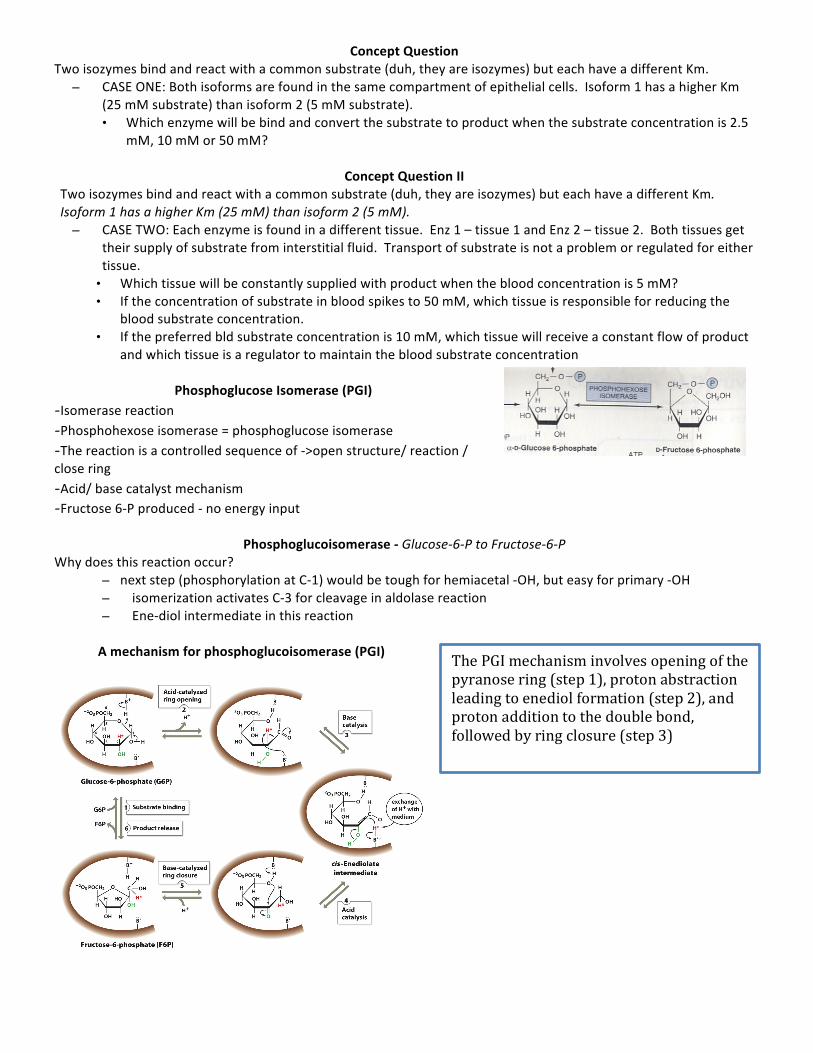
A mechanism for phosphoglucoisomerase (PGI)
The PGI mechanism involves opening of the pyranose ring (step 1), proton abstraction leading to enediol formation (step 2), and proton addition to the double bond, followed by ring closure (step 3)
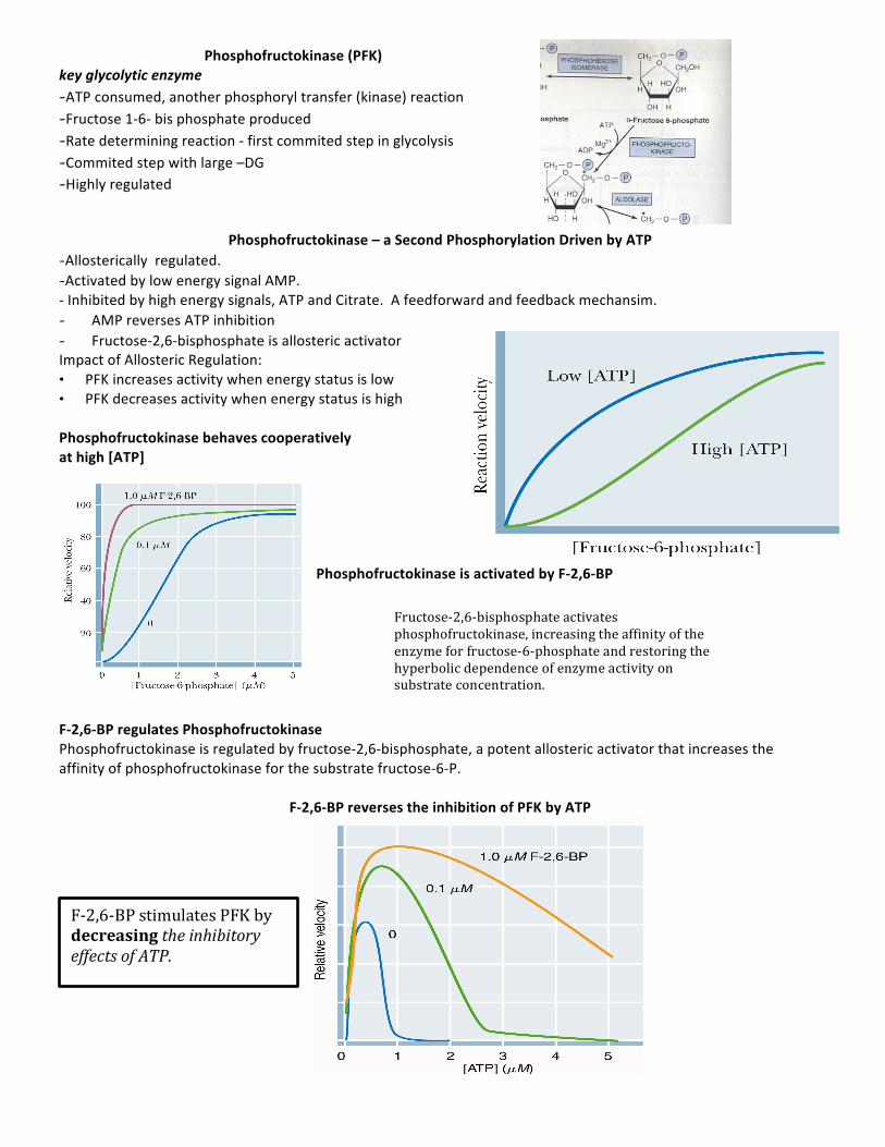
Phosphofructokinase (PFK) key glycolytic enzyme -ATP consumed, another phosphoryl transfer (kinase) reaction -Fructose 1-‐6-‐ bis phosphate produced -Rate determining reaction -‐ first commited step in glycolysis -Commited step with large –DG -Highly regulated
Phosphofructokinase – a Second Phosphorylation Driven by ATP -Allosterically regulated. -Activated by low energy signal AMP. -‐ Inhibited by high energy signals, ATP and Citrate. A feedforward and feedback mechansim. - AMP reverses ATP inhibition - Fructose-‐2,6-‐bisphosphate is allosteric activator Impact of Allosteric Regulation: • PFK increases activity when energy status is low • PFK decreases activity when energy status is high
Phosphofructokinase behaves cooperatively at high [ATP]
Phosphofructokinase is activated by F-‐2,6-‐BP
F-‐2,6-‐BP regulates Phosphofructokinase Phosphofructokinase is regulated by fructose-‐2,6-‐bisphosphate, a potent allosteric activator that increases the affinity of phosphofructokinase for the substrate fructose-‐6-‐P.
F-‐2,6-‐BP reverses the inhibition of PFK by ATP
Fructose-‐2,6-‐bisphosphate activates phosphofructokinase, increasing the affinity of the enzyme for fructose-‐6-‐phosphate and restoring the hyperbolic dependence of enzyme activity on substrate concentration.
F-‐2,6-‐BP stimulates PFK by decreasing the inhibitory effects of ATP.
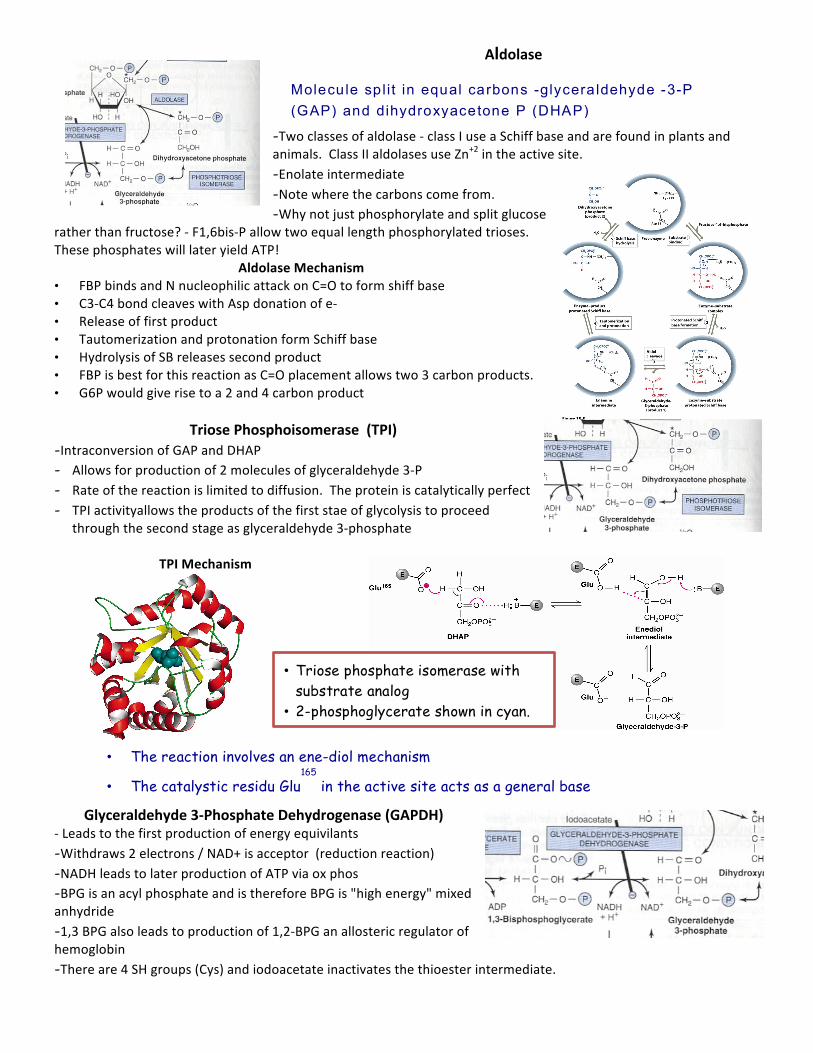
Aldolase
-Two classes of aldolase -‐ class I use a Schiff base and are found in plants and animals. Class II aldolases use Zn+2 in the active site. -Enolate intermediate -Note where the carbons come from. -Why not just phosphorylate and split glucose
rather than fructose? -‐ F1,6bis-‐P allow two equal length phosphorylated trioses. These phosphates will later yield ATP!
Aldolase Mechanism • FBP binds and N nucleophilic attack on C=O to form shiff base • C3-‐C4 bond cleaves with Asp donation of e-‐ • Release of first product • Tautomerization and protonation form Schiff base • Hydrolysis of SB releases second product • FBP is best for this reaction as C=O placement allows two 3 carbon products. • G6P would give rise to a 2 and 4 carbon product
Triose Phosphoisomerase (TPI)
-Intraconversion of GAP and DHAP - Allows for production of 2 molecules of glyceraldehyde 3-‐P - Rate of the reaction is limited to diffusion. The protein is catalytically perfect - TPI activityallows the products of the first stae of glycolysis to proceed
through the second stage as glyceraldehyde 3-‐phosphate
TPI Mechanism
Glyceraldehyde 3-‐Phosphate Dehydrogenase (GAPDH) -‐ Leads to the first production of energy equivilants -Withdraws 2 electrons / NAD+ is acceptor (reduction reaction) -NADH leads to later production of ATP via ox phos -BPG is an acyl phosphate and is therefore BPG is "high energy" mixed anhydride -1,3 BPG also leads to production of 1,2-‐BPG an allosteric regulator of hemoglobin -There are 4 SH groups (Cys) and iodoacetate inactivates the thioester intermediate.
Molecule sp l i t in equal carbons -glycera ldehyde -3-P (GAP) and dihydroxyacetone P (DHAP)
• The reaction involves an ene-diol mechanism
• The catalystic residu Glu165
in the active site acts as a general base
• Triose phosphate isomerase with substrate analog
• 2-phosphoglycerate shown in cyan.
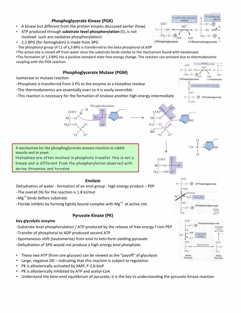
Phosphoglycerate Kinase (PGK)
• A kinase but different from the protein kinases discussed earlier (how) • ATP produced through substrate level phosphorylation (O2 is not
involved such ans oxidative phosphorylation) • 2,3 BPG (for hemoglobin) is made from 3PG -‐ The phosphoryl group of C1 of 1,3 BPG is transferred to the beta phosphoryl of ADP •The active site is closed off from water once the substrate binds similar to the mechanism found with hexokinase •The formation of 1,3 BPG has a positive standard state free energy change. The reaction can proceed due to thermodynamic coupling with the PGK reaction
Phosphoglycerate Mutase (PGM) Isomerase or mutase reaction -Phosphate is transferred from 3 PG to the enzyme at a histadine residue -The thermodynamics are essentially even so it is easily reversible -This reaction is necessary for the formation of enolase another high energy intermediate
Enolase
Dehydration of water -‐ formation of an enol group -‐ high energy product – PEP -The overall DG for the reaction is 1.8 kJ/mol -Mg+2 binds before substrate -Floride inhibits by forming tightly bound complex with Mg+2 at active site
Pyruvate Kinase (PK)
key glycolytic enzyme -Substrate level phosphorylation / ATP produced by the release of free energy f rom PEP -Transfer of phosphoryl to ADP produced second ATP -Spontaneous shift (tautomerize) from enol to keto form yielding pyruvate -Dehydration of 3PG would not produce a high energy enol phosphate. • These two ATP (from one glucose) can be viewed as the "payoff" of glycolysis • Large, negative DG – indicating that this reaction is subject to regulation • PK is allosterically activated by AMP, F-‐1,6-‐bisP • PK is allosterically inhibited by ATP and acetyl-‐CoA • Understand the keto-‐enol equilibrium of pyruvate; it is the key to understanding the pyruvate kinase reaction
A mechanism for the phosphoglycerate mutase reaction in rabbit muscle and in yeast. Histadines are often involved in phosphate transfer this is not a kinase and is different from the phosphorylation observed with serine threonine and tyrosine
Pyruvate Kinase Regulation Two different isozymes: L (liver) and M (muscle) Both are tetramers. §PK is allosterically activated by thesubstrate PEP §Feed forward activation by F 16 BP §ATP and alanine inhibits §Liver form is phosphorylated at C terminal by PKA §The resulting phosphorylation shifts the enzyme from tetrameter (active) to monomer (inactive) form - This type of regulation is only observed in liver -‐
why only in the liver and why is that important? Think in terms of energy needs during fight or flight. Muscle want ATP and liver can use other forms of energy sparing glucose for other tissues.
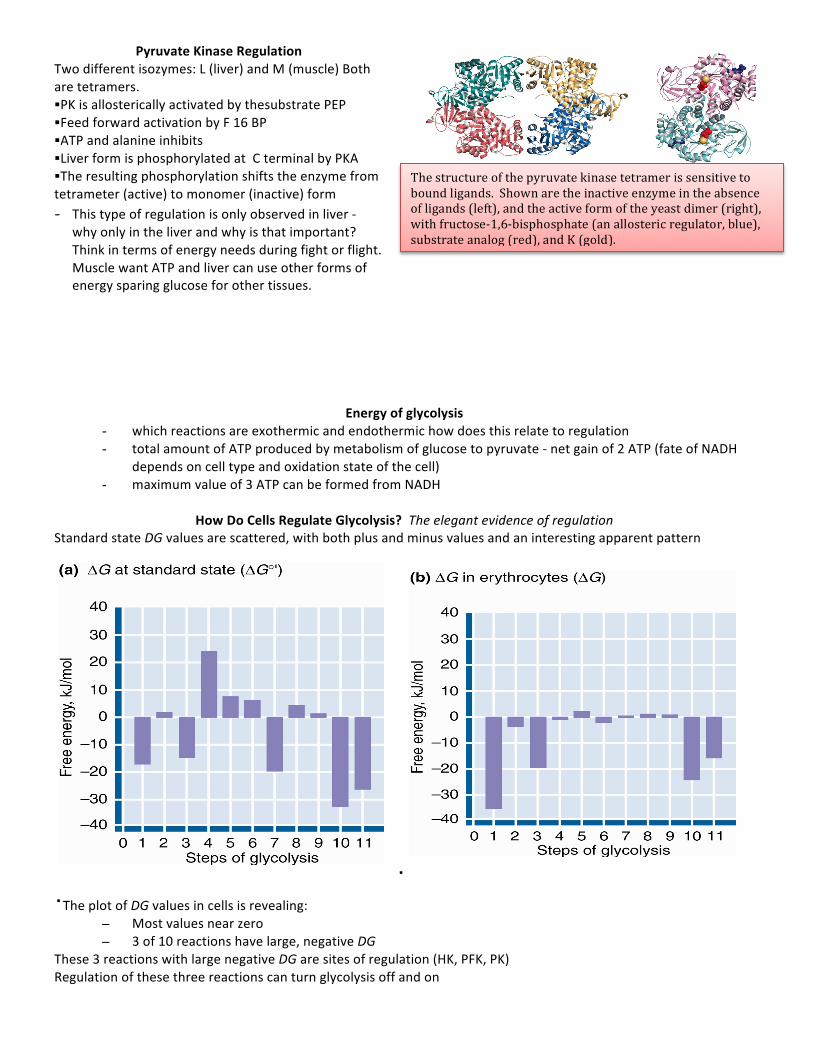
Energy of glycolysis - which reactions are exothermic and endothermic how does this relate to regulation - total amount of ATP produced by metabolism of glucose to pyruvate -‐ net gain of 2 ATP (fate of NADH
depends on cell type and oxidation state of the cell) - maximum value of 3 ATP can be formed from NADH
How Do Cells Regulate Glycolysis? The elegant evidence of regulation
Standard state DG values are scattered, with both plus and minus values and an interesting apparent pattern
· ·The plot of DG values in cells is revealing:
– Most values near zero – 3 of 10 reactions have large, negative DG
These 3 reactions with large negative DG are sites of regulation (HK, PFK, PK) Regulation of these three reactions can turn glycolysis off and on
The structure of the pyruvate kinase tetramer is sensitive to bound ligands. Shown are the inactive enzyme in the absence of ligands (left), and the active form of the yeast dimer (right), with fructose-‐1,6-‐bisphosphate (an allosteric regulator, blue), substrate analog (red), and K (gold).
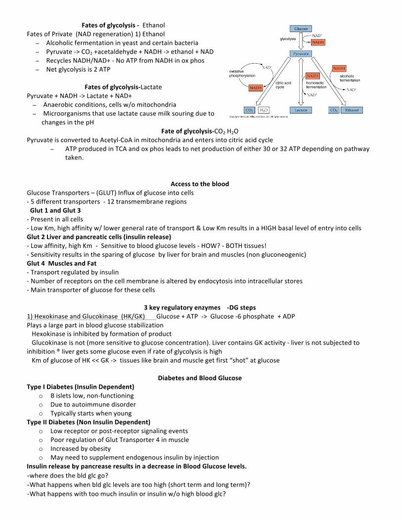
Fates of glycolysis -‐ Ethanol Fates of Private (NAD regeneration) 1) Ethanol
– Alcoholic fermentation in yeast and certain bacteria – Pyruvate -‐> CO2 +acetaldehyde + NADH -‐> ethanol + NAD – Recycles NADH/NAD+ -‐ No ATP from NADH in ox phos – Net glycolysis is 2 ATP
Fates of glycolysis-‐Lactate
Pyruvate + NADH -‐> Lactate + NAD+ – Anaerobic conditions, cells w/o mitochondria – Microorganisms that use lactate cause milk souring due to
changes in the pH Fate of glycolysis-‐CO2 H2O
Pyruvate is converted to Acetyl-‐CoA in mitochondria and enters into citric acid cycle – ATP produced in TCA and ox phos leads to net production of either 30 or 32 ATP depending on pathway
taken.
Access to the blood Glucose Transporters – (GLUT) Influx of glucose into cells -‐ 5 different transporters -‐ 12 transmembrane regions Glut 1 and Glut 3 -‐ Present in all cells -‐ Low Km, high affinity w/ lower general rate of transport & Low Km results in a HIGH basal level of entry into cells Glut 2 Liver and pancreatic cells (insulin release) -‐ Low affinity, high Km -‐ Sensitive to blood glucose levels -‐ HOW? -‐ BOTH tissues! -‐ Sensitivity results in the sparing of glucose by liver for brain and muscles (non gluconeogenic) Glut 4 Muscles and Fat -‐ Transport regulated by insulin -‐ Number of receptors on the cell membrane is altered by endocytosis into intracellular stores -‐ Main transporter of glucose for these cells
3 key regulatory enzymes -‐DG steps
1) Hexokinase and Glucokinase (HK/GK) Glucose + ATP -‐> Glucose -‐6 phosphate + ADP Plays a large part in blood glucose stabilization Hexokinase is inhibited by formation of product Glucokinase is not (more sensitive to glucose concentration). Liver contains GK activity -‐ liver is not subjected to inhibition ® liver gets some glucose even if rate of glycolysis is high Km of glucose of HK << GK -‐> tissues like brain and muscle get first “shot” at glucose
Diabetes and Blood Glucose Type I Diabetes (Insulin Dependent)
o B islets low, non-‐functioning o Due to autoimmune disorder o Typically starts when young
Type II Diabetes (Non Insulin Dependent) o Low receptor or post-‐receptor signaling events o Poor regulation of Glut Transporter 4 in muscle o Increased by obesity o May need to supplement endogenous insulin by injection
Insulin release by pancrease results in a decrease in Blood Glucose levels. -where does the bld glc go? -What happens when bld glc levels are too high (short term and long term)? -What happens with too much insulin or insulin w/o high blood glc?
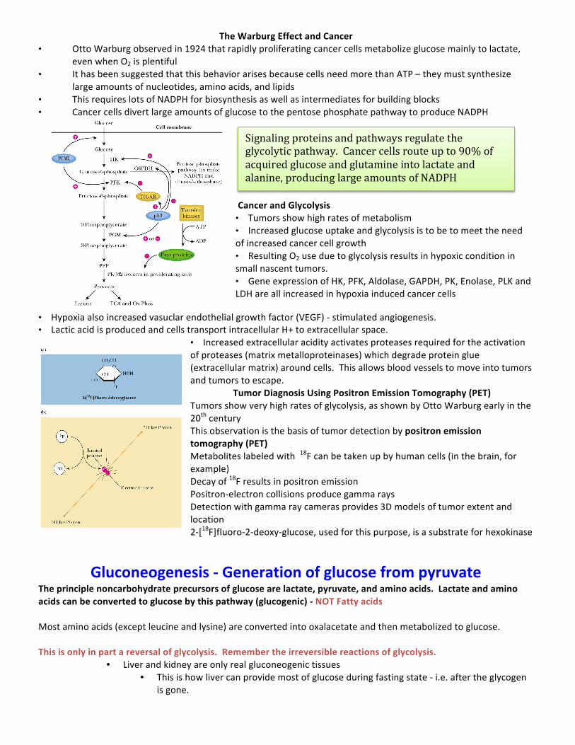
The Warburg Effect and Cancer • Otto Warburg observed in 1924 that rapidly proliferating cancer cells metabolize glucose mainly to lactate,
even when O2 is plentiful • It has been suggested that this behavior arises because cells need more than ATP – they must synthesize
large amounts of nucleotides, amino acids, and lipids • This requires lots of NADPH for biosynthesis as well as intermediates for building blocks • Cancer cells divert large amounts of glucose to the pentose phosphate pathway to produce NADPH
Cancer and Glycolysis • Tumors show high rates of metabolism • Increased glucose uptake and glycolysis is to be to meet the need of increased cancer cell growth • Resulting O2 use due to glycolysis results in hypoxic condition in small nascent tumors. • Gene expression of HK, PFK, Aldolase, GAPDH, PK, Enolase, PLK and LDH are all increased in hypoxia induced cancer cells
• Hypoxia also increased vasuclar endothelial growth factor (VEGF) -‐ stimulated angiogenesis. • Lactic acid is produced and cells transport intracellular H+ to extracellular space.
• Increased extracellular acidity activates proteases required for the activation of proteases (matrix metalloproteinases) which degrade protein glue (extracellular matrix) around cells. This allows blood vessels to move into tumors and tumors to escape.
Tumor Diagnosis Using Positron Emission Tomography (PET) Tumors show very high rates of glycolysis, as shown by Otto Warburg early in the 20th century This observation is the basis of tumor detection by positron emission tomography (PET) Metabolites labeled with 18F can be taken up by human cells (in the brain, for example) Decay of 18F results in positron emission Positron-‐electron collisions produce gamma rays Detection with gamma ray cameras provides 3D models of tumor extent and location 2-‐[18F]fluoro-‐2-‐deoxy-‐glucose, used for this purpose, is a substrate for hexokinase
Gluconeogenesis -‐ Generation of glucose from pyruvate The principle noncarbohydrate precursors of glucose are lactate, pyruvate, and amino acids. Lactate and amino acids can be converted to glucose by this pathway (glucogenic) -‐ NOT Fatty acids Most amino acids (except leucine and lysine) are converted into oxalacetate and then metabolized to glucose. This is only in part a reversal of glycolysis. Remember the irreversible reactions of glycolysis.
• Liver and kidney are only real gluconeogenic tissues • This is how liver can provide most of glucose during fasting state -‐ i.e. after the glycogen
is gone.
Signaling proteins and pathways regulate the glycolytic pathway. Cancer cells route up to 90% of acquired glucose and glutamine into lactate and alanine, producing large amounts of NADPH
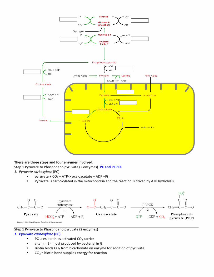
There are three steps and four enzymes involved. Step 1 Pyruvate to Phosphoenolpyruvate (2 enzymes) PC and PEPCK 1. Pyruvate carboxylase (PC)
• pyruvate + CO2 + ATP-‐> oxaloacetate + ADP +Pi • Pyruvate is carboxylated in the mitochondria and the reaction is driven by ATP hydrolysis
Step 1 Pyruvate to Phosphoenolpyruvate (2 enzymes) 1. Pyruvate carboxylase (PC)
• PC uses biotin as activated CO2 carrier • vitamin B -‐ most produced by bacterial in GI • Biotin binds CO2 from bicarbonate on enzyme for addition of pyruvate • CO2 ~ biotin bond supplies energy for reaction
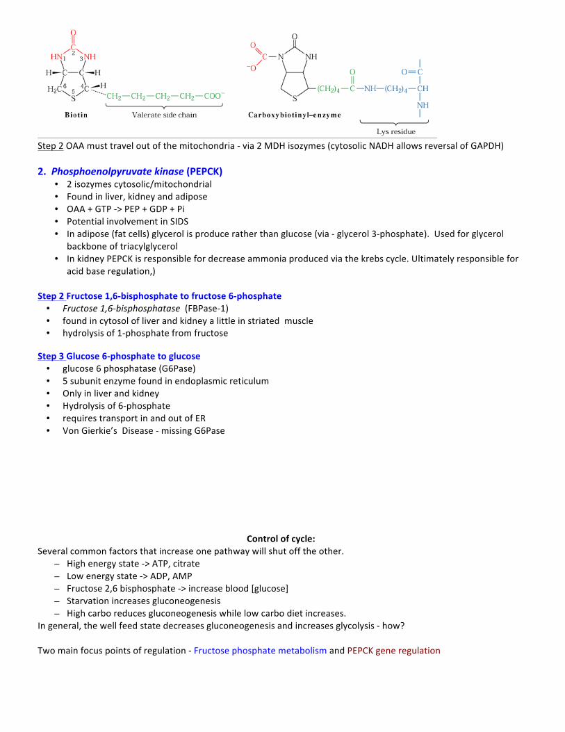
Step 2 OAA must travel out of the mitochondria -‐ via 2 MDH isozymes (cytosolic NADH allows reversal of GAPDH) 2. Phosphoenolpyruvate kinase (PEPCK)
• 2 isozymes cytosolic/mitochondrial • Found in liver, kidney and adipose • OAA + GTP -‐> PEP + GDP + Pi • Potential involvement in SIDS • In adipose (fat cells) glycerol is produce rather than glucose (via -‐ glycerol 3-‐phosphate). Used for glycerol
backbone of triacylglycerol • In kidney PEPCK is responsible for decrease ammonia produced via the krebs cycle. Ultimately responsible for
acid base regulation,) Step 2 Fructose 1,6-‐bisphosphate to fructose 6-‐phosphate • Fructose 1,6-‐bisphosphatase (FBPase-‐1) • found in cytosol of liver and kidney a little in striated muscle • hydrolysis of 1-‐phosphate from fructose
Step 3 Glucose 6-‐phosphate to glucose • glucose 6 phosphatase (G6Pase) • 5 subunit enzyme found in endoplasmic reticulum • Only in liver and kidney • Hydrolysis of 6-‐phosphate • requires transport in and out of ER • Von Gierkie’s Disease -‐ missing G6Pase
Control of cycle: Several common factors that increase one pathway will shut off the other.
– High energy state -‐> ATP, citrate – Low energy state -‐> ADP, AMP – Fructose 2,6 bisphosphate -‐> increase blood [glucose] – Starvation increases gluconeogenesis – High carbo reduces gluconeogenesis while low carbo diet increases.
In general, the well feed state decreases gluconeogenesis and increases glycolysis -‐ how? Two main focus points of regulation -‐ Fructose phosphate metabolism and PEPCK gene regulation
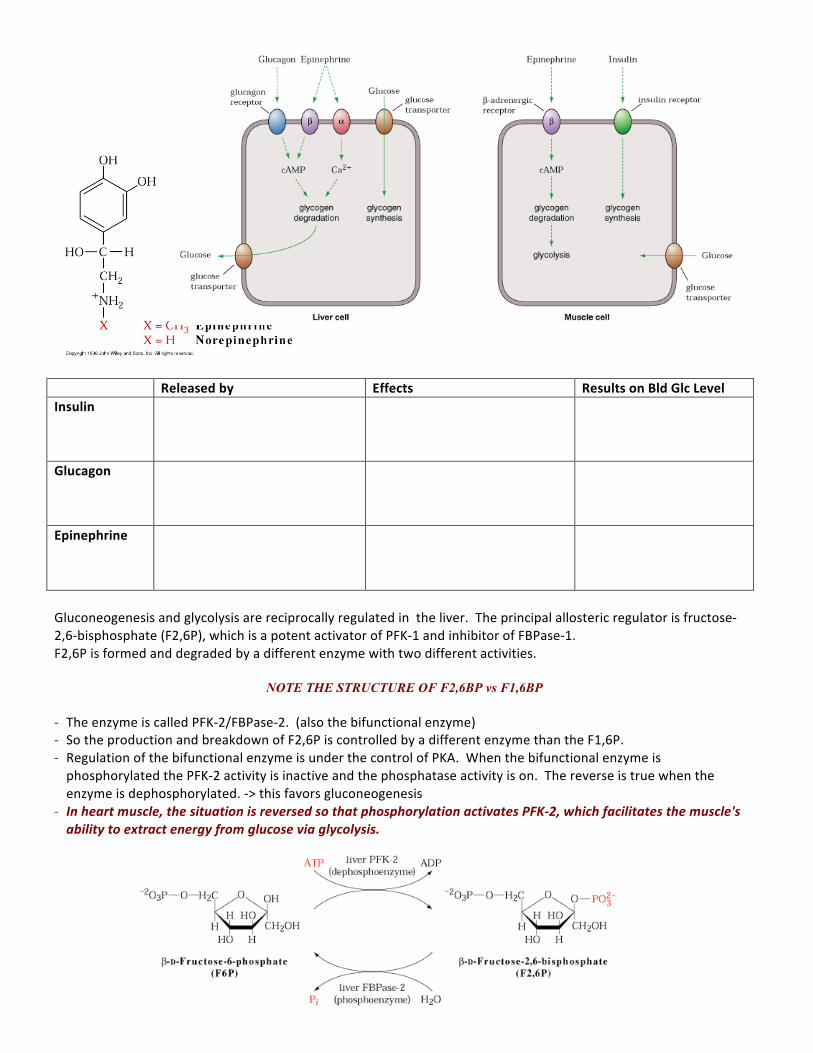
Released by Effects Results on Bld Glc Level Insulin
Glucagon
Epinephrine
Gluconeogenesis and glycolysis are reciprocally regulated in the liver. The principal allosteric regulator is fructose-‐2,6-‐bisphosphate (F2,6P), which is a potent activator of PFK-‐1 and inhibitor of FBPase-‐1. F2,6P is formed and degraded by a different enzyme with two different activities.
NOTE THE STRUCTURE OF F2,6BP vs F1,6BP - The enzyme is called PFK-‐2/FBPase-‐2. (also the bifunctional enzyme) - So the production and breakdown of F2,6P is controlled by a different enzyme than the F1,6P. - Regulation of the bifunctional enzyme is under the control of PKA. When the bifunctional enzyme is phosphorylated the PFK-‐2 activity is inactive and the phosphatase activity is on. The reverse is true when the enzyme is dephosphorylated. -‐> this favors gluconeogenesis
- In heart muscle, the situation is reversed so that phosphorylation activates PFK-‐2, which facilitates the muscle's ability to extract energy from glucose via glycolysis.
Transcriptional control of PEPCK gene promoter region of gene -‐ ultimately controls protein levels - PEPCK is quickly degraded - several hormones can effect the concentration of PEPCK in cell through the promoter including insulin, cAMP.
glucorticoids, thyroid hormones - this all leads to an increase in gluconeogenesis w/long term starvation