Embed Size (px)
Citation preview
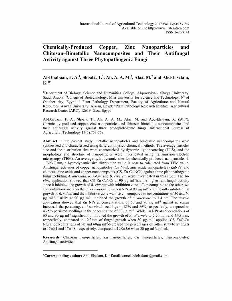
International Journal of Agricultural Technology 2017 Vol. 13(5):753-769 Available online http://www.ijat-aatsea.com
ISSN 1686-9141
Chemically-Produced Copper, Zinc Nanoparticles and Chitosan–Bimetallic Nanocomposites and Their Antifungal Activity against Three Phytopathogenic Fungi Al-Dhabaan, F. A.1, Shoala, T.2, Ali, A. A. M.3, Alaa, M.2 and Abd-Elsalam, K.4*
1Department of Biology, Science and Humanities College, Alquwayiyah, Shaqra University, Saudi Arabia; 2College of Biotechnology, Misr University for Science and Technology, 6th of October city, Egypt; 3 Plant Pathology Department, Faculty of Agriculture and Natural Resources, Aswan University, Aswan, Egypt; 4Plant Pathology Research Institute, Agricultural Research Center (ARC), 12619, Giza, Egypt. Al-Dhabaan, F. A., Shoala, T., Ali, A. A. M., Alaa, M. and Abd-Elsalam, K. (2017). Chemically-produced copper, zinc nanoparticles and chitosan–bimetallic nanocomposites and their antifungal activity against three phytopathogenic fungi. International Journal of Agricultural Technology 13(5):753-769. Abstract In the present study, metallic nanoparticles and bimetallic nanocompsites were synthesized and characterized using different physico-chemical methods. The average particles size and the distribution size were characterized by dynamic light scattering (DLS), and the morphology and structure of nanoparticles were investigated using transmission electron microscopy (TEM). An average hydrodynamic size for chemically-produced nanoparticles is 1.7-23.7 nm, a hydrodynamic size distribution value is near to calculated from TEM value. Antifungal activities of copper nanoparticles (Cu NPs), zinc oxide nanoparticles (ZnNPs) and chitosan, zinc oxide and copper nanocompsites (CS–Zn-Cu NCs) against three plant pathogenic fungi including A. alternata, R. solani and B. cinerea, were investigated in this study. The In-vitro application showed that CS–Zn-CuNCs at 90 µg ml-1has the highest antifungal activity since it inhibited the growth of B. cinerea with inhibition zone 1.7cm compared to the other two concentrations and also the other nanoparticles. Zn NPs at 90 µg ml-1 significantly inhibited the growth of R. solani and the inhibition zone was 1.6 cm compared to concentrations of 30 and 60 µg ml-1. CuNPs at 90 µg ml-1 inhibited the growth of A. alternate to 1.4 cm. The in-vivo application showed that Zn NPs at concentrations of 60 and 90 µg ml-1 against R. solani increased the percentages of survived seedlings to 85% and 86%, respectively, compared to 43.5% persisted seedlings in the concentration of 30 µg ml-1. While Cu NPs at concentrations of 60 and 90 µg ml-1 significantly inhibited the growth of A. alternate to 5.20 mm and 4.95 mm, respectively, compared to 12.3mm of fungal growth when 30 µg ml-1 applied. CS–ZnO-Cu NCsat concentrations of 90 and 60µg ml-1decreased the percentages of rotten strawberry fruits to 15±6.1 and 17±4.8, respectively, compared to19.0±5.6 when 30 µg ml-1applied. Keywords: Chitosan nanoparticles, Zn nanoparticles, Cu nanoparticles, nancomposites, Antifungal activities
*Corresponding author: Abd-Elsalam, K.; Email:[email protected]
754
Introduction
Fungal pathogens are the main cause of significant economic loss during the growth of the crop and postharvest handling of fruits. R. solani, A. alternata and Botrytis cinerea can cause severe damage for number of economic crops either in the field or postharvest fruit diseases even when the most advanced preharvest and postharvest technologies are applied (Spadaro et al., 2004). Many recent research studies have already demonstrated antimicrobial activities of various nanoparticles such as silver (Kim et al., 2008; Kumar et al., 2008), copper (Cioffi et al., 2005, Youssef et al., 2017), chitosan (Xing et al., 2011) and zinc oxide (Liu et al., 2009). Copper nanoparticles had a high activity against Gram-positive bacteria, standard and clinical strains, including methicillin-resistant Staphylococcus aureus, comparable to silver nanoparticles and some antibiotics (Kruk et al., 2015). They also exhibited antifungal activity against Candida species. Also, the foliar applications of CuO NPs limit growth of the oomycete Phytopthora (Giannousi et al., 2013). Zinc oxide (ZnO) and magnesium oxide (MgO) nanoparticles at different concentrations brought about significant inhibition in the germination of spores of A. alternata, F. oxysporum, R. stolonifer and Mucor plumbeus (Wani and Shah, 2012). Zinc oxide (ZnO) and magnesium oxide (MgO) nanoparticles have been proposed as an anti-microbial preservative for wood or food products (Aruoja et al., 2009; Huang et al., 2006; Sharma et al., 2009). ZnO NPs toward plant pathogenic fungi including Penicillium expansum and B. cinerea was demonstrated in recent papers (He et al., 2011). Also, it demonstrated against Aspergillus isolate (Jayaseelan et al. 2012). Dimkpa et al., 2013 reported that ZnO NPs are toxic to the wheat pathogen, F. graminearum both in medium and in a solid sand matrix. The combined copper-chitosan colloids are used as a new generation of copper-based bio-pesticides (Brunel et al., 2013). Chitosan-based copper nanomaterials have been used as antifungal, antibacterial as well as plant growth promoting agents (Hardy et al., 2004; Jaiswal et al., 2012, Saharan et al., 2013 and 2015). The aim of this work was to synthesize and characterize three types of nano-agrochemicals, also to determine theantifungal their activities against three plant pathogenic fungi A. alternata, R. solani and B. cinerea. Material and methods Chemicals
Copper sulfate (Purity 98%, M. wt. 249.68) was purchased from El-Nasr Pharmaceutical Co. for Chemicals (Egypt). Polyethylene glycol 6000
International Journal of Agricultural Technology 2017 Vol. 13(5):753-769
755
(H(OCH2CH2)nOH- Sigma-Aldrich) was used as the capping agent. Sodium borohydride (NaBH4, Sigma-Aldrich) was used as the main reducing agent, while ascorbic acid (C6H8O6, Sigma-Aldrich) was used as the antioxidant of colloidal copper. Sodium hydroxide (NaOH, Sigma-Aldrich) was used to adjust the pH. Preparation of Chitosan nanoparticles
Chitosan (0.25g) was dissolved in 2% acetic acid, while stirring followed by sonication for 15 min. This was then filtered to obtain a clear solution. 20% Na2SO4 was then added drop wise to the chitosan solution, which was simultaneously stirred and sonicated. The solution was left undisturbed, centrifuged at 8,000 rpm to collect the CS nanoparticles. Synthesis of Cu-based NPs
CuNPs were synthesized by reduction of copper sulphate with isopropyl alcohol (IPA) in the presence of the cationic surfactant CTAB following Athawale et al., (2005) technique with slight modification. Copper (II) sulfate nanoparticles were synthesis according to the method of Kanhed et al. (2014) with minor modification. Firstly, 0.0030 M copper (II) sulfate and 0.09 M of CTAB were prepared in IPA. Reaction was carried out in clean dry 250 mL Erlenmeyer flasks open to air. Copper (II) sulfate was added drop-wise to CTAB/ (IPA) solution. The reaction mixture was stirred vigorously on a magnetic stir plate. IPA was used as a reducing agent in the synthesis of CuNPs. CTAB molecules catalyze the reduction of Cu2+ ions to Cu0 with IPA. CTAB acts as a capping agent by surrounding the surface of CuNPs. Synthesis of ZnO Nanoparticle
ZnO nanoparticle was prepared by conventional precipitation method with NaOH as precipitation agent. 0.4mg NaOH was added drop wise continually to aqueous 5.75mg Zinc sulphate solution in the molar ratio 1:2 with continuous stirring. The resulting slurry was continuously stirred for 24 h and precipitate obtained was filtered, washed thoroughly with distilled H2O. The washing procedure was repeated several times and then collected residue was dried in oven at 100ºC for 12h grinned to fine powder.
756
Synthesis of Chitosan–bimetallic nanocomposites
For the synthesis of CS–Zn-Cu nanocomposite, 0.5g of chitosan was dissolved in 2% acetic acid. 20% Na2SO4 solution was added drop wise to the above solution under simultaneous stirring and sonication. 0.1g of synthesized ZnO and CuO nanoparticles were added to the solution which was stirred under sonication. The suspension was centrifuged and washed several times. Finally, the nanocomposite was collected and dried at 60 °C. Characterization of NPs
Dynamic light scattering (DLS) Measurement of Cu NPs distribution and size was performed by a dynamic light scattering method using Zetasizer Nano ZS (Malvern Instruments, UK) at room temperature. Prior to measurement, 30µl of the nanoparticle was diluted with 3ml of water at 25°C. Particle size data were expressed as the mean of the Z-average of 3 independent batches of the nanoparticles.
Transmission electron microscopy (TEM)
Twenty microliters of diluted samples was placed on a film-coated 200-mesh copper specimen grid for 10 min and the fluid excess was eliminated using filter paper. The grid was then stained with one drop of 3% phosphor tungstic acid (PTA) and allowed to dry for 3 min. The coated grid was dried and examined under the TEM microscope (Philips, CM 12). Then the samples were observed by operating at 120kV. The effect of three types of NPs in vitro against R. solani, A. alternata and B. cinerea
The effect of three types of NPs on the mycelial growth R. solani, A. alternata and B. cinerea was evaluated. Agar plugs (5mm in diam.) containing mycelia from the growing edge of 1-week-old cultures of R. solani, A. alternata and B. cinerea were placed in the center of Petri dishes containing PDA. Three different concentrations (30, 60 and 90µg mL-1) were used, three Petri dishes were utilized as replicates and the entire experiment was repeated twice. The plates were incubated at 23±1 °C for one week. Colony diameter (mm) for each fungus was calculated as the average of the longest and the shortest diameter. Non-amended PDA served as a control while copper sulfate was used as raw material for comparison.
International Journal of Agricultural Technology 2017 Vol. 13(5):753-769
757
Fungal pathogen suspension
To produce fungal inoculums for A. alternata or B. cinerea, isolates were grown on PDA plates at 24°C in the dark. Conidia were collected from two week-old plates by scraping them with a sterile spatula, and then suspended in sterile distilled water containing 0.05% Tween 80 (v/v). The resulting suspension was filtered through two layers of sterile gauze. The spore counts were made by a Thoma counting chamber (HGB Henneberg-Sander GmbH, Lutzellinden, Germany) and the suspension was diluted with sterile distilled water to obtain a final concentration of 104 conidia ml-1. Indirect antifungal activity of Cu NPs and Cu-Zn-Chitosan NPs
Tomato or strawberry fruits were wounded once (5 mm depth × 3 mm wide) with a sterile nail-head along the equatorial axis. For each treatment, 30 µl of copper nanoparticles solution were applied into each wound. Treated fruit were placed in a tray and individually wrapped into a plastic bag. After 48 hour of incubation at 20±1°C and high relative humidity, another wound was made approximately 5 mm apart from the previous one. This wound was air-dried and inoculated with 10 µl of a 104 conidia ml-1 suspension of A. alternata or B. cinerea (Youssef et al., 2014). The control consisted of fruits treated in the first wound with sterile distilled water and then inoculated with the pathogen conidial suspension in the other one. Each treatment was replicated 3 times and each replicate consisted of 5 fruits. Replicates were individually wrapped in a plastic bag and incubated at 20 ±1°C and high relative humidity for two weeks. The percentage of infected fruit (rot incidence, %) and decay lesions diameter (mm) was recorded for each fungal pathogen. Effect of seed dressing treatment with ZnNPs on R. solani in greenhouse
Fifty seeds were surface sterilized by gently shaking in 1% sodium hypchlorite solution for 3 min and rinsed six times for 5 min in sterile deionized water. Seeds were soaked in 30, 60, and 90µg for about 30 min. Infested soil was dispensed in 15 cm diameter clay pots and each pot was planted with 10 seeds. Pots were distributed on greenhouse benches in a randomized complete block design. The greenhouse was equipped with a heating system assuring that the minimum temperature in the greenhouse was maintained at 28°C. The test was repeated once with almost the same results. Percentage of infected seedlings was recorded 45 days from sowing.
758
Fragmentation of fungal genomic DNA
Fungal mycelium was produced in 20ml of liquid medium (24gl-1 of potato dextrose broth [PDB, Difco Laboratories]. Mycelium was harvested by filtration through mesh sieves (40 µm), washed with sterile water, and deposited onto Whatman paper no. 1 to remove excess water. Mycelium was ground to a fine powder in liquid nitrogen using a mortar and DNA was extracted by the method of Moslem et al., (2010). Five micrograms of fungal DNA for A. alternate, R. solani and B. cinerea was treated with NPs and NCs (60µg ml-1). Seven microliters of the isolated DNA and 3 µl of 10X loading dye were loaded in a lane of 1.5% (w/v) agarose gel containing 0.05 µgml-1 ethidium bromide, to check the quality of the DNA. For quantitative measurements, a charge-coupled device camera imaging system and UVIsoft analysis (Gel Documentation and Analysis Systems, Uvitec, Cambridge, UK) were used to capture the image. Results Nanomaterials Characterization
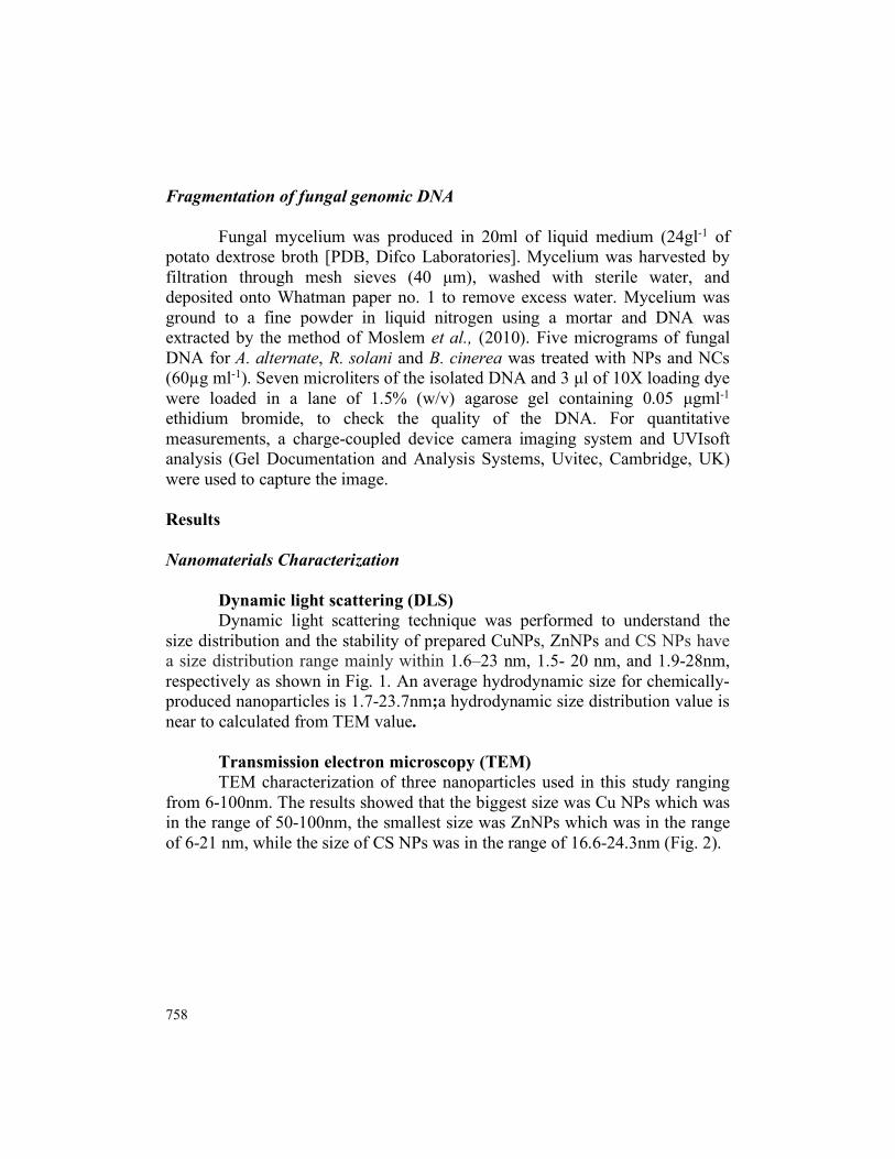
Dynamic light scattering (DLS) Dynamic light scattering technique was performed to understand the
size distribution and the stability of prepared CuNPs, ZnNPs and CS NPs have a size distribution range mainly within 1.6–23 nm, 1.5- 20 nm, and 1.9-28nm, respectively as shown in Fig. 1. An average hydrodynamic size for chemically-produced nanoparticles is 1.7-23.7nm;a hydrodynamic size distribution value is near to calculated from TEM value.
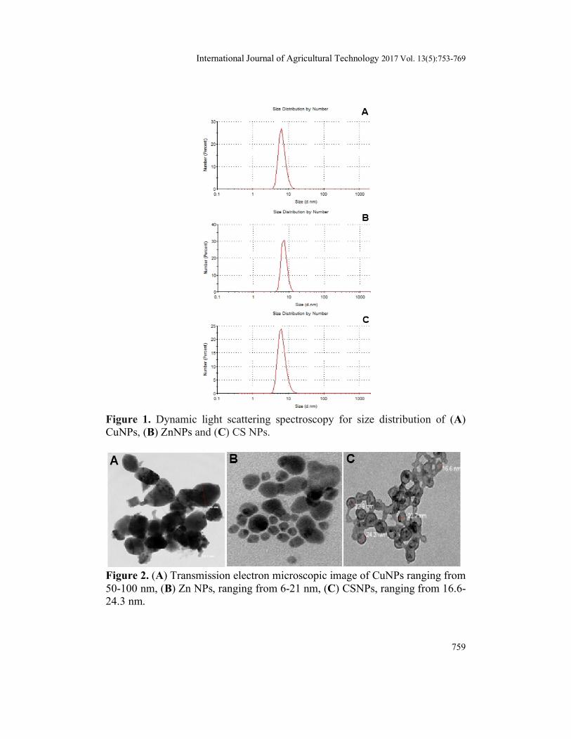
Transmission electron microscopy (TEM) TEM characterization of three nanoparticles used in this study ranging
from 6-100nm. The results showed that the biggest size was Cu NPs which was in the range of 50-100nm, the smallest size was ZnNPs which was in the range of 6-21 nm, while the size of CS NPs was in the range of 16.6-24.3nm (Fig. 2).
International Journal of Agricultural Technology 2017 Vol. 13(5):753-769
759
Figure 1. Dynamic light scattering spectroscopy for size distribution of (A) CuNPs, (B) ZnNPs and (C) CS NPs.
Figure 2. (A) Transmission electron microscopic image of CuNPs ranging from 50-100 nm, (B) Zn NPs, ranging from 6-21 nm, (C) CSNPs, ranging from 16.6-24.3 nm.
760
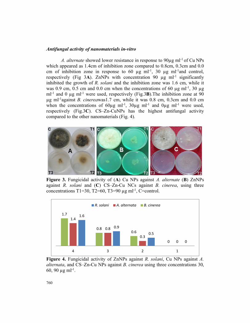
Antifungal activity of nanomaterials in-vitro A. alternate showed lower resistance in response to 90µg ml-1 of Cu NPs
which appeared as 1.4cm of inhibition zone compared to 0.8cm, 0.3cm and 0.0 cm of inhibition zone in response to 60 µg ml-1, 30 µg ml-1and control, respectively (Fig 3A). ZnNPs with concentration 90 µg ml-1 significantly inhibited the growth of R. solani and the inhibition zone was 1.6 cm, while it was 0.9 cm, 0.5 cm and 0.0 cm when the concentrations of 60 µg ml-1, 30 µg ml-1 and 0 µg ml-1 were used, respectively (Fig.3B).The inhibition zone at 90 µg ml-1against B. cinereawas1.7 cm, while it was 0.8 cm, 0.3cm and 0.0 cm when the concentrations of 60µg ml-1, 30µg ml-1 and 0µg ml-1 were used, respectively (Fig.3C). CS–Zn-CuNPs has the highest antifungal activity compared to the other nanomaterials (Fig. 4).
Figure 3. Fungicidal activity of (A) Cu NPs against A. alternate (B) ZnNPs against R. solani and (C) CS–Zn-Cu NCs against B. cinerea, using three concentrations T1=30, T2=60, T3=90 µg ml-1, C=control.
Figure 4. Fungicidal activity of ZnNPs against R. solani, Cu NPs against A. alternata, and CS–Zn-Cu NPs against B. cinerea using three concentrations 30, 60, 90 µg ml-1.
0
0.50.9
1.6
00.3
0.8
1.4
0
0.60.8
1.7
1234
R. solani A. alternata B. cinerea
International Journal of Agricultural Technology 2017 Vol. 13(5):753-769
761
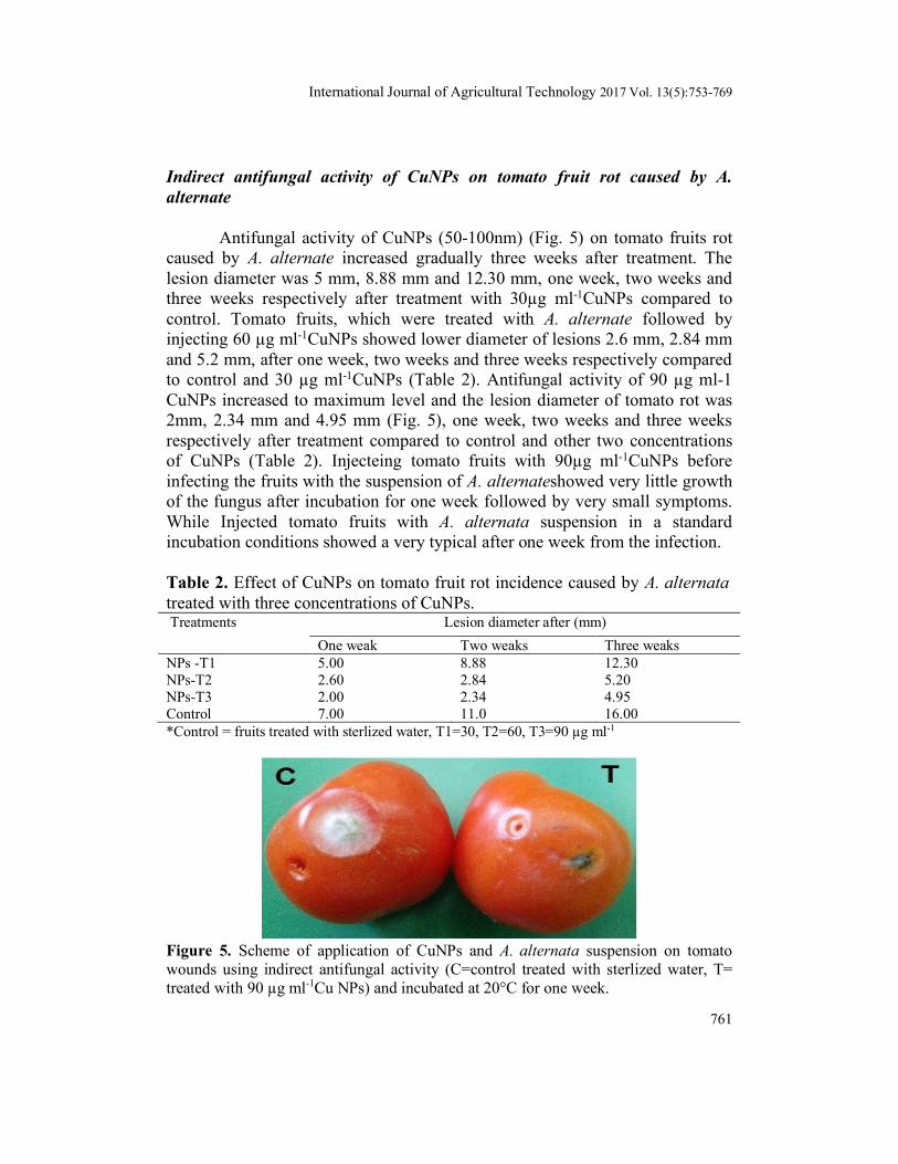
Indirect antifungal activity of CuNPs on tomato fruit rot caused by A. alternate
Antifungal activity of CuNPs (50-100nm) (Fig. 5) on tomato fruits rot caused by A. alternate increased gradually three weeks after treatment. The lesion diameter was 5 mm, 8.88 mm and 12.30 mm, one week, two weeks and three weeks respectively after treatment with 30µg ml-1CuNPs compared to control. Tomato fruits, which were treated with A. alternate followed by injecting 60 µg ml-1CuNPs showed lower diameter of lesions 2.6 mm, 2.84 mm and 5.2 mm, after one week, two weeks and three weeks respectively compared to control and 30 µg ml-1CuNPs (Table 2). Antifungal activity of 90 µg ml-1 CuNPs increased to maximum level and the lesion diameter of tomato rot was 2mm, 2.34 mm and 4.95 mm (Fig. 5), one week, two weeks and three weeks respectively after treatment compared to control and other two concentrations of CuNPs (Table 2). Injecteing tomato fruits with 90µg ml-1CuNPs before infecting the fruits with the suspension of A. alternateshowed very little growth of the fungus after incubation for one week followed by very small symptoms. While Injected tomato fruits with A. alternata suspension in a standard incubation conditions showed a very typical after one week from the infection. Table 2. Effect of CuNPs on tomato fruit rot incidence caused by A. alternata treated with three concentrations of CuNPs. Treatments Lesion diameter after (mm)
One weak Two weaks Three weaks NPs -T1 5.00 8.88 12.30 NPs-T2 2.60 2.84 5.20 NPs-T3 2.00 2.34 4.95 Control 7.00 11.0 16.00 *Control = fruits treated with sterlized water, T1=30, T2=60, T3=90 µg ml-1
Figure 5. Scheme of application of CuNPs and A. alternata suspension on tomato wounds using indirect antifungal activity (C=control treated with sterlized water, T= treated with 90 µg ml-1Cu NPs) and incubated at 20°C for one week.
762
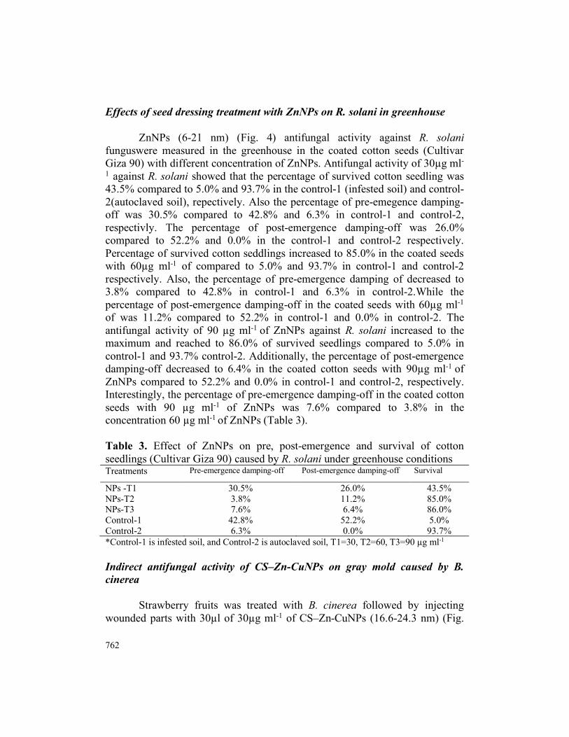
Effects of seed dressing treatment with ZnNPs on R. solani in greenhouse
ZnNPs (6-21 nm) (Fig. 4) antifungal activity against R. solani funguswere measured in the greenhouse in the coated cotton seeds (Cultivar Giza 90) with different concentration of ZnNPs. Antifungal activity of 30µg ml-
1 against R. solani showed that the percentage of survived cotton seedling was 43.5% compared to 5.0% and 93.7% in the control-1 (infested soil) and control-2(autoclaved soil), repectively. Also the percentage of pre-emegence damping-off was 30.5% compared to 42.8% and 6.3% in control-1 and control-2, respectivly. The percentage of post-emergence damping-off was 26.0% compared to 52.2% and 0.0% in the control-1 and control-2 respectively. Percentage of survived cotton seddlings increased to 85.0% in the coated seeds with 60µg ml-1 of compared to 5.0% and 93.7% in control-1 and control-2 respectively. Also, the percentage of pre-emergence damping of decreased to 3.8% compared to 42.8% in control-1 and 6.3% in control-2.While the percentage of post-emergence damping-off in the coated seeds with 60µg ml-1
of was 11.2% compared to 52.2% in control-1 and 0.0% in control-2. The antifungal activity of 90 µg ml-1 of ZnNPs against R. solani increased to the maximum and reached to 86.0% of survived seedlings compared to 5.0% in control-1 and 93.7% control-2. Additionally, the percentage of post-emergence damping-off decreased to 6.4% in the coated cotton seeds with 90µg ml-1 of ZnNPs compared to 52.2% and 0.0% in control-1 and control-2, respectively. Interestingly, the percentage of pre-emergence damping-off in the coated cotton seeds with 90 µg ml-1 of ZnNPs was 7.6% compared to 3.8% in the concentration 60 µg ml-1 of ZnNPs (Table 3).
Table 3. Effect of ZnNPs on pre, post-emergence and survival of cotton seedlings (Cultivar Giza 90) caused by R. solani under greenhouse conditions Treatments Pre-emergence damping-off Post-emergence damping-off Survival
NPs -T1 30.5% 26.0% 43.5% NPs-T2 3.8% 11.2% 85.0% NPs-T3 7.6% 6.4% 86.0% Control-1 42.8% 52.2% 5.0% Control-2 6.3% 0.0% 93.7% *Control-1 is infested soil, and Control-2 is autoclaved soil, T1=30, T2=60, T3=90 µg ml-1 Indirect antifungal activity of CS–Zn-CuNPs on gray mold caused by B. cinerea
Strawberry fruits was treated with B. cinerea followed by injecting wounded parts with 30µl of 30µg ml-1 of CS–Zn-CuNPs (16.6-24.3 nm) (Fig.
International Journal of Agricultural Technology 2017 Vol. 13(5):753-769
763
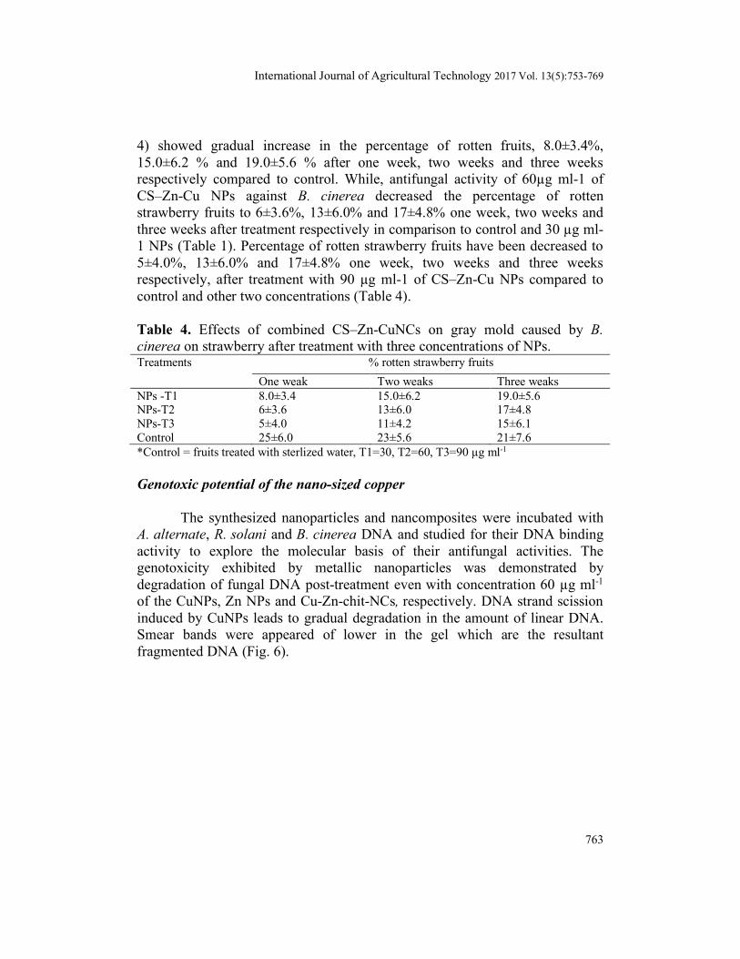
4) showed gradual increase in the percentage of rotten fruits, 8.0±3.4%, 15.0±6.2 % and 19.0±5.6 % after one week, two weeks and three weeks respectively compared to control. While, antifungal activity of 60µg ml-1 of CS–Zn-Cu NPs against B. cinerea decreased the percentage of rotten strawberry fruits to 6±3.6%, 13±6.0% and 17±4.8% one week, two weeks and three weeks after treatment respectively in comparison to control and 30 µg ml-1 NPs (Table 1). Percentage of rotten strawberry fruits have been decreased to 5±4.0%, 13±6.0% and 17±4.8% one week, two weeks and three weeks respectively, after treatment with 90 µg ml-1 of CS–Zn-Cu NPs compared to control and other two concentrations (Table 4).
Table 4. Effects of combined CS–Zn-CuNCs on gray mold caused by B. cinerea on strawberry after treatment with three concentrations of NPs. Treatments % rotten strawberry fruits
One weak Two weaks Three weaks NPs -T1 8.0±3.4 15.0±6.2 19.0±5.6 NPs-T2 6±3.6 13±6.0 17±4.8 NPs-T3 5±4.0 11±4.2 15±6.1 Control 25±6.0 23±5.6 21±7.6 *Control = fruits treated with sterlized water, T1=30, T2=60, T3=90 µg ml-1 Genotoxic potential of the nano-sized copper
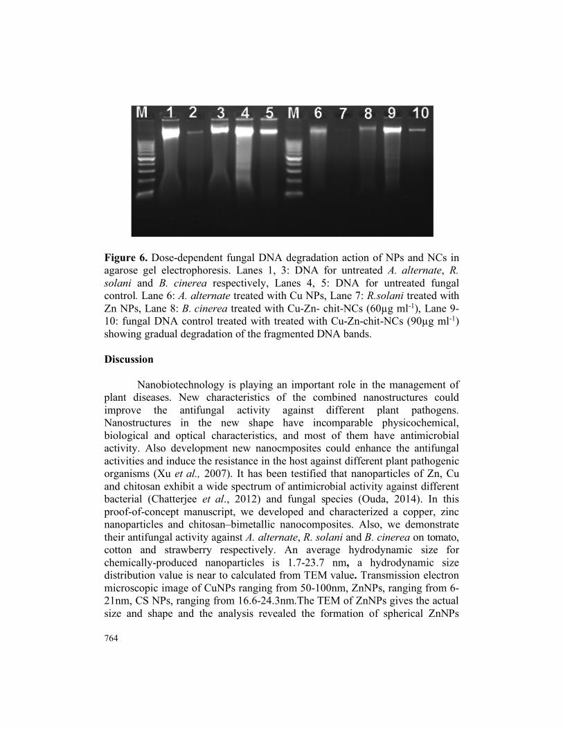
The synthesized nanoparticles and nancomposites were incubated with A. alternate, R. solani and B. cinerea DNA and studied for their DNA binding activity to explore the molecular basis of their antifungal activities. The genotoxicity exhibited by metallic nanoparticles was demonstrated by degradation of fungal DNA post-treatment even with concentration 60 µg ml-1 of the CuNPs, Zn NPs and Cu-Zn-chit-NCs, respectively. DNA strand scission induced by CuNPs leads to gradual degradation in the amount of linear DNA. Smear bands were appeared of lower in the gel which are the resultant fragmented DNA (Fig. 6).
764
Figure 6. Dose-dependent fungal DNA degradation action of NPs and NCs in agarose gel electrophoresis. Lanes 1, 3: DNA for untreated A. alternate, R. solani and B. cinerea respectively, Lanes 4, 5: DNA for untreated fungal control. Lane 6: A. alternate treated with Cu NPs, Lane 7: R.solani treated with Zn NPs, Lane 8: B. cinerea treated with Cu-Zn- chit-NCs (60µg ml-1), Lane 9-10: fungal DNA control treated with treated with Cu-Zn-chit-NCs (90µg ml-1) showing gradual degradation of the fragmented DNA bands. Discussion
Nanobiotechnology is playing an important role in the management of plant diseases. New characteristics of the combined nanostructures could improve the antifungal activity against different plant pathogens. Nanostructures in the new shape have incomparable physicochemical, biological and optical characteristics, and most of them have antimicrobial activity. Also development new nanocmposites could enhance the antifungal activities and induce the resistance in the host against different plant pathogenic organisms (Xu et al., 2007). It has been testified that nanoparticles of Zn, Cu and chitosan exhibit a wide spectrum of antimicrobial activity against different bacterial (Chatterjee et al., 2012) and fungal species (Ouda, 2014). In this proof-of-concept manuscript, we developed and characterized a copper, zinc nanoparticles and chitosan–bimetallic nanocomposites. Also, we demonstrate their antifungal activity against A. alternate, R. solani and B. cinerea on tomato, cotton and strawberry respectively. An average hydrodynamic size for chemically-produced nanoparticles is 1.7-23.7 nm, a hydrodynamic size distribution value is near to calculated from TEM value. Transmission electron microscopic image of CuNPs ranging from 50-100nm, ZnNPs, ranging from 6-21nm, CS NPs, ranging from 16.6-24.3nm.The TEM of ZnNPs gives the actual size and shape and the analysis revealed the formation of spherical ZnNPs
International Journal of Agricultural Technology 2017 Vol. 13(5):753-769
765
measuring about 21 nm in diameter, uniformly distributed without any significant aggregation. Particle size and zeta potential are the important properties which may influence the antifungal activity of nanoparticles. Nanoparticles with different particle size or zeta potential may have different mechanisms of inhibition against fungi.
A. alternate showed lower resistance in response to 90 µg ml-1 of Cu NPs which appeared as 1.4 cm of inhibition zone compared to 0.8 cm, 0.3 cm and 0.0 cm of inhibition zone in response to 60 µg ml-1, 30 µg ml-1, 0.0 µg ml-1 of Cu NPs respectively. CuNPs have been demonstrated previously to suppress vegetative growth of some fungal species such as A. flavus, A. niger, A. alternata, F. solani, P. chrysogenum and Candida albicans.
In vitro studies showed that the antifungal activities of Zn NPs in the concentration of 90 µg ml-1reached the highest activities against R. solani compared to 30 and 60 µg ml-1of Zn NPs and the control. Zn NPs showed great enhancement in the antimicrobial activity due to their unique properties such as large surface area. However, Kasemets et al. (2009) found nano and bulk ZnO were of comparable toxicity against S. cerevisiae. Number of studies proposed that ZnNPs may cause structural modifications of microbial cell membrane, causing cytoplasm outflow and ultimately the death of bacterial cells (Brayner et al., 2006). In vitro studies on Cu-chitosan NCs proved their uniform size and stability, which may contribute to their higher antifungal activity against number of pathogenic fungi like, A. alternata, M. phaseolina and R. solani. Also, Cu-chitosan NPs showed maximum inhibition rate of spore germination of A. alternate (Saharan et al., 2013).
Also, current research study showed that application of 90 µg ml-1CS–Zn-Cu nanocomposites (ranging from 16.6-24.3 nm) on strawberry fruits decreased the percentage of rotten strawberry fruits to 5±4.0%, 13±6.0% and 17±4.8% one week, two weeks and three weeks respectively. Application 60µg ml-1CS–Zn-CuNPs NCs on strawberry fruits showed antifungal activities against B. cinerea but not as significant as the highest concentration of CS–Zn-Cu nanomaterials. Chitosan, zinc oxide and copper nanomaterials, each one of them has its unique and distinctive characters and mechanism as antimicrobial agent against wide range of microorganisms.
This new combinations of CS–Zn-CuNPs nanocomposites increased the antifungal activities against B. cinerea and increased the shelve life for strawberry fruits. This new combination was effective reducing strawberry fruits decay as a result of chitosan fungistatic effect plus the fungicide activities of both Cu and ZnO. Current study demonstrates the potential of chitosan nanomaterials as fruits preservatives against B. cinerea plus the antifungal activity of the new combination CS–Zn-Cu nanomaterials which may increase
766
the positive effect of nanomaterials against different post-harvest fungi. On the other hand new combinations of nanomaterials might be toxic to plant, pathogens, animal and human.
The biocidal activity of CuNPs could be attributed to the effect of the CuNPs and/or the copper ions discharged from CuNPs. Because of the great surface area of the nanoparticles, it could be strongly adsorbed onto the surface of the microbial cells resulting in; i) disruption of cell permeability and release of integral components (Raffi et al., 2010), ii) denaturing of number of functional biomolecules (Wei et al., 2010), iii) stimulation of oxidative damage to the microbial cells. However, some studies have reported that Cu2+ is the stimulating force behind the antimicrobial properties of polymers containing Cu-nanocomposites (Delgado et al., 2011). At the same time, the discharged copper ions might be moved inside the microbial cells or attached to their outer surfaces resulting in cell apoptosis via protein denaturation and disruption of cell membrane (Palza et al., 2015). Briefly, Copper has strong biocidal activity with non-specific mode of action against microbial cells that make it ideal antimicrobial agent. Cu-chitosan nanoparticles enhanced enzymes activities involved in plant defense by chitosan participate in reactive oxygen species (ROS) scavenging system (Saharan et al., 2013; Saharan et al., 2015).CuNPs and Zn-chitosan nanocomposites nearly have the same mode of action for Cu-chitosan nanocomposites such as the production of reactive oxygen species and membrane disruption (Xie et al., 2011; Ingle et al., 2013). Therefore, ZnNPs may exhibit different antifungal activities against B. cinerea and other fungi. ZnNPs could be used as an effective fungicide in agricultural and food safety applications.
In the current work, the genotoxicity exhibited by the three types of nano-agrochemicals was demonstrated by degradation of fungal DNA post-treatment even with A. alternate treated with CuNPs, R. solani treated with Zn NPs, B. cinerea treated with Cu-Zn- chit-NCs (60 µg ml-1), respectively. DNA strand scission induced by CuNPs leads to gradual degradation in the amount of linear DNA. Cu-chitosan NCs could penetrate into cell and interacts with cellular DNA or mRNA directly regulates the metabolisms and reproduction which could ultimately lead to death of fungal pathogens
In conclusion, antifungal activities of Cu NPs, ZnONPs and CS–Zn-CuNCs against three plant pathogenic fungi A. alternata, R. solani and B. cinerea, were investigated in this study. The In-vitro application showed that CS–Zn-CuNCs at 90 µg ml-1has the highest antifungal activity since it inhibited the growth of B. cinerea. The in-vivo application showed that Zn NPs at concentrations of 60 and 90 µg ml-1 against R. solani increased the percentages of survived seedlings to 85% and 86%, respectively. Every type of nanoparticle
International Journal of Agricultural Technology 2017 Vol. 13(5):753-769
767
used in the current work has its own unique properties which make it destructive to specific organisms with a specific mode of action. Combination between more than one nanoparticle may enhance the antifungal activities of the new compound or decrease the activities of the nanomaterials because of interference between two different modes of action. Further studies are needed to investigate the toxicology of each size of nanoparticles and also discover the unique activities of new combinations of nanomaterials. Acknowledgment
This research was supported by a grant from Science and Technology Development Fund (STDF), Egypt (STDF-RFBR program) [grant no. 13791]. Also, this work was partially funded by Russian Foundation for Basic Research grant (RFBR-15-53-61030). References Aruoja, V., Dubourguier, H., Kasamets, C. and Kahru, K. A. (2009). Toxicity of nanoparticles
of CuO, ZnO and TiO2 to microalgae, Pseudokirchneriella subcapitata. Science of the Total Environment 407:1461-1468.
Brayner, R., Ferrari-Iliou, R., Brivois, N., Djediat, S., Benedetti, M. F. And Fievet, F. (2006). Toxicological impact studies based on Escherichia coli bacteria in ultrafine ZnO nanoparticles colloidal medium. Nano Letters 6:866-870.
Brunel, F., Gueddari, NE. and Moerschbacher, BM. (2013). Complexation of copper (II) with chitosan nanogels: Toward control of microbial growth. Carbohydrate Polymers 92:1348-1356.
Chatterjee, A. K., Sarkar, R. K., Chattopadhyay, A. P., Aich, P., Chakraborty, R. and Basu, T. (2012). Asimple robust method for synthesis of metallic copper nanoparticles of high antibacterial potency against E. coli. Nanotechnology 23:1-11.
Cioffi, N., Torsi, L., Ditaranto, N., Tantillo, G., Ghibelli, L. and Sabbatini, L. (2005). Copper nanoparticle/polymer composites with antifungal and bacteriostatic properties. Chemistry of Materials 17:5255-62.
Delgado, K., Quijada, R., Palma, R. and Palza, H. (2011). Polypropylene with embedded copper metal or copper oxide nanoparticles as a novel plastic antimicrobial agent. Letter of Applied and Microbiology 53:50-54.
Dimkpa, C. O., McLean, J. E., Britt, D. W. and Anderson, A. J. (2013). Antifungal activity of ZnO nanoparticles and their interactive effect with a biocontrol bacterium on growth antagonism of the plant pathogen Fusarium graminearum. Biometals 26:913-924.
Giannousi, K., Avramidis, I. and Dendrinou-Samara, C. (2013). Synthesis, characterization and evaluation of copper based nanoparticles as agrochemicals against. Phytophthora infestans. RSC Advances 3:21743-21752.
Hardy, JJE., Hubert, S., Macquarrie, DJ. and Wilson, AJ. (2004). Chitosan-based heterogeneous catalysts for Suzuki and Heck reactions. Green Chemistry 6:53–56.
He, L., Liu, Y., Mustapha, A. and Lin, M. (2011). Antifungal activity of zinc oxide nanoparticles against Botrytis cinerea and Penicillium expansum. Microbiology Research 166:207-215.
768
Huang, L., Dian-Qing, L., Yan-Jun, W., Min David, G. and Xue, E. D. (2005). Controllable preparation of Nano-MgO and investigation of its bactericidal properties. Journal of Inorganic Biochemistry 99:986-993
Ingle, A. P., Duran, N. and Rai, M. (2013). Bioactivity, mechanism of action, and cytotoxicity of copper-based nanoparticles: a review. Applied of Microbiology and Biotechnology 98:1001-1009.
Jaiswal, M., Chauhan, D. and Sankararamakrishnan, N. (2012). Copper chitosan nanocomposite: synthesis, characterization, and application in removal of organophosphorous pesticide from agricultural runoff. Environmental Science and Pollution Research 19:2055-2062.
Jayaseelan, C., Rahuman, A. A., Kirthi, A. V., Marimuthu, S., Santhoshkumar, T., Bagavan, A., Gaurav, K., Karthik, L. and Rao, K. V. (2012). Novel microbial route to synthesize ZnO nanoparticles using Aeromonas hydrophila and their activity against pathogenic bacteria and fungi. Spectrochima Acta A 90:78-84.
Kasemets, K., Ivask, A., Dubourguier, H. C. and Kahru, A. (2009). Toxicity of nanoparticles of ZnO, CuO and TiO2 to yeast Saccharomyces cerevisiae. Toxicolology In Vitro 23:1116-22.
Kim, K. J., Sung, W. S., Moon, S. K., Choi, J. S., Kim, J. G. and Lee, D. G. (2008). Antifungal effect of silver nanoparticles on dermatophytes. Journal of Microbiology and Biotechnology 18:1482-1484.
Kruk, T., Szczepanowicz, K., Stefa´nska, J., Socha, RP. and Warszy´nski, P. (2015). Synthesis and antimicrobial activity of monodisperse copper nanoparticles. Colloids and Surfaces B 128:17-22.
Kumar, A., Vemula, P. K., Ajayan, P. M. and John, G. (2008). Silver nanoparticle- embedded antimicrobial paints based on vegetable oil. Nature Materials 7:236-241.
Liu, Y., He, L., Mustapha, A., Li, H. and Lin, M. (2009). Antibacterial activities of zinc oxide nanoparticles against Escherichia coliO157:H7. Journal of Applied Microbiology 107:1193-201.
Ouda, S. M. (2014). Antifungal activity of silver and copper nanoparticles on two plant pathogens, Alternaria alternata and Botrytis cinerea. Research Journal of Microbiology 9:34-42.
Palza, H. (2015). Antimicrobial polymers with metal nanoparticles. International Journal of Molecular Science 19:2099-2116.
Raffi, M., Mehrwan, S., Bhatti, T. M., Akhter, J. I., Hameed, A. and Yawar, W. (2010). Investigations into the antibacterial behavior of copper nanoparticles against Escherichia coli. Annals of Microbiology 60:75-80.
Saharan, V., Mehrotra, A., Khatik, R., Rawal, P., Sharma, S. S. and Pal, A. (2013). Synthesis of chitosan based nanoparticles and their in vitro evaluation against phytopathogenic fungi. International Journal of Biology and Macromolcule 62:677-683.
Saharan, V., Mehrotra, A., Khatik, R., Rawal, P., Sharma, S. S. and Pal, A. (2013). Synthesis of chitosan based nanoparticles and their in vitro evaluation against phytopathogenic fungi. International Journal of Biological Macromolecules 62:677-683.
Saharan, V., Sharma, G., Yadav, M., Choudhary, M. K., Sharma, S. S., Pal, A., Raliya, R. and Biswas, P. (2015). Synthesis and in vitro antifungal efficacy of Cu-chitosan nanoparticles against pathogenic fungi of tomato. International Journal of Biological Macromolecules 75:346-353.
Sharma, V. K., Yngard, R. A. and Lin, Y. (2009). Silver nanoparticles: green synthesis and their antimicrobial activities. Advances of Colloid Interface Science 145:83-96.
International Journal of Agricultural Technology 2017 Vol. 13(5):753-769
769
Spadaro, D., Garibaldi, A. and Martines, G. F. (2004). Control of Penicillium expansum and Botrytis cinerea on apple combining a biocontrol agent with hot water dipping and acibenzolar-S-methyl, baking soda, or ethanol application. Postharvest Biology and Technology 33:141-51.
Wani, A. H. and Shah, M. A. (2012). A unique and profound effect of MgO and ZnO nanoparticles on some plant pathogenic fungi. Journal of Applied Pharmaceutical Science 02:40-44.
Wei, Y., Chen, S., Kowalczyk, B., Huda, S., Gray, T. P. And Grzybowski, B. A. (2010). Synthesis of stable, low dispersity copper nanoparticles and nanorods and their antifungal and catalytic properties. The Journal of Physical Chemistry 114:15612-15616.
Xie, Y., He, Y., Irwin, P. L., Jin, T. and Shi, X. (2011). Antibacterial activity and mode of action of ZnO, Applied and Environmental Microbiology 77:2325-2331.
Xing, Y., Li, X., Xu, Q., Yun, J., Lu, Y. and Tang, Y. (2011). Effects of chitosan coating enriched with cinnamon oil on qualitative properties of sweet pepper (Capsicum annuum L.). Food Chemistry 124:1443-1450.
Xu, J., Zhao, X., Han, X. and Du, Y. (2007). Antifungal activity of oligo chitosan against Phytophthora capsici and other plant pathogenic fungi in vitro. Pesticide Biochemistry and Physiology 87:220-228.
Youssef, K., Hashim, A. F., Margarita, R., Alghuthaymi, M. A. and Abd-Elsalam, K. A. (2017). Fungicidal efficacy of chemically-produced copper nanoparticles against Penicilium digitatum and Fusarium solani on citrus fruit. Philippine Agricultural Scientist 100:69-78.
Youssef, K., Sanzani, S. M., Ligorio, A., Ippolito, A. and Terry, L. A. (2014). Sodium carbonate and bicarbonate treatments induce resistance to postharvest green mold on citrus fruit. Postharvest Biology and Technology 87:61-69.
(Received: 17 May 2017, accepted: 30 July 2017)