Embed Size (px)
Citation preview
CHEST Special FeaturesCHEST IMAGING
journal.publications.chestnet.org CHEST / 143 / 3 / MARCH 2013 825
The solitary pulmonary nodule (SPN) is defi ned as a radiographic opacity � 3 cm in diameter with at
least two-thirds of its margins surrounded by lung parenchyma. 1,2 Implied in this defi nition is the exclu-sion of lymph nodes, atelectasis, and postobstructive pneumonia. However, it may be diffi cult at times to exclude intraparenchymal lymph nodes based on just radiologic appearance. SPNs have been noted in 0.09% to 7% of all chest radiographs. 3-5 A review of eight large studies on lung cancer screening using CT imaging 6-13 documented the prevalence of SPN from 8% to 51%, and the prevalence of malignancy from 1.1% to 12%. 14 The etiologic spectrum of SPN represents a veritable minefi eld of diseases, including benign conditions such as hamartomas to potentially fatal ones such as primary lung cancer ( Table 1 ). Establishing the etiology of a SPN in a timely and accurate manner, therefore, assumes critical importance, since surgical resection in a patient with early-stage
lung cancer provides the highest chance of cure. By the same token, avoiding thoracic surgery for a benign SPN whenever possible does obviate signifi cant mor-bidity. We will describe an algorithmic approach to diagnosis of SPN in Part 2 (see page 840). 15
Clinical Evaluation
A SPN does not typically herald its presence with clin-ical symptoms, nor does it lend itself to self-awareness like melanoma or a breast lump. Although the SPN may be insidious, a variety of clinical risk factors such as advancing age and history of smoking have been associated with a higher OR of the SPN being malig-nant. 15-19 Elucidating a thorough history of prior malig-nancy is crucial; the majority of SPNs detected in patients with a history of prior malignancy are malig-nant. 20-22 Interestingly, the malignant SPNs are equally or more likely to represent primary lung cancer rather
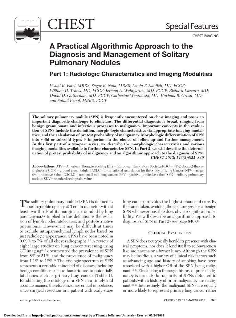
The solitary pulmonary nodule (SPN) is frequently encountered on chest imaging and poses an important diagnostic challenge to clinicians. The differential diagnosis is broad, ranging from benign granulomata and infectious processes to malignancy. Important concepts in the evalua-tion of SPNs include the defi nition, morphologic characteristics via appropriate imaging modal-ities, and the calculation of pretest probability of malignancy. Morphologic differentiation of SPN into solid or subsolid types is important in the choice of follow-up and further management. In this fi rst part of a two-part series, we describe the morphologic characteristics and various imaging modalities available to further characterize SPN. In Part 2, we will describe the determi-nation of pretest probability of malignancy and an algorithmic approach to the diagnosis of SPN. CHEST 2013; 143(3):825–839
Abbreviations: ATS 5 American Thoracic Society; ERS 5 European Respiratory Society; FDG 5 18 F-2-deoxy-2-fl uoro- d -glucose; GGN 5 ground glass nodule; IASLC 5 International Association for the Study of Lung Cancer; NPV 5 nega-tive predictive value; NSCLC 5 non-small cell lung cancer; PPV 5 positive predictive value; SPN 5 solitary pulmonary nodule; SUV 5 standardized uptake value
A Practical Algorithmic Approach to the Diagnosis and Management of Solitary Pulmonary Nodules Part 1: Radiologic Characteristics and Imaging Modalities
Vishal K. Patel , MBBS ; Sagar K. Naik , MBBS ; David P. Naidich , MD , FCCP ; William D. Travis , MD , FCCP ; Jeremy A. Weingarten , MD , FCCP ; Richard Lazzaro , MD ; David D. Gutterman , MD , FCCP ; Catherine Wentowski , MD ; Horiana B. Grosu , MD ; and Suhail Raoof , MBBS , FCCP
Downloaded From: http://journal.publications.chestnet.org/ by a Thomas Jefferson University User on 05/24/2013
826 Special Features
than metastasis from the extrapulmonary malignancy, with the notable exceptions of sarcoma, melanoma, and testicular carcinoma ( Table 2 ). The presence of mediastinal lymph node enlargement on CT scan strongly suggests a new primary lung cancer rather than metastasis. 20 Interstitial lung diseases, such as idiopathic pulmonary fi brosis, asbestosis, and sclero-derma are associated with an increased incidence of lung cancer. 23 The prevalence of lung cancer in idio-pathic pulmonary fi brosis, for example, ranges from 9% to 38%, with a predilection for peripheral lung areas in the lower lobes in elderly male smokers. 24-29 Finally, residence in or travel to an area with endemic fungal pathogens could suggest a benign, infectious SPN in the correct clinical context. For example, coccidioidomycosis is endemic in the southwestern United States and Mexico, and often presents as a SPN on chest CT scans. 30 Cryptococcus 31-33 infection and histoplasmosis can also present as a SPN.
Radiographic Characteristics: CT Scan
Specifi c morphologic characteristics of SPNs on imaging may help differentiate benign from malig-nant SPNs. It is recommended that CT images be thin section, with contiguous 1-mm images through nodules. Both lung and mediastinal windows should be obtained, the former for the edges, the latter for solid components. Low-dose (milliamperes second [mAs] , 80) CT scan can be used for this purpose.
Growth Rate
Malignant, solid SPNs have a volume doubling time of 20-400 days, 34-38 with a majority having volume doubling times of signifi cantly , 100 days. 39 A volume doubling time . 400 days suggests slow growth and is usually associated with benign SPNs, whereas volume doubling time , 20 days indicates very rapid growth, usually attributable to infectious processes. 40 It is important to realize that since the volume of a sphere equals 4 p r 3 /3, an increase in nodule diameter by only 26% indicates doubling of volume. In other words, a
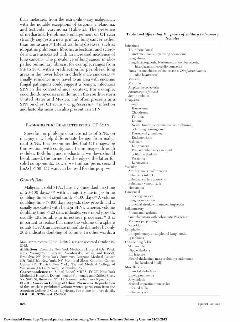
Table 1— Differential Diagnosis of Solitary Pulmonary Nodules
Infectious TB (tuberculoma) Round pneumonia, organizing pneumonia Lung abscess Fungal: aspergillosis, blastomycosis, cryptococcosis,
histoplasmosis, coccidioidomycosis Parasitic: amoebiasis, echinococcosis, Dirofi laria immitis
(dog heartworm) Measles Nocardia Atypical mycobacteria Pneumocystis jiroveci Septic embolusNeoplastic Benign Hamartoma Chondroma Fibroma Lipoma Neural tumor (Schwannoma, neurofi broma) Sclerosing hemangioma Plasma cell granuloma Endometriosis Malignant Lung cancer Primary pulmonary carcinoid Solitary metastasis Teratoma LeiomyomaVascular Arteriovenous malformation Pulmonary infarct Pulmonary artery aneurysm Pulmonary venous varix HematomaCongenital Bronchogenic cyst Lung sequestration Bronchial atresia with mucoid impactionInfl ammatory Rheumatoid arthritis Granulomatosis with polyangiitis (Wegener) Microscopic polyangiitis SarcoidosisLymphatic Intrapulmonary or subpleural lymph node LymphomaOutside lung fi elds Skin nodule Nipple shadows Rib fracture Pleural thickening, mass or fl uid (pseudotumor
[ie, loculated fl uid])Miscellaneous Rounded atelectasis Lipoid pneumonia Amyloidosis Mucoid impaction (mucocele) Infected bulla Pulmonary scar
Manuscript received June 12, 2012; revision accepted October 10, 2012 . Affi liations : From the New York Methodist Hospital (Drs Patel, Naik, Weingarten, Lazzaro, Wentowski, Grosu, and Raoof), Brooklyn, NY; New York University Langone Medical Center (Dr Naidich), New York, NY; Memorial Sloan-Kettering Cancer Center (Dr Travis), New York, NY; and Medical College of Wisconsin (Dr Gutterman), Milwaukee, WI. Correspondence to: Suhail Raoof, MBBS, FCCP, New York Methodist Hospital, Department of Pulmonary and Critical Care, 506 Sixth St, Brooklyn, NY 11215; e-mail: [email protected] © 2013 American College of Chest Physicians . Reproduction of this article is prohibited without written permission from the American College of Chest Physicians. See online for more details. DOI: 10.1378/chest.12-0960
Downloaded From: http://journal.publications.chestnet.org/ by a Thomas Jefferson University User on 05/24/2013
journal.publications.chestnet.org CHEST / 143 / 3 / MARCH 2013 827
as pure ground glass nodules (GGNs), as will be dis-cussed separately.
Size
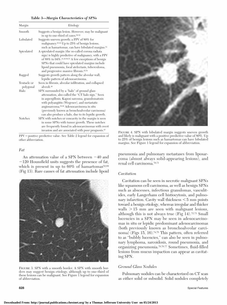
The likelihood of malignancy in a SPN increases with the nodule diameter 47,48 ( Fig 2 ). Lesions larger than 3 cm are more likely to be malignant. However, a smaller size does not exclude malignancy.
Location
SPNs in the upper lobes have been observed to be malignant in a larger number of cases. 17,49,50 This is possibly because there is a higher concentration of inhaled carcinogens in the upper lobes resulting from cigarette smoking. 40
Margin/Border/Edge Characteristics
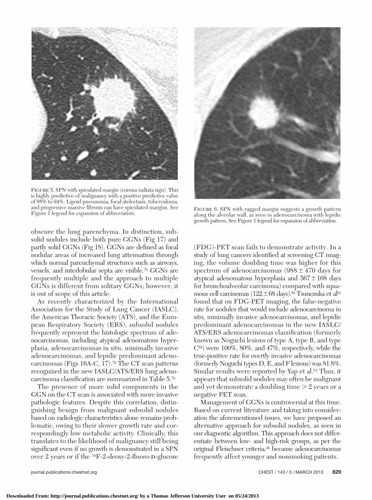
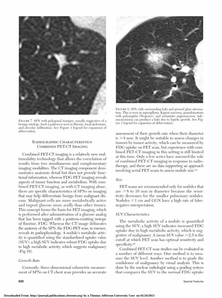
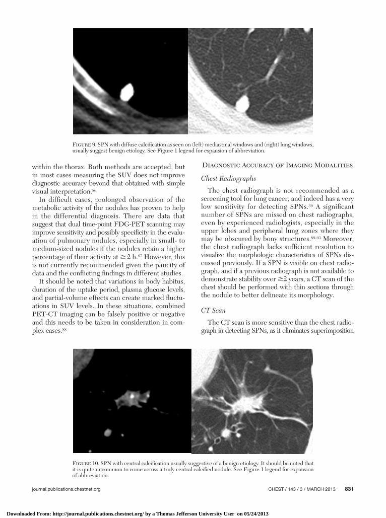
Furuya et al 51 divided margins of SPNs into seven categories: smooth, lobulated, spiculated, ragged, ten-tacle, polygonal, and with surrounding halo and with notches or concavity. The described margin types and their possible etiologies are shown in Table 3 and Figures 3-8 . 16,47,51-59
Calcifi cation and Attenuation
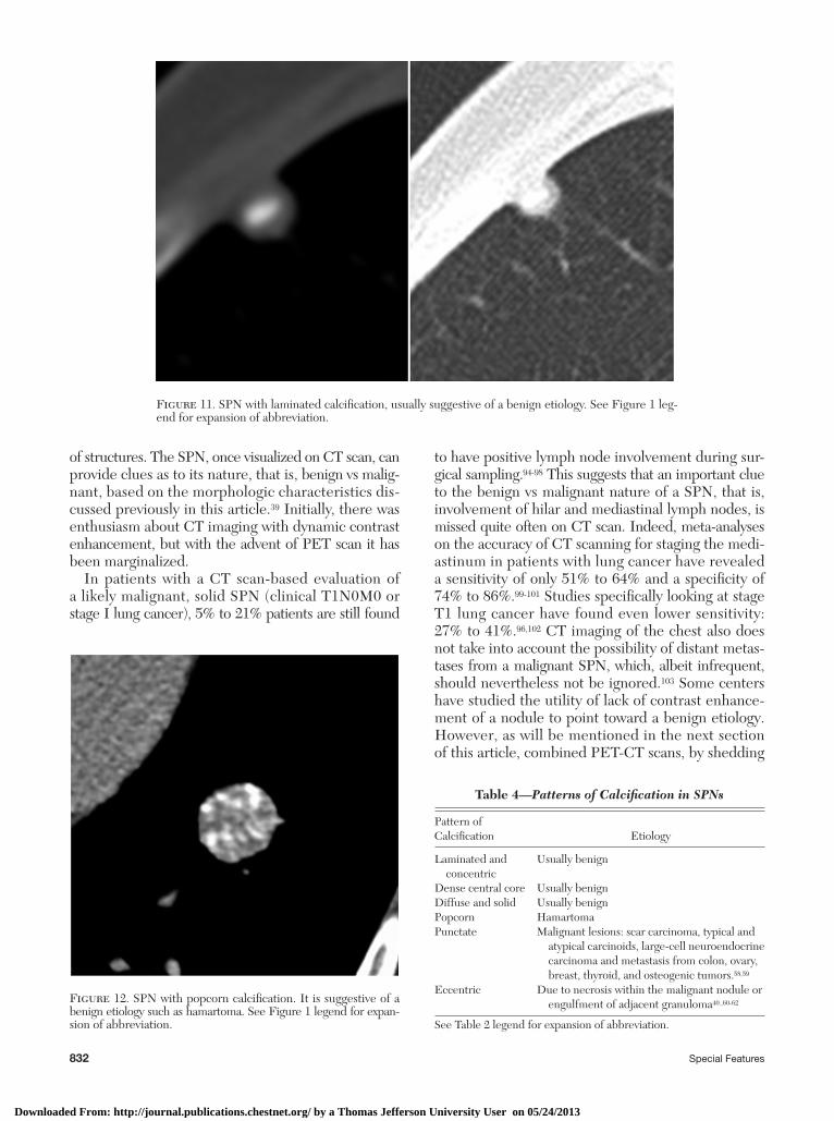
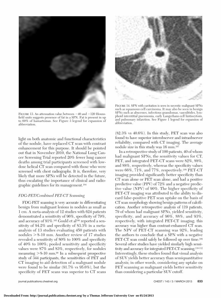
Specifi c patterns of calcifi cation within a SPN (dif-fuse, central [bull’s eye], laminated or concentric, or popcorn) can suggest a benign lesion ( Figs 9-12 ). However, there is no pattern of calcifi cation that is considered specifi c for malignancy 60 ( Table 4 40,61-65 ). The plain chest roentgenogram is not very sensitive in detecting calcification within a SPN, demonstrat-ing a sensitivity of 50% and a specifi city of 87%. 66,67 Hence, a noncontrast CT scan with 1 to 3 mm sections through the nodule is recommended to assess the SPN and the calcifi cation within the SPN. Attenua-tion values . 200 Hounsfi eld units in a SPN indicate the presence of calcium in the nodule. 68
4-mm nodule may double in volume, but its size will barely increase to 5 mm, a change often too subtle to detect reliably on CT scans. 41 Three-dimensional volumetric measurement of a SPN on a CT scan of the chest can provide a more accurate assessment of growth rate compared with the SPN diameter, espe-cially in small ( , 1 cm) SPNs ( Fig 1 ). 42-44 However, it must be pointed out that there is signifi cant contro-versy regarding the most accurate method to follow the rate of growth of nodules. There are those who advocate for volumetric analysis, but this is generally applicable for solid lesions . 8 to 10 mm. The data for smaller lesions are less compelling. It should also be stated that for ground glass and subsolid nodules, assessment of growth rate is even less clear. These lesions may be followed by assessing their mass, that is, a combination of both volume and density. 45,46 To date, there is no consensus on the best way to follow these lesions. In general, a solid SPN, stable in volume over a period of 2 years (which indicates a doubling time . 730 days) can be reliably considered benign; important exceptions to this rule are slow-growing ade-nocarcinomas of the lung, especially those presenting
Table 2— Extrapulmonary Malignancy and Ratio of Primary vs Metastatic SPNs 20-22
Extrapulmonary Malignancy
Ratio of Malignant SPNs Representing Primary Lung
Cancer vs Metastasis From the Extrapulmonary Malignancy
Carcinomas of the head and neck 25:3Cancers of the bladder, breast,
cervix, bile ducts, esophagus, ovary, prostate, or stomach
26:8
Cancers of the salivary glands, adrenal gland, colon, parotid gland, kidney, thyroid gland, thymus, or uterus
13:16
Melanoma, sarcoma, or testicular carcinoma
9:23
SPN 5 solitary pulmonary nodule. See text for explanation.
Figure 1. Solitary pulmonary nodule (SPN) doubling time. A 4-mm nodule can double in size over a period of time but the diameter will increase only approximately 1 mm to 5 mm, which may not be reliably detected on the CT scan. However, an increase in a bigger mass is very well appreciated by just looking at the diameter on the CT scan.
Figure 2. SPN size and corresponding likelihood of malignancy. See Figure 1 legend for expansion of abbreviation.
Downloaded From: http://journal.publications.chestnet.org/ by a Thomas Jefferson University User on 05/24/2013
828 Special Features
pneumonia and pulmonary metastases from liposar-coma (almost always solid-appearing lesions), and renal cell carcinoma. 70,71
Cavitation
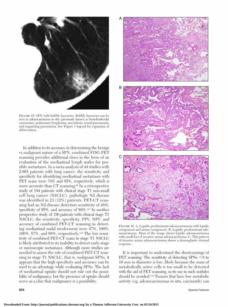
Cavitation can be seen in necrotic malignant SPNs like squamous cell carcinoma, as well as benign SPNs such as abscesses, infectious granulomas, vasculit-ides, early Langerhans cell histiocytosis, and pulmo-nary infarction. Cavity wall thickness , 5 mm points toward a benign etiology, whereas irregular and thicker walls . 15 mm are seen with malignant lesions, although this is not always true ( Fig 14 ). 72,73 Small lucencies in a SPN may be seen in adenocarcino-mas in situ or lepidic predominant adenocarcinomas (both previously known as bronchoalveolar carci-noma) ( Figs 15, 16 ). 74,75 This pattern, often referred to as “bubbly lucencies,” can also be seen in pulmo-nary lymphoma, sarcoidosis, round pneumonia, and organizing pneumonia. 70,76,77 Sometimes, fl uid-fi lled lesions from mucus impaction can appear as cavitat-ing SPN.
Ground Glass Nodules
Pulmonary nodules can be characterized on CT scan as either solid or subsolid. Solid nodules completely
Fat
An attenuation value of a SPN between 2 40 and 2 120 Hounsfi eld units suggests the presence of fat, which is present in up to 60% of hamartomas 52,69 ( Fig 13 ). Rare causes of fat attenuation include lipoid
Table 3— Margin Characteristics of SPNs
Margin Etiology
Smooth Suggests a benign lesion. However, may be malignant in up to one-third of cases. 50,52
Lobulated Suggests uneven growth; a PPV of 80% for malignancy. 45,49 Up to 25% of benign lesions, such as hamartomas, can have lobulated margins. 51
Spiculated A spiculated margin (the so-called corona radiata sign) is highly predictive of malignancy, with a PPV of 88% to 94%. 16,49,50,53 A few exceptions of benign SPNs that could have spiculated margins include lipoid pneumonia, focal atelectasis, tuberculoma, and progressive massive fi brosis. 45,54
Ragged Suggests growth pattern along the alveolar wall; lepidic pattern of adenocarcinoma.
Tentacle or polygonal
Seen in fi brosis, alveolar infi ltration, and collapsed alveoli. 49
Halo SPN surrounded by a “halo” of ground glass attenuation, also called the “CT halo sign.” Seen in aspergillosis, Kaposi sarcoma, granulomatosis with polyangiitis (Wegener), and metastatic angiosarcoma. 55,56 Adenocarcinoma in situ (previously known as bronchoalveolar carcinoma) can also produce a halo, due to its lepidic growth.
Notches SPN with notches or concavity in the margin is seen in some SPNs with tumor growth. These notches are frequently found in adenocarcinomas with overt invasion and are associated with poor prognosis. 57
PPV 5 positive predictive value. See Table 2 legend for expansion of other abbreviation.
Figure 3. SPN with a smooth border. A SPN with smooth bor-ders may suggest benign etiology, although up to one-third of these lesions can be malignant. See Figure 1 legend for expansion of abbreviation.
Figure 4. SPN with lobulated margin suggests uneven growth and likely is malignant with a positive predictive value of 80%. Up to 25% of benign lesions such as hamartomas can have lobulated margins. See Figure 1 legend for expansion of abbreviation.
Downloaded From: http://journal.publications.chestnet.org/ by a Thomas Jefferson University User on 05/24/2013
journal.publications.chestnet.org CHEST / 143 / 3 / MARCH 2013 829
(FDG)-PET scan fails to demonstrate activity. In a study of lung cancers identifi ed at screening CT imag-ing, the volume doubling time was higher for this spectrum of adenocarcinomas (988 � 470 days for atypical adenomatous hyperplasia and 567 � 168 days for bronchoalveolar carcinoma) compared with squa-mous cell carcinomas (122 � 68 days). 80 Tsunezuka et al 81 found that on FDG-PET imaging, the false-negative rate for nodules that would include adenocarcinoma in situ, minimally invasive adenocarcinomas, and lepidic predominant adenocarcinomas in the new IASLC/ATS/ERS adenocarcinomas classifi cation (formerly known as Noguchi lesions of type A, type B, and type C 82 ) were 100%, 80%, and 47%, respectively, while the true-positive rate for overtly invasive adenocarcinomas (formerly Noguchi types D, E, and F lesions) was 81.8%. Similar results were reported by Yap et al. 83 Thus, it appears that subsolid nodules may often be malignant and yet demonstrate a doubling time . 2 years or a negative PET scan.
Management of GGNs is controversial at this time. Based on current literature and taking into consider-ation the aforementioned issues, we have proposed an alternative approach for subsolid nodules, as seen in our diagnostic algorithm. This approach does not differ-entiate between low- and high-risk groups, as per the original Fleischner criteria, 48 because adenocar cinomas frequently affect younger and nonsmoking patients.
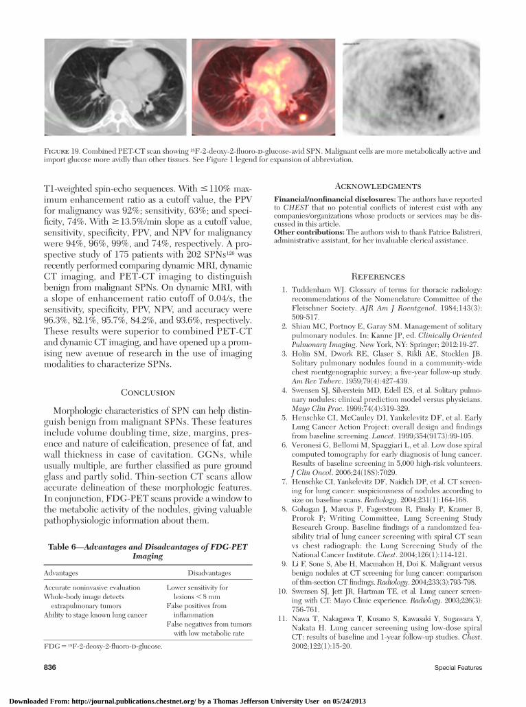
obscure the lung parenchyma. In distinction, sub-solid nodules include both pure GGNs ( Fig 17 ) and partly solid GGNs ( Fig 18 ). GGNs are defi ned as focal nodular areas of increased lung attenuation through which normal parenchymal structures such as airways, vessels, and interlobular septa are visible. 78 GGNs are frequently multiple and the approach to multiple GGNs is different from solitary GGNs; however, it is out of scope of this article.
As recently characterized by the International Association for the Study of Lung Cancer (IASLC), the American Thoracic Society (ATS), and the Euro-pean Respiratory Society (ERS), subsolid nodules frequently represent the histologic spectrum of ade-nocarcinomas, including atypical adenomatous hyper-plasia, adenocarcinomas in situ, minimally invasive adenocarcinomas, and lepidic predominant adeno-carcinomas ( Figs 16A-C, 17 ). 79 The CT scan patterns recognized in the new IASLC/ATS/ERS lung adeno-carcinoma classifi cation are summarized in Table 5 . 79
The presence of more solid components in the GGN on the CT scan is associated with more invasive pathologic features. Despite this correlation, distin-guishing benign from malignant subsolid nodules based on radiologic characteristics alone remains prob-lematic, owing to their slower growth rate and cor-respondingly low metabolic activity. Clinically, this translates to the likelihood of malignancy still being signifi cant even if no growth is demonstrated in a SPN over 2 years or if the 18 F-2-deoxy-2-fl uoro- d -glucose
Figure 5. SPN with spiculated margin (corona radiata sign). This is highly predictive of malignancy with a positive predictive value of 88% to 94%. Lipoid pneumonia, focal atelectasis, tuberculoma, and progressive massive fi brosis can have spiculated margins. See Figure 1 legend for expansion of abbreviation.
Figure 6. SPN with ragged margin suggests a growth pattern along the alveolar wall, as seen in adenocarcinoma with lepidic growth pattern. See Figure 1 legend for expansion of abbreviation.
Downloaded From: http://journal.publications.chestnet.org/ by a Thomas Jefferson University User on 05/24/2013
830 Special Features
assessment of their growth rate when their diameter is . 8 mm. It might be suitable to assess changes in tumors by tumor activity, which can be measured by FDG uptake on PET scan, but experience with com-bined PET-CT imaging in this setting is still limited at this time. Only a few series have assessed the role of combined PET-CT imaging in response to radio-therapy, and there are no data supporting an approach involving serial PET scans to assess nodule size. 84
Size
PET scans are recommended only for nodules that are . 8 to 10 mm in diameter because the sensi-tivity decreases for the smaller pulmonary nodules. Nodules , 1 cm and GGN have a high rate of false-negative interpretation.
SUV Characteristics
The metabolic activity of a nodule is quantifi ed using the SUV; a high SUV indicates increased FDG uptake due to high metabolic activity, which is sug-gestive of malignancy. A mean SUV value . 2.5 is the cutoff at which PET scan has optimal sensitivity and specifi city. 85
Combined PET-CT scan studies can be evaluated in a number of different ways. One method is to mea-sure the SUV level. Another method is to grade the confi dence of malignancy by visual analysis. This is done by the nuclear radiologist using a grading system that compares the SUV to the normal FDG uptake
Radiographic Characteristics: Combined PET-CT Imaging
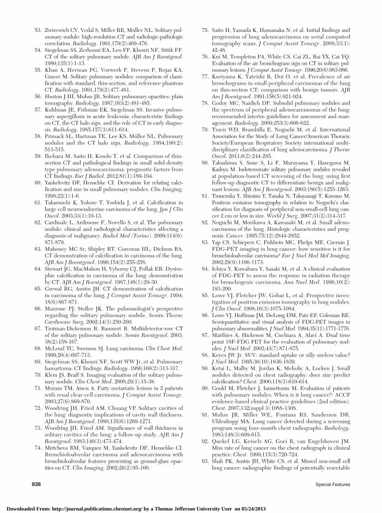
Combined PET-CT imaging is a relatively new mul-timodality technology that allows the correlation of results from two simultaneous and complementary imaging modalities. The CT imaging component dem-onstrates anatomic detail but does not provide func-tional information, whereas FDG-PET imaging reveals aspects of tumor function and metabolism. With com-bined PET-CT imaging, as with CT imaging alone, there are specifi c characteristics of SPNs on imaging that may help differentiate benign from malignant dis-ease. Malignant cells are more metabolically active and import glucose more avidly than other tissues. This concept forms the basis for PET imaging, which is performed after administration of a glucose analog that has been tagged with a positron-emitting isotope of fl uorine, FDG. Whereas the CT image delineates the anatomy of the SPN, the FDG-PET scan, in essence, reveals its pathophysiology. A nodule’s metabolic activ-ity is quantifi ed using the standardized uptake value (SUV); a high SUV indicates robust FDG uptake due to high metabolic activity, which suggests malignancy ( Fig 19 ).
Growth Rate
Currently, three-dimensional volumetric measure-ment of SPNs on CT chest scan provides an accurate
Figure 7. SPN with polygonal margins, usually suggestive of a benign etiology. Such a pattern is seen in fi brosis, focal atelectasis, and alveolar infi ltration. See Figure 1 legend for expansion of abbreviation.
Figure 8. SPN with surrounding halo and ground glass attenua-tion . This is seen in aspergillosis, Kaposi sarcoma, granulomatosis with polyangiitis (Wegener), and metastatic angiosarcoma. Ade-nocarcinoma can produce a halo due to lepidic growth. See Fig-ure 1 legend for expansion of abbreviation.
Downloaded From: http://journal.publications.chestnet.org/ by a Thomas Jefferson University User on 05/24/2013
journal.publications.chestnet.org CHEST / 143 / 3 / MARCH 2013 831
Diagnostic Accuracy of Imaging Modalities
Chest Radiographs
The chest radiograph is not recommended as a screening tool for lung cancer, and indeed has a very low sensitivity for detecting SPNs. 39 A signifi cant number of SPNs are missed on chest radiographs, even by experienced radiologists, especially in the upper lobes and peripheral lung zones where they may be obscured by bony structures. 89-93 Moreover, the chest radiograph lacks suffi cient resolution to visualize the morphologic characteristics of SPNs dis-cussed previously. If a SPN is visible on chest radio-graph, and if a previous radiograph is not available to demonstrate stability over � 2 years, a CT scan of the chest should be performed with thin sections through the nodule to better delineate its morphology.
CT Scan
The CT scan is more sensitive than the chest radio-graph in detecting SPNs, as it eliminates superimposition
within the thorax. Both methods are accepted, but in most cases measuring the SUV does not improve diagnostic accuracy beyond that obtained with simple visual interpretation. 86
In diffi cult cases, prolonged observation of the metabolic activity of the nodules has proven to help in the differential diagnosis. There are data that suggest that dual time-point FDG-PET scanning may improve sensitivity and possibly specifi city in the evalu-ation of pulmonary nodules, especially in small- to medium-sized nodules if the nodules retain a higher percentage of their activity at � 2 h. 87 However, this is not currently recommended given the paucity of data and the confl icting fi ndings in different studies.
It should be noted that variations in body habitus, duration of the uptake period, plasma glucose levels, and partial-volume effects can create marked fl uctu-ations in SUV levels. In these situations, combined PET-CT imaging can be falsely positive or negative and this needs to be taken in consideration in com-plex cases. 88
Figure 9. SPN with diffuse calcifi cation as seen on (left) mediastinal windows and (right) lung windows, usually suggest benign etiology. See Figure 1 legend for expansion of abbreviation.
Figure 10. SPN with central calcifi cation usually suggestive of a benign etiology. It should be noted that it is quite uncommon to come across a truly central calcifi ed nodule. See Figure 1 legend for expansion of abbreviation.
Downloaded From: http://journal.publications.chestnet.org/ by a Thomas Jefferson University User on 05/24/2013
832 Special Features
to have positive lymph node involvement during sur-gical sampling. 94-98 This suggests that an important clue to the benign vs malignant nature of a SPN, that is, involvement of hilar and mediastinal lymph nodes, is missed quite often on CT scan. Indeed, meta-analyses on the accuracy of CT scanning for staging the medi-astinum in patients with lung cancer have revealed a sensitivity of only 51% to 64% and a specifi city of 74% to 86%. 99-101 Studies specifi cally looking at stage T1 lung cancer have found even lower sensitivity: 27% to 41%. 96,102 CT imaging of the chest also does not take into account the possibility of distant metas-tases from a malignant SPN, which, albeit infrequent, should nevertheless not be ignored. 103 Some centers have studied the utility of lack of contrast enhance-ment of a nodule to point toward a benign etiology. However, as will be mentioned in the next section of this article, combined PET-CT scans, by shedding
of structures. The SPN, once visualized on CT scan, can provide clues as to its nature, that is, benign vs malig-nant, based on the morphologic characteristics dis-cussed previously in this article. 39 Initially, there was enthusiasm about CT imaging with dynamic contrast enhancement, but with the advent of PET scan it has been marginalized.
In patients with a CT scan-based evaluation of a likely malignant, solid SPN (clinical T1N0M0 or stage I lung cancer), 5% to 21% patients are still found
Figure 11. SPN with laminated calcifi cation, usually suggestive of a benign etiology. See Figure 1 leg-end for expansion of abbreviation.
Figure 12. SPN with popcorn calcifi cation. It is suggestive of a benign etiology such as hamartoma. See Figure 1 legend for expan-sion of abbreviation.
Table 4— Patterns of Calcifi cation in SPNs
Pattern of Calcifi cation Etiology
Laminated and concentric
Usually benign
Dense central core Usually benignDiffuse and solid Usually benignPopcorn HamartomaPunctate Malignant lesions: scar carcinoma, typical and
atypical carcinoids, large-cell neuroendocrine carcinoma and metastasis from colon, ovary, breast, thyroid, and osteogenic tumors. 58,59
Eccentric Due to necrosis within the malignant nodule or engulfment of adjacent granuloma 40. ,60-62
See Table 2 legend for expansion of abbreviation.
Downloaded From: http://journal.publications.chestnet.org/ by a Thomas Jefferson University User on 05/24/2013
journal.publications.chestnet.org CHEST / 143 / 3 / MARCH 2013 833
(82.3% vs 40.6%). In this study, PET scan was also found to have superior interobserver and intraobserver reliability, compared with CT imaging. The average nodule size in this study was 16 mm. 107
In a retrospective study of 100 patients, 40 of whom had malignant SPNs, the sensitivity values for CT, PET, and integrated PET-CT scans were 82%, 88%, and 88%, respectively, whereas the specifi city values were 66%, 71%, and 77%, respectively. 108 PET-CT imag ing provided signifi cantly better specifi city than CT scan alone or PET scan alone, and had a positive predictive value (PPV) of 72% and a negative predic-tive value (NPV) of 90%. The higher specifi city of PET-CT imaging was attributed to the ability to dis-card false-positive PET scan uptake on the basis of CT scan morphology showing benign patterns of calcifi -cation. Another retrospective analysis of 119 patients, 79 of whom had malignant SPNs, yielded sensitivity, specifi city, and accuracy of 96%, 88%, and 93%, respectively, with integrated PET-CT imaging; this accuracy was higher than contrast-enhanced CT scan. The NPV of PET-CT scanning was 92%, leading the authors to conclude that a SPN with a negative PET-CT scan could safely be followed up over time. 109 Several other studies have yielded similarly high sensi-tivity and accuracy for integrated PET-CT scan ning. 110-112 Interestingly, these studies found that visual analysis of SUV yields better accuracy than semiquantitative analysis; in other words, considering any uptake on PET scanning as malignant yields better sensitivity than considering a particular SUV cutoff.
light on both anatomic and functional characteristics of the nodule, have replaced CT scan with contrast enhancement for this purpose. It should be pointed out that in November 2010, the National Lung Can-cer Screening Trial reported 20% fewer lung cancer deaths among trial participants screened with low-dose helical CT scan compared with those who were screened with chest radiographs. It is, therefore, very likely that more SPNs will be detected in the future, thus escalating the importance of clinical and radio-graphic guidelines for its management. 104
FDG-PET/Combined PET-CT Scanning
FDG-PET scanning is very accurate in differentiating benign from malignant lesions in nodules as small as 1 cm. A meta-analysis of 12 studies with 624 patients demonstrated a sensitivity of 96%, specifi city of 79%, and accuracy of 91%. 105 Gould et al 106 reported a sen-sitivity of 94.2% and specifi city of 83.3% in a meta-analysis of 13 studies evaluating 450 patients with nodules . 8-10 mm. Another review of 17 studies revealed a sensitivity of 80% to 100% and specifi city of 40% to 100%; pooled sensitivity and specifi city values were 87% and 83%, respectively, for nodules measuring . 8-10 mm. 90 In a subsequent prospective study of 344 participants, the sensitivities of PET and CT imaging to aid detection of a malignant nodule were found to be similar (91.7% vs 95.6%), but the specifi city of PET scans was superior to CT scans
Figure 13. An attenuation value between 2 40 and 2 120 Houns-fi eld units suggests presence of fat in a SPN. Fat is present in up to 60% of hamartomas. See Figure 3 legend for expansion of abbreviation.
Figure 14. SPN with cavitation is seen in necrotic malignant SPNs such as squamous cell carcinoma. It may also be seen in benign SPNs such as abscesses, infectious granulomas, vasculitides, lym-phoid interstitial pneumonia , early Langerhans-cell histiocytosis, and pulmonary infarction. See Figure 1 legend for expansion of abbreviation.
Downloaded From: http://journal.publications.chestnet.org/ by a Thomas Jefferson University User on 05/24/2013
834 Special Features
It is important to understand the shortcomings of PET scanning. The sensitivity of detecting SPNs , 8 to 10 mm in diameter is low, likely because the mass of metabolically active cells is too small to be detected with the aid of PET scanning, so its use in such nodules should be avoided. 115 Tumors that have low metabolic activity (eg, adenocarcinomas in situ, carcinoids) can
In addition to its accuracy in determining the benign vs malignant nature of a SPN, combined FDG-PET scanning provides additional clues in the form of an evaluation of the mediastinal lymph nodes for pos-sible metastases. In a meta-analysis of 44 studies with 2,865 patients with lung cancer, the sensitivity and specifi city for identifying mediastinal metastases with PET scans were 74% and 85%, respectively, which is more accurate than CT scanning. 99 In a retrospective study of 184 patients with clinical stage T1 non-small cell lung cancer (NSCLC), pathologic N2 disease was identifi ed in 23 (12%) patients. PET-CT scan-ning had an N2-disease detection sensitivity of 48%, specifi city of 95%, and accuracy of 89%. 113 In another prospective study of 150 patients with clinical stage T1 NSCLC, the sensitivity, specifi city, PPV, NPV, and accuracy of combined PET-CT scanning in detect-ing mediastinal nodal involvement were 47%, 100%, 100%, 87%, and 88%, respectively. 114 The low sensi-tivity of combined PET-CT scans in stage T1 NSCLC is likely attributed to its inability to detect early-stage or microscopic metastases. Although more studies are needed to assess the role of combined PET-CT scan-ning in stage T1 NSCLC, that is, malignant SPNs, it appears that the high specifi city and accuracy can be used to an advantage while evaluating SPNs. The lack of mediastinal uptake should not rule out the possi-bility of malignancy, but the presence of uptake should serve as a clue that malignancy is a possibility.
Figure 15. SPN with bubbly lucencies. Bubbly lucencies can be seen in adenocarcinoma in situ (previously known as bronchoalveolar carcinoma), pulmonary lymphoma, sarcoidosis, round pneumonia, and organizing pneumonia. See Figure 1 legend for expansion of abbreviation.
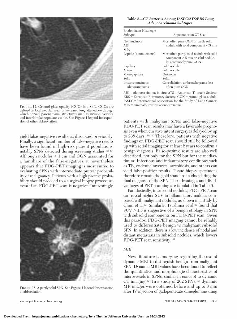
Figure 16. A, Lepidic predominant adenocarcinoma with lepidic component and acinar component. B, Lepidic predominant ade-nocarcinoma. Most of the image shows lepidic adenocarcinoma with small foci of invasive acinar adenocarcinoma. C, This pattern of invasive acinar adenocarcinoma shows a desmoplastic stromal response.
Downloaded From: http://journal.publications.chestnet.org/ by a Thomas Jefferson University User on 05/24/2013
journal.publications.chestnet.org CHEST / 143 / 3 / MARCH 2013 835
patients with malignant SPNs and false-negative FDG-PET scan results may have a favorable progno-sis even when curative intent surgery is delayed by up to 238 days. 119,120 Therefore, patients with negative fi ndings on FDG-PET scan should still be followed up with serial imaging for at least 2 years to confi rm a benign diagnosis. False-positive results are also well described, not only for the SPN but for the medias-tinum: Infectious and infl ammatory conditions such as TB, endemic mycoses, sarcoidosis, and others can yield false-positive results. Tissue biopsy specimens therefore remain the gold standard in elucidating the fi nal diagnosis of the SPN. The advantages and disad-vantages of PET scanning are tabulated in Table 6 .
Paradoxically, in subsolid nodules, FDG-PET scan can reveal higher SUV in infl ammatory nodules com-pared with malignant nodules, as shown in a study by Chun et al. 121 Similarly, Tsushima et al 122 found that SUV . 1.5 is suggestive of a benign etiology in SPN with subsolid components on FDG-PET scan. Given this paradox, FDG-PET imaging cannot be reliably used to differentiate benign vs malignant subsolid SPN. In addition, there is a low incidence of nodal and distant metastasis in subsolid nodules, which lowers FDG-PET scan sensitivity. 123
MRI
New literature is emerging regarding the use of dynamic MRI to distinguish benign from malignant SPN. Dynamic MRI values have been found to refl ect the quantitative and morphologic characteristics of microvessels in SPNs, similar in concept to dynamic CT imaging. 124 In a study of 202 SPNs, 125 dynamic MR images were obtained before and up to 8 min after IV injection of gadopentetate dimeglumine using
yield false-negative results, as discussed previously. Finally, a signifi cant number of false-negative results have been found in high-risk patient populations, notably SPNs detected during screening studies. 116-118 Although nodules , 1 cm and GGN accounted for a fair share of the false-negatives, it nevertheless appears that FDG-PET imaging is most suited to evaluating SPNs with intermediate pretest probabil-ity of malignancy. Patients with a high pretest proba-bility should proceed to a surgical biopsy procedure even if an FDG-PET scan is negative. Interestingly,
Figure 17. Ground glass opacity (GGO) in a SPN. GGOs are defi ned as focal nodular areas of increased lung attenuation through which normal parenchymal structures such as airways, vessels, and interlobular septa are visible. See Figure 1 legend for expan-sion of other abbreviation.
Figure 18. A partly solid SPN. See Figure 1 legend for expansion of abbreviation.
Table 5— CT Patterns Among IASLC/ATS/ERS Lung Adenocarcinoma Subtypes
Predominant Histologic Subtype Appearance on CT Scan
Nonmucinous Most often pure GGN or partly solid nodule with solid component , 5 mmAIS
MIALepidic (nonmucinous) Most often partly solid nodule with solid
component . 5 mm or solid nodule; less commonly pure GGN
Papillary Solid noduleAcinar Solid noduleMicropapillary UnknownSolid SolidInvasive mucinous
adenocarcinomaConsolidation, air bronchograms; less
often pure GGN
AIS 5 adenocarcinoma in situ; ATS 5 American Thoracic Society; ERS 5 European Respiratory Society; GGN 5 ground glass nodule; IASLC 5 International Association for the Study of Lung Cancer; MIA 5 minimally invasive adenocarcinoma.
Downloaded From: http://journal.publications.chestnet.org/ by a Thomas Jefferson University User on 05/24/2013
836 Special Features
Acknowledgments Financial/nonfi nancial disclosures: The authors have reported to CHEST that no potential confl icts of interest exist with any companies/organizations whose products or services may be dis-cussed in this article. Other contributions: The authors wish to thank Patrice Balistreri, administrative assistant, for her invaluable clerical assistance.
References 1 . Tuddenham WJ . Glossary of terms for thoracic radiology:
recommendations of the Nomenclature Committee of the Fleischner Society . AJR Am J Roentgenol . 1984 ; 143 ( 3 ): 509 - 517 .
2 . Shiau MC , Portnoy E , Garay SM . Management of solitary pulmonary nodules . In: Kanne JP , ed. Clinically Oriented Pulmonary Imaging . New York, NY : Springer ; 2012 : 19 - 27 .
3 . Holin SM , Dwork RE , Glaser S , Rikli AE , Stocklen JB . Solitary pulmonary nodules found in a community-wide chest roentgenographic survey; a fi ve-year follow-up study . Am Rev Tuberc . 1959 ; 79 ( 4 ): 427 - 439 .
4 . Swensen SJ , Silverstein MD , Edell ES , et al . Solitary pulmo-nary nodules: clinical prediction model versus physicians . Mayo Clin Proc . 1999 ; 74 ( 4 ): 319 - 329 .
5 . Henschke CI , McCauley DI , Yankelevitz DF , et al . Early Lung Cancer Action Project: overall design and fi ndings from baseline screening . Lancet . 1999 ; 354 ( 9173 ): 99 - 105 .
6 . Veronesi G , Bellomi M , Spaggiari L , et al . Low dose spiral computed tomography for early diagnosis of lung cancer. Results of baseline screening in 5,000 high-risk volunteers . J Clin Oncol . 2006 ; 24 (18S): 7029 .
7 . Henschke CI , Yankelevitz DF , Naidich DP , et al . CT screen-ing for lung cancer: suspiciousness of nodules according to size on baseline scans . Radiology . 2004 ; 231 ( 1 ): 164 - 168 .
8 . Gohagan J , Marcus P , Fagerstrom R , Pinsky P , Kramer B , Prorok P ; Writing Committee, Lung Screening Study Research Group . Baseline fi ndings of a randomized fea-sibility trial of lung cancer screening with spiral CT scan vs chest radiograph: the Lung Screening Study of the National Cancer Institute . Chest . 2004 ; 126 ( 1 ): 114 - 121 .
9 . Li F , Sone S , Abe H , Macmahon H , Doi K . Malignant versus benign nodules at CT screening for lung cancer: comparison of thin-section CT fi ndings . Radiology . 2004 ; 233 ( 3 ): 793 - 798 .
10 . Swensen SJ , Jett JR , Hartman TE , et al . Lung cancer screen-ing with CT: Mayo Clinic experience . Radiology . 2003 ; 226 ( 3 ): 756 - 761 .
11 . Nawa T , Nakagawa T , Kusano S , Kawasaki Y , Sugawara Y , Nakata H . Lung cancer screening using low-dose spiral CT: results of baseline and 1-year follow-up studies . Chest . 2002 ; 122 ( 1 ): 15 - 20 .
T1-weighted spin-echo sequences. With � 110% max-imum enhancement ratio as a cutoff value, the PPV for malignancy was 92%; sensitivity, 63%; and speci-fi city, 74%. With � 13.5%/min slope as a cutoff value, sensitivity, specifi city, PPV, and NPV for malignancy were 94%, 96%, 99%, and 74%, respectively. A pro-spective study of 175 patients with 202 SPNs 126 was recently performed comparing dynamic MRI, dynamic CT imaging, and PET-CT imaging to distinguish benign from malignant SPNs. On dynamic MRI, with a slope of enhancement ratio cutoff of 0.04/s, the sensitivity, specifi city, PPV, NPV, and accuracy were 96.3%, 82.1%, 95.7%, 84.2%, and 93.6%, respectively. These results were superior to combined PET-CT and dynamic CT imaging, and have opened up a prom-ising new avenue of research in the use of imaging modalities to characterize SPNs.
Conclusion
Morphologic characteristics of SPN can help distin-guish benign from malignant SPNs. These features include volume doubling time, size, margins, pres-ence and nature of calcifi cation, presence of fat, and wall thickness in case of cavitation. GGNs, while usually multiple, are further classifi ed as pure ground glass and partly solid. Thin-section CT scans allow accurate delineation of these morphologic features. In conjunction, FDG-PET scans provide a window to the metabolic activity of the nodules, giving valuable pathophysiologic information about them.
Figure 19. Combined PET-CT scan showing 18 F-2-deoxy-2-fl uoro- d -glucose-avid SPN. Malignant cells are more metabolically active and import glucose more avidly than other tissues. See Figure 1 legend for expansion of abbreviation.
Table 6— Advantages and Disadvantages of FDG-PET Imaging
Advantages Disadvantages
Accurate noninvasive evaluationWhole-body image detects
extrapulmonary tumorsAbility to stage known lung cancer
Lower sensitivity for lesions , 8 mm
False positives from infl ammation
False negatives from tumors with low metabolic rate
FDG 5 18 F-2-deoxy-2-fl uoro- d -glucose.
Downloaded From: http://journal.publications.chestnet.org/ by a Thomas Jefferson University User on 05/24/2013

journal.publications.chestnet.org CHEST / 143 / 3 / MARCH 2013 837
31 . Lindell RM , Hartman TE , Nadrous HF , Ryu JH . Pulmonary cryptococcosis: CT fi ndings in immunocompetent patients . Radiology . 2005 ; 236 ( 1 ): 326 - 331 .
32 . Fox DL , Müller NL . Pulmonary cryptococcosis in immuno-competent patients: CT fi ndings in 12 patients . AJR Am J Roentgenol . 2005 ; 185 ( 3 ): 622 - 626 .
33 . Murayama S , Sakai S , Soeda H , et al . Pulmonary cryptococ-cosis in immunocompetent patients: HRCT characteristics . Clin Imaging . 2004 ; 28 ( 3 ): 191 - 195 .
34 . Nathan MH , Collins VP , Adams RA . Differentiation of benign and malignant pulmonary nodules by growth rate . Radiology . 1962 ; 79 : 221 - 232 .
35 . Garland LH , Coulson W , Wollin E . The rate of growth and apparent duration of untreated primary bronchial carci-noma . Cancer . 1963 ; 16 : 694 - 707 .
36 . Steele JD , Buell P . Asymptomatic solitary pulmonary nod-ules. Host survival, tumor size, and growth rate . J Thorac Cardiovasc Surg . 1973 ; 65 ( 1 ): 140 - 151 .
37 . Weiss W . Tumor doubling time and survival of men with bronchogenic carcinoma . Chest . 1974 ; 65 ( 1 ): 3 - 8 .
38 . Friberg S , Mattson S . On the growth rates of human malig-nant tumors: implications for medical decision making . J Surg Oncol . 1997 ; 65 ( 4 ): 284 - 297 .
39 . Bach PB , Silvestri GA , Hanger M . Screening for lung cancer: ACCP evidence-based clinical practice guidelines (2nd edition) . Chest . 2007 ; 132 ( suppl 3 ): 69S - 77S .
40 . Soubani AO . The evaluation and management of the solitary pulmonary nodule . Postgrad Med J . 2008 ; 84 ( 995 ): 459 - 466 .
41 . Nietert PJ , Ravenel JG , Leue WM , et al . Imprecision in auto-mated volume measurements of pulmonary nodules and its effect on the level of uncertainty in volume doubling time estimation . Chest . 2009 ; 135 ( 6 ): 1580 - 1587 .
42 . Yankelevitz DF , Henschke CI . Small solitary pulmonary nodules . Radiol Clin North Am . 2000 ; 38 ( 3 ): 471 - 478 .
43 . Erasmus JJ , McAdams HP , Connolly JE . Solitary pulmonary nodules: part II. Evaluation of the indeterminate nodule . Radiographics . 2000 ; 20 ( 1 ): 59 - 66 .
44 . Revel MP , Merlin A , Peyrard S , et al . Software volumetric evaluation of doubling times for differentiating benign ver-sus malignant pulmonary nodules . AJR Am J Roentgenol . 2006 ; 187 ( 1 ): 135 - 142 .
45 . Park CM , Goo JM , Lee HJ , Kim KG , Kang MJ , Shin YH . Persistent pure ground-glass nodules in the lung: interscan variability of semiautomated volume and attenuation mea-surements . AJR Am J Roentgenol . 2010 ; 195 ( 6 ): W408 - W414 .
46 . Singh S , Pinsky P , Fineberg NS , et al . Evaluation of reader variability in the interpretation of follow-up CT scans at lung cancer screening . Radiology . 2011 ; 259 ( 1 ): 263 - 270 .
47 . Zerhouni EA , Stitik FP , Siegelman SS , et al . CT of the pulmonary nodule: a cooperative study . Radiology . 1986 ; 160 ( 2 ): 319 - 327 .
48 . MacMahon H , Austin JH , Gamsu G , et al ; Fleischner Society . Guidelines for management of small pulmonary nod-ules detected on CT scans: a statement from the Fleischner Society . Radiology . 2005 ; 237 ( 2 ): 395 - 400 .
49 . Swensen SJ , Viggiano RW , Midthun DE , et al . Lung nod-ule enhancement at CT: multicenter study . Radiology . 2000 ; 214 ( 1 ): 73 - 80 .
50 . Winer-Muram HT . The solitary pulmonary nodule . Radiology . 2006 ; 239 ( 1 ): 34 - 49 .
51 . Furuya K , Murayama S , Soeda H , et al . New classifi cation of small pulmonary nodules by margin characteristics on high-resolution CT . Acta Radiol . 1999 ; 40 ( 5 ): 496 - 504 .
52 . Seemann MD , Seemann O , Luboldt W , et al . Differentiation of malignant from benign solitary pulmonary lesions using chest radiography, spiral CT and HRCT . Lung Cancer . 2000 ; 29 ( 2 ): 105 - 124 .
12 . Henschke CI , Yankelevitz DF , Libby DM , et al . Early lung cancer action project: annual screening using single-slice helical CT . Ann N Y Acad Sci . 2001 ; 952 : 124 - 134 .
13 . Diederich S , Wormanns D , Lenzen H , Semik M , Thomas M , Peters PE . Screening for asymptomatic early bronchogenic carcinoma with low dose CT of the chest . Cancer . 2000 ; 89 ( suppl 11 ): 2483 - 2484 .
14 . Wahidi MM , Govert JA , Goudar RK . Evidence for the treat-ment of patients with pulmonary nodules: when is it lung cancer?: ACCP evidence-based clinical practice guidelines (2nd edition) . Chest . 2007 ; 132 ( suppl 3 ): 94S - 107S .
15 . Patel VK, Naik SK, Naidich DP, et al . A practical algorith-mic approach to the diagnosis of solitary pulmonary nod-ules: part 2: pretest probability and algorithm . Chest . 2013 ; 143 ( 3 ): 840 - 846 .
16 . Gurney JW . Determining the likelihood of malignancy in solitary pulmonary nodules with Bayesian analysis. Part I. Theory . Radiology . 1993 ; 186 ( 2 ): 405 - 413 .
17 . Swensen SJ , Silverstein MD , Ilstrup DM , Schleck CD , Edell ES . The probability of malignancy in solitary pulmo-nary nodules. Application to small radiologically indetermi-nate nodules . Arch Intern Med . 1997 ; 157 ( 8 ): 849 - 855 .
18 . Gould MK , Ananth L , Barnett PG ; Veterans Affairs SNAP Cooperative Study Group . A clinical model to estimate the pretest probability of lung cancer in patients with solitary pulmonary nodules . Chest . 2007 ; 131 ( 2 ): 383 - 388 .
19 . Peto R , Lopez AD , Boreham J . Mortality From Smoking in Developed Countries 1950-2000. Indirect Estimates From National Vital Statistics . Oxford, England : Oxford University Press ; 1994 .
20 . Quint LE , Park CH , Iannettoni MD . Solitary pulmo-nary nodules in patients with extrapulmonary neoplasms . Radiology . 2000 ; 217 ( 1 ): 257 - 261 .
21 . Sortini A , Carcoforo P , Ascanelli S , Sortini D , Pozza E . Signifi cance of a single pulmonary nodule in patients with previous history of malignancy . Eur J Cardiothorac Surg . 2001 ; 20 ( 6 ): 1101 - 1105 .
22 . Mery CM , Pappas AN , Bueno R , et al . Relationship between a history of antecedent cancer and the probability of malig-nancy for a solitary pulmonary nodule . Chest . 2004 ; 125 ( 6 ): 2175 - 2181 .
23 . Bouros D , Hatzakis K , Labrakis H , Zeibecoglou K . Asso-ciation of malignancy with diseases causing interstitial pul-monary changes . Chest . 2002 ; 121 ( 4 ): 1278 - 1289 .
24 . Kawai T , Yakumaru K , Suzuki M , Kageyama K . Diffuse interstitial pulmonary fi brosis and lung cancer . Acta Pathol Jpn . 1987 ; 37 ( 1 ): 11 - 19 .
25 . Nagai A , Chiyotani A , Nakadate T , Konno K . Lung cancer in patients with idiopathic pulmonary fi brosis . Tohoku J Exp Med . 1992 ; 167 ( 3 ): 231 - 237 .
26 . Aubry ME , Myers JL , Douglas WW . Primary pulmonary carcinomas in patients with idiopathic pulmonary fi brosis . Mayo Clin Proc . 2002 ; 77 ( 8 ): 77 .
27 . Kawasaki H , Nagai K , Yokose T , et al . Clinicopathological characteristics of surgically resected lung cancer associated with idiopathic pulmonary fi brosis . J Surg Oncol . 2001 ; 76 ( 1 ): 53 - 57 .
28 . Turner-Warwick M , Lebowitz M , Burrows B , Johnson A . Cryptogenic fi brosing alveolitis and lung cancer . Thorax . 1980 ; 35 ( 7 ): 496 - 499 .
29 . Kishi K , Homma S , Kurosaki A , Motoi N , Yoshimura K . High-resolution computed tomography fi ndings of lung can-cer associated with idiopathic pulmonary fi brosis . J Comput Assist Tomogr . 2006 ; 30 ( 1 ): 95 - 99 .
30 . Kim KI , Leung AN , Flint JD , Müller NL . Chronic pulmonary coccidioidomycosis: computed tomographic and pathologic fi ndings in 18 patients . Can Assoc Radiol J . 1998 ; 49 ( 6 ): 401 - 407 .
Downloaded From: http://journal.publications.chestnet.org/ by a Thomas Jefferson University User on 05/24/2013
838 Special Features
75 . Saito H , Yamada K , Hamanaka N , et al . Initial fi ndings and progression of lung adenocarcinoma on serial computed tomography scans . J Comput Assist Tomogr . 2009 ; 33 ( 1 ): 42 - 48 .
76 . Kui M , Templeton PA , White CS , Cai ZL , Bai YX , Cai YQ . Evaluation of the air bronchogram sign on CT in solitary pul-monary lesions . J Comput Assist Tomogr . 1996 ; 20 ( 6 ): 983 - 986 .
77 . Kuriyama K , Tateishi R , Doi O , et al . Prevalence of air bronchograms in small peripheral carcinomas of the lung on thin-section CT: comparison with benign tumors . AJR Am J Roentgenol . 1991 ; 156 ( 5 ): 921 - 924 .
78 . Godoy MC , Naidich DP . Subsolid pulmonary nodules and the spectrum of peripheral adenocarcinomas of the lung: recommended interim guidelines for assessment and man-agement . Radiology . 2009 ; 253 ( 3 ): 606 - 622 .
79 . Travis WD , Brambilla E , Noguchi M , et al . International Association for the Study of Lung Cancer/American Thoracic Society/European Respiratory Society international multi-disciplinary classifi cation of lung adenocarcinoma . J Thorac Oncol . 2011 ; 6 ( 2 ): 244 - 285 .
80 . Takashima S , Sone S , Li F , Maruyama Y , Hasegawa M , Kadoya M . Indeterminate solitary pulmonary nodules revealed at population-based CT screening of the lung: using fi rst follow-up diagnostic CT to differentiate benign and malig-nant lesions . AJR Am J Roentgenol . 2003 ; 180 ( 5 ): 1255 - 1263 .
81 . Tsunezuka Y , Shimizu Y , Tanaka N , Takayanagi T , Kawano M . Positron emission tomography in relation to Noguchi’s clas-sifi cation for diagnosis of peripheral non-small-cell lung can-cer 2 cm or less in size . World J Surg . 2007 ; 31 ( 2 ): 314 - 317 .
82 . Noguchi M , Morikawa A , Kawasaki M , et al . Small adeno-carcinoma of the lung. Histologic characteristics and prog-nosis . Cancer . 1995 ; 75 ( 12 ): 2844 - 2852 .
83 . Yap CS , Schiepers C , Fishbein MC , Phelps ME , Czernin J . FDG-PET imaging in lung cancer: how sensitive is it for bronchioloalveolar carcinoma? Eur J Nucl Med Mol Imaging . 2002 ; 29 ( 9 ): 1166 - 1173 .
84 . Ichiya Y , Kuwabara Y , Sasaki M , et al . A clinical evaluation of FDG-PET to assess the response in radiation therapy for bronchogenic carcinoma . Ann Nucl Med . 1996 ; 10 ( 2 ): 193 - 200 .
85 . Lowe VJ , Fletcher JW , Gobar L , et al . Prospective inves-tigation of positron emission tomography in lung nodules . J Clin Oncol . 1998 ; 16 ( 3 ): 1075 - 1084 .
86 . Lowe VJ , Hoffman JM , DeLong DM , Patz EF , Coleman RE . Semiquantitative and visual analysis of FDG-PET images in pulmonary abnormalities . J Nucl Med . 1994 ; 35 ( 11 ): 1771 - 1776 .
87 . Matthies A , Hickeson M , Cuchiara A , Alavi A . Dual time point 18F-FDG PET for the evaluation of pulmonary nod-ules . J Nucl Med . 2002 ; 43 ( 7 ): 871 - 875 .
88 . Keyes JW Jr . SUV: standard uptake or silly useless value? J Nucl Med . 1995 ; 36 ( 10 ): 1836 - 1839 .
89 . Ketai L , Malby M , Jordan K , Meholic A , Locken J . Small nodules detected on chest radiography: does size predict calcifi cation? Chest . 2000 ; 118 ( 3 ): 610 - 614 .
90 . Gould M , Fletcher J , Iannettonin M . Evaluation of patients with pulmonary nodules: When is it lung cancer?: ACCP evidence-based clinical practice guidelines (2nd edition) . Chest . 2007 ; 132 ( suppl 3 ): 108S - 130S .
91 . Muhm JR , Miller WE , Fontana RS , Sanderson DR , Uhlenhopp MA . Lung cancer detected during a screening program using four-month chest radiographs . Radiology . 1983 ; 148 ( 3 ): 609 - 615 .
92 . Quekel LG , Kessels AG , Goei R , van Engelshoven JM . Miss rate of lung cancer on the chest radiograph in clinical practice . Chest . 1999 ; 115 ( 3 ): 720 - 724 .
93 . Shah PK , Austin JH , White CS , et al . Missed non-small cell lung cancer: radiographic fi ndings of potentially resectable
53 . Zwirewich CV , Vedal S , Miller RR , Müller NL . Solitary pul-monary nodule: high-resolution CT and radiologic-pathologic correlation . Radiology . 1991 ; 179 ( 2 ): 469 - 476 .
54 . Siegelman SS , Zerhouni EA , Leo FP , Khouri NF , Stitik FP . CT of the solitary pulmonary nodule . AJR Am J Roentgenol . 1980 ; 135 ( 1 ): 1 - 13 .
55 . Khan A , Herman PG , Vorwerk P , Stevens P , Rojas KA , Graver M . Solitary pulmonary nodules: comparison of classi-fi cation with standard, thin-section, and reference phantom CT . Radiology . 1991 ; 179 ( 2 ): 477 - 481 .
56 . Huston J III , Muhm JR . Solitary pulmonary opacities: plain tomography . Radiology . 1987 ; 163 ( 2 ): 481 - 485 .
57 . Kuhlman JE , Fishman EK , Siegelman SS . Invasive pulmo-nary aspergillosis in acute leukemia: characteristic fi ndings on CT, the CT halo sign, and the role of CT in early diagno-sis . Radiology . 1985 ; 157 ( 3 ): 611 - 614 .
58 . Primack SL , Hartman TE , Lee KS , Müller NL . Pulmonary nodules and the CT halo sign . Radiology . 1994 ; 190 ( 2 ): 513 - 515 .
59 . Ikehara M , Saito H , Kondo T , et al . Comparison of thin-section CT and pathological fi ndings in small solid-density type pulmonary adenocarcinoma: prognostic factors from CT fi ndings . Eur J Radiol . 2012 ; 81 ( 1 ): 189 - 194 .
60 . Yankelevitz DF , Henschke CI . Derivation for relating calci-fi cation and size in small pulmonary nodules . Clin Imaging . 1998 ; 22 ( 1 ): 1 - 6 .
61 . Takamochi K , Yokose T , Yoshida J , et al . Calcifi cation in large cell neuroendocrine carcinoma of the lung . Jpn J Clin Oncol . 2003 ; 33 ( 1 ): 10 - 13 .
62 . Cardinale L , Ardissone F , Novello S , et al . The pulmonary nodule: clinical and radiological characteristics affecting a diagnosis of malignancy . Radiol Med (Torino) . 2009 : 114 ( 6 ): 871 - 879 .
63 . Mahoney MC Sr , Shipley RT , Corcoran HL , Dickson BA . CT demonstration of calcifi cation in carcinoma of the lung . AJR Am J Roentgenol . 1990 ; 154 ( 2 ): 255 - 258 .
64 . Stewart JG , MacMahon H , Vyborny CJ , Pollak ER . Dystro-phic calcifi cation in carcinoma of the lung: demonstration by CT . AJR Am J Roentgenol . 1987 ; 148 ( 1 ): 29 - 30 .
65 . Grewal RG , Austin JH . CT demonstration of calcifi cation in carcinoma of the lung . J Comput Assist Tomogr . 1994 ; 18 ( 6 ): 867 - 871 .
66 . Mazzone PJ , Stoller JK . The pulmonologist’s perspective regarding the solitary pulmonary nodule . Semin Thorac Cardiovasc Surg . 2002 ; 14 ( 3 ): 250 - 260 .
67 . Trotman-Dickenson B , Baumert B . Multidetector-row CT of the solitary pulmonary nodule . Semin Roentgenol . 2003 ; 38 ( 2 ): 158 - 167 .
68 . McLoud TC , Swenson SJ . Lung carcinoma . Clin Chest Med . 1999 ; 20 ( 4 ): 697 - 713 .
69 . Siegelman SS , Khouri NF , Scott WW Jr , et al . Pulmo nary hamartoma: CT fi ndings . Radiology . 1986 ; 160 ( 2 ): 313 - 317 .
70 . Klein JS , Braff S . Imaging evaluation of the solitary pulmo-nary nodule . Clin Chest Med . 2008 ; 29 ( 1 ): 15 - 38 .
71 . Muram TM , Aisen A . Fatty metastatic lesions in 2 patients with renal clear-cell carcinoma . J Comput Assist Tomogr . 2003 ; 27 ( 6 ): 869 - 870 .
72 . Woodring JH , Fried AM , Chuang VP . Solitary cavities of the lung: diagnostic implications of cavity wall thickness . AJR Am J Roentgenol . 1980 ; 135 ( 6 ): 1269 - 1271 .
73 . Woodring JH , Fried AM . Signifi cance of wall thickness in solitary cavities of the lung: a follow-up study . AJR Am J Roentgenol . 1983 ; 140 ( 3 ): 473 - 474 .
74 . Mirtcheva RM , Vazquez M , Yankelevitz DF , Henschke CI . Bronchioloalveolar carcinoma and adenocarcinoma with bronchioloalveolar features presenting as ground-glass opac-ities on CT . Clin Imaging . 2002 ; 26 ( 2 ): 95 - 100 .
Downloaded From: http://journal.publications.chestnet.org/ by a Thomas Jefferson University User on 05/24/2013
journal.publications.chestnet.org CHEST / 143 / 3 / MARCH 2013 839
111 . Kagna O , Solomonov A , Keidar Z , et al . The value of FDG-PET/CT in assessing single pulmonary nodules in patients at high risk of lung cancer . Eur J Nucl Med Mol Imaging . 2009 ; 36 ( 6 ): 997 - 1004 .
112 . Kim SK , Allen-Auerbach M , Goldin J , et al . Accuracy of PET/CT in characterization of solitary pulmonary lesions . J Nucl Med . 2007 ; 48 ( 2 ): 214 - 220 .
113 . Shin KM , Lee KS , Shim YM , et al . FDG PET/CT and mediastinal nodal metastasis detection in stage T1 non-small cell lung cancer: prognostic implications . Korean J Radiol . 2008 ; 9 ( 6 ): 481 - 489 .
114 . Kim BT , Lee KS , Shim SS , et al . Stage T1 non-small cell lung cancer: preoperative mediastinal nodal staging with integrated FDG PET/CT—a prospective study . Radiology . 2006 ; 241 ( 2 ): 501 - 509 .
115 . Herder GJ , Golding RP , Hoekstra OS , et al . The perfor-mance of (18)F-fl uorodeoxyglucose positron emission tomog-raphy in small solitary pulmonary nodules . Eur J Nucl Med Mol Imaging . 2004 ; 31 ( 9 ): 1231 - 1236 .
116 . Pastorino U , Bellomi M , Landoni C , et al . Early lung-cancer detection with spiral CT and positron emission tomography in heavy smokers: 2-year results . Lancet . 2003 ; 362 ( 9384 ): 593 - 597 .
117 . Nomori H , Watanabe K , Ohtsuka T , Naruke T , Suemasu K , Uno K . Evaluation of F-18 fl uorodeoxyglucose (FDG) PET scanning for pulmonary nodules less than 3 cm in diameter, with special reference to the CT images . Lung Cancer . 2004 ; 45 ( 1 ): 19 - 27 .
118 . Lindell RM , Hartman TE , Swensen SJ , et al . Lung can-cer screening experience: a retrospective review of PET in 22 non-small cell lung carcinomas detected on screening chest CT in a high-risk population . AJR Am J Roentgenol . 2005 ; 185 ( 1 ): 126 - 131 .
119 . Marom EM , Sarvis S , Herndon JE 2nd, Patz EF Jr . T1 lung cancers: sensitivity of diagnosis with fl uorodeoxyglucose PET . Radiology . 2002 ; 223 ( 2 ): 453 - 459 .
120 . Cheran SK , Nielsen ND , Patz EF Jr . False-negative fi nd-ings for primary lung tumors on FDG positron emission tomography: staging and prognostic implications . AJR Am J Roentgenol . 2004 ; 182 ( 5 ): 1129 - 1132 .
121 . Chun EJ , Lee HJ , Kang WJ , et al . Differentiation between malignancy and infl ammation in pulmonary ground-glass nodules: the feasibility of integrated (18)F-FDG PET/CT . Lung Cancer . 2009 ; 65 ( 2 ): 180 - 186 .
122 . Tsushima Y , Tateishi U , Uno H , et al . Diagnostic performance of PET/CT in differentiation of malignant and benign non-solid solitary pulmonary nodules . Ann Nucl Med . 2008 ; 22 ( 7 ): 571 - 577 .
123 . Kim TJ , Park CM , Goo JM , Lee KW . Is there a role for FDG PET in the management of lung cancer manifesting pre-dominantly as ground-glass opacity? AJR Am J Roentgenol . 2012 ; 198 ( 1 ): 83 - 88 .
124 . Zou Y , Zhang M , Wang Q , Shang D , Wang L , Yu G . Quan-titative investigation of solitary pulmonary nodules: dynamic contrast-enhanced MRI and histopathologic analysis . AJR Am J Roentgenol . 2008 ; 191 ( 1 ): 252 - 259 .
125 . Kono R , Fujimoto K , Terasaki H , et al . Dynamic MRI of solitary pulmonary nodules: comparison of enhancement patterns of malignant and benign small peripheral lung lesions . AJR Am J Roentgenol . 2007 ; 188 ( 1 ): 26 - 36 .
126 . Ohno Y , Koyama H , Takenaka D , et al . Dynamic MRI, dynamic multidetector-row computed tomography (MDCT), and coregistered 2-[fl uorine-18]-fl uoro-2-deoxy-D-glucose-positron emission tomography (FDG-PET)/CT: comparative study of capability for management of pulmonary nodules . J Magn Reson Imaging . 2008 ; 27 ( 6 ): 1284 - 1295 .
lesions evident only in retrospect . Radiology . 2003 ; 226 ( 1 ): 235 - 241 .
94 . Bayraktaroglu S , Savaș R , Basoglu OK , et al . Dynamic com-puted tomography in solitary pulmonary nodules . J Comput Assist Tomogr . 2008 ; 32 ( 2 ): 222 - 227 .
95 . The American Thoracic Society and The European Respira-tory Society . Pretreatment evaluation of non-small-cell lung cancer . Am J Respir Crit Care Med . 1997 ; 156 ( 1 ): 320 - 332 .
96 . Seely JM , Mayo JR , Miller RR , Müller NL . T1 lung can-cer: prevalence of mediastinal nodal metastases and diag-nostic accuracy of CT . Radiology . 1993 ; 186 ( 1 ): 129 - 132 .
97 . Tahara RW , Lackner RP , Graver LM . Is there a role for routine mediastinoscopy in patients with peripheral T1 lung cancers? Am J Surg . 2000 ; 180 ( 6 ): 488 - 491 .
98 . Choi YS , Shim YM , Kim J , Kim K . Mediastinoscopy in patients with clinical stage I non-small cell lung cancer . Ann Thorac Surg . 2003 ; 75 ( 2 ): 364 - 366 .
99 . Silvestri GA , Gould MK , Margolis ML , et al . Noninvasive staging of non-small cell lung cancer: ACCP evidence-based clinical practice guidelines (2nd edition) . Chest . 2007 ; 132 ( suppl 3 ): 178S - 201S .
100 . Gould MK , Kuschner WG , Rydzak CE , et al . Test performance of positron emission tomography and computed tomography for mediastinal staging in patients with non-small-cell lung can-cer: a meta-analysis . Ann Intern Med . 2003 ; 139 ( 11 ): 879 - 892 .
101 . Dwamena BA , Sonnad SS , Angobaldo JO , Wahl RL . Metas-tases from non-small cell lung cancer: mediastinal staging in the 1990s—meta-analytic comparison of PET and CT . Radiology . 1999 ; 213 ( 2 ): 530 - 536 .
102 . Shim SS , Lee KS , Chung MJ , Kim H , Kwon OJ , Kim S . Do hemodynamic studies of stage T1 lung cancer enable the prediction of hilar or mediastinal nodal metastasis? AJR Am J Roentgenol . 2006 ; 186 ( 4 ): 981 - 988 .
103 . Reed CE , Harpole DH , Posther KE , et al ; American College of Surgeons Oncology Group Z0050 trial . Results of the American College of Surgeons Oncology Group Z0050 trial: the utility of positron emission tomography in staging potentially operable non-small cell lung cancer . J Thorac Cardiovasc Surg . 2003 ; 126 ( 6 ): 1943 - 1951 .
104 . Aberle DR , Adams AM , Berg CD , et al ; National Lung Screening Trial Research Team . Reduced lung-cancer mor-tality with low-dose computed tomographic screening . N Engl J Med . 2011 ; 365 ( 5 ): 395 - 409 .
105 . Vansteenkiste JF , Stroobants SS . PET scan in lung cancer: current recommendations and innovation . J Thorac Oncol . 2006 ; 1 ( 1 ): 71 - 73 .
106 . Gould MK , Maclean CC , Kuschner WG , Rydzak CE , Owens DK . Accuracy of positron emission tomography for diagnosis of pulmonary nodules and mass lesions: a meta-analysis . JAMA . 2001 ; 285 ( 7 ): 914 - 924 .
107 . Fletcher JW , Kymes SM , Gould M , et al ; VA SNAP Coopera-tive Studies Group . A comparison of the diagnostic accuracy of 18F-FDG PET and CT in the characterization of solitary pulmonary nodules . J Nucl Med . 2008 ; 49 ( 2 ): 179 - 185 .
108 . Jeong SY , Lee KS , Shin KM , et al . Effi cacy of PET/CT in the characterization of solid or partly solid solitary pulmo-nary nodules . Lung Cancer . 2008 ; 61 ( 2 ): 186 - 194 .
109 . Yi CA , Lee KS , Kim BT , et al . Tissue characterization of solitary pulmonary nodule: comparative study between heli-cal dynamic CT and integrated PET/CT . J Nucl Med . 2006 ; 47 ( 3 ): 443 - 450 .
110 . Bar-Shalom R , Kagna O , Israel O , Guralnik L . Noninvasive diagnosis of solitary pulmonary lesions in cancer patients based on 2-fl uoro-2-deoxy-D-glucose avidity on positron emission tomography/computed tomography . Cancer . 2008 ; 113 ( 11 ): 3213 - 3221 .
Downloaded From: http://journal.publications.chestnet.org/ by a Thomas Jefferson University User on 05/24/2013