Embed Size (px)
Citation preview
Cholestasis in Infancy
Pathologic Findings and
Differential Diagnosis
Atif Ahmed, MD
Children’s Mercy Hospital, Kansas City
University of Missouri
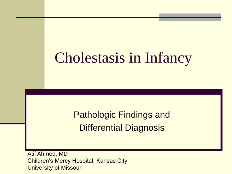
Infantile Cholestasis Conjugated hyperbilirubinemia and jaundice
May be noted in the first 2 wk of life.
Infants present with jaundice, dark urine (conjugated bilirubin), acholic stools, and hepatomegaly.
In chronic cases: chronic pruritus, symptoms and signs of fat-soluble vitamin deficiency, slow or decreased growth charts.
In older infants: signs of hepatic fibrosis and cirrhosis: portal hypertension, abdominal distention from ascites, dilated abdominal veins, and esophageal varices may develop.
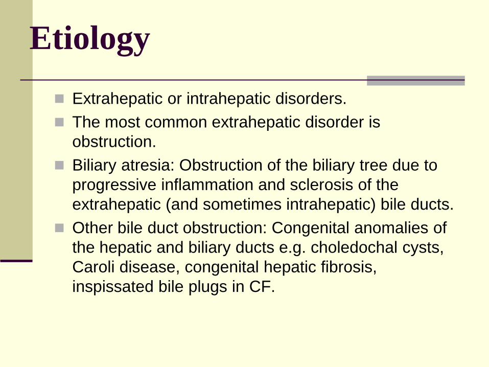
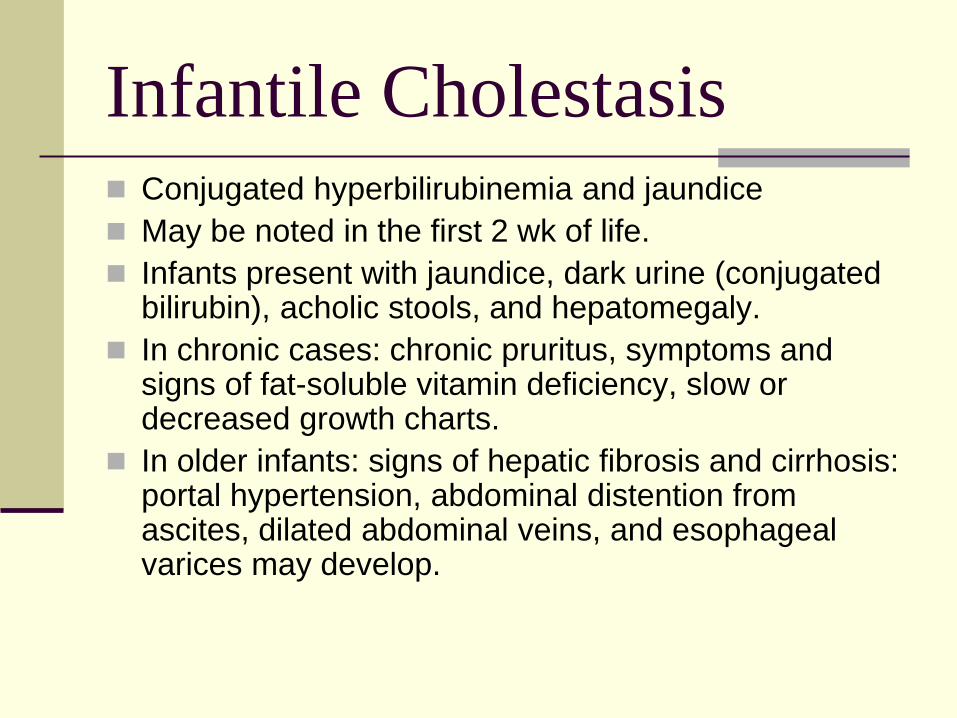
Etiology
Extrahepatic or intrahepatic disorders.
The most common extrahepatic disorder is
obstruction.
Biliary atresia: Obstruction of the biliary tree due to
progressive inflammation and sclerosis of the
extrahepatic (and sometimes intrahepatic) bile ducts.
Other bile duct obstruction: Congenital anomalies of
the hepatic and biliary ducts e.g. choledochal cysts,
Caroli disease, congenital hepatic fibrosis,
inspissated bile plugs in CF.
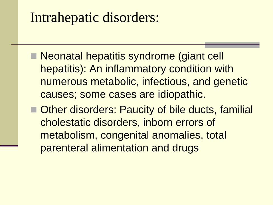
Intrahepatic disorders:
Neonatal hepatitis syndrome (giant cell
hepatitis): An inflammatory condition with
numerous metabolic, infectious, and genetic
causes; some cases are idiopathic.
Other disorders: Paucity of bile ducts, familial
cholestatic disorders, inborn errors of
metabolism, congenital anomalies, total
parenteral alimentation and drugs
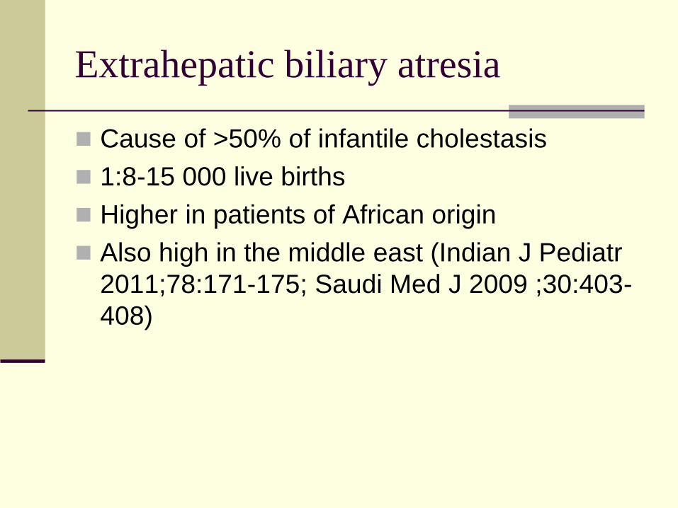
Extrahepatic biliary atresia
Cause of >50% of infantile cholestasis
1:8-15 000 live births
Higher in patients of African origin
Also high in the middle east (Indian J Pediatr
2011;78:171-175; Saudi Med J 2009 ;30:403-
408)
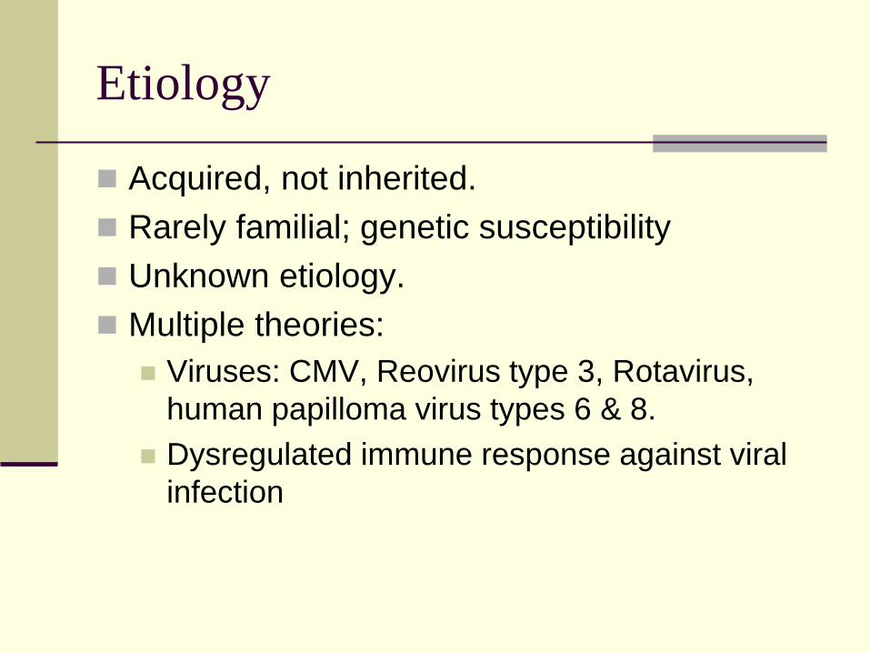
Etiology
Acquired, not inherited.
Rarely familial; genetic susceptibility
Unknown etiology.
Multiple theories:
Viruses: CMV, Reovirus type 3, Rotavirus,
human papilloma virus types 6 & 8.
Dysregulated immune response against viral
infection
Diagnostic procedures
Cholangiography, ERCP, MRCP
Radionuclide imaging
Ultrasound
Liver biopsy: diagnostic in >60% of cases
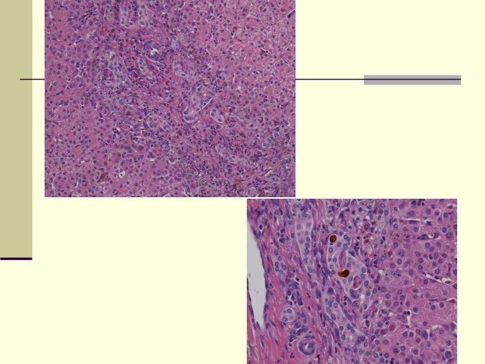
Pathology
Features of bile duct obstruction
Periportal ductular proliferation
Cholestasis
Bile plugs in cholangioles and interlobular bile
ducts
Mixed inflammatory infiltrate with neutrophils
infiltration of cholangioles
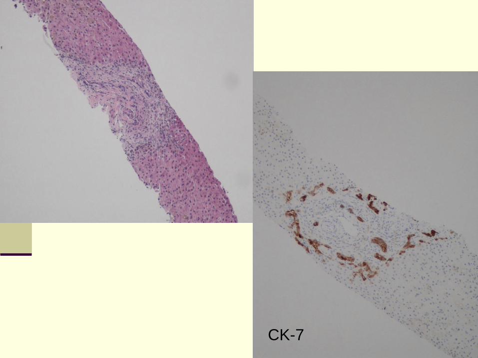
CK-7
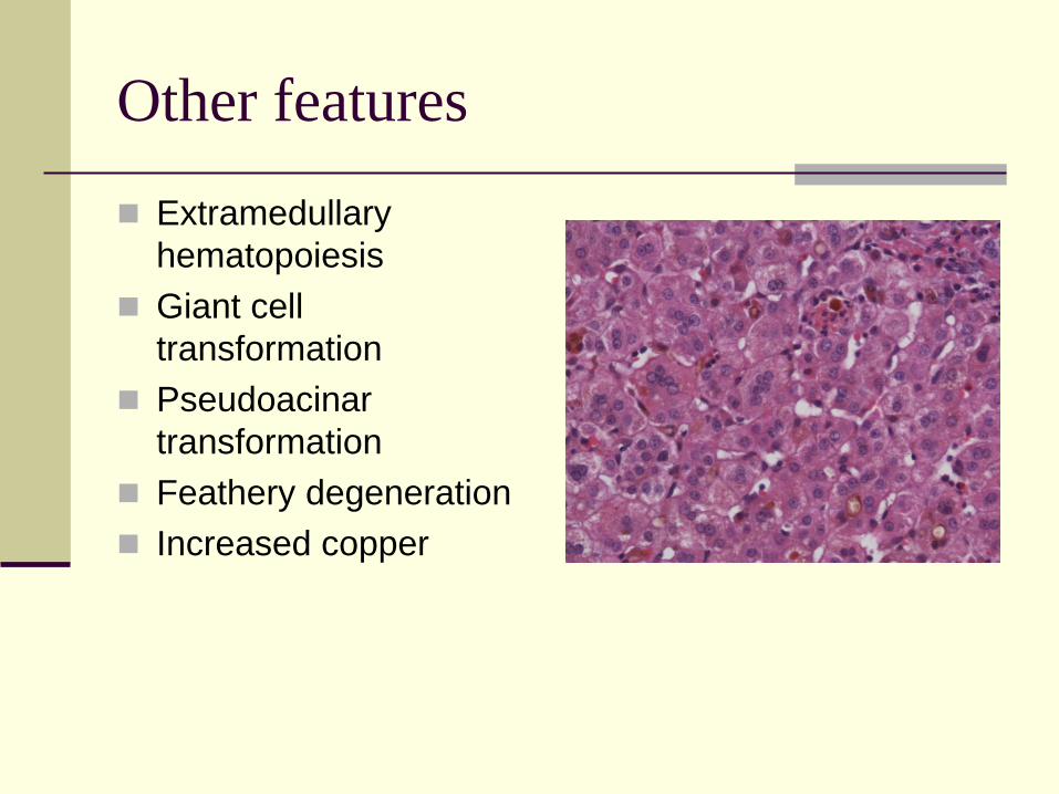
Other features
Extramedullary
hematopoiesis
Giant cell
transformation
Pseudoacinar
transformation
Feathery degeneration
Increased copper
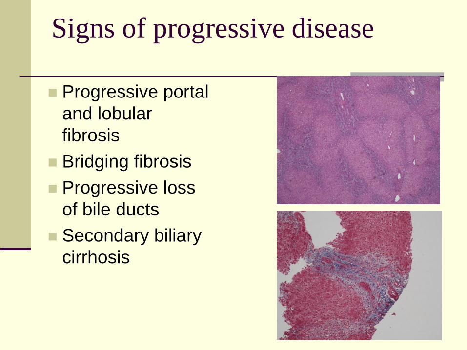
Signs of progressive disease
Progressive portal
and lobular
fibrosis
Bridging fibrosis
Progressive loss
of bile ducts
Secondary biliary
cirrhosis
Treatment
Porto-enterostomy (Kasai): Re-establish bile drainage after resection of atretic ducts
Age and degree of liver damage are the most determinant factors
Has good results especially if done before 60 days of age and before significant fibrosis develops
There is no correlation between success of portoenterostomy and size of remaining bile ducts
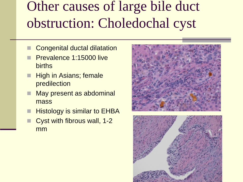
Other causes of large bile duct
obstruction: Choledochal cyst
Congenital ductal dilatation
Prevalence 1:15000 live
births
High in Asians; female
predilection
May present as abdominal
mass
Histology is similar to EHBA
Cyst with fibrous wall, 1-2
mm
Neonatal hepatitis
Idiopathic (>50% of cases): Diagnosis of
exclusion
Infectious: CMV, rubella, Hep B, HSV,
varicella, coxsackie virus, toxoplasma and
Treponema pallidum.
Familial (10% of cases): Autosomal recessive
Metabolic/genetic
Toxins
Alloimmune
Miscellaneous
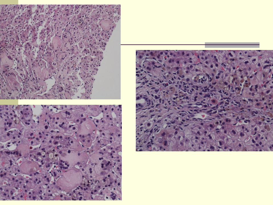
Pathology
Liver may be enlarged
Parenchymal changes: Ballooning, acidophilic bodies
Giant cell transformation:
Diffuse
Cytoplasm contains bile
May become necrotic and surrounded by neutrophils
Cholestasis +/- rosetting (usually in zone 3)
Lobular and portal inflammation
Extramedullary hematopoiesis
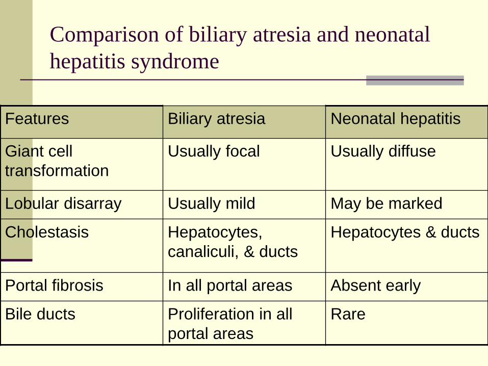
Comparison of biliary atresia and neonatal
hepatitis syndrome
Features Biliary atresia Neonatal hepatitis
Giant cell
transformation
Usually focal Usually diffuse
Lobular disarray Usually mild May be marked
Cholestasis Hepatocytes,
canaliculi, & ducts
Hepatocytes & ducts
Portal fibrosis In all portal areas Absent early
Bile ducts Proliferation in all
portal areas
Rare
Prognosis
Depends on etiology
Variable in idiopathic cases (75% recovery)
Progression to chronic liver disease in 7% of
cases (usually in familial cases)
Persistent cholestasis
Alagille Syndrome
Alpha-1 antitrypsin deficiency
Metabolic causes
Familial Cholestatic Syndromes:
Progressive Familial Intrahepatic Cholestasis
Benign Recurrent Intrahepatic Cholestasis
North American Indian Cholestasis
Norwegian Cholestasis
Greenland Eskimo cholestasis
Turkish nonsyndromic paucity of interlobular bile ducts
Childhood cirrhosis in Arab Israelis
Tyrolean infantile cirrhosis
Navajo neurohepatopathy
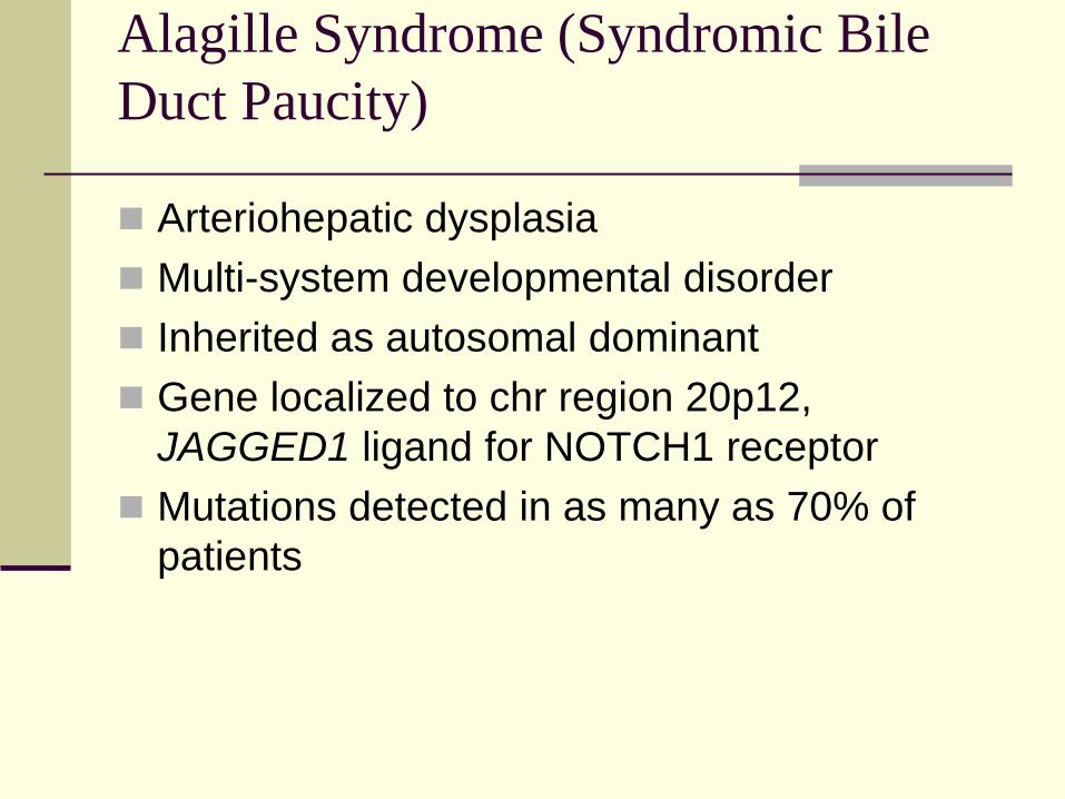
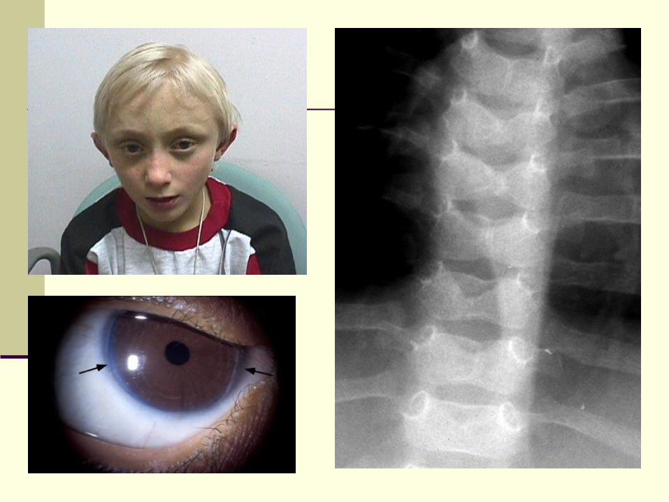
Alagille Syndrome (Syndromic Bile
Duct Paucity)
Arteriohepatic dysplasia
Multi-system developmental disorder
Inherited as autosomal dominant
Gene localized to chr region 20p12,
JAGGED1 ligand for NOTCH1 receptor
Mutations detected in as many as 70% of
patients
Pathology
Variable degree of cholestasis
Portal spaces unexpanded
Bile ducts may be normal in initial biopsies
<3 months: Portal inflammation, bile ductular proliferation (may not be present in non-syndromic cases)
>3 months: bile duct paucity, fibrosis, cholestasis; may be minimal in non-syndromic cases.
> 1year: 90% paucity
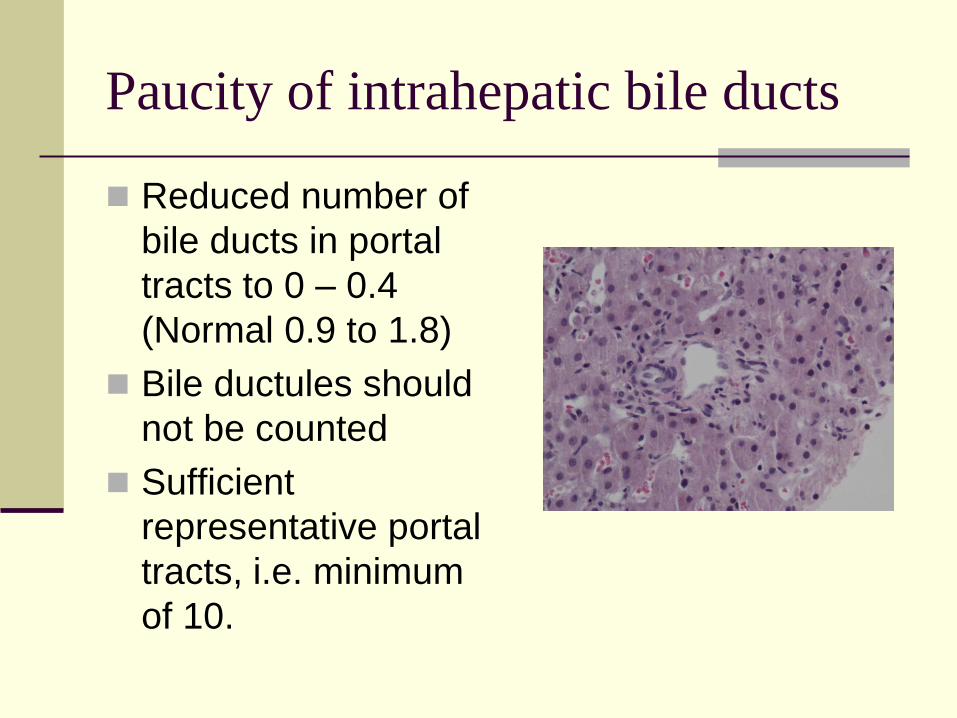
Paucity of intrahepatic bile ducts
Reduced number of
bile ducts in portal
tracts to 0 – 0.4
(Normal 0.9 to 1.8)
Bile ductules should
not be counted
Sufficient
representative portal
tracts, i.e. minimum
of 10.
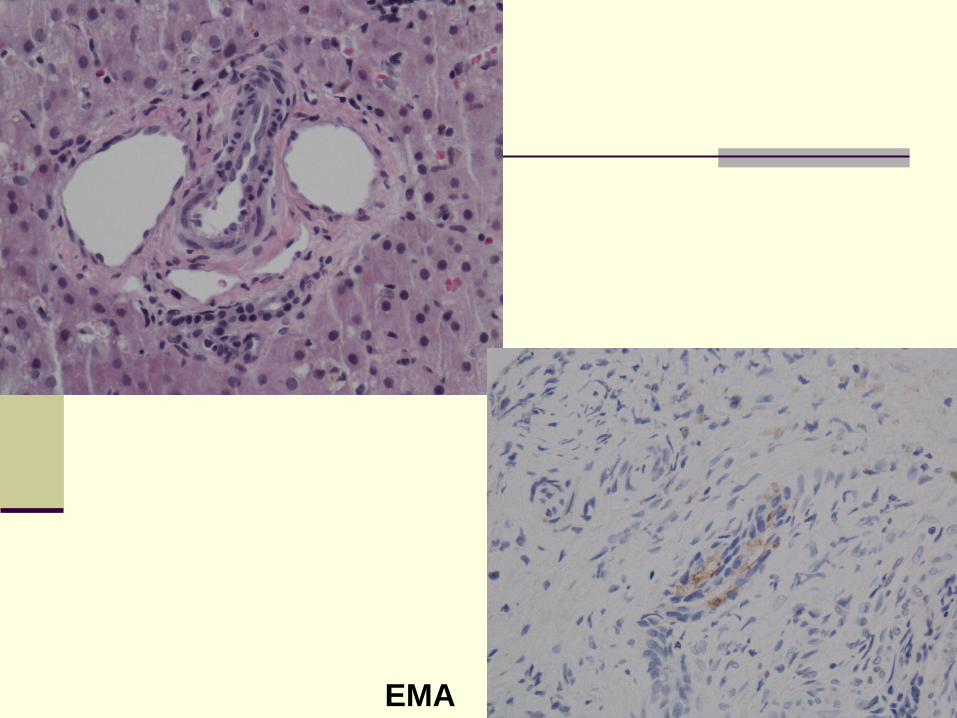
EMA
Additional findings
Non-specific parenchymal changes:
pseudoxanthomatous hepatocytes, giant cell
transformation, periportal fibrosis,
accumulation of copper, EMH
Fibrosis starts early in non-syndromic cases
Progression to cirrhosis is rare.
Non-syndromic bile ducts paucity
Inherited metabolic diseases: α1-antitrypsin
deficiency, inborn errors of bile acid metabolism
Congenital infections: Rubella, CMV
Chromosomal abnormalities (Turner, trisomy 21,
trisomy 17-18)
Prune belly syndrome
Acquired: primary sclerosing cholangitis, Langerhans’
cell histiocytosis, drug-induced, vanishing bile duct
injury, allograft rejection, and graft versus host
disease
Alpha-1 Antitrypsin Deficiency
The most common genetic cause of liver
disease in childhood
The most common variant is homozygous Z
phenotype (PiZ) or compound heterozygous
(SZ)
Deficiency occurs in 1:2000 – 1:5000
individuals
May be associated with glomerulonephritis
Alpha-1 Antitrypsin Deficiency
Conjugated hyperbilirubinemia, acholic stools
Cholestasis resolves by 6 months of age
Chronic liver disease with elevated
transaminases occur in minority of patients
Death may occur in 2.5% of cases
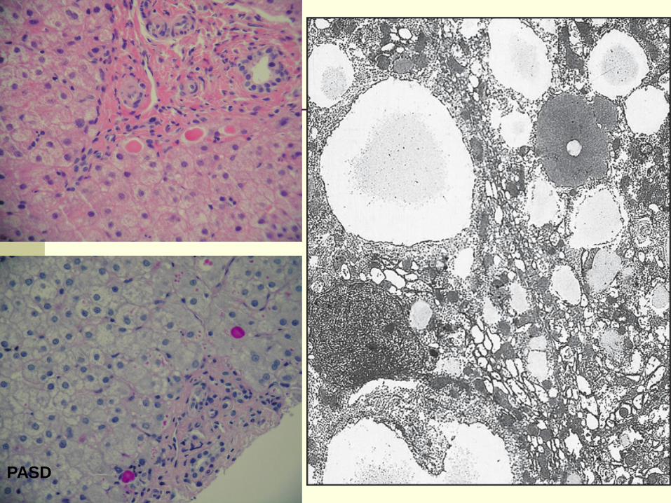
PASD
Progressive Familial Intrahepatic
Cholestasis (PFIC)
Persistent cholestasis in first year of life
No evidence of bile duct obstruction
Three different disease types caused by three different genes:
PFIC 1 deficiency (ATP8B1 gene at 18q21): Byler disease
BSEP (Bile salt export pump) deficiency (ABCB11 gene at chr 2q24): Byler-like disease
MRD3 deficiency (ABCB4 gene at 7q21): high serum λ-glutamate transferase
Clinical features
Jaundice, therapy-resistant pruritis
Malabsorption and failure to thrive
Marked elevation in bile acid levels
Bilirubin normal or elevated
Elevations in serum aminotransferases
λ-GTP normal in types 1 and 2; elevated in
type 3
Complications by biliary cirrhosis and portal
hypertension
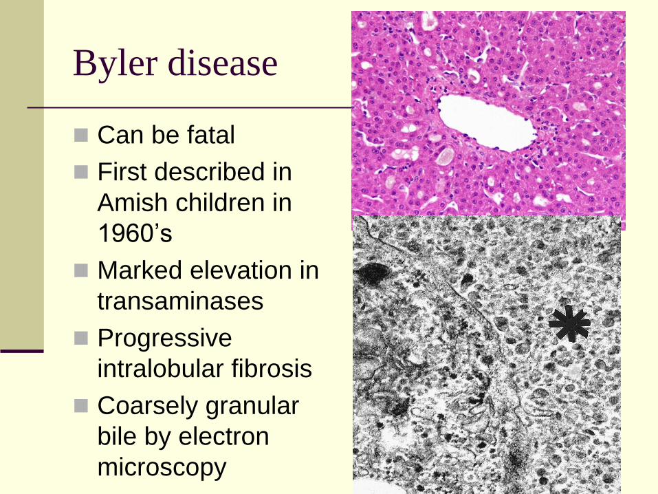
Byler disease
Can be fatal
First described in
Amish children in
1960’s
Marked elevation in
transaminases
Progressive
intralobular fibrosis
Coarsely granular
bile by electron
microscopy
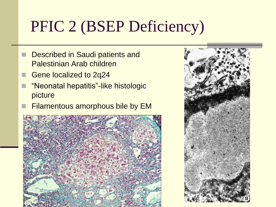
PFIC 2 (BSEP Deficiency)
Described in Saudi patients and
Palestinian Arab children
Gene localized to 2q24
“Neonatal hepatitis”-like histologic
picture
Filamentous amorphous bile by EM
Benign Recurrent Intrahepatic
Cholestasis
Similar to Byler’s disease, with episodic
cholestasis
Mild disease, permanent liver damage does
not develop
Low or normal serum γ-GT
BRIC gene localized to 18q21-q22, same like
Byler’s (ATP8B1 gene)
Inborn Errors of Bile Acid Metabolism
Incidence 1-2% of cholestatic disorders
Familial disorders ?autosomal recessive
Jaundice, poor growth, hepatomegaly
Elevated serum ALT, AST
Low urine bile acids
Chronic cholestatic neonatal hepatitis of
indeterminate etiology; varies from mild to
severe
Rapid evolution of neonatal hepatitis to cirrhosis
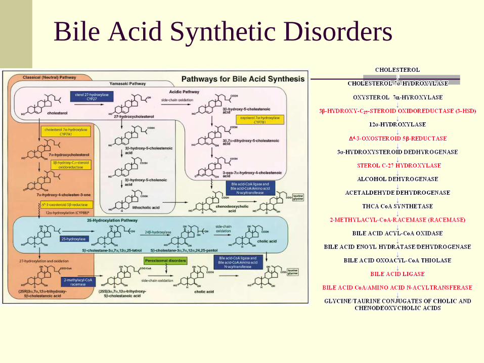
Bile Acid Synthetic Disorders
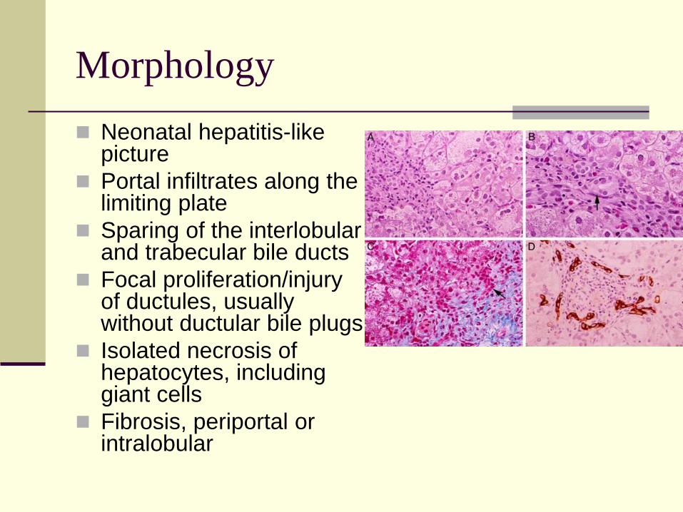
Morphology
Neonatal hepatitis-like picture
Portal infiltrates along the limiting plate
Sparing of the interlobular and trabecular bile ducts
Focal proliferation/injury of ductules, usually without ductular bile plugs
Isolated necrosis of hepatocytes, including giant cells
Fibrosis, periportal or intralobular
Other Inborn errors of metabolism
Galactosemia, hereditary fructose intolerance
and tyrosinemia
Mitochondrial hepatopathies: Multi-organ
disease
Hereditary defects of bilirubin metabolism:
Crigler-Najjar, Gilbert, Dubin-Johnson
syndrome
Acquired Cholestatic Disorders
Septicemia
Drug-induced
Viral hepatitis


![[2015] post lt cholestasis](https://img.pdfslide.net/doc/110x75/58ee0ee21a28ab92198b4665/2015-post-lt-cholestasis.jpg)