Embed Size (px)
Citation preview

Gut, 1962, 3, 252
Chronic painless pancreatitisS. J. M. GOULSTON AND N. D. GALLAGHER
From the Gastroenterology Unit, Royal Prince Alfred Hospital, Sydney, Australia
EDITORIAL SYNOPSIS This paper describes four patients whose chief symptoms were steatorrhoeaand loss of weight. Despite the absence of a history of abdominal pain investigations showedthat these patients had chronic pancreatitis, which responded to medical treatment. The patho-logical findings in two of these cases and in six which came to necropsy are reported.
Pain is the outstanding symptom of pancreatitis inall its stages. The pain is usually severe, situated inthe upper abdomen and may radiate to the back.Less typical attacks of pain may also occur. How-ever, the post-mortem demonstration of acutepancreatitis in patients without a recorded historyof pain by Evans, Gross, and Baggenstoss (1958)suggests that pain may be completely absent. Ahistory of pain may also be absent in patients withchronic pancreatitis and although this was recognizedby Friedreich (1878) and Opie (1902), its recognitionto-day followed the paper by Bartholomew andComfort (1956) describing the clinical features of10 patients. Our interest in this condition resultedfrom the discovery of four patients with chronicpainless pancreatitis who were in a group of 35patients with chronic pancreatitis admitted to thishospital in 1959 and 1960. A study of post-mortemrecords from the period 1950 to 1960 revealed sixpatients in whom the diagnosis of chronic painlesspancreatitis was not considered during life in viewof the absence of any symptoms or signs suggestiveof pancreatic disease. Our experience is reported inthis paper.The four living patients were men in the sixth or
seventh decades who complained of fatty diarrhoeaand weight loss. Diarrhoea was the most distressingsymptom and had been present for periods rangingfrom four months to six years. Two patients hadlost more than 50 lb. in weight but loss of weight wasless marked in the remainder. No members of thegroup complained of a loss of appetite, appetitebeing either unchanged or increased during theperiod of illness. Clinical evidence of diabetesmellitus was found in only one patient but theremaining three patients had pre-diabetic glucosetolerance tests. There was no history of episodes ofabdominal pain nor was there a history suggestive
of biliary disease. The intake of alcohol by the fourpatients was insignificant.
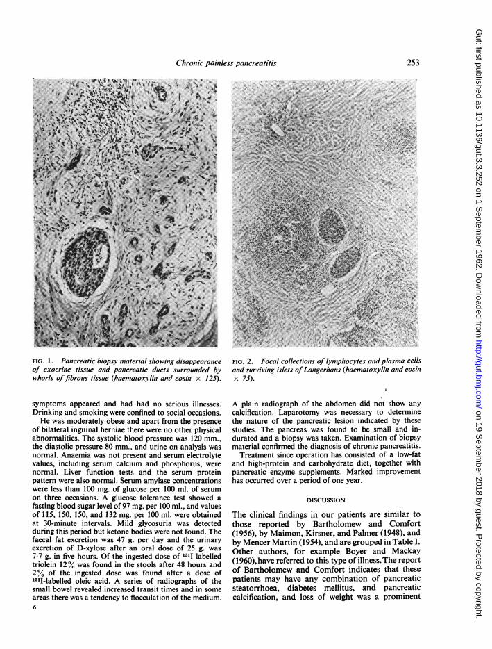
Steatorrhoea was shown to be present by thefinding of abnormal values for faecal fat excretionand in two patients faecal fat excretion exceeded100 g. per day. The results of 1311-labelled triolein andoleic acid tests indicated pancreatic steatorrhoea.Plain x-ray examination of the abdomen revealedpancreatic calcification in only one patient. Mal-absorption states such as idiopathic steatorrhoea andWhipple's disease were excluded by the findings ofnormal jejunal mucosa after intestinal biopsies wereperformed or normal values for the urinary excretionof an oral dose of D-xylose. Local lesions of theintestine such as stricture and diverticulosis were notdemonstrated by radiological studies. Laparotomywas necessary in two patients in whom symptomshad been present for four and 12 months respectivelyto exclude carcinoma of the pancreas or duct ofWirsung. Pancreatic tissue obtained by biopsyshowed that although the outline of the pancreaticlobules and their ducts remained intact, exocrinecells were almost completely absent (Fig. 1) andwere replaced by discrete collections of lymphocytesand plasma cells, neutrophils being conspicuouslyabsent (Fig. 2). Pancreatic fibrosis was marked andwas both perilobular and intralobular. Some islets ofLangerhans were preserved and vascular changeswere minor. The case history of one of these patientsis as follows.
CASE HISTORY
A male engineer, married and aged 62, was admitted tohospital in August 1960 with complaints of diarrhoeaand weight loss. The diarrhoea had begun abruptly fourmonths previously and after a short time the stoolsappeared fatty. Despite a good appetite he had lost21 lb. in weight. He had been in good health until these
252
on 19 Septem
ber 2018 by guest. Protected by copyright.
http://gut.bmj.com
/G
ut: first published as 10.1136/gut.3.3.252 on 1 Septem
ber 1962. Dow
nloaded from
Chronic painless pancreatitis
FIG. 1. Pancreatic biopsy material showing disappearance FIG. 2. Focal collections of lymphocytes and plasma cellsof exocrine tissue and pancreatic ducts surrounded by and surviving islets ofLangerhans (haematoxylin and eosinwhorls offibrous tissue (haematoxYlin and eosin x 125). x 75).
symptoms appeared and had had no serious illnesses.Drinking and smoking were confined to social occasions.He was moderately obese and apart from the presence
of bilateral inguinal herniae there were no other physicalabnormalities. The systolic blood pressure was 120 mm.,the diastolic pressure 80 mm., and urine on analysis wasnormal. Anaemia was not present and serum electrolytevalues, including serum calcium and phosphorus, werenormal. Liver function tests and the serum proteinpattern were also normal. Serum amylase concentrationswere less than 100 mg. of glucose per 100 ml. of serumon three occasions. A glucose tolerance test showed afasting blood sugar level of 97 mg. per 100 ml., and valuesof 115, 150, 150, and 132 mg. per 100 ml. were obtainedat 30-minute intervals. Mild glycosuria was detectedduring this period but ketone bodies were not found. Thefaecal fat excretion was 47 g. per day and the urinaryexcretion of D-xylose after an oral dose of 25 g. was7-7 g. in five hours. Of the ingested dose of 1311-labelledtriolein 12% was found in the stools after 48 hours and2% of the ingested dose was found after a dose of1311-labelled oleic acid. A series of radiographs of thesmall bowel revealed increased transit times and in someareas there was a tendency to flocculation of the medium.6
A plain radiograph of the abdomen did not show anycalcification. Laparotomy was necessary to determinethe nature of the pancreatic lesion indicated by thesestudies. The pancreas was found to be small and in-durated and a biopsy was taken. Examination of biopsymaterial confirmed the diagnosis of chronic pancreatitis.Treatment since operation has consisted of a low-fat
and high-protein and carbohydrate diet, together withpancreatic enzyme supplements. Marked improvementhas occurred over a period of one year.
DISCUSSION
The clinical findings in our patients are similar tothose reported by Bartholomew and Comfort(1956), by Maimon, Kirsner, and Palmer (1948), andby Mencer Martin (1954), and are grouped in Table I.Other authors, for example Boyer and Mackay(1960), have referred to this type of illness.The reportof Bartholomew and Comfort indicates that thesepatients may have any combination of pancreaticsteatorrhoea, diabetes mellitus, and pancreaticcalcification, and loss of weight was a prominent
253
on 19 Septem
ber 2018 by guest. Protected by copyright.
http://gut.bmj.com
/G
ut: first published as 10.1136/gut.3.3.252 on 1 Septem
ber 1962. Dow
nloaded from

254 S. J. M. Goulston and N. D. Gallagher
TABLE ICLINICAL FINDINGS IN 18 REPORTED CASES
Author Steatorrhoea Diabetes Calcification
Maimon et al. (1948) 3 2 3Mencer Martin (1954) 1 1 1Bartholomew and Comfort(1956) 10 7 8Present series 4 1 1
symptom in their patients. Moreover, they notedthe presence of a pseudocyst in one patient andobstructive jaundice in another. These reports alsoindicate that both sexes may be affected and that theillness occurs predominantly in the sixth andseventh decades. The incidence of chronic painlesspancreatitis is probably small, although Gross (1958)found four patients in a group of 75 patients withchronic pancreatitis and our incidence is also high.The histopathology of the pancreatic tissue
obtained from two patients showed marked pan-creatic fibrosis; exocrine cells had disappeared andbeen replaced by collections of lymphocytes andfibrous tissue. Examination of post-mortem materialfrom six other patients gave similar findings. Twoof the latter group also had small areas of acuteinflammation and fat necrosis. These examplesoffer further support for the occurrence of acute,subacute, and chronic pancreatitis unaccompaniedby pain. Although in our material it was notable thatthe ducts were not dilated, the histological featuresdo not allow a distinction to be made betweenchronic painless and chronic relapsing pancreatitis.Furthermore the appearances we have describedare similar to those found by Bartholomew, Baggen-stoss, Morlock, and Comfort (1959) in pancreaticatrophy and lipomatosis but the massive fattyinfiltration which may be present in the lattercondition was absent.Numerous factors have been described in the
aetiology of pancreatitis and these have recently beenreviewed by Blumenthal and Probstein (1959). Theseinclude biliary tract disease, chronic alcoholism,protein malnutrition, pancreatic duct obstruction,hyperparathyroidism, and hyperlipaemia. Our ex-perience did not suggest that any of these had asignificant aetiological role nor was there a historyof abdominal trauma, infective panlcreatitis, or afamily history of pancreatic disease. Whether chronicpainless pancreatitis is the end-result of repeatedepisodes of silent acute pancreatitis or the result ofan insidious chronic inflammatory process from thebeginning is unknown. The latter process suggeststhe possibility of an auto-immune mechanism but theresults of antigen-antibody studies in chronicpancreatitis are inconclusive (Murray and Thal,1960). The role of vascular disease is also uncertain
but attention was drawn to this possibility by ourfinding widespread hypertensive and degenerativevascular disease at necropsy in six patients. Joske(1955) has suggested that arterial disease maycontribute to the development ofchronic pancreatitis,and Blumenthal and Probstein (1959) report peri-lobular and intralobular fibrosis in the pancreas ofpatients with degenerative vascular disease inamounts increasing with age. However, much of thethought devoted to the origins of pancreatic diseaseremains speculative, and the reasons for the absenceof pain in these patients are also unknown.The treatment of chronic painless pancreatitis is
entirely medical as the absence of pain removes themain indication for surgery. Our patients respondedwell to a high-protein and carbohydrate, low-fat diet,together with pancreatic enzyme supplements andvitamins. Although this regime helped in the controlof diabetes mellitus in one patient, his insulin require-ments remained high. Clinical evidence of diabeteshas not appeared in the remainder during a follow-up period of one year. Our experience and that ofBartholomew and Comfort (1956) indicates thatthese patients have a satisfactory prognosis. Deathdue to vascular disease in our cases coming tonecropsy suggests that a number may eventually diein this way.
We would like to acknowledge the help of Dr. V. J.McGovern with the pancreatic histology; Mr. F. Millsperformed the laparotomies. The Gastroenterology Unitis supported by the Bushell Trust.
REFERENCES
Bartholomew, L. G., Baggenstoss, A. H., Morlock, C. G., andComfort, M. W. (1959). Primary atrophy and lipomatosis ofthe pancreas. Gastroenterology, 36, 563-572.and Comfort, M. W. (1956). Chronic pancreatitis without pain.Ibid., 31, 727-743.
Blumenthal, H. T., and Probstein, J. G. (1959). Pancreatitis, pp.15-63, 247. Springfield, Illinois: Thomas.
Boyer, J. T., and Mackay, I. R. (1960). The aetiology, course andsurgical aspects of pancreatitis. A review of 108 cases. Aust.N.Z. J. Surg., 30, 150-157.
Evans, H. W., Gross, J. B., and Baggenstoss, A. H. (1958). Acuteand subacute interstitial pancreatitis: A clinicopathologicstudy. Gastroenterology, 35, 457-464.
Friedreich, N. (1878). "Diseases of the Pancreas." In Cyclopaedia ofthe Practice of Medicine, ed. H. Ziemssen, Vol. 8, p. 551.Sampson Low, London.
Gross, J. B. (1958). Some recent developments pertaining to pancreatitis. Ann. intern. Med., 49, 796-819.
Joske, R. A. (1955). Aetiological factors in the pancreatitis syndrome.Brit. med. J., 2, 1477-1481.
Maimon, S. N., Kirsner, J. B., and Palmer, W. L. (1948). Chronicrecurrent pancreatitis. Arch. intern. Med., 81, 56-72.
Martin, M. Mencer (1954). Pancreatic lithiasis. Diabetes mellitus.Pulmonary tuberculosis. Intermittent claudication. Proc. roy.Soc. Med., 47, 513-515.
Murray, M. J., and Thal, A. P. (1960). The clinical significance ofcirculating pancreatic antibodies. Ann. intern. Med., 53,548-555.
Opie, E. L. (1902). The causes and varieties of chronic interstitialpancreatitis. Amer. J. med. Sci., 123, 845-868.
on 19 Septem
ber 2018 by guest. Protected by copyright.
http://gut.bmj.com
/G
ut: first published as 10.1136/gut.3.3.252 on 1 Septem
ber 1962. Dow
nloaded from