Embed Size (px)
Citation preview
Chronic Pulmonary Granulomatosis
Report of Ten Cases
J. M. DENARDI, M.D.. H. S. VAN ORDSTRAND, M.D. and M. G. CARMoDY, M.D.
Cleveland. Ohio
N 1943 two of us first reported the oc- currence in the United States of an acute pulmonary involvement occur-
ring in workers extracting beryllium oxide. ’ A subsequent, more comprehensive report of acute manifestations in these same pa- tients was published in 1945.2 Since the initial publication, beryllium and its com- pounds have been under investigation as a possible source of both acute and chronic conditions in man although final convincing proof of a relationship is still lacking.
Industrial concerns employing and manu- facturing beryllium and its compounds have not waited for conclusive evidence of beryl- lium toxicity but began at once to develop measures to decrease exposure of their workers to this element. The industry has also taken steps to reduce the amount of beryllium compounds escaping through its various exhausts into the surrounding atmosphere.
The syndrome under discussion is most commonly known as chronic pulmonary granulomatosis. It has been described by Hardy and Tabershaw3 and by Higgins.4 It has also been referred to as miliary sar- coidosis, chronic granulomatous pneumo- nitis, delayed chemical pneumonitis and beryllium sarcoid. The purpose of this report is to make available clinical, labora- tory and roentgenographic data to aid in the early diagnosis and management of this chronic pulmonary disease.
It is noteworthy that since 1944 or 1945 exposure to beryllium has been much less frequent because of the steps taken by manu- facturing plants. Consequently, in pre- paring this report we wish to point out that the majority of the case histories presented
in this paper were of those who were exposed to beryllium compounds prior to 1946. Of the many who were exposed before 1946 only a small number have shown any symptoms.
ETIOLOGY AND PATHOLOOY
At present no etiologic factor or combina- tion of factors has been definitely established as the cause of chronic pulmonary granulo- matosis. Previously reported cases have been closely associated with the handling of phos- phorus in the manufacture of fluorescent lamps.” In the series of cases presented four of the patients worked in a plant producing beryllium and four of the remaining six lived near the plant. However, consideration must also be given to agents other than beryllium such as infection, copper, man- ganese and zinc. On his last visit to Cleve- land, Dr. L. U. Gardner suggested the possibility of a diphtheroid organism or other bacteria or viruses becoming virulent in the presence of certain chemical com- pounds. In spite of the failure to identify the cause with certainty, public health officials as well as industrial sanitary engineers have drawn attention to beryllium.
Because of lack of knowledge of the exact etiologic factors, little has been advanced theoretically as to the pathogenesis of the disease. On the theory that the etiologic agent is an insoluble organic or inorganic compound it is possible that phagocytes, in the usual response to irritants, engulf the minute particles of the compound in the finer bronchioles and respiratory alveoli. Some of the cells die locally owing to the toxicity of the agent with the characteristic tissue reaction of formation of sarcoid-like
SEPTBMRER, 1949 345
346 Chronic Pulmonary Granulomatosis--DeJVardz’ et al.
nodules. Other cells are capable of migrat- ing into the local lymphatic system and still others into the blood capillaries, a mecha- nism which would explain the nodular reaction.
HISTORY
Six of the ten persons reported had not worked with metallic beryllium or its com- pounds but four of them lived within a radius of 300 meters from a beryllium plant for periods of from one to four years. The fifth lived about three miles from the plant, where her husband was employed, but at no time did she handle his work clothing. Probability of direct exposure was ques- tioned but denied. The sixth patient lived approximately one-half mile from the plant for about one year. She had no complaints nor did she manifest any respiratory diffi- culty while in this area. Her residence since has been two to three miles from the plant and her only possible contact during this time has been with a friend whose father was employed in a beryllium plant. The entire exhaust material from the plant has not been thoroughly investigated; however, preliminary samples in the proximity re- vealed minimal contamination with beryl- lium compounds for a short distance only.
All six of the patients hereby reported as out-plant cases were brunette women who ranged in age between twenty-two and thirty-five years. In five instances the disease had manifested itself from one to eight months postpartum. In the sixth instance the interval between delivery and onset of the disease was almost two years. Of the four remaining patients one worked in a beryllium plant for six weeks during the summer of 1941 and his earliest symptoms appeared while he was in the army in 1944. The second patient was employed in the laboratory of a beryllium plant for twelve weeks from November, 1943 to February 1944, and her first symptoms appeared in August, 1946. Neither had any manifesta- tions of the acute form of the disease while working in the beryllium plant. The third patient worked in a beryllium plant for
eight weeks in 1941. He left this employ- ment after having developed a cough and shortness of breath which he attributed to chemical bronchitis. His first symptoms of chronic pulmonary granulomatosis appeared in January, 1946, and during the interven- ing years he had not been exposed to any other chemical or occupational hazards.
The fourth patient developed an acute chemical bronchitis in May, 1945. Upon complete recovery two months later he returned to work as a beryl ore grinder. Within ten days he again disclosed clinical symptoms and findings of acute bronchitis. After full recovery he was released from the industry in September, 1945 and has since avoided chemical contact.
Clinical and Roentgenographic Data. The clinical history was characteristic in all cases. The onset was rather insidious and because of the mildness of the process in the early months the patients failed to seek medical attention. The typical history was a mild productive or non-productive morn- ing cough usually following an acute coryza. In a few months the cough became more pronounced and dry in character and sudden changes in air temperature and humidity sometimes elicited a severe attack of paroxysmal coughing. Substernal pain and discomfort were frequently noticed during the coughing attacks or upon attempted deep breathing.
Concomitant with the paroxysmal cough there was exertional dyspnea which became more pronounced as the disease progressed. Acrocyanosis, “watchglass” fingernails and clubbing of the finger tips were present in the later stages.
Physical examination in all patients revealed some weight loss, depending upon the stage of the disease. The vital capacity was diminished greatly, chest expansion was definitely limited and substernal pain was experienced at the height of inspiration. The percussion note was resonant through- out and fine, crackling rales were audible in the hilar areas in the advanced stages of the disease. Abdominal examination proved entirely negative in all except Case I during
Chronic Pulmonary Granulomatosis-DeNurdi et al. 347 the terminal months of the illness. No such skin or glandular lesions as were described by Higgins4 have been observed.
Laboratory studies, including complete blood counts, Kline and Kahn tests, uri- nalysis, sedimentation rate, non-protein nitrogen and blood sugar determinations were within normal limits in all cases. Albumin globulin ratio was within normal limits in all cases.
Chest roentgenograms revealing a ground- glass appearance with minute nodules throughout the lungs is one of the outstand- ing characteristic diagnostic features of the disease. The small nodules are unquestion- ably the result of coalescing granules. The fact that symptomatic onset of the disease precedes positive roentgenographic findings by several months is verified in the histories of the reported cases. Roentgenograms of the hands in all living patients did not reveal any abnormalities.
Two of these patients died and autopsies were performed. Nodular lesions were con- fined to the lungs, hilar glands and the liver. The pathologist’s reports are given in detail in connection with Cases I and II. Beryllium was not recovered from the tissues of one of the patients in whom an autopsy was performed; in the other quantitative studies were equivocal. Beryllium analysis of the urine revealed small amounts in two patients (Cases VI and VII) and none in the other two patients (Cases III and v).
D#erential Diagnosis. There is need for great caution in making a specific roent- genologic diagnosis, especially on single plate observation. All other possibilities must be eliminated by exhaustive exposure investigation, by serial roentgenogram study, by clinical course pattern and by laboratory procedures. Furthermore, the possible co- existence of multiple etiologic factors must be given consideration. Differential diag- nosis must include consideration of miliary tuberculosis, tuberculosilicosis, Boeck’s sarcoid,6,7 miliary carcinomatosis,8 pneu- moconiosis,g,‘O erythema nodosum,l’ coc- cidioidomycosis12 and acute chemical pneumonitis. r2*r3 Differential highlights
SEPTEMRER, 1949
have been given by Pascuccii4 in his recent report of similar cases.
Prognosis. The prognosis remains guar- ded. Most cases have been diagnosed in the later stages of the disease while only a few have been detected in an earlier phase. At the present time the first patient seen in con- sultation with Dr. L. U. Gardner in 1945 is living and while she has improved clinically, roentgenograms of the lungs remain un- changed. Three others (Cases VI, VII and VIII) have manifested clinical improvement in the last six months and are allowed minimal physical exertion.
Treatment. No specific treatment has been suggested up to the present. Since the cause or causes are not yet established, treatment is empirical. BAL (2.3 dimer- captopropanol) has been used without particular success. Aminophyllin, benadryl and cough sedation have been used sympto- matically with some degree of relief. In two patients during attacks of chills, high fever, shortness of breath and severe acrocyanosis intravenous injection of 10 to 50 mg. of benadryl produced an amazing alleviation of symptoms immediately and a sudden drop in the temperature to normal, a con- dition which persisted for several days.
The most effective treatment at present depends upon early diagnosis and consists of prescribing minimal exertion so that the patient’s physical reserve is not impaired. It has been observed that a clear, dry and warm environment is conducive to clinical improvement even though roentgenographic findings remain unchanged. Because of de- creased vital capacity in these patients, great caution must be observed to avoid any acute respiratory infection.
CASE REPORTS *
CASE I. A white woman, who was thirty- eight years old at the time of her death on .July 15, 1946, complained of a progressive respiratory difficulty of two and one-half years’ duration. The apparent onset was in -January,
* Clinical data furnished through the courtesy of Dr. M. E. Kishman, Dr. L. Hait, Dr. H. Marsico of Lorain. Ohio, and Dr. Russell Dickason of Vermilion, Ohio.
348 Chronic Pulmonary Granulomatosis-DeNurdi et al,
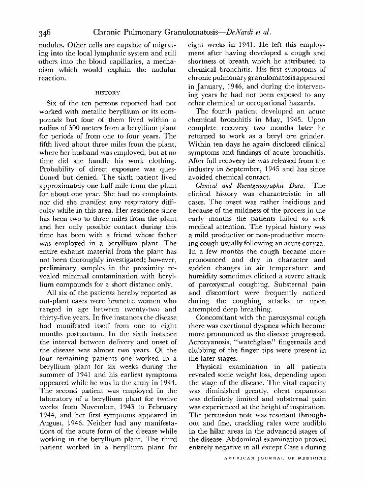
FIG. 1. Case I. Fine diffuse granular infiltration through- out lung fields, with cardiac silhouette of secondary car pulmonale; five months prior to death.
1944 when the patient suffered a cold after which she experienced persistent weakness, a slight but constant cough, dyspnea on exertion and a slight loss of weight. She did not have chills or fever. On April 13, 1945, she was de- livered of her second child and on October 12th stated that she had lost over 20 pounds during the preceding summer. Her cough had become more pronounced and also spasmodic and while it had been productive during the summer was dry at the time of examination. She complained of severe dyspnea and of substernal pain on inspiration. Examination revealed acrocyanosis and slight clubbing of the fingers and toes. Expansion of the chest was diminished, but percussion and auscultation revealed no ab- normality. There was some retraction of the supraclavicular spaces during inspiration. With the patient in the sitting position, distention of the jugular veins was evident almost to the angle of the jaw bilaterally.
Two weeks later the pulse rate was 96. The temperature and blood pressure were normal. Urinalysis, blood count and Kahn and Wasser- mann tests were also normal. An electrocardio- gram showed slight right axis deviation.
Three months later, in February, 1946, the patient’s condition had become worse. She complained of a painful, pulling sensation in the mediastinum when in the recumbent posi- tion. Severe exertional dyspnea was present.
About five months later and five weeks before her death examination of the chest revealed fine crepitant rales at the bases with sibilant rales over the hilar areas. Chest expansion was limited. Edge of the liver was palpable 2 cm. below the costal margin. There was edema of the ankles and feet. A month later on July 13, 1946, there were signs of early right-sided cardiac failure. The patient died two days later.
During 1944 and until August, 1945 the patient had lived about two blocks from a plant extracting and using beryllium. She then moved approximately one and one-half miles away. She first noticed symptoms on foggy days in <January, 1944 when she could smell fumes from the plant. From September, 1944 to April, 1946, some three months preceding her death, five roentgenograms * of the chest were taken, one of which is shown in Figure 1.
An autopsy was performed. The significant observations made on microscopic examination of the lungs by the staff pathologist are as follows:
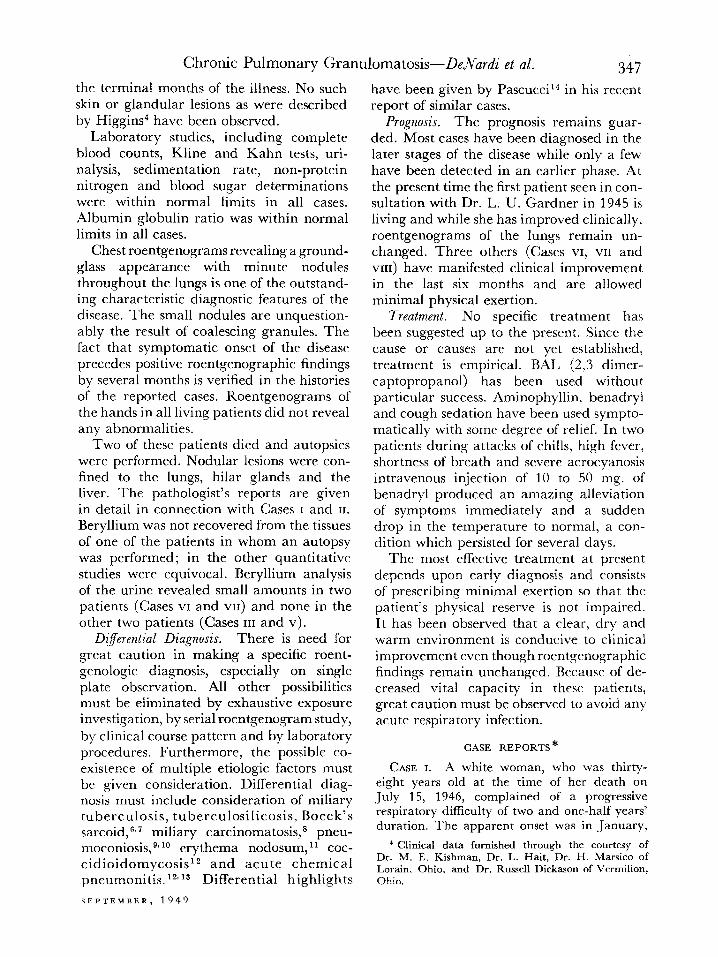
The alveolar walls were irregularly thickened by infiltration of lymphocytes and focal accumu- lation of histiocytes and foreign body giant cells. No necrosis was apparent. Occasional small purplish staining (calcified) lamellated bodies, 50-75 micra, were present in infiltrate. In patches small irregular alveoli with cuboidal epithelium enclosed by thickened walls were evident, and other patches of dilated alveoli with partly thin, partly thickened walls. Con- nective tissue septa were only partly involved by infiltrate; pleura showed a moderate lympho- cyte infiltrate and rare histiocyte focus.
One parabronchial lymph node showed fre- quent, poorly demarcated foci of large mono- nuclear cells, frequent giant cells of Langhans’ type and occasional calcified bodies. A small branch of pulmonary artery reveaIed marked intimal thickening due principally to connective tissue increase but with small foci and single lipophages. The anatomic diagnosis was chronic granulomatous interstitial pneumonitis and chronic granulomatous lymphadenitis. A typical area of granuloma of the lung is shown in Figure 2.
There was severe parenchymatous degenera- tion of the liver. Occasional nodules with giant and round cells were found. There were in- creased numbers of round cells in the portal
* All roentgenograms were read and interpreted by Dr. Delbert Russell, Lorain, Ohio.
AMERICAN JOURNA’. OF MEDICINE
Chronic Pulmonary Granulomatosis-DeNad et al. 349
FIG. 2. Case I. Typical area of pulmonary granuloma- tosis. A focal accumulation of histiocytes and foreign body giant cells with infiltration of lymphocytes; no apparent necrosis.
tracts. The pathologic diagnosis was chronic granulomatous interstitial pneumonitis.
The results of beryllium determinations per- formed on lung tissue were as follows: mg. Be/sample-Nil or <0.000015; mg. Be/100 Gm.-Nil or <0.00008; results of similar determinations on the formalin in which the tissue was sent were as follows: mg. Be/sample -Nil and mg. Be/L.-Nil.
CASE II. A white woman, aged twenty-five at the time of her death February 17, 1948, complained of a mild non-productive cough and steady loss of weight over a period of nearly two years. The loss of weight in the two-year period was 11 pounds. About one and one-half years before she died in June, 1946 she noticed dyspnea on exertion and intermittent fever and arthralgia.
On August 12, 1946, this patient had a pulse rate of 100 with normal temperature and blood pressure. There were a few fine crepitant rales over the lung bases posteriorly but no other physical findings in the chest. Vital capacity was approximately 70 per cent of normal. Urinalysis and blood studies were normal. At this time a roentgenogram of the chest revealed
SEPTEMBER, 1949
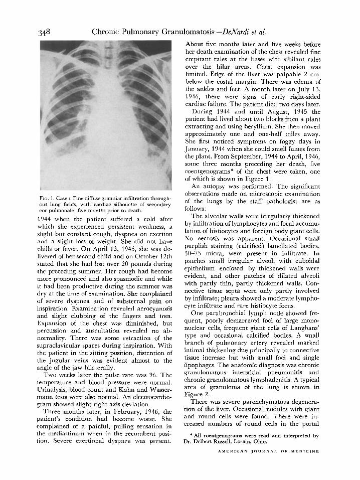
FIG. 3. Case II. Minute nodulation throughout all lung areas; five months prior to death.
accentuated hilar shadows especially on the right side with a diffuse, generalized granular appearance of all lobes of the lungs.
By June, 1947, about seven months prior to death, the patient had developed purpuric areas of the skin and mucous membranes. She had suffered from a severe pulmonary hemorrhage. Laboratory studies at this time revealed a deficiency of platelets and secondary anemia; roentgenograms of the chest showed some clearing of the lung field without associated evidence of clinical improvement. An x-ray of September 9, 1947, is shown in Figure 3.
The final roentgenograms in November, 1947 about three months prior to her death revealed a generalized granular infiltration throughout both lungs. Shortly preceding death acrocyano- sis became evident, aggravated by slight exer- tions. Clubbing of the fingers was present. Excursions of the chest were rapid and shallow. Heart rate was 124. Fine and coarse, moist rales were audible throughout both lung fields. Occupational exposure to chemicals could not be determined. During 1941 the patient had lived approximately one-half mile from a beryllium plant. She had two normal preg- nancies and deliveries in 194’2 and 1944.
An autopsy was performed and the results of the microscopic examination of the tissue was made by the staff pathologist. Focal accumula- tions of histiocytes of foreign body and Langer- hans’ type with variable numbers of lympho-
350 Chronic Pulmonary Granulomatosis-DeNad et al.
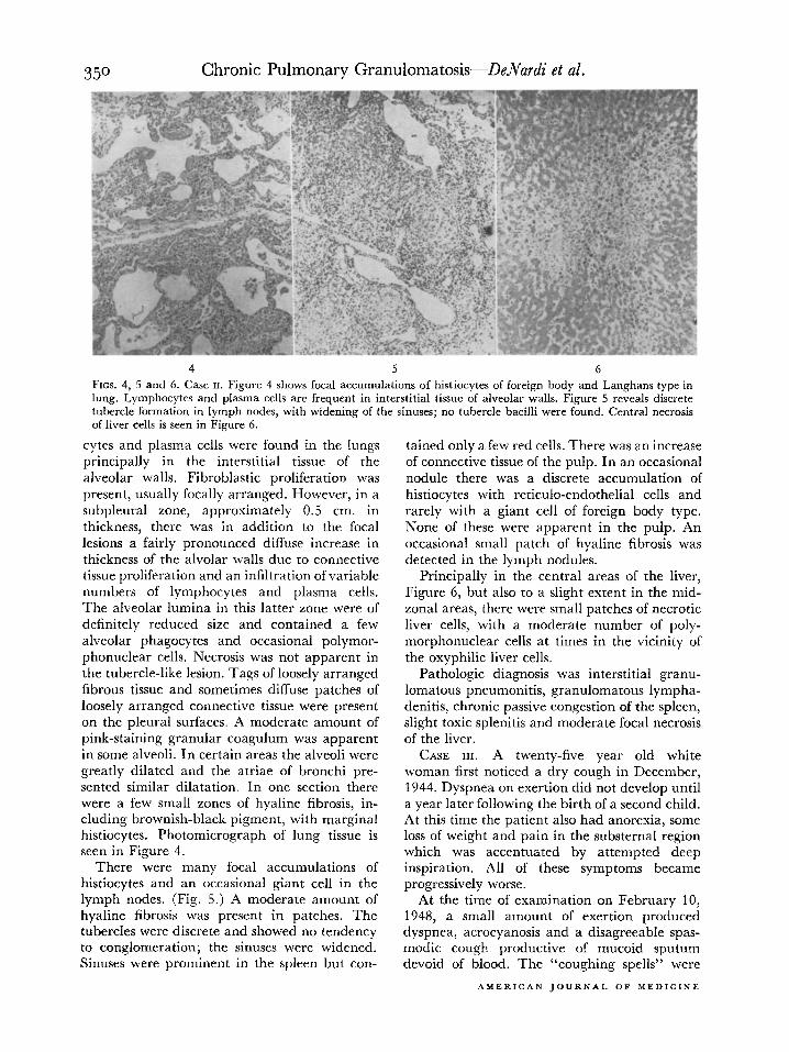
4 5 6 FIGS. 4, 5 and 6. Case II. Figure 4 shows focal accumulations of histiocytes of foreign body and Langhans type in lung. Lymphocytes and plasma cells arc frequent in interstitial tissue of alveolar walls. Figure 5 reveals discrete tubercle formation in lymph nodes, with widening of the sinuses; no tubercle bacilli were found. Central necrosis of liver cells is seen in Figure 6.
cytes and plasma cells were found in the lungs principally in the interstitial tissue of the alveolar walls. Fibroblastic proliferation was present, usually focally arranged. However, in a subpleural zone, approximately 0.5 cm. in thickness, there was in addition to the focal lesions a fairly pronounced diffuse increase in thickness of the alvolar walls due to connective tissue proliferation and an infiltration of variable numbers of lymphocytes and plasma cells. The alveolar lumina in this latter zone were of definitely reduced size and contained a few alveolar phagocytes and occasional polymor- phonuclear cells. Necrosis was not apparent in the tubercle-like lesion. Tags of loosely arranged fibrous tissue and sometimes diffuse patches of loosely arranged connective tissue were present on the pleural surfaces. A moderate amount of pink-staining granular coagulum was apparent in some alveoli. In certain areas the alveoli were greatly dilated and the atriae of bronchi pre- sented similar dilatation. In one section there were a few small zones of hyaline fibrosis, in- cluding brownish-black pigment, with marginal histiocytes. Photomicrograph of lung tissue is seen in Figure 4.
There were many focal accumulations of histiocytes and an occasional giant cell in the lymph nodes. (Fig. 5.) A moderate amount of hyaline fibrosis was present in patches. The tubercles were discrete and showed no tendency to conglomeration; the sinuses were widened. Sinuses were prominent in the spleen but con-
tained only a few red cells. There was an increase of connective tissue of the pulp. In an occasional nodule there was a discrete accumulation of histiocytes with reticula-endothelial cells and rarely with a giant cell of foreign body type. None of these were apparent in the pulp. An occasional small patch of hyaline fibrosis was detected in the lymph nodules.
Principally in the central areas of the liver, Figure 6, but also to a slight extent in the mid- zonal areas, there were small patches of necrotic liver cells, with a moderate number of poly- morphonuclear cells at times in the vicinity of the oxyphilic liver cells.
Pathologic diagnosis was interstitial granu- lomatous pneumonitis, granulomatous lympha- denitis, chronic passive congestion of the spleen, slight toxic splenitis and moderate focal necrosis of the liver.
CASE III. A twenty-five year old white woman first noticed a dry cough in December, 1944. Dyspnea on exertion did not develop until a year later following the birth of a second child. At this time the patient also had anorexia, some loss of weight and pain in the substernal region which was accentuated by attempted deep inspiration. All of these symptoms became progressively worse.
At the time of examination on February 10, 1948, a small amount of exertion produced dyspnea, acrocyanosis and a disagreeable spas- modic cough productive of mucoid sputum devoid of blood. The “coughing spells” were
AMERICAN JOURNAL OF MEDICINE
Chronic Pulmonary Granulomatosis-DeNad et al.
FIG. 7 FIG. 8
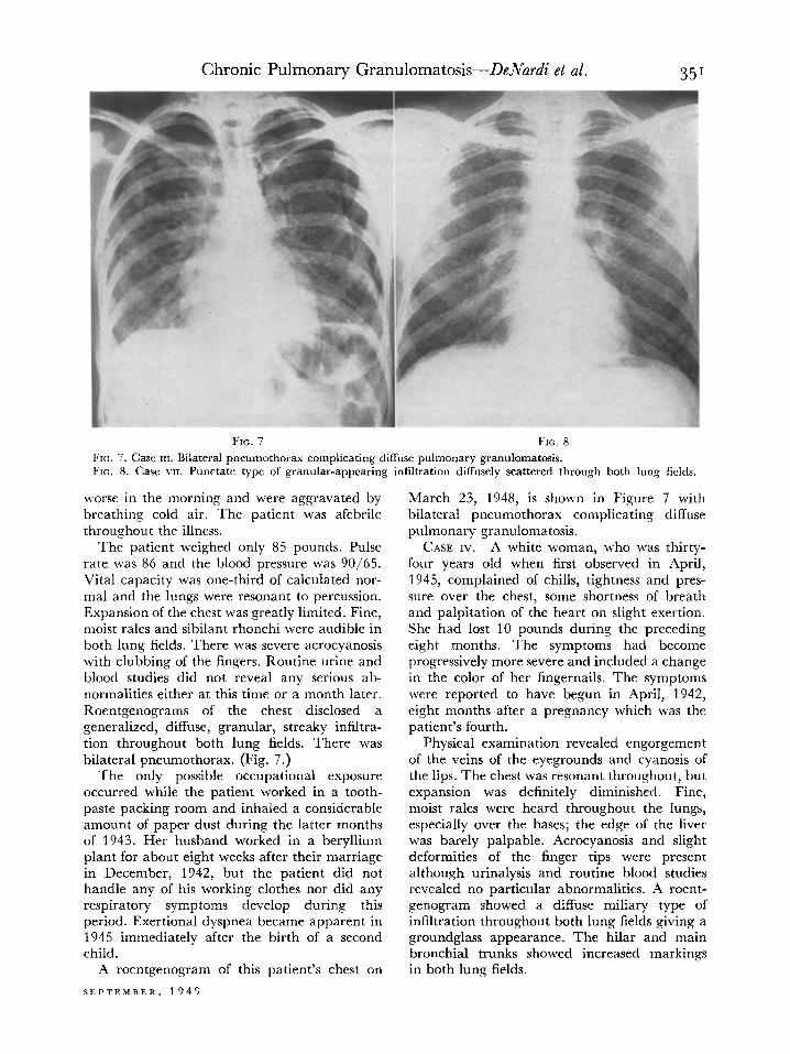
FIG. 7. Case III. Bilateral pneumothorax complicating diffuse pulmonary granulomatosis. FE. 8. Case VII. Punctate type of granular-appearing infiltration diffusely scattered through both lung fields.
worse in the morning and were aggravated by March 23, 1948, is shown in Figure 7 with breathing cold air. The patient was afebrile bilateral pneumothorax complicating diffuse throughout the illness. pulmonary granulomatosis.
The patient weighed only 85 pounds. Pulse rate was 86 and the blood pressure was 90/65. Vital capacity was one-third of calculated nor- mal and the lungs were resonant to percussion. Expansion of the chest was greatly limited. Fine, moist rales and sibilant rhonchi were audible in both lung fields. There was severe acrocyanosis with clubbing of the fingers. Routine urine and blood studies did not reveal any serious ab- normalities either at this time or a month later. Roentgenograms of the chest disclosed a generalized, diffuse, granular, streaky infiltra- tion throughout both lung fields. There was bilateral pneumothorax. (Fig. 7.)
CASE IV. A white woman, who was thirty- four years old when first observed in April, 1945, complained of chills, tightness and pres- sure over the chest, some shortness of breath and palpitation of the heart on slight exertion. She had lost 10 pounds during the preceding eight months. The symptoms had become progressively more severe and included a change in the color of her fingernails. The symptoms were reported to have begun in April, 1942, eight months after a pregnancy which was the patient’s fourth.
The only possible occupational exposure occurred while the patient worked in a tooth- paste packing room and inhaled a considerable amount of paper dust during the latter months of 1943. Her husband worked in a beryllium plant for about eight weeks after their marriage in December, 1942, but the patient did not handle any of his working clothes nor did any respiratory symptoms develop during this period. Exertional dyspnea became apparent in 1945 immediately after the birth of a second child.
A roentgenogram of this patient’s chest on
Physical examination revealed engorgement of the veins of the eyegrounds and cyanosis of the lips. The chest was resonant throughout, but expansion was definitely diminished. Fine, moist rales were heard throughout the lungs, especially over the bases; the edge of the liver was barely palpable. Acrocyanosis and slight deformities of the finger tips were present although urinalysis and routine blood studies revealed no particular abnormalities. A roent- genogram showed a diffuse miliary type of infiltration throughout both lung fields giving a groundglass appearance. The hilar and main bronchial trunks showed increased markings in both lung fields.
SEPTEMRER. t 949
352 Chronic Pulmonary Granulomatosis-DeNurdz’ et al.
During her three week hospitalization she received calcium gluconate intravenously and aminophylline. For two weeks she was given 20,000 units of penicillin every four hours. Slight improvement occurred with some clear- ing of the apices of the lung, lessened fatigue and shortness of breath on exertion and diminu- tion in the amount of sputum. The patient was somewhat improved six months later but still complained of dyspnea on exertion. Chest examination revealed some limitation of ex- pansion and sibilant rhonchi were heard throughout the hilar areas and bases; vital capacity was 82 per cent of calculated normal. Laboratory studies were essentially within normal limits and roentgenograms of the chest disclosed a diffuse miliary type of infiltration throughout both lung fields. During the eight months preceding her initial examination the patient had been exposed to inhalation of fumes from a beryllium plant located about 100 feet from her residence. She was not, however, em- ployed by the plant or apparently otherwise exposed.
On June 29, 1948, she gave birth to a normal boy following an uneventful pregnancy. When re-examined on October 1, 1948, she stated that she had improved considerably since leaving the neighborhood of the beryllium plant. The vital capacity was increased to 90 per cent of the computed normal and the acrocyanosis and clubbing of the fingers were comparatively less pronounced.
CASE v. A twenty-one year old white woman was first examined in September, 1947, her symptoms dating from about nine months previously when she had developed a spasmodic cough with some production of mucus. Shortly thereafter she noticed some dyspnea on exertion which was aggravated on damp days. Coughing spells were also more acute on damp days and in the mornings. Substernal pain and tightness became apparent, and a month or two before her visit her fingernails turned blue and showed a tendency to curve downward. Chills and fever were also reported.
On the day of admission to the hospital in September, 1947 the patient developed dyspnea with chills and fever. Her temperature was 104’~., pulse 134 and respiration 35. Acro- cyanosis with clubbing of the fingers were evi- dent. A persistent cough existed, perspiration was profuse and the patient’s breathing was rapid and shallow. Fine and coarse moist rales
were heard over the right lung and sibilant rhonchi over both lungs. Chest expansion was greatly limited. Urinalysis was negative and the blood count was within normal limits except for a leukocytosis of 10,450.
Roentgenograms, Figure 8, revealed a wide- spread, diffuse, miliary-like, granular-appearing infiltration throughout both lungs. Each granule appeared to be discrete. The patient was given oxygen almost constantly for a week and peni- cillin for two weeks. Streptomycin was ad- ministered for five days. A roentgenogram taken at the time of discharge from the hospital seventeen days after admission did not show any appreciable change.
The patient was re-examined about two months later. Her vital capacity was 86 per cent of calculated normal and the laboratory tests were essentially normal.
This patient for the approximate five years from 1940 to 1945 had lived within one block of a beryllium plant. In November, 1945 she moved about 1 mile away but returned to the earlier address in January, 1947 shortly before the first symptoms became manifest. The same month she gave birth to a son. She was ex- amined on June 29, 1948, and stated that since moving to her new residence three months ago she had noticed a definite improvement. The vital capacity was 90 per cent of normal but physical examination of the chest was the same as on the last examination.
CASE VI. A white woman, twenty-eight years old, first noticed symptoms in December, 1946. These symptoms of weakness and loss of ambi- tion became progressively worse. Approxi- mately five months later in May, 1947 she developed a spasmodic non-productive cough, more pronounced in the mornings on awaken- ing. About one month later the cough became productive with mucus occasionally slightly tinged with blood. The cough grew worse when she inhaled fumes from a beryllium plant near which she lived. Fever and chills at frequent intervals developed in July, 1947. At this time she had a fever of over 101O~. The symptoms of dyspnea were more evident on humid days while the patient had lost 22 pounds between the onset of her symptoms and the time when she was first observed on November 24, 1947. A bluish color of her fingernails was noted during the summer of 1947 as well as downward curving of the fingernails. On May 28, 1945, the patient completed her second pregnancy.
AMERICAN JOURNAL OF MEDICINE
Chronic Pulmonary Granulomatosis-De.Nd~ et al. 353 On examination acrocyanosis was noted with
slight clubbing of the finger tips. Systolic pres- sure was 100, pulse rate was 120, the vital capacity was 68 per cent of normal and the chest expansion was definitely diminished. Fine crackling rales and sibilant rhonchi were audible, especially in the hilar and basal areas of the lungs. Resonance of the lungs was normal.
Roentgenograms of the chest revealed a diffuse granular type of infiltration throughout both lung fields. The granular areas were some- what discrete and generalized haziness was shown throughout both lungs. Examination of the urine and blood did not reveal any serious abnormalities except a white blood cell count of 13,300.
The patient had worked in and around a beryllium plant for six weeks during the sum- mer of 1941. His work consisted principally in the construction and handling of bags of beryllium oxide and what he thought was beryllium sulfate. He cleaned out several sulfate vats. He did not complain of any illness during this period, but dust from the bags irritated his throat and produced a cough. With the excep- tion of these six weeks, he was either working on a railroad or serving in the armed forces until February, 1946. Since that time, he has been employed in a radiator plant where his work consists of the chemical testing of iron for sulfur, silica and carbon. He has worked with calcium carbide frequently.
This patient had lived approximately one block from a beryllium plant for the f&r years preceding her first examination in November, 1947, i.e., over three years before any symptoms became manifest. She was last seen in June 1, 1948, and had improved definitely since leaving the neighborhood of the beryllium plant. How- ever, she still complained of cough and shortness of breath on exertion. Examination of the chest did not reveal anything new. The vital capacity was 68 per cent of computed normal.
He was last examined on June 29, 1948, at which time he complained of frequent attacks of hyperpyrexia associated with dyspnea. These attacks had occurred at intervals of from two to three times a week during the previous two months. Examination at that time revealed the same chest findings as on the last visit. The vital capacity was 47 per cent of normal. The weight was 124 pounds and the temperature 98’~.
CASE VII. A white man aged twenty-seven developed a cough with chills and fever in December, 1944 while he was in the army. Roentgenogram of the chest at that time was negative. The chills, fever and cough persisted with the symptoms most pronounced early in the morning. The cough was non-productive. The patient had lost 8 pounds during the six months prior to December 1, 1947, at which time he complained of weakness and exertional dyspnea with some substernal pain and tightness of the chest on inspiration. The symptoms were more evident on humid days.
CASE VIII. A white woman thirty-nine years old began to exhibit symptoms in August, 1946. At that time she developed a cold with a cough which persisted until December 1, 1947, the time of her first examination by us. At first thr cough was non-productive but after several months sputum was present in the morning. During inspiration and the coughing attacks she was aware of substernal pain and tightness of the chest. In July, 1947 she experienced dyspnea on exertion which has grown progres- sively worse. By December, 1947 she was able to do only light housework without discomfort. During the entire illness her appetite was good and she gained 8 pounds from <June, 1947 to December, 1947.
On examination there was definite acrocyano- Physical examination did not disclose any sis with slight clubbing of the fingers and toes. acrocyanosis or abnormal changes of the Pulse rate was 78. Chest expansion was restricted fingers. Pulse rate was 76. Expansion of both and the vital capacity was 74 per cent. Sibilant lungs was adequate and equal. Vital capacity rhonchi were heard intermittently over both was 95 per cent of computed normal although lung bases. Roentgenograms of the chest (Fig. 8) a few wheezy rales were audible throughout showed slight enlargement of the hilar shadows the hilar areas. Routine laboratory tests were but there was no suggestion of nodulation. A essentially normal. Roentgenograms on Decem- punctate type of granular-appearing infiltration ber 1, 1947, Figure 9, revealed a slight enlarge- was diffusely scattered through both lung fields. ment of the hilar shadows but no nodulation. The patient has been under observation since There was a punctate type of granular-appear- that time and has grown progressively worse. ing infiltration diffusely scattered through both Laboratory tests are essentially normal. lung fields.
SEPTEMBER, 1949
354 Chronic Pulmonary Granulomatosis-DeNardi et al.
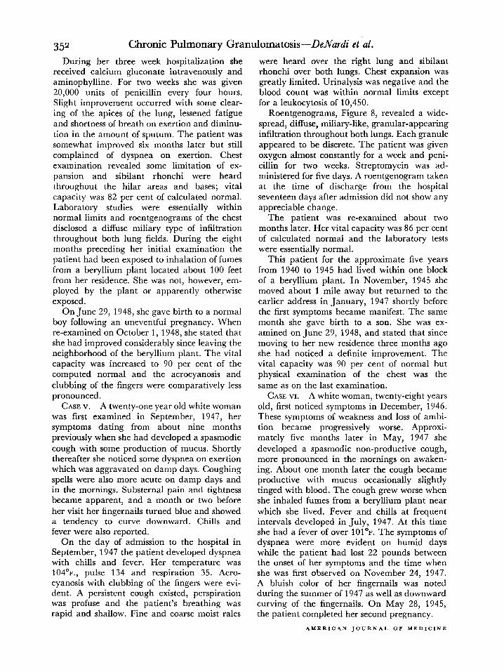
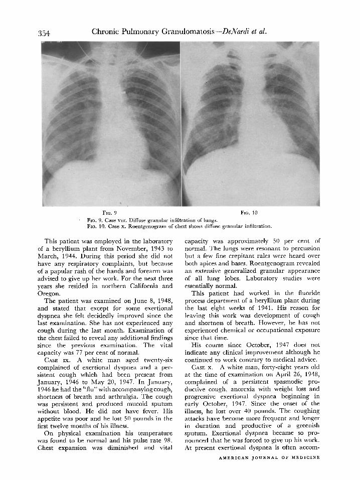
FIG. 9 FIG. 10 i FIG. 9. Case VIII. Diffuse granular infiltration of lungs.
FIG. 10. Case x. Roentgenogram of chest shows diffuse granular infiltration.
This patient was employed in the laboratory of a beryllium plant from November, 1943 to March, 1944. During this period she did not have any respiratory complaints, but because of a papular rash of the hands and forearm was advised to give up her work. For the next three years she resided in northern California and Oregon.
The patient was examined on June 8, 1948, and stated that except for some exertional dyspnea she felt decidedly improved since the last examination. She has not experienced any cough during the last month. Examination of the chest failed to reveal any additional findings since the previous examination. The vital capacity was 77 per cent of normal.
CASE IX. A white man aged twenty-six complained of exertional dyspnea and a per- sistent cough which had been present from January, 1946 to May 20, 1947. In January, 1946 he had the ‘Ylu” with accompanying cough, shortness of breath and arthralgia. The cough was persistent and produced mucoid sputum without blood. He did not have fever. His appetite was poor and he lost 50 pounds in the first twelve months of his illness.
On physical examination his temperature was found to be normal and his pulse rate 98. Chest expansion was diminished and vital
capacity was approximately 50 per cent of normal. The lungs were resonant to percussion but a few fine crepitant rales were heard over both apices and bases. Roentgenogram revealed an extensive generalized granular appearance of all lung lobes. Laboratory studies were essentially normal.
This patient had worked in the fluoride process department of a beryllium plant during the last eight weeks of 1941. His reason for leaving this work was development of cough and shortness of breath. However, he has not experienced chemical or occupational exposure since that time.
His course since October, 1947 does not indicate any clinical improvement although he continued to work contrary to medical advice.
CASE x. A white man, forty-eight years old at the time of examination on April 26, 1948, complained of a persistent spasmodic pro- ductive cough, anorexia with weight loss and progressive exertional dyspnea beginning in early October, 1947. Since the onset of the illness, he lost over 40 pounds. The coughing attacks have become more frequent and longer in duration and productive of a greenish sputum. Exertional dyspnea became so pro- nounced that he was forced to give up his work. At present exertional dyspnea is often accom-
AMERICAN JOURNAL OF MEDICINE
Chronic Pulmonary Granulomatosis-DeJVurdi et al. 355 panied by vertigo. He has experienced some mild chills and at times stated that he “felt hot.”
Physical examination revealed a normal tem- perature, pulse rate of 75 and respiratory rate of 20. There was no respiratory distress or acrocyanosis and changes of the finger tips. The blood pressure was 96 mm. Hg systolic and 70 mm. Hg diastolic. The vital capacity was 82 per cent of normal and the breath holding test was 20 seconds.
The chest expansion was slightly diminished with the patient’s complaint of substernal dis- comfort at the limit of inspiration. The per- cussion note was resonant and equal throughout. Breath sounds were of normal character throughout both lungs and no adventitious sounds were audible. Pulmonary roentgeno- grams showed a diffuse, granular and fibrous- appearing infiltration throughout the paren- chyma of both lungs. There was some associated prominence of the hilar shadows but no nodula- tion at the hila. (Fig. 10.) Laboratory studies of the blood and urine were essentially normal.
The patient was employed in a beryllium plant on April 30, 1945, and first worked for seven days handling beryllium fluoride until transferred to the crystallizing sulfating depart- ment for a period of nine days previous to onset of symptoms on May 30, 1945, when he de- veloped exertional dyspnea and a productive spasmodic cough. The clinical and roentgen- ologic diagnosis at that time was acute chemical bronchitis probably due to beryllium salts. He recovered from this initial attack and returned to work in the same plant on July 9, 1945, in the ore grinding mill. After an additional ten days of work he developed a second attack of acute bronchitis and following complete re- covery he was given a medical release from the industry on September 11, 1948, because of his recent illness.
This patient was seen on June 28, 1948, and still complained of a spasmodic cough, shortness of breath and loss of weight. Examination of the chest failed to reveal any changes. The vital capacity was 71 per cent of normal. His weight was 145 pounds.
SUMMARY *
From a review of the pertinent data in ten cases, including two patients who died,
* Since this report was submitted three additional patients have been seen. One patient died and necropsy was performed with findings similar to those previously reported.
SEPTEMBER, 1949
it is apparent that a characteristic sequence of clinical symptoms and the typical ap- pearance of roentgenograms permit ac- curate diagnosis in chronic pulmonary granulomatosis. A history of exposure to certain chemical hazards may aid in the diagnosis, but conclusive evidence of ex- posure cannot always be secured. Specific treatment is not available. Certain drugs may be used symptomatically with some degree of relief of subjective symptoms. The most effective treatment at present is ade- quate rest and the avoidance of respiratory infection. All evidence points to early diagnosis as the 3rime requisite for proper treatment.
REFERENCES
1. VAN ORDSTRAND, H. S., HUGHES, R. and CARMODY, M. G. Chemical pneumonia in workers extracting beryllium oxide; report of 3 cases. Cle~~lnnd Clin. Quart., 10: 10-18, 1943.
2. VAN ORDSTRAND, H. S., HUGHES, R., DENARDI, J. M. and CARMODY, M. G. Beryllium poisoning. 3. A. M. A., 129: 1084-1090, 1945.
3. HARDY, H. L. and TABERSHAW, I. R. Delayed chemical pneumonitis occurring in workers exposed to beryllium compounds. 3. Indust. Hyg. L? Toxical.. 28: 197-211. 1946.
4. HIGGINS, H.’ L. Pulmonary sarcoidosis. Connecticut hf. .jf., 11: 330-339, 1947.
5. KRESS, J. E. and CRISPELL, K. R. Chemical pneu- monitis in men working with fluorescent powders containing beryllium. Gut/z& Clin. Bull., 13: 91-95, 1944.
6. KING, D. S. Sarcoid disease as revealed in chest roentgenogram. Am. 3. Roentgenol., 45: 505-512, 1941.
7. REISNER, D. Boeck’s sarcoid and systemic sarcoido- sis, (Besnier-Boeck-Schaumann disease; study of 35 cases. Am. Rev. ‘Tuberc., 49: 289-307, 1944.
8. CULVER, G. J. Miliary carcinosis of lungs secondary to primary cancer of gastrointestinal tract. Am. 3. Roentgenol., 54: 474-482, 1945.
9. DUNNER, L., HERMON, R. and BAGNALL, D. J. T. Pneumoconiosis in radiator and boiler finishers. Brit. 3. Radial., 18: 377-381, 1945.
10. GARDNER, L. U. Pathology and roentgenographic manifestations of pneumoconiosis. 3. A. M. A., 114: 535-545, 1940.
11. KERLEY, P. Significance of radiological manifesta- tions of erythema nodosum. Brit. .7. Rndiol., 15: 155-165, 1942.
._
12. CARTER, R. A. Roentgen diagnosis of fungous infec- tions of lungs with special reference to coccidioido- mycosis. Rodiolqey, 38: 649-659, 1942.
13. CAMIEI., M. R. and BERICAN, H. S. Inhalation pneumonia from nitric fumes. Kndiolop. 42: 175-182, 1944.
14. PASCUCCI, L. M. Pulmonary disease in workers exposed to beryllium compounds; its roentgen characteristics. Radiology, 50: 23-35, 1948.


![Chronic Obstructive Pulmonary Diseaseopenaccessebooks.com/chronic-obstructive-pulmonary...Chronic Obstructive Pulmonary Disease 5 a-MCI is made [32]. COPD patients without significant](https://img.pdfslide.net/doc/110x75/5f853ccf82a2412fd65b9e28/chronic-obstructive-pulmonary-dis-chronic-obstructive-pulmonary-disease-5-a-mci.jpg)