Embed Size (px)
Citation preview
42
postoperative imaging.
Case report A 64-year-old female was diagnosed with idiopathic
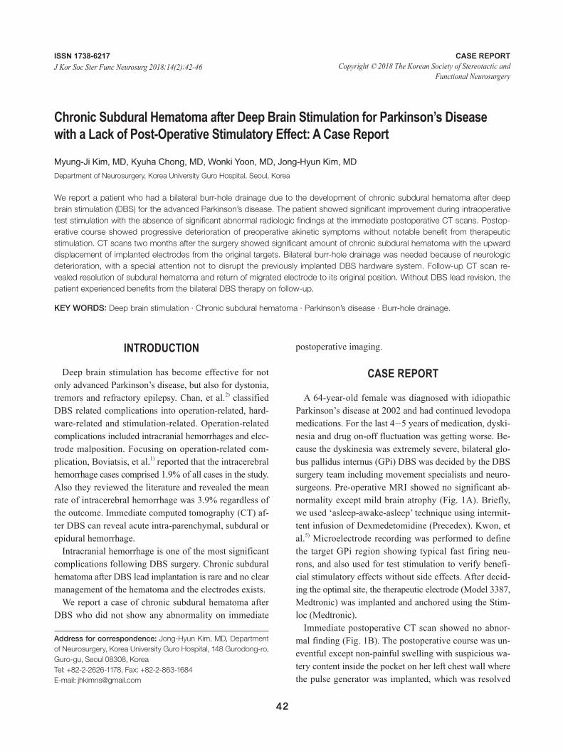
Parkinson’s disease at 2002 and had continued levodopa medications. For the last 4-5 years of medication, dyski-nesia and drug on-off fluctuation was getting worse. Be-cause the dyskinesia was extremely severe, bilateral glo-bus pallidus internus (GPi) DBS was decided by the DBS surgery team including movement specialists and neuro-surgeons. Pre-operative MRI showed no significant ab-normality except mild brain atrophy (Fig. 1A). Briefly, we used ‘asleep-awake-asleep’ technique using intermit-tent infusion of Dexmedetomidine (Precedex). Kwon, et al.5) Microelectrode recording was performed to define the target GPi region showing typical fast firing neu-rons, and also used for test stimulation to verify benefi-cial stimulatory effects without side effects. After decid-ing the optimal site, the therapeutic electrode (Model 3387, Medtronic) was implanted and anchored using the Stim-loc (Medtronic).
Immediate postoperative CT scan showed no abnor-mal finding (Fig. 1B). The postoperative course was un-eventful except non-painful swelling with suspicious wa-tery content inside the pocket on her left chest wall where the pulse generator was implanted, which was resolved
Chronic subdural Hematoma after Deep Brain stimulation for parkinson’s Disease with a Lack of post-operative stimulatory effect: a Case report
Myung-Ji Kim, MD, Kyuha Chong, MD, Wonki Yoon, MD, Jong-Hyun Kim, MDDepartment of Neurosurgery, Korea University Guro Hospital, Seoul, Korea
We report a patient who had a bilateral burr-hole drainage due to the development of chronic subdural hematoma after deep brain stimulation (DBS) for the advanced Parkinson’s disease. The patient showed significant improvement during intraoperative test stimulation with the absence of significant abnormal radiologic findings at the immediate postoperative CT scans. Postop-erative course showed progressive deterioration of preoperative akinetic symptoms without notable benefit from therapeutic stimulation. CT scans two months after the surgery showed significant amount of chronic subdural hematoma with the upward displacement of implanted electrodes from the original targets. Bilateral burr-hole drainage was needed because of neurologic deterioration, with a special attention not to disrupt the previously implanted DBS hardware system. Follow-up CT scan re-vealed resolution of subdural hematoma and return of migrated electrode to its original position. Without DBS lead revision, the patient experienced benefits from the bilateral DBS therapy on follow-up.
KEY WORDS: Deep brain stimulation · Chronic subdural hematoma · Parkinson’s disease · Burr-hole drainage.
IntroDuCtIon
Deep brain stimulation has become effective for not only advanced Parkinson’s disease, but also for dystonia, tremors and refractory epilepsy. Chan, et al.2) classified DBS related complications into operation-related, hard-ware-related and stimulation-related. Operation-related complications included intracranial hemorrhages and elec-trode malposition. Focusing on operation-related com-plication, Boviatsis, et al.1) reported that the intracerebral hemorrhage cases comprised 1.9% of all cases in the study. Also they reviewed the literature and revealed the mean rate of intracerebral hemorrhage was 3.9% regardless of the outcome. Immediate computed tomography (CT) af-ter DBS can reveal acute intra-parenchymal, subdural or epidural hemorrhage.
Intracranial hemorrhage is one of the most significant complications following DBS surgery. Chronic subdural hematoma after DBS lead implantation is rare and no clear management of the hematoma and the electrodes exists.
We report a case of chronic subdural hematoma after DBS who did not show any abnormality on immediate
Address for correspondence: Jong-Hyun Kim, MD, Department of Neurosurgery, Korea University Guro Hospital, 148 Gurodong-ro, Guro-gu, Seoul 08308, KoreaTel: +82-2-2626-1178, Fax: +82-2-863-1684E-mail: [email protected]
ISSN 1738-6217J Kor Soc Ster Func Neurosurg 2018;14(2):42-46
CASE REPORTCopyright © 2018 The Korean Society of Stereotactic and
Functional Neurosurgery
Myung-Ji Kim, et al : Chronic Subdural Hematoma after Deep Brain Stimulation
43
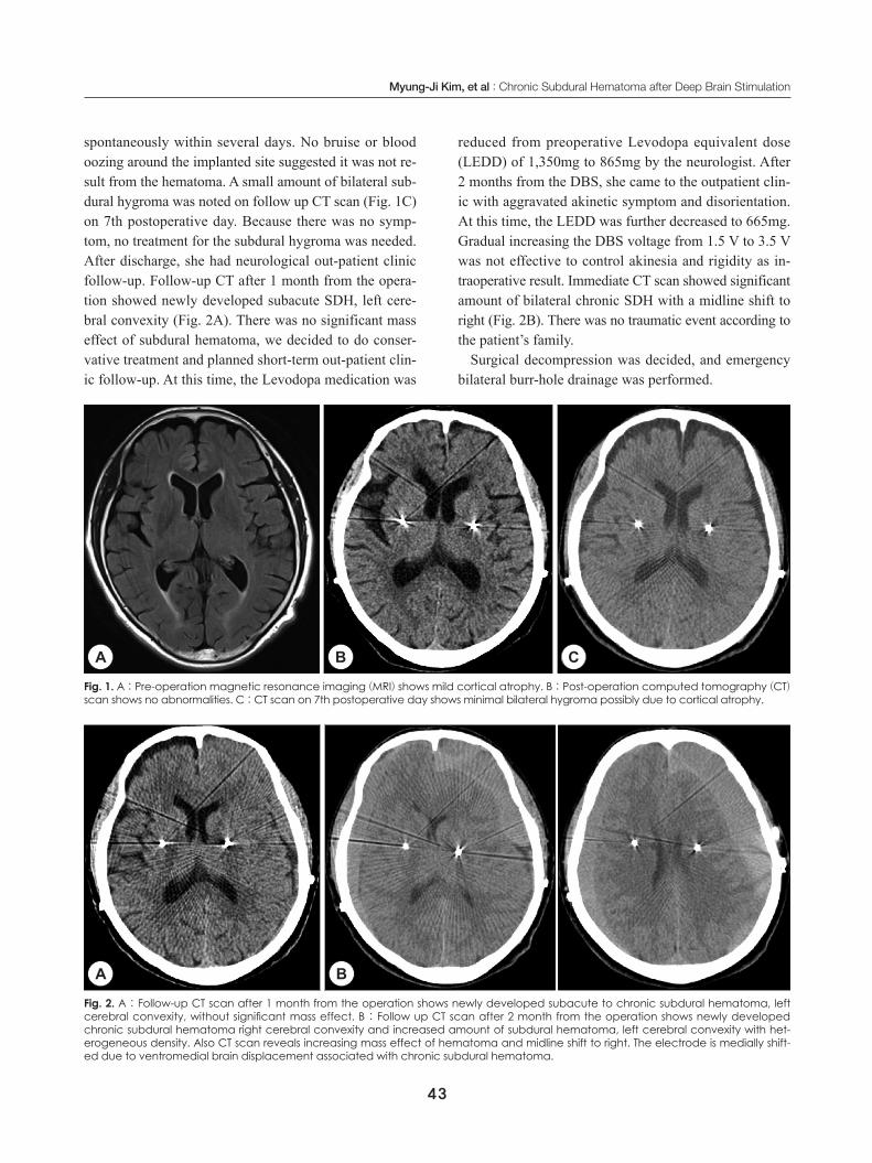
spontaneously within several days. No bruise or blood oozing around the implanted site suggested it was not re-sult from the hematoma. A small amount of bilateral sub-dural hygroma was noted on follow up CT scan (Fig. 1C) on 7th postoperative day. Because there was no symp-tom, no treatment for the subdural hygroma was needed. After discharge, she had neurological out-patient clinic follow-up. Follow-up CT after 1 month from the opera-tion showed newly developed subacute SDH, left cere-bral convexity (Fig. 2A). There was no significant mass effect of subdural hematoma, we decided to do conser-vative treatment and planned short-term out-patient clin-ic follow-up. At this time, the Levodopa medication was
reduced from preoperative Levodopa equivalent dose (LEDD) of 1,350mg to 865mg by the neurologist. After 2 months from the DBS, she came to the outpatient clin-ic with aggravated akinetic symptom and disorientation. At this time, the LEDD was further decreased to 665mg. Gradual increasing the DBS voltage from 1.5 V to 3.5 V was not effective to control akinesia and rigidity as in-traoperative result. Immediate CT scan showed significant amount of bilateral chronic SDH with a midline shift to right (Fig. 2B). There was no traumatic event according to the patient’s family.
Surgical decompression was decided, and emergency bilateral burr-hole drainage was performed.
Fig. 1. A : Pre-operation magnetic resonance imaging (MRI) shows mild cortical atrophy. B : Post-operation computed tomography (CT) scan shows no abnormalities. C : CT scan on 7th postoperative day shows minimal bilateral hygroma possibly due to cortical atrophy.
A B C
Fig. 2. A : Follow-up CT scan after 1 month from the operation shows newly developed subacute to chronic subdural hematoma, left cerebral convexity, without significant mass effect. B : Follow up CT scan after 2 month from the operation shows newly developed chronic subdural hematoma right cerebral convexity and increased amount of subdural hematoma, left cerebral convexity with het-erogeneous density. Also CT scan reveals increasing mass effect of hematoma and midline shift to right. The electrode is medially shift-ed due to ventromedial brain displacement associated with chronic subdural hematoma.
A B
44
J Kor Soc Ster Func Neurosurg 2018;14(2):42-46
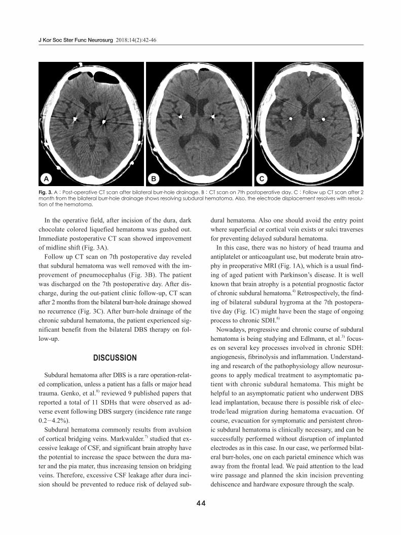
In the operative field, after incision of the dura, dark chocolate colored liquefied hematoma was gushed out. Immediate postoperative CT scan showed improvement of midline shift (Fig. 3A).
Follow up CT scan on 7th postoperative day reveled that subdural hematoma was well removed with the im-provement of pneumocephalus (Fig. 3B). The patient was discharged on the 7th postoperative day. After dis-charge, during the out-patient clinic follow-up, CT scan after 2 months from the bilateral burr-hole drainage showed no recurrence (Fig. 3C). After burr-hole drainage of the chronic subdural hematoma, the patient experienced sig-nificant benefit from the bilateral DBS therapy on fol-low-up.
DIsCussIon Subdural hematoma after DBS is a rare operation-relat-
ed complication, unless a patient has a falls or major head trauma. Genko, et al.8) reviewed 9 published papers that reported a total of 11 SDHs that were observed as ad-verse event following DBS surgery (incidence rate range 0.2-4.2%).
Subdural hematoma commonly results from avulsion of cortical bridging veins. Markwalder.7) studied that ex-cessive leakage of CSF, and significant brain atrophy have the potential to increase the space between the dura ma-ter and the pia mater, thus increasing tension on bridging veins. Therefore, excessive CSF leakage after dura inci-sion should be prevented to reduce risk of delayed sub-
dural hematoma. Also one should avoid the entry point where superficial or cortical vein exists or sulci traverses for preventing delayed subdural hematoma.
In this case, there was no history of head trauma and antiplatelet or anticoagulant use, but moderate brain atro-phy in preoperative MRI (Fig. 1A), which is a usual find-ing of aged patient with Parkinson’s disease. It is well known that brain atrophy is a potential prognostic factor of chronic subdural hematoma.4) Retrospectively, the find-ing of bilateral subdural hygroma at the 7th postopera-tive day (Fig. 1C) might have been the stage of ongoing process to chronic SDH.6)
Nowadays, progressive and chronic course of subdural hematoma is being studying and Edlmann, et al.3) focus-es on several key processes involved in chronic SDH: angiogenesis, fibrinolysis and inflammation. Understand-ing and research of the pathophysiology allow neurosur-geons to apply medical treatment to asymptomatic pa-tient with chronic subdural hematoma. This might be helpful to an asymptomatic patient who underwent DBS lead implantation, because there is possible risk of elec-trode/lead migration during hematoma evacuation. Of course, evacuation for symptomatic and persistent chron-ic subdural hematoma is clinically necessary, and can be successfully performed without disruption of implanted electrodes as in this case. In our case, we performed bilat-eral burr-holes, one on each parietal eminence which was away from the frontal lead. We paid attention to the lead wire passage and planned the skin incision preventing dehiscence and hardware exposure through the scalp.
Fig. 3. A : Post-operative CT scan after bilateral burr-hole drainage. B : CT scan on 7th postoperative day. C : Follow up CT scan after 2 month from the bilateral burr-hole drainage shows resolving subdural hematoma. Also, the electrode displacement resolves with resolu-tion of the hematoma.
A B C
Myung-Ji Kim, et al : Chronic Subdural Hematoma after Deep Brain Stimulation
45
Follow-up CT scan after 2 months from the burr-hole drainage revealed that the electrode displacement resolved with resolution of the hematoma and no hardware revi-sion was necessary (Fig. 3C). Yang, et al.10) commented that gliosis formed along the track of the electrodes may function as a potential space. As a result, once the subdu-ral hematoma is evacuated, the migrated electrode may glide back to its original location. Consequently, the DBS stimulation can remain effective without DBS revision surgery. When asymptomatic chronic subdural hemato-ma is managed conservatively without operation, it is bothersome to wait the electrode returns to near its initial position after spontaneous resolution of subdural hema-toma.
Usually, it is known that levodopa dose can be reduced after subthalamic nucleus (STN) DBS but not after GPi DBS.9) The anti-dyskinetic effect of STB DBS is believed through reducing the levodopa dose, but through direct effect in GPi DBS. The reduction of levodopa equivalent daily dose (LEDD) after the surgery in this case is not usu-al. The reason of dose reduction in this case was due to side effects of dopamine receptor agonist, mild psycho-sis and hallucination after the surgery. Simultaneous in-creasing the DBS voltage was not as effective to control patient’s akinetic symptoms as have been observed in-traoperatively. This might be due to gradual upward dis-placement of implanted electrodes from the original site by the mass effect due to chronic SDH (Fig. 2B).
We could learn several lessons from this case. Surgeons are generally careful avoiding CSF leakage during DBS surgery. Too much CSF leak may result in brain shift which may result in changes of target coordinates measured pre-operatively. Brain shift may lead to acute subdural hem-orrhage due to bridging-vein damage. We were also care-ful not to drain CSF by applying sufficient irrigation and surgical glue on the incised dura during the surgery, and immediate postoperative CT did not show any evidence of pneumocephalus or hygroma. The delayed subdural hy-groma 1 week after the surgery might have been a warn-ing sign of subsequent chronic SDH. The non-painful swelling on chest wall might have resulted from CSF leakage from the burr-hole site. The Stimloc device uses screws to secure it to the skull, but it does not always fit the curvature in our experience. We speculate the possi-bility of CSF leakage from the unfitted gap between the device and skull might have resulted in CSF collection on the swollen chest site, but there was no evidence.
Chronic SDH is characterized by insidious symptoms. Evolution from the subdural hygroma to chronic SDH is common which has been suggested as a natural course previously.6) As seen in this case, even small amount of subdural hygroma may evolve to significant amount of chronic SDH. Serious results may occur in case of chron-ic SDH after DBS, if the physician does not identify the brain image and misinterprets the patient’s akinetic symp-toms as a result of reduced levodopa in the dose adjust-ment process.
ConCLusIon
Chronic SDH after DBS is a rare complication. A pa-tient with over moderate brain atrophy needs more at-tention for chronic subdural hematoma. It may be pre-vented by avoiding excessive CSF leakage and a careful decision of an entry point in the operation, but not always possible as seen in the present case. The present case shows even small amount of postoperative subdural hy-groma may change into a significant chronic subdural he-matoma. Therefore, special attention and subsequent brain imaging are necessary, especially if there is a lack of an-ticipated DBS stimulatory effect.
REfERENCES1. Boviastsis EJ, Stavrinou LC, Themistocleous M, Kouyiallis AT,
Sakas DE: Surgical and hardware complication of deep brain stim-ulation: A seven-year experience and review of the literature. Acta Neurochirurgica (Wien) 152:2053-2062, 2010
2. Chan DTM, Zhu XL, Yeung JHM, Mok VCT, Wong E, Lau C, et al: Complication of deep brain stimulation: A collective review. Asian J Surgery 32:258-263, 2009
3. Edlmann E, Giorgi-Coll S, Whittfield PC, Carpenter KLH, Hutchin-son PJ: Pathophysiology of chronic subdural hematoma: inflam-mation, angiogenesis and implications for pharmacotherapy. J Neu-roinflammation 14:108-121, 2017
4. Jeong EO, Choi SW, Lim JW, Kwon HJ, Kim SH, Koh HS, et al: Effectiveness of cortical atrophy scale and indirect indices of brain atrophy to predict chronic subdural hematoma in older patients. Korean J Neurotrauma 12:112-117, 2016
5. Kwon WG, Kim JH, Lee JH, Lim BG, Lee IO, Koh SB, et al: Mi-croelectrode recording (MER) findings during sleep-awake anes-thesia using dexmedetomidine in deep brain stimulation surgery for Parkinson’s disease. Clin Neurol Neurosurg 143:27-33, 2016
6. Lee KS: Natural history of chronic subdural hematoma: Review. Brain injury 18:351-358, 2004
7. Markwalder TM: Chronic subdural hematomas: a review. J Neuro-surgery 54:637-645, 1981
8. Oyama G, Okun MS, Zesiewicz TA, Tamse T, Romrell J, Zeilman P, et al: Delayed clinical improvement after deep brain stimula-tion-related subdural hematoma. J Neurosurgery 115:289-294, 2011
9. Williams NR, Foote KD, Okum MS: Subthalamic nucleus versus

46
J Kor Soc Ster Func Neurosurg 2018;14(2):42-46
globus pallidus internus deep brain stimulation: Translating the re-match into clinical practice. Movement disorder: Clinical Practice 1:24-35, 2014
10. Yang YJ, Jhang SW, Chen CM, Chen YH, Cheng CY: Functional
preservation of deep brain stimulation electrodes after brain shift induced by traumatic subdural hematoma-case report. British J Neurosurgery 27:128-129, 2013