Embed Size (px)
Citation preview

Cirrhosis By: Kirsten Dyck, Emily Horn,
Kim Jenkins, and Natalie White
Liver
Physiology By: Kim Jenkins
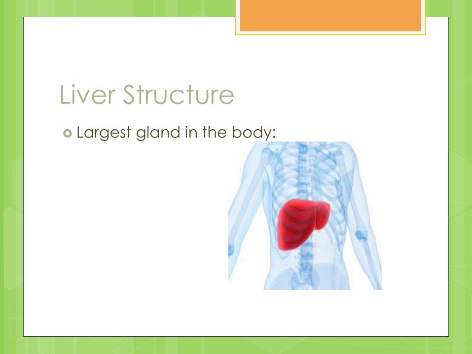
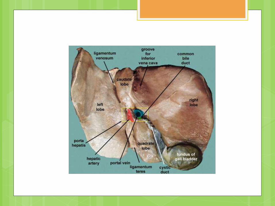
Liver Structure
Largest gland in the body:
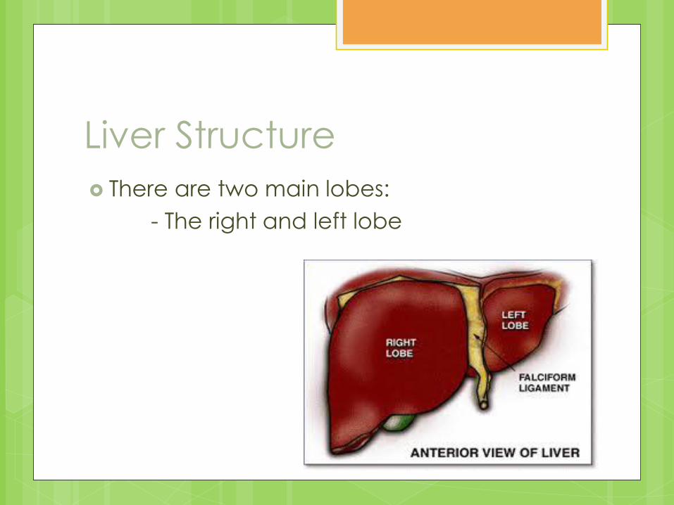
Liver Structure
There are two main lobes:
- The right and left lobe
Liver Structure
The liver gets blood from the hepatic
artery and the portal vein.
The liver has bile ducts. Bile is formed in
the liver cells, exits the liver through a
series of bile ducts that increase in size as
they approach the common bile duct.
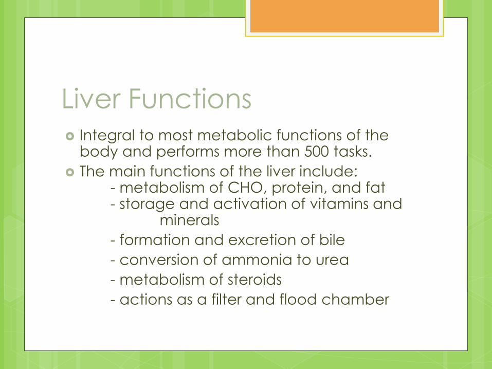
Liver Functions Integral to most metabolic functions of the
body and performs more than 500 tasks.
The main functions of the liver include: - metabolism of CHO, protein, and fat - storage and activation of vitamins and minerals
- formation and excretion of bile
- conversion of ammonia to urea
- metabolism of steroids
- actions as a filter and flood chamber
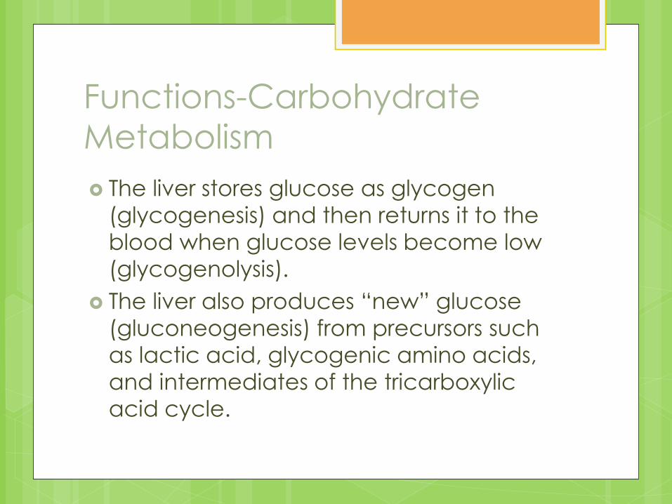
Functions-Carbohydrate
Metabolism
The liver stores glucose as glycogen
(glycogenesis) and then returns it to the
blood when glucose levels become low
(glycogenolysis).
The liver also produces “new” glucose
(gluconeogenesis) from precursors such
as lactic acid, glycogenic amino acids,
and intermediates of the tricarboxylic
acid cycle.
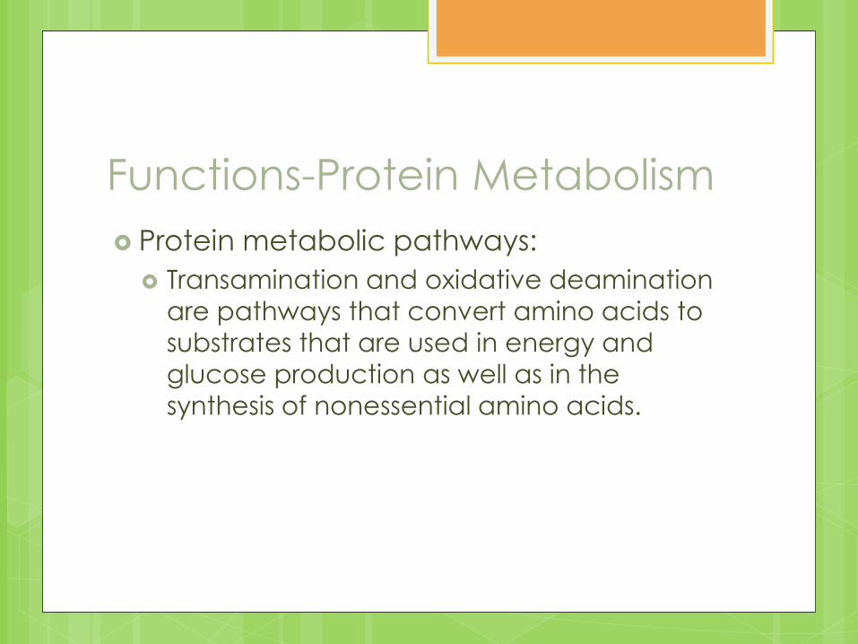
Functions-Protein Metabolism
Protein metabolic pathways:
Transamination and oxidative deamination
are pathways that convert amino acids to
substrates that are used in energy and
glucose production as well as in the
synthesis of nonessential amino acids.
Functions- Fat Metabolism
Fatty acids from the diet and adipose
tissue are converted in the liver to acetyl-
coenzyme A by the process of beta-
oxidation to produce energy. Ketones
are also produced.
The liver synthesizes and hydrolyzes
triglycerides, phospholipids, cholesterol,
and lipoproteins.
Functions- Vitamins and
Minerals
Storage:
Storage of all the fat-soluble vitamins in addition to vitamin B12 and the minerals zinc, iron, copper, and magnesium.
Transportation:
Hepatically synthesized proteins transport vitamin A, iron, zinc, and copper in the bloodstream.
Activation:
Carotene is converted to vitamin A, folate to 5-methyl tetrahydrofolic acid, and vitamin D to an active form (25 hyroxycholecalciferol).
Functions- Bile
The liver forms and excretes bile.
Bile salts are metabolized and used for the
digestion and absorption of fats and fat-
soluble vitamins.
Bilirubin is a metabolic end produce from
red blood cell destruction; it is conjugated
and excreted in the bile.
Functions- Other Hepatocytes detoxify ammonia by converting to
the urea, 75% which is excreted by the kidneys. The remaining goes back to the GI tract.
The liver metabolizes steroids: it inactivates and excretes aldosterone, glucocorticoids, estrogen, progesterone, and testosterone.
It is responsible for the detoxification of drugs and alcohol and other substances.
Acts as a filter and flood chamber by removing bacteria and debris from blood through the phagocytic action of kupffer cells located in the sinusoids and by storing blood backed up from the vena cava as in right heart failure.
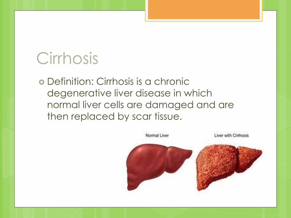
Cirrhosis
Definition: Cirrhosis is a chronic
degenerative liver disease in which
normal liver cells are damaged and are
then replaced by scar tissue.
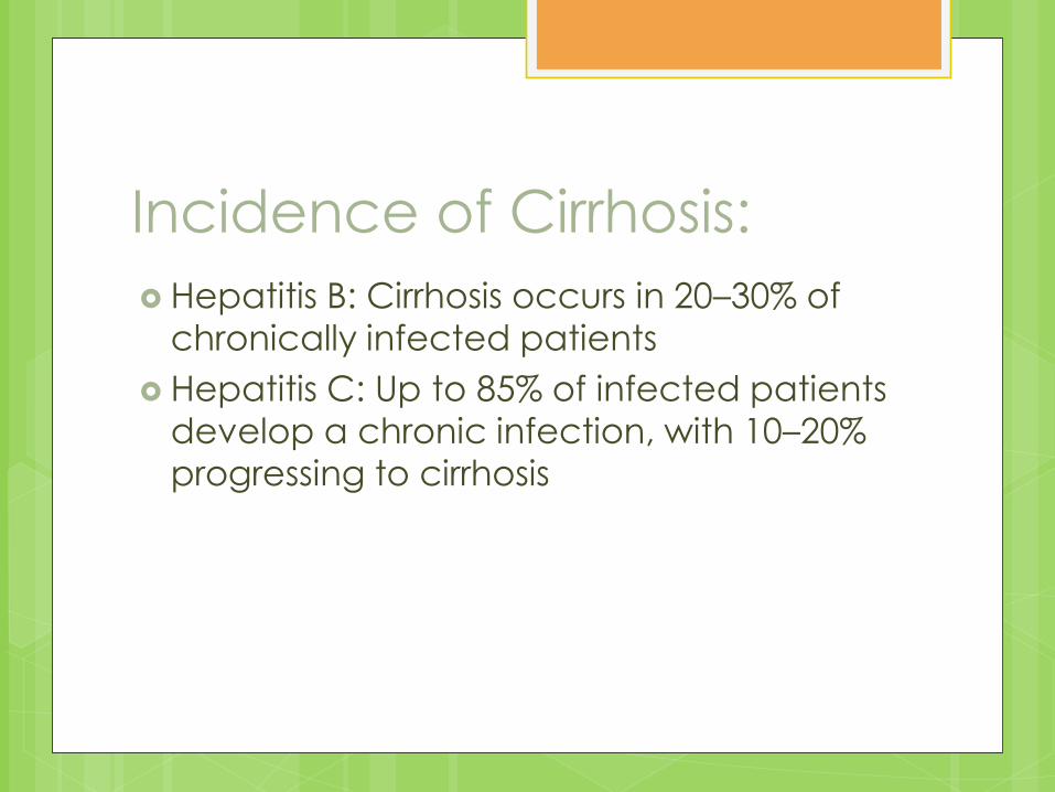
Incidence of Cirrhosis:
Hepatitis B: Cirrhosis occurs in 20–30% of
chronically infected patients
Hepatitis C: Up to 85% of infected patients
develop a chronic infection, with 10–20%
progressing to cirrhosis
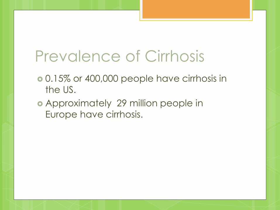
Prevalence of Cirrhosis
0.15% or 400,000 people have cirrhosis in
the US.
Approximately 29 million people in
Europe have cirrhosis.
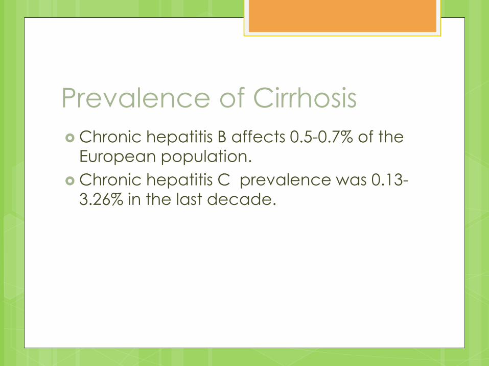
Prevalence of Cirrhosis
Chronic hepatitis B affects 0.5-0.7% of the
European population.
Chronic hepatitis C prevalence was 0.13-
3.26% in the last decade.
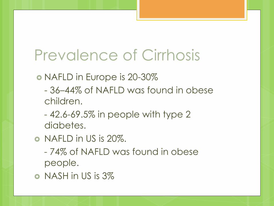
Prevalence of Cirrhosis
NAFLD in Europe is 20-30%
- 36–44% of NAFLD was found in obese
children.
- 42.6-69.5% in people with type 2
diabetes.
NAFLD in US is 20%.
- 74% of NAFLD was found in obese
people.
NASH in US is 3%
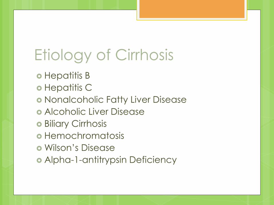
Etiology of Cirrhosis
Hepatitis B
Hepatitis C
Nonalcoholic Fatty Liver Disease
Alcoholic Liver Disease
Biliary Cirrhosis
Hemochromatosis
Wilson’s Disease
Alpha-1-antitrypsin Deficiency

Hepatitis B and C
Can lead to chronic and carrier states.
Chronic active hepatitis can also
develop, which leads to cirrhosis and liver
failure.
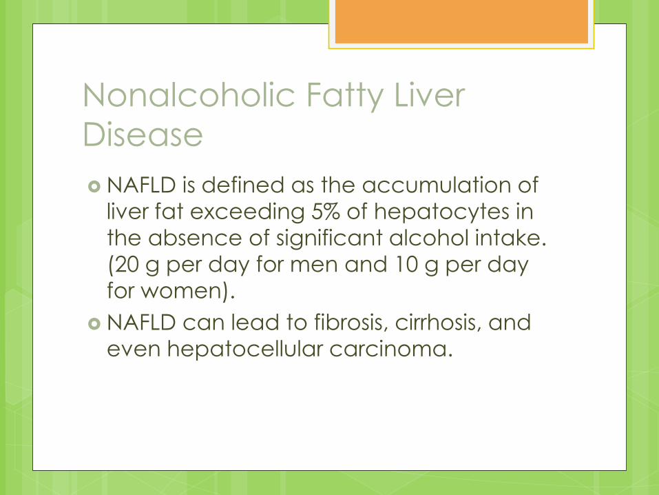
Nonalcoholic Fatty Liver
Disease
NAFLD is defined as the accumulation of
liver fat exceeding 5% of hepatocytes in
the absence of significant alcohol intake.
(20 g per day for men and 10 g per day
for women).
NAFLD can lead to fibrosis, cirrhosis, and
even hepatocellular carcinoma.
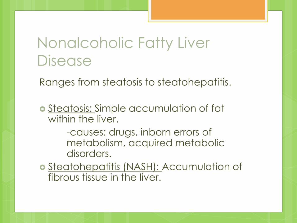
Nonalcoholic Fatty Liver
Disease
Ranges from steatosis to steatohepatitis.
Steatosis: Simple accumulation of fat within the liver.
-causes: drugs, inborn errors of metabolism, acquired metabolic disorders.
Steatohepatitis (NASH): Accumulation of fibrous tissue in the liver.
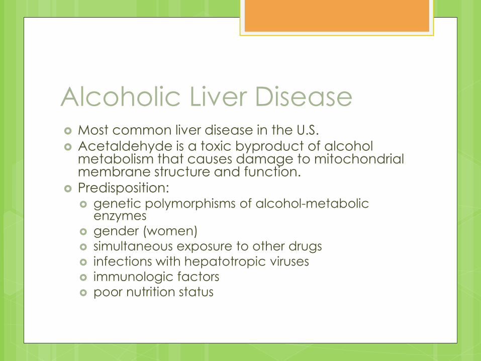
Alcoholic Liver Disease Most common liver disease in the U.S.
Acetaldehyde is a toxic byproduct of alcohol metabolism that causes damage to mitochondrial membrane structure and function.
Predisposition: genetic polymorphisms of alcohol-metabolic
enzymes
gender (women)
simultaneous exposure to other drugs
infections with hepatotropic viruses
immunologic factors
poor nutrition status
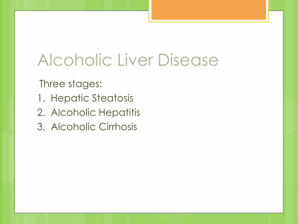
Alcoholic Liver Disease
Three stages:
1. Hepatic Steatosis
2. Alcoholic Hepatitis
3. Alcoholic Cirrhosis
Biliary Cirrhosis
Chronic cholestatic disease caused by
progressive destruction of small and
intermediate-size intrahepatic bile ducts. (not extrahepatic or larger intrahepatic bile ducts)
Results in Cirrhosis, disease progresses
slowly, eventually resulting in cirrhosis,
portal hypertension, liver transplantation,
or death.

Hemochromatosis
Inherited disease of iron overload
associated with the gene HFE.
Patients absorb excessive iron from the
gut and may store 20-40g compared to
the normal .3-.8g.
Life expectancy is normal if phlebotomy is
initiated before the development of
cirrhosis or diabetes mellitus.
Wilson’s Disease
Autosomal-recessive disorder associated with impaired biliary copper excretion.
Copper accumulateds in the liver, brain, cornea, and kidneys.
Copper chelation improves survival but does ot prevent cirrhosis, liver transplantation will correct the metabolic defect.
Alpha1 –Antitrypsin Deficiency
Inherited disorder.
Alpha1-Antitrypsin is a glycoprotein found
in serum and body fluids, it inhibits
neutrophil proteinases.
Cholestasis or cirrhosis is caused by this
deficiency.
Risk Factors
Obesity
Overweight
Type 2 diabetes
Hemochromatosis
Hepatitis B or C

Pathophysiology By: Kirsten Dyck
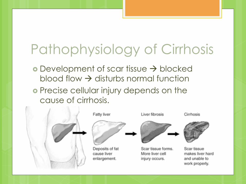
Pathophysiology of Cirrhosis
Development of scar tissue blocked
blood flow disturbs normal function
Precise cellular injury depends on the
cause of cirrhosis.
Alcoholic Cirrhosis
Characterized by:
Inflammation
Degeneration and necrosis of hepatocytes
Infiltration of leukocytes and lymphocytes
Immunologic alterations
Lipid peroxidation
Fibrosis
Serum IgA is often elevated.
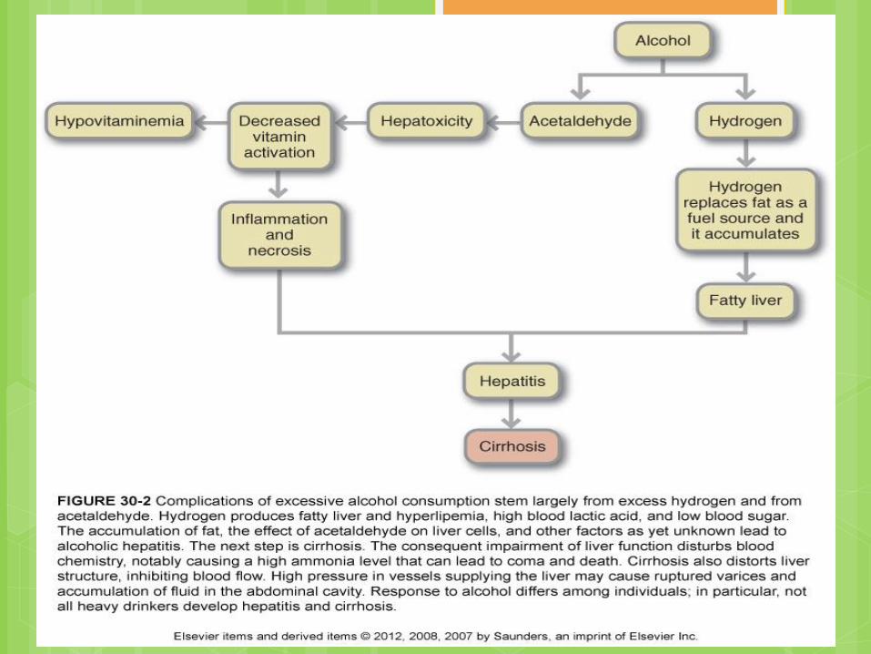
Alcoholic Cirrhosis
Begins with hepatic steatosis:
Alcohol acetaldehyde lipid
peroxidation disruption in membrane
function.
Mitochondrial function is impaired.
Enzyme and protein synthesis may be
depressed or altered.
Hormone and ammonia degradation is
diminished.
Alcoholic Cirrhosis cont.
Acetaldehyde causes:
Altered metabolism of vitamins and minerals.
Liver fibrosis
Malnutrition
Hepatic specific autoantibodies
TNF, IL-6, IL-8 and IL-18 are associated with
ALD
Inflammation and necrosis collagen
formation.
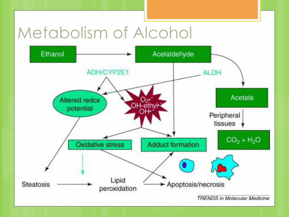
Metabolism of Alcohol
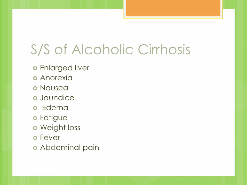
S/S of Alcoholic Cirrhosis Enlarged liver
Anorexia
Nausea
Jaundice
Edema
Fatigue
Weight loss
Fever
Abdominal pain
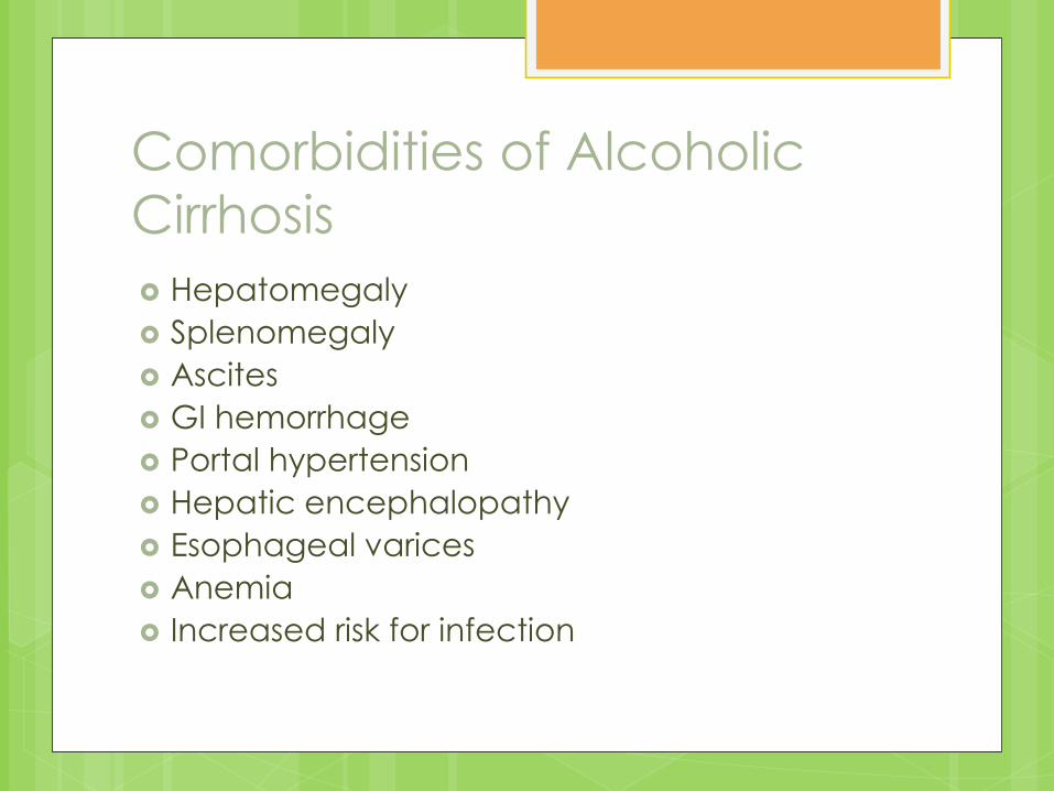
Comorbidities of Alcoholic
Cirrhosis
Hepatomegaly
Splenomegaly
Ascites
GI hemorrhage
Portal hypertension
Hepatic encephalopathy
Esophageal varices
Anemia
Increased risk for infection
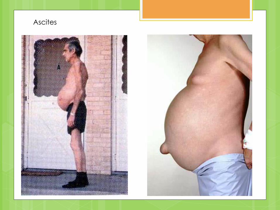
Ascites
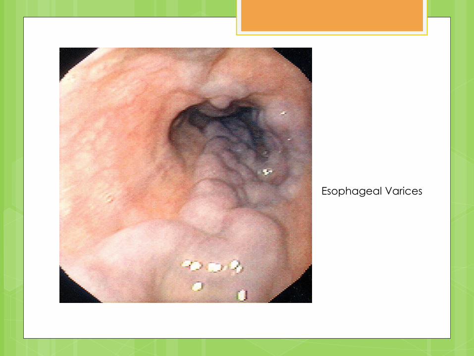
Esophageal Varices
Biliary Cirrhosis
Damage begins in the bile canaliculi and
bile ducts, not hepatocytes.
Primary Biliary Cirrhosis
Autoimmune
Mitochondrial autoantibodies present
Characterized by
Inflammation and destruction of small
intrahepatic bile ducts
Fibrosis
S/S of Primary Biliary Cirrhosis
Elevated alkaline phosphatase levels
Pruritus
Fatigue
Abdominal pain
Jaundice
Light colored stools
Steatorrhea
Fat soluble vitamin deficiencies
Hyperbilirubinemia
Hyperlipidemia
Comorbidities of Primary Biliary
Cirrhosis
Portal hypertension
Hepatic encephalopathy
Liver failure
Osteomalacia
Osteoporosis
Secondary Biliary Cirrhosis
Obstruction of the common bile duct or its
branches
Obstruction increased hepatic pressure
accumulation of bile
Necrosis proliferation and inflammation
edema and fibrosis
S/S of Secondary Biliary
Cirrhosis
Similar to primary, especially jaundice and
pruritus.
Elevated conjugated bilirubin and
alkaline phosphatase levels.
Postnecrotic Cirrhosis
Consequence of many, chronic, severe liver diseases.
25% of people with hepatitis C develop this.
Drugs or toxins, inherited metabolic disorders, and alpha antitrypsin deficiency can also lead to this.
Fibrosis separates islands of liver cells nodular appearance
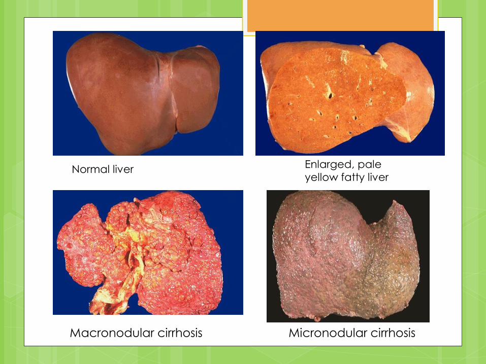
Enlarged, pale
yellow fatty liver Normal liver
Macronodular cirrhosis Micronodular cirrhosis
S/S of Postnecrotic Cirrhosis
Portal hypertension
Ascites
Varices
Hypersplenism
Hepatic encephalopathy
Nonalcoholic Fatty Liver
Disease (NAFLD) Accumulation of fat droplets in the hepatocytes Can lead to
Fibrosis
Cirrhosis
Hepatocellular carcinoma
Caused by: Drugs
Inborn errors of metabolism
Acquired metabolic disorders
Associated with: Obesity
Diabetes mellitus
Dyslipidemia
Insulin resistance
Nonalcoholic Steatohepatitis
(NASH)
More severe form of NAFLD
Accumulation of fibrous tissue
Patients may experience:
Malaise
Weakness
Hepatomegaly
Progression to cirrhosis is variable.
Acute Liver Failure
Severe complications rapidly after the first
signs of liver disease (such as jaundice).
Indicates severe liver damage (80-90% of
liver cells do not function)

Acute Liver Failure
S/S:
Cerebral edema
Hepatic encephalopathy
Coma
Brain herniation
Medical Diagnosis Liver Biopsy
Though unnecessary if clinical, laboratory and radiologic data indicates cirrhosis.
Biopsy poses a small but significant risk and cirrhosis increases risk for complications with biopsy.
Imaging
CT
MRI
Ultrasound
Endoscopy
Labs
Important Lab Values Alanine Aminotransferase (ALT)
Alkaline Phosphatase
Ammonia
Amylase
Aspartate aminotransferase
Gamma Glutamyl Transpeptidase
Lactic Dehydrogenase
Prothrombin Time
BUN
Bilirubin
Glucose
MELD (model for end stage liver
disease)
Based on three blood tests
International normalized ratio (INR)
Bilirubin
Creatinine
MELD scores usually range between 6 and
40, with a score of 6 indicating the best
likelihood of 90-day survival.

Medical
Therapies By: Natalie White
Current Medical Therapies
Management of portal hypertensive bleeding
Pharmacologic therapy
Adrenergic blockers to decrease heart rate
Somatostatin analog to decrease bleeding
Endoscopic banding or variceal ligation
Shunts (surgically placed)
Medications used for biliary cirrhosis
Actigall
Chenix
Current Medical Therapies
Medication for encephalopathy
Rifaximin: nonabsorbable antibiotic which kills ammonia-producing bacteria in the colon
Neomycin: nonabsorbable antibiotic which kills
ammonia-producing bacteria in the colon
Lactulose: nonabsorbable disaccharide which
acidifies the colon and keeps ammonia as
ammonium ion; also acts as an osmotic laxative
to help remove the ammonia
Current Medical Therapies Treatment of ascites
Large-volume paracentesis Fluid drained from the abdomen through a needle
Diuretic therapy Lasix (furosemide): loop diuretic; limits Na+
reabsorption Possible side effects include hyponatremia, hypokalemia,
hypomagnesemia, hypocalcemia, hypochloremic acidosis
Spironolactone: K+-sparing diuretic; limits Na+ reabsorption Serum levels of K+ must be monitored closely
Must monitor weight, abdominal girth, urinary Na+, serum nitrogen, creatinine, albumin, uric acid, & electrolytes during use of diuretics
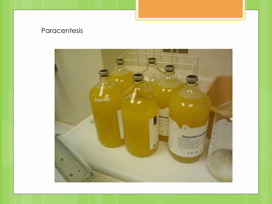
Paracentesis
Current Medical Therapies
Liver transplant
An established treatment for ESLD
The liver can regenerate itself, so a
transplant of a partial liver can grow to full
size
Complicated surgery
Post-transplant medications have nutritional
implications
Nutritional Assessment and
MNT for Cirrhosis and ESLD “The pancreas and liver are essential to digestion
and metabolism….When they are diseased, the
necessary medical nutrition therapy (MNT) is complex” (Krause, 645).
Nutritional Assessment Moderate to severe malnutrition is common
among cirrhosis and ESLD patients Inadequate oral intake
Anorexia, dysgeusia, early satiety, nausea, vomiting
Maldigestion and malabsorption
Abnormal macronutrient metabolism
Increased energy expenditure
Protein loss from paracentesis
Appropriate therapy can reverse malnutrition and improve outcomes
Nutritional Assessment Performed to determine the cause and the extent of malnutrition
Subjective global assessment (SGA) is used Why SGA?
1. Traditional markers of nutrition status can be affected by liver disease, making interpretation difficult:
Body weight is affected by edema, ascites, and diuretic use
Visceral protein levels are affected by decreased protein synthesis
2. Based on both anthropometrics and dietary intake
Nutritional Assessment: SGA
Subjective Global Assessment (Box 30-1, pg
657)
History
Physical Findings
Existing Conditions
Nutritional Rating Based on Results
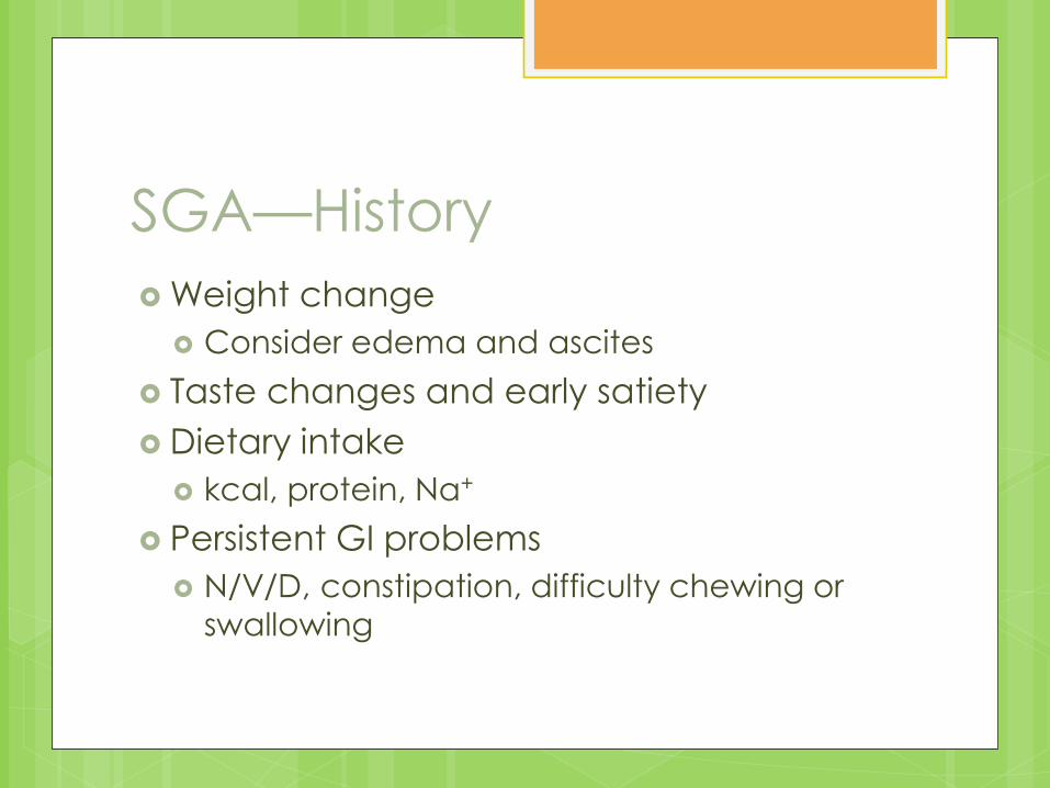
SGA—History
Weight change
Consider edema and ascites
Taste changes and early satiety
Dietary intake
kcal, protein, Na+
Persistent GI problems
N/V/D, constipation, difficulty chewing or
swallowing
SGA—Physical Findings
Muscle wasting
Fat stores
Ascites or edema
SGA—Existing Conditions
Disease state
Problems that could influence nutrition
status
Hepatic encephalopathy
GI bleeding
Renal insufficiency
Infection
SGA—Nutritional Rating
Well nourished
Moderately (or suspected) malnourished
Severely malnourished
Nutritional Assessment
SGA gives a broad perspective, but is not
sensitive to changes in nutrition status
Lab tests for nutritional deficiencies such
as vitamins, Mg2+, and iron may also be
helpful in identifying needed intervention
Nutrient Requirements
Energy
REE varies among patients
Could have normal, hypo-, or hyper-
metabolism
Lipids
Preferred source of energy increased lipolysis
Despite increased use, lipid storage not
impaired
25-40% kcal recommended
Nutrient Requirements
Carbohydrates
Difficult to determine because of liver’s role
in carbohydrate metabolism
Decrease in gluconeogenesis
Increased use of lipids and amino acids as
fuel
Alterations in insulin, glucagon, cortisol, and
epinephrine
Insulin resistance can also be a factor
Nutrient Requirements Protein
Most controversial and most complex to manage
Nitrogen losses with fulminant hepatic failure or decompensated disease, but not with stable cirrhosis
Uncomplicated cirrhosis w/o encephalopathy 0.8-1.0 g/kg dry body weight
To promote positive nitrogen balance 1.2-1.3 g/kg dry body weight
Alcoholic hepatitis or decompensated disease (sepsis, infection, GI bleeding, severe ascites) ≥ 1.5 g/kg dry body weight
Nutrient Requirements Vitamins and minerals
Supplementation necessary for all ESLD patients
Liver plays key role in nutrient transport, storage, and metabolism
Deficiencies can contribute to complications
Both fat- and water-soluble vitamins needed
Copper and manganese should not be given in supplements
Primarily excreted with bile, which is often impaired
Zinc, magnesium, and calcium should be given
Medical Nutrition Therapy
General recommendations:
Increased energy intake via small, frequent
meals
Restriction of Na+ for fluid retention
Carbohydrate-controlled diets for
hyperglycemia
Vitamin and mineral supplements
Oral liquid supplements or enteral (tube)
feeding
MNT—Special Considerations
Complications with nutrition implications: Portal hypertension
EN cannot be used during acute bleeding episodes
PN is used if patient will be taking nothing orally for 5 days
Ascites Na+ restriction
Adequate protein intake to replace losses from paracentesis
Hyponatremia Fluid restriction
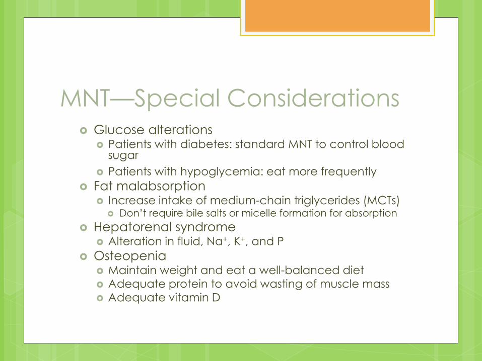
MNT—Special Considerations Glucose alterations
Patients with diabetes: standard MNT to control blood sugar
Patients with hypoglycemia: eat more frequently
Fat malabsorption Increase intake of medium-chain triglycerides (MCTs)
Don’t require bile salts or micelle formation for absorption
Hepatorenal syndrome Alteration in fluid, Na+, K+, and P
Osteopenia Maintain weight and eat a well-balanced diet Adequate protein to avoid wasting of muscle mass Adequate vitamin D
MNT—Hepatic
Encephalopathy
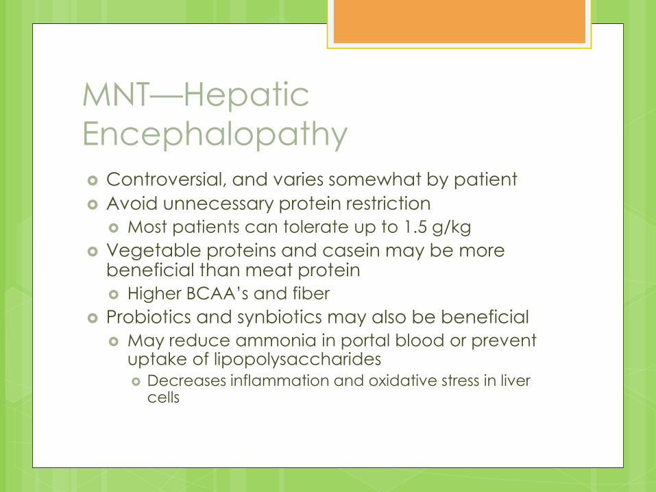
Controversial, and varies somewhat by patient
Avoid unnecessary protein restriction
Most patients can tolerate up to 1.5 g/kg
Vegetable proteins and casein may be more beneficial than meat protein
Higher BCAA’s and fiber
Probiotics and synbiotics may also be beneficial
May reduce ammonia in portal blood or prevent uptake of lipopolysaccharides
Decreases inflammation and oxidative stress in liver cells
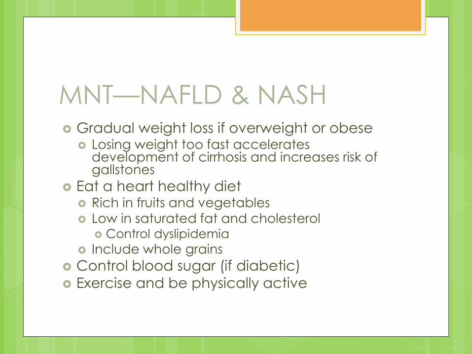
MNT—NAFLD & NASH Gradual weight loss if overweight or obese
Losing weight too fast accelerates development of cirrhosis and increases risk of gallstones
Eat a heart healthy diet Rich in fruits and vegetables
Low in saturated fat and cholesterol Control dyslipidemia
Include whole grains
Control blood sugar (if diabetic)
Exercise and be physically active
MNT—Liver Transplants
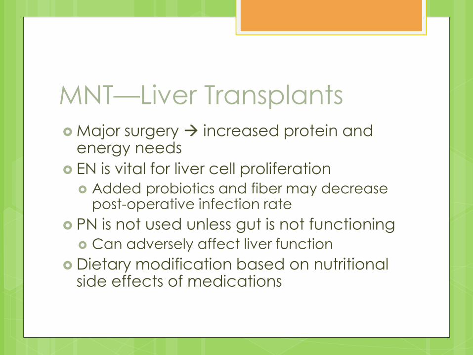
Major surgery increased protein and energy needs
EN is vital for liver cell proliferation
Added probiotics and fiber may decrease post-operative infection rate
PN is not used unless gut is not functioning
Can adversely affect liver function
Dietary modification based on nutritional side effects of medications
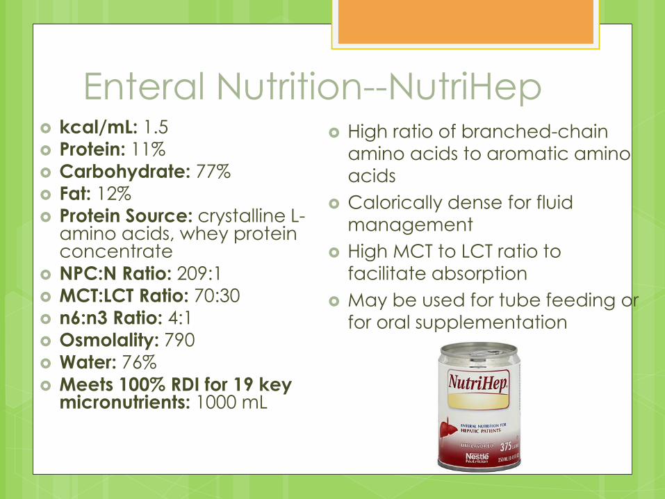
Enteral Nutrition--NutriHep kcal/mL: 1.5
Protein: 11%
Carbohydrate: 77%
Fat: 12%
Protein Source: crystalline L-amino acids, whey protein concentrate
NPC:N Ratio: 209:1
MCT:LCT Ratio: 70:30
n6:n3 Ratio: 4:1
Osmolality: 790
Water: 76%
Meets 100% RDI for 19 key micronutrients: 1000 mL
High ratio of branched-chain
amino acids to aromatic amino
acids
Calorically dense for fluid
management
High MCT to LCT ratio to
facilitate absorption
May be used for tube feeding or
for oral supplementation

Prognosis By: Emily Horn
Prognosis
Fibrosis/scarring are irreversible
Life expectancy depends on underlying
disease, extent of liver damage, number
of complications
Alcoholic cirrhosis
Continued drinking worsens prognosis
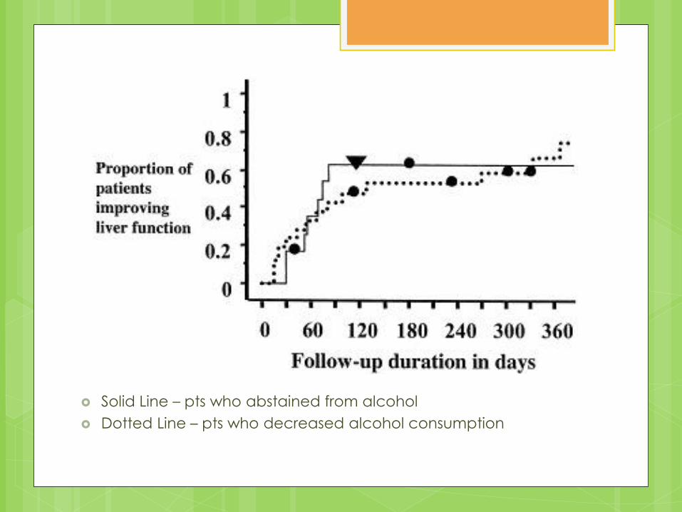
Solid Line – pts who abstained from alcohol
Dotted Line – pts who decreased alcohol consumption
Prognosis, cont.
Primary Biliary Cirrhosis
Patients usually live 8-10 years after first symptoms appear (1)
Medications can prolong life
Liver transplant
Secondary Biliary Cirrhosis
Surgery can be performed to reopen bile channels – prolongs survival and relieves symptoms
Prognosis, cont.
End-Stage Liver Disease
Ascites – 25% of people “who develop
ascites caused by cirrhosis die within 1
year” (1)
Hepatorenal syndrome –prognosis worse
depending on severity of liver dysfunction
Hepatic encephalopathy – if not treated,
can lead to coma and death
Liver transplants usually necessary
Liver Transplants
Demand is higher than supply
Model for End-Stage Liver Disease (MELD)
Prognostic tool to choose liver recipients
Post-op survival predicted by prothrombin time, serum bilirubin and creatinine
High score = bad
Patients with decompensated cirrhosis and MELD score <20 get livers
7-year survival rate of 60% (2)
Living Donor Liver Transplants
Transplant often taken from right lobe
“Almost 60% of the liver mass of the donor must be transplanted”
Worth the risk?
Lower survival rate in recipients of LDLT vs. deceased donor liver transplant (DDLT)
Puts donors at risk
However, reduces waiting list mortality from low availability of DDLT
Herbal Supplements Milk Thistle
Contains silymarin
Supposedly reduces oxidation
S-adenosyl-L-methionine (SAMe) Supposedly involved with glutathione production
Small studies – helpful for cholestasis?
Astragalus Not well understood; possibly dangerous side effects
Licorice Root May help treat hepatitis C
Dangerous side effects
None are overwhelmingly effective

Case Study

Teresa Wilcox – Grad Student and
Teacher
26 years old
C/O - Fatigue, anorexia, N/V, weakness,
bruising unrelated to injury
Nutrition Assessment
Anthropometric
86% of IBW, with 7% wt loss in last 6 mo.
BMI = 18.49
Biochemical
alb – severely depleted
prealb – mildly depleted
bilirubin – high
ALT, AST – both high
TG - high
Nutrition Assessment, cont.
Clinical
Diagnosed w/ hepatitis C 3 years ago
Dx: probable cirrhosis secondary to chronic
hepatitis C
Dietary
Protein: 1-1.2g/kg = 57 – 68.4g
Energy: (USE INDIRECT CALORIMETRY!)
1704-1988 kcals (30-35 kcal/kg)
PES Statement
Inadequate oral intake related to loss of
appetite secondary to cirrhosis as
evidenced by 7% weight loss in last 6
months.
Sample Diet – 1805 kcals,
68.6 g protein
Breakfast: Carnation instant breakfast mixed in 8 oz. low fat milk and medium banana
Snack: 8 oz. OJ, 1 Tbsp. peanut butter, 6 Ritz crackers
Lunch: 1 c. tomato basil soup w/1oz. shredded cheese, 1 wheat dinner roll, 8 oz. diet coke
Snack: ¾ c. potato salad
Dinner: 1 serving lasagna, ½ c. steamed zucchini, 8oz. OJ


![[Dyck, Andrew]Cicero's en](https://img.pdfslide.net/doc/110x75/577d1dca1a28ab4e1e8cf631/dyck-andrewciceros-en.jpg)
