Embed Size (px)
Citation preview
C
Cd
Aa
b
AA
KCBdC
1
bcbd
2
pbmwegbietio
i
1
Orthopaedics & Traumatology: Surgery & Research 101 (2015) 119–122
Available online at
ScienceDirectwww.sciencedirect.com
ase report
leidocranial dysplasia with bilateral posterior glenohumeralislocation: A case-report
. Hardya,∗, M.W. Thiong’oa, A. Leroya, P. Hardyb, C. Laportea
Service de chirurgie orthopédique, centre hospitalier de Meaux, 6-8, rue Saint-Fiacre, 77100 Meaux, FranceService de chirurgie orthopédique, hôpital Ambroise-Paré, 9, avenue Charles-de-Gaule, 92100 Boulogne-Billancourt, France
a r t i c l e i n f o
rticle history:ccepted 14 November 2014
a b s t r a c t
A 31-year-old man experienced bilateral posterior glenohumeral dislocation during seizures. He had clei-docranial dysplasia with complete absence of both clavicles. Cleidocranial dysplasia is a rare inherited
eywords:lavicular agenesisilateral posterior glenohumeralislocationleidocranial dysplasia
disease also known as Marie-Sainton syndrome and responsible for dental abnormalities well-known tostomatologists and dentists. Other manifestations include defective development of the skull bones andhypoplastic or aplastic clavicles. We found no previous reports of bilateral posterior glenohumeral dislo-cation in patients with cleidocranial dysplasia. The objective of this work was to look for an associationbetween clavicular aplasia and posterior glenohumeral dislocation.
© 2015 Elsevier Masson SAS. All rights reserved.
. Introduction
We report a case of acute, traumatic, posterior dislocation ofoth glenohumeral joints during seizures in a patient with bilaterallavicular agenesis. The possibility that an association may existetween clavicular agenesis and bilateral posterior glenohumeralislocation is discussed.
. Case-report
A 31-year-old man receiving follow-up for cleidocranial dys-lasia (CCD) was admitted for a staphylococcal infection of the faceelieved to be a complication of recent surgery to extract supernu-erary teeth. He had three febrile seizures associated with alcoholithdrawal and probably related to delirium tremens. The clinical
valuation indicated posterior dislocation of both shoulders. Radio-raphs confirmed this diagnosis, showing posterior dislocation ofoth glenohumeral joints, as well as anteromedial humeral head
mpaction fractures (McLaughlin fractures) (Fig. 1A and B). Bilat-ral clavicular agenesis was a previously undiagnosed feature ofhe CCD. He denied any history of anterior or posterior shouldernstability. Appropriate measures were taken to ensure resolution
f the seizures and delirium tremens.Relocation of the shoulders was then achieved in the operat-ng room, under general anaesthesia, using external manoeuvres
∗ Corresponding author. Tel.: +33 615 101 016.E-mail address: [email protected] (A. Hardy).
http://dx.doi.org/10.1016/j.otsr.2014.11.011877-0568/© 2015 Elsevier Masson SAS. All rights reserved.
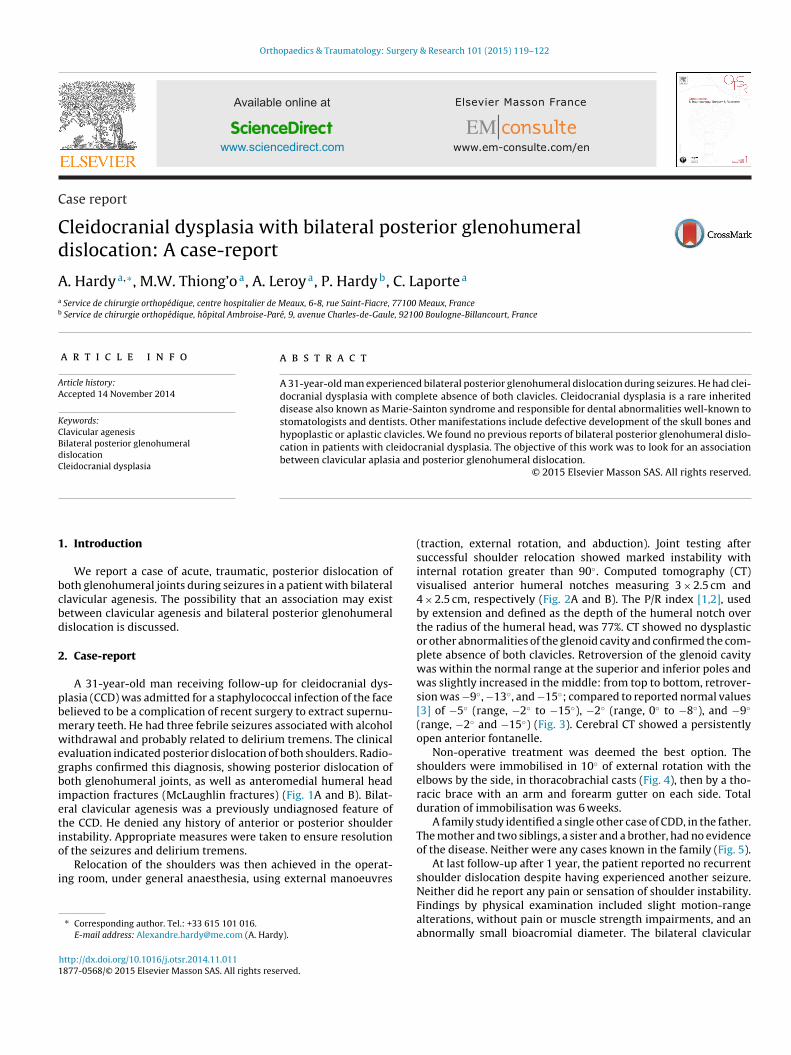
(traction, external rotation, and abduction). Joint testing aftersuccessful shoulder relocation showed marked instability withinternal rotation greater than 90◦. Computed tomography (CT)visualised anterior humeral notches measuring 3 × 2.5 cm and4 × 2.5 cm, respectively (Fig. 2A and B). The P/R index [1,2], usedby extension and defined as the depth of the humeral notch overthe radius of the humeral head, was 77%. CT showed no dysplasticor other abnormalities of the glenoid cavity and confirmed the com-plete absence of both clavicles. Retroversion of the glenoid cavitywas within the normal range at the superior and inferior poles andwas slightly increased in the middle: from top to bottom, retrover-sion was −9◦, −13◦, and −15◦; compared to reported normal values[3] of −5◦ (range, −2◦ to −15◦), −2◦ (range, 0◦ to −8◦), and −9◦
(range, −2◦ and −15◦) (Fig. 3). Cerebral CT showed a persistentlyopen anterior fontanelle.
Non-operative treatment was deemed the best option. Theshoulders were immobilised in 10◦ of external rotation with theelbows by the side, in thoracobrachial casts (Fig. 4), then by a tho-racic brace with an arm and forearm gutter on each side. Totalduration of immobilisation was 6 weeks.
A family study identified a single other case of CDD, in the father.The mother and two siblings, a sister and a brother, had no evidenceof the disease. Neither were any cases known in the family (Fig. 5).
At last follow-up after 1 year, the patient reported no recurrentshoulder dislocation despite having experienced another seizure.
Neither did he report any pain or sensation of shoulder instability.Findings by physical examination included slight motion-rangealterations, without pain or muscle strength impairments, and anabnormally small bioacromial diameter. The bilateral clavicular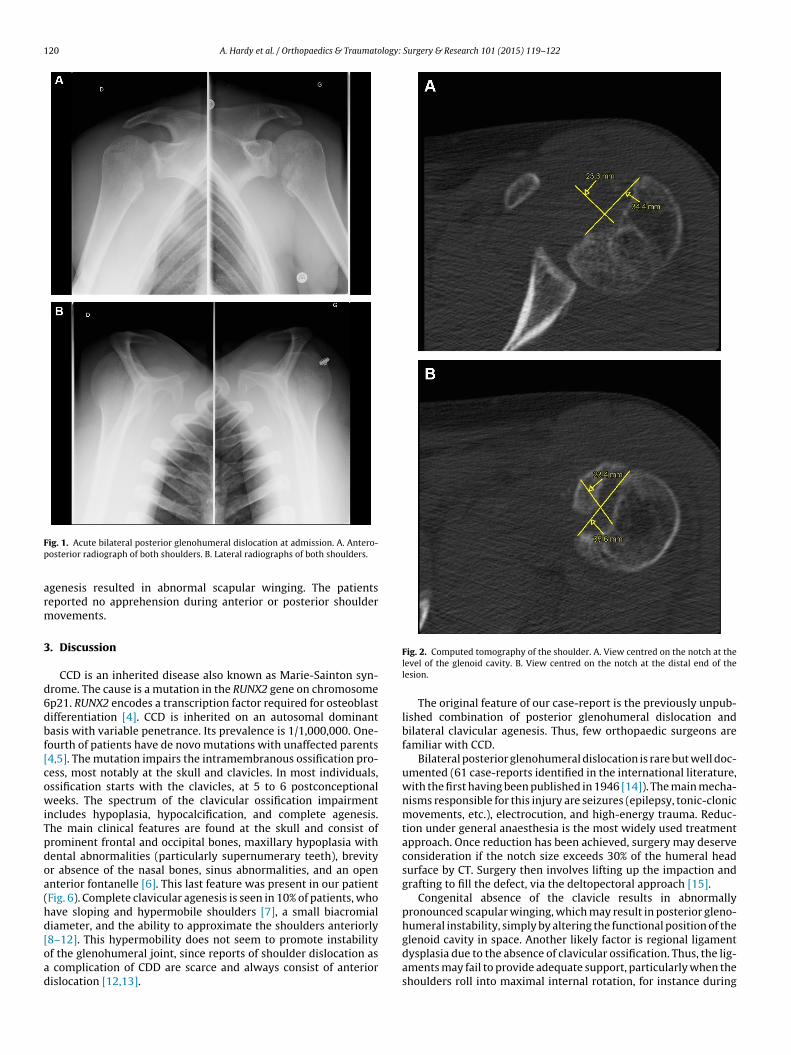
120 A. Hardy et al. / Orthopaedics & Traumatology: Surgery & Research 101 (2015) 119–122
Fp
arm
3
d6dbf[cowiTpdoa(hd[oad
ig. 1. Acute bilateral posterior glenohumeral dislocation at admission. A. Antero-osterior radiograph of both shoulders. B. Lateral radiographs of both shoulders.
genesis resulted in abnormal scapular winging. The patientseported no apprehension during anterior or posterior shoulderovements.
. Discussion
CCD is an inherited disease also known as Marie-Sainton syn-rome. The cause is a mutation in the RUNX2 gene on chromosomep21. RUNX2 encodes a transcription factor required for osteoblastifferentiation [4]. CCD is inherited on an autosomal dominantasis with variable penetrance. Its prevalence is 1/1,000,000. One-ourth of patients have de novo mutations with unaffected parents4,5]. The mutation impairs the intramembranous ossification pro-ess, most notably at the skull and clavicles. In most individuals,ssification starts with the clavicles, at 5 to 6 postconceptionaleeks. The spectrum of the clavicular ossification impairment
ncludes hypoplasia, hypocalcification, and complete agenesis.he main clinical features are found at the skull and consist ofrominent frontal and occipital bones, maxillary hypoplasia withental abnormalities (particularly supernumerary teeth), brevityr absence of the nasal bones, sinus abnormalities, and an opennterior fontanelle [6]. This last feature was present in our patientFig. 6). Complete clavicular agenesis is seen in 10% of patients, whoave sloping and hypermobile shoulders [7], a small biacromialiameter, and the ability to approximate the shoulders anteriorly
8–12]. This hypermobility does not seem to promote instabilityf the glenohumeral joint, since reports of shoulder dislocation ascomplication of CDD are scarce and always consist of anteriorislocation [12,13].
Fig. 2. Computed tomography of the shoulder. A. View centred on the notch at thelevel of the glenoid cavity. B. View centred on the notch at the distal end of thelesion.
The original feature of our case-report is the previously unpub-lished combination of posterior glenohumeral dislocation andbilateral clavicular agenesis. Thus, few orthopaedic surgeons arefamiliar with CCD.
Bilateral posterior glenohumeral dislocation is rare but well doc-umented (61 case-reports identified in the international literature,with the first having been published in 1946 [14]). The main mecha-nisms responsible for this injury are seizures (epilepsy, tonic-clonicmovements, etc.), electrocution, and high-energy trauma. Reduc-tion under general anaesthesia is the most widely used treatmentapproach. Once reduction has been achieved, surgery may deserveconsideration if the notch size exceeds 30% of the humeral headsurface by CT. Surgery then involves lifting up the impaction andgrafting to fill the defect, via the deltopectoral approach [15].
Congenital absence of the clavicle results in abnormallypronounced scapular winging, which may result in posterior gleno-humeral instability, simply by altering the functional position of the
glenoid cavity in space. Another likely factor is regional ligamentdysplasia due to the absence of clavicular ossification. Thus, the lig-aments may fail to provide adequate support, particularly when theshoulders roll into maximal internal rotation, for instance during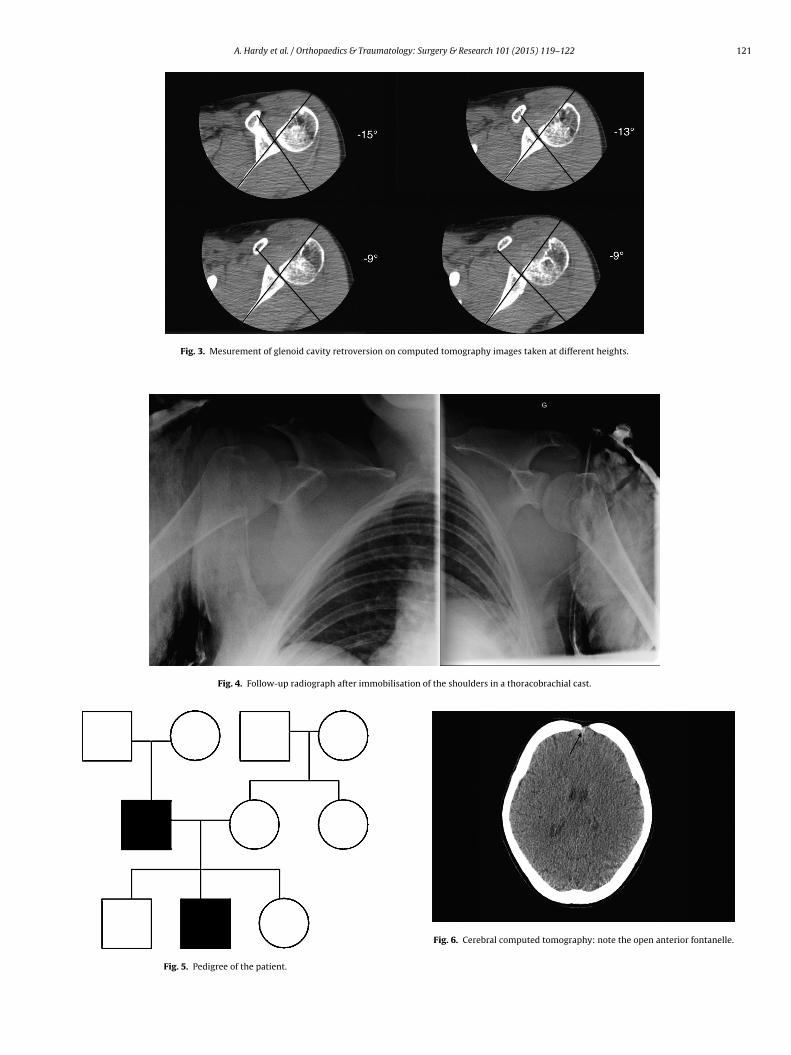
A. Hardy et al. / Orthopaedics & Traumatology: Surgery & Research 101 (2015) 119–122 121
Fig. 3. Mesurement of glenoid cavity retroversion on computed tomography images taken at different heights.
Fig. 4. Follow-up radiograph after immobilisation of the shoulders in a thoracobrachial cast.
Fig. 5. Pedigree of the patient.
Fig. 6. Cerebral computed tomography: note the open anterior fontanelle.
1 logy:
sirnmins
pIfisdt
D
c
R
[[
[
[
22 A. Hardy et al. / Orthopaedics & Traumato
eizures. In normal individuals, the clavicle may limit the range ofnternal shoulder rotation, thereby acting as a barrier against poste-ior dislocation. The small biacromial diameter combined with thearrow shoulders and small scapulae result in sloping and hyper-obile shoulders. Acquired absence of the clavicle does not seem to
ncrease the risk of posterior shoulder dislocation, as this event hasot been reported after clavicle removal during chest or vascularurgery.
Thus, whether the CDD contributed to the occurrence of bilateralosterior glenohumeral dislocation in our patient remains unclear.
n addition, CCD is extremely rare and case-series are therefore dif-cult to assemble. Nevertheless, the combination of seizures andcapular winging in a patient with clavicular agenesis and shoul-er hypermobility supports a relationship between the CDD andhe bilateral shoulder dislocation.
isclosure of interest
The authors declare that they have no conflicts of interest con-erning this article.
eferences
[1] Charousset C, Beauthier V, Bellaïche L, Guillin R, Thomazeau H. Canwe improve radiological analysis of osseous lesions in chronic anterior
[
[
Surgery & Research 101 (2015) 119–122
shoulder instability. Orthop Traumatol Surg Res 2010;96(8 Suppl.):S88–93,http://dx.doi.org/10.1016/j.otsr.2010.09.006 [Epub 2010 Oct 28].
[2] Sommaire C, Penz C, Clavert P, Klouche S, Hardy P, Kempf JF. Recurrenceafter arthroscopic Bankart repair: is quantitative radiological analysis of boneloss of any predictive value? Orthop Traumatol Surg Res 2012;98(5):514–9,http://dx.doi.org/10.1016/j.otsr.2012.03.015 [Epub 2012 Aug 10].
[3] Randelli M, Gambrioli PL. Glenohumeral osteometry by computed tomographyin normal and unstable shoulders. Clin Orthop Relat Res 1986;208:151–6.
[4] Karagüzel G, Aktürk FA, Okur E, Gümele HR, Gedik Y, Okten A. Cleidocranialdysplasia: a case report. J Clin Res Pediatr Endocrinol 2010;2:134–6.
[5] Mohan RP, Suma GN, Vashishth S, Goel S. Cleidocranial dysplasia: clinico-radiological illustration of a rare case. J Oral Sci 2010;52:161–6.
[6] Garg RK, Agrawal P. Clinical spectrum of cleidocranial dysplasia: a case report.Cases J 2008;1:377.
[7] Mehta DN, Vachhani RV, Patel MB. Cleidocranial dysplasia: a report of two cases.J Indian Soc Pedod Prev Dent 2011;29:251–4.
[8] Maroteaux P. La dysostose cleido-cranienne. E.M.C. (Elsevier Masson SAS Ed,Paris). Pediatrie 1983;3:4006A.
[9] Seze (de) S. Malformation des os d’origine genotypique – Maladies des os etdes articulations. CMC Flammarion; 1964.
10] Maroteaux P. Les maladies osseuses de l’enfant. Flammarion; 1974.11] Faure C, Maroteaux P. Progress in pediatric radiology. Intrinsic diseases of
bones. Cleidocranial dysplaisia, IV. Basel Karger; 1973 [p. 211].12] Mendoza-Londono R, Lee B. In: Pagon RA, Adam MP, Bird TD, et al., editors.
Cleidocranial dysplasia. Gene Reviews; 2006.13] Koischwitz D, Sobbe A. Dysplasia cleido-cranialis. Fortschr Geb Rontgenstr Nuk-
learmed 1973;119:438–42.14] Mollerud A. A case of bilateral habitual luxation in the posterior part of the
shoulder-joint. Acta Chir Scand 1946;94:181–6.15] Maynou C, Hardy P. Traitement chirurgical de l’instabilité postérieure de
l’épaule. EMC Tech Chir Orthop Traumatol 2006:44–263.


![Cleidocranial Dysplasia Case Report: Remodeling of …...CaseReportsinDentistry 5 Periodontalaspectsbeforetherestorativetreatmentare important and must be evaluated [26]. Oral instruction](https://img.pdfslide.net/doc/110x75/5e8ce698ec9b376e740bcd89/cleidocranial-dysplasia-case-report-remodeling-of-casereportsindentistry-5.jpg)