Embed Size (px)
Citation preview
Eur Respir J1990, 3, 937-974
Clinical guidelines and indications for bronchoalveolarlavage (BAL) : Report of the European Society of
Pneumology Task Group on BAL
This group report was developed jointly by: U . Costabel (Essen), C . Danel (Paris), L.M. Fabbri(Padova), P.L . Haslam (London), C. Hutter (Vienna) secretary, D. Israel-Biet (Paris), H. Klech(Vienna) chairman, W. Pohl (Vienna) secretary, L.W . Poulter (London), E. Pozzi (Torino), S .I .Rennard (Omaha), V. De Rose (Torino), G.A. Rossi (Genova), M. Rust (Frankfurt), G. Semenzato(Padova), B. Wallaert (Lille) .
Edited by H. Klech' and C. Hutter
Further contributors : C. Albera (Torino), W. Bauer (Bern), L. Bjermer (Umea), L . Carratu (Napoli),C.F . Donner (Veruno), H. Eckert (Berlin), P.H . Godard (Montpellier), T. Izumi (Kyoto), J . Linder(Omaha), D. Olivieri (Parma), M. Pirozynski (Warsaw), W. Pohl, G. Rizzato (Milano), A.J.A .Robalo-Cordeiro (Coibra), C . Sanguinetti (Ancona), N . Secen (Novisad), Y. Sibille (Brussels), M.Spiteri (London), I . Striz (Prague), H. Teschler (Essen), M. Tonnel (Lille), G . Velluti (Modena),H. Worth (Duesseldorf) .
*Request for reprints : H. Klech, 2nd Medical Department, Wilhelminenspital, Montleartstrasse37, A-1160 Vienna, Austria.
938
Contents
H. Klech 939 Introduction
H. Klech, C. Hutter 939 Side-effects and safety of BAL
P.L . Haslam, L.W . Poulter, G.A . Rossi, 940 Idiopathic pulmonary fibrosisW. Bauer,V. de Rose, H. Eckert,D. Olivieri, H. Teschler
B. Wallaert, G.A . Rossi, Y. Sibille 942 Collagen-vascular diseases
L.W. Poulter, G.A . Rossi, L. Bjermer, 943 SarcoidosisU. Costabel, D. Israel-Biet, H. Klech,W. Pohl, G. Velluti
G. Semenzato, L. Bjermer, U. Costabel, 945 Extrinsic allergic alveolitisP.L . Haslam, D. Olivieri
U. Costabel, C.F. Donner, P.L . Haslam, 946 Occupational lung diseases due to inhalation of inorganic dustG. Rizzato, H. Teschler, G. Velluti,B. Wallaert
C. Danel, D. Israel-Biet, U. Costabel, 949 Pulmonary histiocytosis XG.A . Rossi, B. Wallaert
C. Danel, D. Israel-Biet, U. Costabel, 950 Eosinophilic lung diseasesG.A . Rossi, B. Wallaert
C. Danel, D. Israel-Biet, U. Costabel, 950 Alveolar proteinosisG.A . Rossi, B. Wallaert
C. Danel, D. Israel-Biet, U. Costabel, 951 Pulmonary haemorrhagesG.A . Rossi, B. Wallaert
C. Danel, D. Israel-Biet, U. Costabel, 952 Drug induced pneumonitisG.A . Rossi, B. Wallaert
M. Rust, C. Albera, L. Carratu, C. Danel, 954 Pulmonary infectionsD. Israel-Biet, H. Klech, S.I . Rennard,A.J.A . Robalo-Cordeiro, G. Semenzato,G. Velluti, H. Worth
S .I . Rennard, C. Albera, W. Bauer, 956 Pulmonary malignanciesL. Carratu, H. Eckert, J. Linder,B. Pozzi, M. Pirozynski,A.J.A . Robalo-Cordeiro, C. Sanguinetti,G. Semenzato, N. Secen, I. Striz,H. Teschler, G. Velluti
L.M. Fabbri, V. De Rose, Ph . Godard, 958 Bronchial asthmaG.A . Rossi
E. Pozzi, V. De Rose, S .I . Rennard, 959 Chronic bronchitis and emphysemaL.M. Fabbri
C. Danel, D. Israel-Biet, U. Costabel, 960 Therapeutic applications of BALL.M. Fabbri, H. Klech
961 References
As for many clinical tools, there is at the present timeno clear agreement on the appropriate clinical use ofBAL. Undoubtedly, the recent and encouraging clini-cal experiences with BAL for diagnosis of opportunisticinfections in the immunocompromised patient haveencouraged a universal acceptance and interest in BAL.Because of the low morbidity of the lavage procedureand the significant yield of clinically important infor-mation, many physicians have been encouraged toperform a lavage during bronchoscopies undertaken fora variety of indications . This has resulted in a consid-erable body of experience with BAL in a number ofclinical settings . For many years, one of the main ob-stacles for general acceptance of BAL as a clinical toolhas been the vast disparity among centres world-wide regarding the technique and the processing ofthe BAL material .
In order to address this important issue of standardi-zation the European Society of Pneumology (SEP), in1988, set up a Task Force on Bronchoalveolar Lavage .The first report of the group focused specifically ontechnical recommendations and guidelines on how toperform BAL and how to process BAL material andwas published in 1989 in this journal [1] .
This is the second joint report of the SEP Task Groupon BAL and gives appropriate guidelines and infor-mation about the clinical indications and use of BAL invarious diseases of the lung . The members of the TaskGroup have collected all relevant information so faravailable about the clinical usefulness and indications
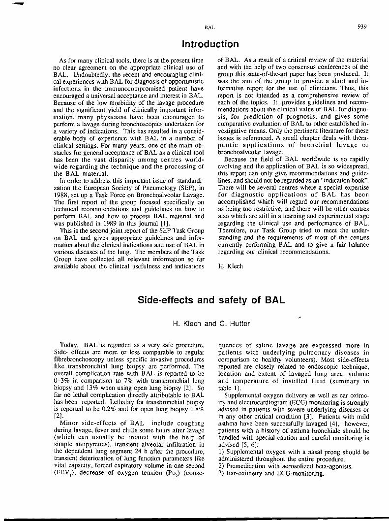
Today, BAL is regarded as a very safe procedure .Side- effects are more or less comparable to regularfibrebronchoscopy unless specific invasive procedureslike transbronchial lung biopsy are performed . Theoverall complication rate with BAL is reported to be0-3% in comparison to 7% with transbronchial lungbiopsy and 13% when using open lung biopsy [2] . Sofar no lethal complication directly attributable to BALhas been reported . Lethality for transbronchial biopsyis reported to be 0.2% and for open lung biopsy 1 .8%[2] .Minor side-effects of BAL include coughing
during lavage, fever and chills some hours after lavage(which can usually be treated with the help ofsimple antipyretics), transient alveolar infiltration inthe dependent lung segment 24 h after the procedure,transient deterioration of lung function parameters likevital capacity, forced expiratory volume in one second(FEV,), decrease of oxygen tension (Po2) (conse-
BAL
Introductionof BAL. As a result of a critical review of the materialand with the help of two consensus conferences of thegroup this state-of-the-art paper has been produced . Itwas the aim of the group to provide a short and in-formative report for the use of clinicians . Thus, thisreport is not intended as a comprehensive review ofeach of the topics . It provides guidelines and recom-mendations about the clinical value of BAL for diagno-sis, for prediction of prognosis, and gives somecomparative evaluation of BAL to other established in-vestigative means. Only the pertinent literature for theseissues is referenced . A small chapter deals with thera-peutic applications of bronchial lavage orbronchoalveolar lavage .Because the field of BAL worldwide is so rapidly
evolving and the application of BAL is so widespread,this report can only give recommendations and guide-lines, and should not be regarded as an "indication book".There will be several centres where a special expertisefor diagnostic applications of BAL has beenaccomplished which will regard our recommendationsas being too restrictive; and there will be other centresalso which are still in a learning and experimental stageregarding the clinical use and performance of BAL.Therefore, our Task Group tried to meet the under-standing and the requirements of most of the centrescurrently performing BAL and to give a fair balanceregarding our clinical recommendations.
H. Klech
Side-effects and safety of BAL
H . Klech and C. Hutter
939
quences of saline lavage are expressed more inpatients with underlying pulmonary diseases incomparison to healthy volunteers) . Most side-effectsreported are closely related to endoscopic technique,location and extent of lavaged lung area, volumeand temperature of instilled fluid (summary intable 1) .Supplemental oxygen delivery as well as ear oxime-
try and electrocardiogram (ECG) monitoring is stronglyadvised in patients with severe underlying diseases orin any other critical condition [3]. Patients with mildasthma have been successfully lavaged [4], however,patients with a history of asthma bronchiale should behandled with special caution and careful monitoring isadvised [5, 6] :1) Supplemental oxygen with a nasal prong should beadministered throughout the entire procedure.2) Premedication with aerosolized beta-agonists.3) Ear-oximetry and ECG-monitoring .
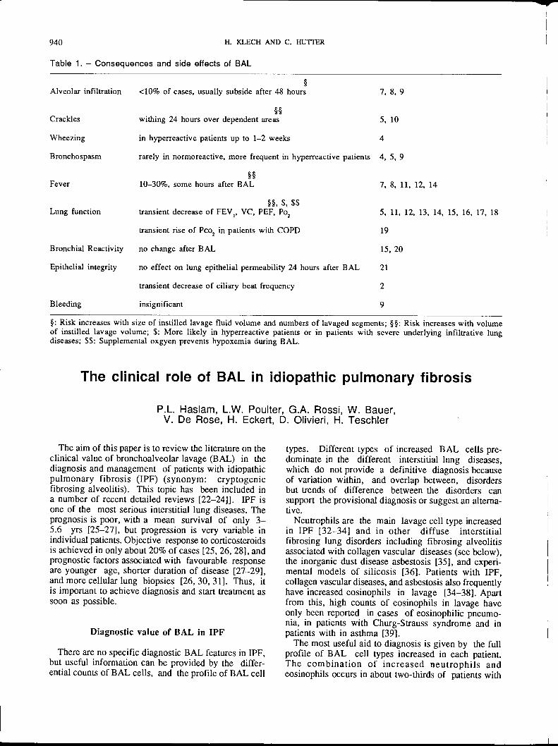
Lung function
transient decrease of FEV,, VC, PEF, Pot
5, 11, 12, 13, 14, 15, 16, 17, 18
transient rise of Pco2 in patients with COPD
19
no change after BAL
15, 20
no effect on lung epithelial permeability 24 hours after BAL
21
transient decrease of ciliary beat frequency
2
insignificant
9
Bronchial Reactivity
Epithelial integrity
Bleeding
§ : Risk increases with size of instilled lavage fluid volume and numbers of lavaged segments ; §§ : Risk increases with volumeof instilled lavage volume; $ : More likely in hyperreactive patients or in patients with severe underlying infiltrative lungdiseases ; $$ : Supplemental oxgyen prevents hypoxemia during BAL.
The clinical role of BAL in idiopathic pulmonary fibrosis
P .L . Haslam, L.W. Poulter, G .A. Rossi, W . Bauer,V. De Rose, H . Eckert, D . Olivieri, H . Teschler
The aim of this paper is to review the literature on theclinical value of bronchoalveolar lavage (BAL) in thediagnosis and management of patients with idiopathicpulmonary fibrosis (IPF) (synonym : cryptogenicfibrosing alveolitis) . This topic has been included ina number of recent detailed reviews [22-24]] . IPF isone of the most serious interstitial lung diseases . Theprognosis is poor, with a mean survival of only 3-5.6 yrs [25-27], but progression is very variable inindividual patients . Objective response to corticosteroidsis achieved in only about 20% of cases [25, 26, 28], andprognostic factors associated with favourable responseare younger age, shorter duration of disease [27-29],and more cellular lung biopsies [26, 30, 31] . Thus, itis important to achieve diagnosis and start treatment assoon as possible .
Diagnostic value of BAL in IPF
There are no specific diagnostic BAL features in IPF,but useful information can be provided by the differ-ential counts of BAL cells, and the profile of BAL cell
types .
Different types of increased BAL cells pre-dominate in the different interstitial lung diseases,which do not provide a definitive diagnosis becauseof variation within, and overlap between, disordersbut trends of difference between the disorders cansupport the provisional diagnosis or suggest an alterna-tive .Neutrophils are the main lavage cell type increased
in IPF [32-34] and in other diffuse interstitialfibrosing lung disorders including fibrosing alveolitisassociated with collagen vascular diseases (see below),the inorganic dust disease asbestosis [35], and experi-mental models of silicosis [36] . Patients with IPF,collagen vascular diseases, and asbestosis also frequentlyhave increased eosinophils in lavage [34-38] . Apartfrom this, high counts of eosinophils in lavage haveonly been reported in cases of eosinophilic pneumo-nia, in patients with Churg-Strauss syndrome and inpatients with in asthma [39] .The most useful aid to diagnosis is given by the full
profile of BAL cell types increased in each patient.The combination of increased neutrophils andeosinophils occurs in about two-thirds of patients with
940 H. KLECH AND C . HUTIER
Table 1 . - Consequences and side effects of BAL
Alveolar infiltration <10% of cases, usually subside after 48 hours 7, 8, 9
§§Crackles wishing 24 hours over dependent areas 5, 10
Wheezing in hyperreactive patients up to 1-2 weeks 4
Bronchospasm rarely in normoreactive, more frequent in hyperreactive patients 4, 5, 9
§§Fever 10-30%, some hours after BAL 7, 8, 11, 12,
IPF [34, 40] and in asbestosis [35], but is very rare inpatients with granulomatous lung diseases where lym-phocytes are the predominant increased BAL cell type .Furthermore, the distinction between IPF and asbestosisis aided by the identification of asbestos bodies amongstthe lavage cells, which indicate that exposure has takenplace and that the diagnosis of occupational lung dis-ease must be considered [25, 35, 41]. Lone neutrophilincreases occur in many patients with IPF but cautionmust be taken regarding the diagnostic interpretation,since moderate increases can arise for many reasons,and very high counts occurring alone can suggest bac-terial infection . However, it is of interest that neutrophilcounts increase and lymphocyte counts tend to fall asthe grade of radiographic shadowing and fibrosis in-creases in patients with sarcoidosis [42-44].A minority of IPF patients show a less typical BAL
cell profile. In particular, the subset who respondfavourably to corticosteroids frequently have slight tomoderate increases in BAL lymphocytes in associationwith neutrophils but very rarely with eosinophils [34,45-47] . Increases in BAL lymphocytes have also beenreported in workers exposed to asbestos or silica at astage prior to the development of symptoms [48, 49].
Increased T-helper/suppressor BAL lymphocyte ra-tios have recently been reported in IPF, contrastingwith reduced ratios in patients with associated colla-gen vascular diseases [50, 516], but the diagnosticvalue of this approach is restricted since increases inBAL lymphocytes are relatively infrequent in these dis-eases . Measurement of carcinoembryonic antigen inBAL fluid has recently been claimed to be a possiblemarker of early malignant change in the clinical courseof IPF [52] . Physicians should also be aware that al-veolar lipoproteinosis can very occasionally developin patients with IPF following treatment withcorticosteroids [53] . It is also important to be awarethat findings similar to those in patients with IPF haverecently been reported in clinically unaffected familymembers, namely increased numbers of neutrophils,evidence of macrophage activation, and growth factorsfor lung fibroblasts [54] .
In conclusion, inclusion of lavage in the pre-treat-ment investigation of patients with IPF, although it isnot pathognomonic, can give some support to the di-agnosis, when considered in the full clinical context.However, once patients have commenced therapy thiscan influence the lavage findings (see below) .
Prognostic value of BAL in IPF
Pre-treatment BAL cell counts may be of somevalue in the clinical management of IPF patients as aprognostic indicator of response to therapy .
Patients with increased percentage counts of BALlymphocytes have a significantly better chance ofresponding to corticosteroids than the remainder [34,45-47] . By contrast, percentages of neutrophils andeosinophils are significantly higher in those who fail torespond to steroids [34, 45, 55] and patients with in-
BAL
creased eosinophils have an especially poor response[34, 40, 45, 46, 56, 57] . However, there is a recentreport that some patients with increased eosinophils canrespond to cyclophosphamide (100 mg per day)combined with prednisolone (20 mg per alternate day)[58] . It is hoped that future prospective trials may showthat pre-treatment lavage cell counts may be of valueto indicate the most appropriate drug for the indi-vidual patient.Numerous other markers can be measured in BAL
samples, but there is little information on their cor-relations with clinical features . It has recently beenreported that IPF patients with high concentrations ofmyeloperoxidase [59], and those with higher levels ofhyaluronate and type III procollagen peptide [60] inBAL fluid deteriorate more rapidly than those withlow levels ; that patients with increased histamine inBAL fluid have higher grades of fibrosis in their lungbiopsies [41] ; and that patients with late stage IPFhave low levels of proteolytic activity in the BALfluids [62] . Factors released from activated alveolarmacrophages may play the major role in stimulatingthe growth of fibroblastss in IPF [63], but the clinicalvalue of measuring such markers is unknown. How-ever, since colchicine can suppress the production ofthese factors in vitro, it has been suggested that thisdrug may have a potential role in the treatment of IPF[64] .
In conclusion, the current evidence on the prognos-tic value of lavage findings in IPF suggests that theinformation may be of some value in guiding the selec-tion of therapeutic agents .
The value of BAL in monitoring andsurveillance of therapy in IPF
94 1
The safety of BAL makes it an ideal technique tomonitor changes occurring with disease progressionand under the influence of therapy, but there is stillrelatively little information on serial lavage studiesin patients with IPF. One series of patients has beenfollowed from 1-7 yrs, mean 4 yrs [58] . Patients re-sponding to high dose prednisolone showed a signifi-cant fall in the percentages of all inflammatory cell types,but most notably in neutrophils, while counts remainedelevated or increased in the non-responders; patientsfollowed on treatment with cyclophosphamide plus lowdose prednisolone, showed a significant fall ineosinophils in the responders, but not in the non-re-sponders. Another study has also found that corticoster-oid treatment does not suppress BAL neutrophils innon-responders after 3 mths or 6 mths of therapy, butstated that patients failing to respond to cyclophospha-mide alone or plus corticosteroids showed a significantreduction in neutrophils at 3 mths and at 6 mths [65] .By contrast, a third study has observed that BAL neu-trophil counts increased after 3 mths prednisolone insmokers, but not in nonsmokers, with IPF who showedclinical improvement [66] . However, the follow-up pe-riods were very different in the three studies, up to 7
942
yrs in the former [58], but only 3 mths [65, 66] and 6mths [65] in the latter . Thus, it is still prematureto draw conclusions on the clinical value oflavage cell counts in monitoring the progress of IPFpatients .
Serial lavage studies have also recently shown thatproportions of phosphatidylglycerol, which are reducedin the BAL fluids of many untreated IPF patients [67,68], return to normal in patients responding to pred-nisolone but not in non-responders [67] . It has beensuggested that such changes may reflect the extent ofdamage to the alveolar epithelium in IPF.
In conclusion, preliminary reports indicate that BALmay be of clinical value to monitor changes in thelungs associated with therapeutic response in IPF, butfurther information is required . In particular, inde-pendent prospective studies are needed where patientsare evaluated over comparable long-term periods, anddetails are required of survival as well as radiologicaland functional response to therapy.
Inflammatory processes that develop in the lung inmany of the collagen vascular diseases (CVD) usuallyresult in a diffuse interstitial lung disease (ILD) similarto idiopathic pulmonary fibrosis . Chronic alveolitis, asassessed by bronchoalveolar lavage, revealed the samecharacteristic pattern of alveolar inflammation associatedwith idiopathic pulmonary fibrosis ; which is evidenceof neutrophil accumulation and macrophage activation[38, 45, 50, 69-85] . However, there is a considerableoverlap for each disease and type of alveolitis . In ad-dition, inflammatory alveolitis may also be present in ahigh proportion of patients with CVD and withoutclinical or radiological evidence of pulmonary involve-ment, suggesting the presence of an ongoing subclinicalalveolitis .
Cellular characteristics of alveolitis
Total number of recovered cells is increased in patientswith overt ILD but not in patients without ILD. Inaddition, total number of cells is progressively reducedin advanced progressive systemic sclerosis [77] .The distribution of BAL cell type according to thedisease and to the presence of an associated ILDis summarized in table 1 . In addition, alveolarmacrophages are "spontaneously" activated and releasevarious bioactive mediators that could be relevantto the pathogenesis of ILD: superoxide anion(various CVD), neutrophil chemotactic factors (variousCVD), fibronectin (various CVD), alveolar macrophagederived growth factor for fibrosis (AMDGF) (progres-
H. KLECH AND C. HUTFER
Collagen-vascular diseases
B. Wallaert, G.A . Rossi, Y. Sibille
Conclusions
Current published evidence suggests that lavage is ofvalue to aid the diagnosis and management of patientswith IPF. BAL cell counts are only a guide to thedifferential diagnosis of IPF because of the variabilitywithin and overlap between diseases. Nevertheless, BALis of particular value to identify and exclude some ofthe rarer lung diseases which must be considered in theprovisional diagnosis . BAL can provide some usefulprognostic indicators in IPF which may aid therapeuticdecisions, and serial BAL measurements may have aplace in assessing suppression of inflammation inpatients responding to therapy. However, at this stagein our knowledge caution should be given to theinterpretation of BAL findings, and they are most usefulwhen considered and interpreted in the context of theoverall clinical and other investigatory techniques usedin the diagnosis and management of patients with thisserious lung disease .
sive systemic sclerosis) and tumour necrosis factor (TNF)(rheumatoid arthritis) .
It appears that symptomless patients with CVD canhave a similar pattern of alveolar inflammation includingaccumulation of neutrophils and/or lymphocytes andactivated alveolar macrophages [86-91] .On the other hand, some cell activities maybe defec-
tive : since decreased antibacterial activity of alveolarmacrophages has been reported in systemic lupus ery-thematosus but not in other CVD [92, 93] .
Biochemical characteristics of alveolitis
The biochemical analysis of BAL fluid shows anincreased transudation of serum factors and/or anincreased secretion of mediators: albumin, immu-noglobulin G (IgG), IgM, alpha-2 macroglobulin,plasminogen activator, procollagen peptide (progres-sive systemic sclerosis), collagenase, elastase [73, 76,83, 84, 94, 95] . So far, the value of biochemical analy-sis of BAL fluid in diagnosis and management of ILDCVD remains to be established .
Clinical significance of alveolitis in CVD
Since alveolar inflammation is a characteristicfeature of CVD patients with or without associated ILD,the BAL cytology is by no means a reliable argumentfor the diagnosis of ILD in this context. However,BAL may be useful for the diagnosis of an associated
BAL
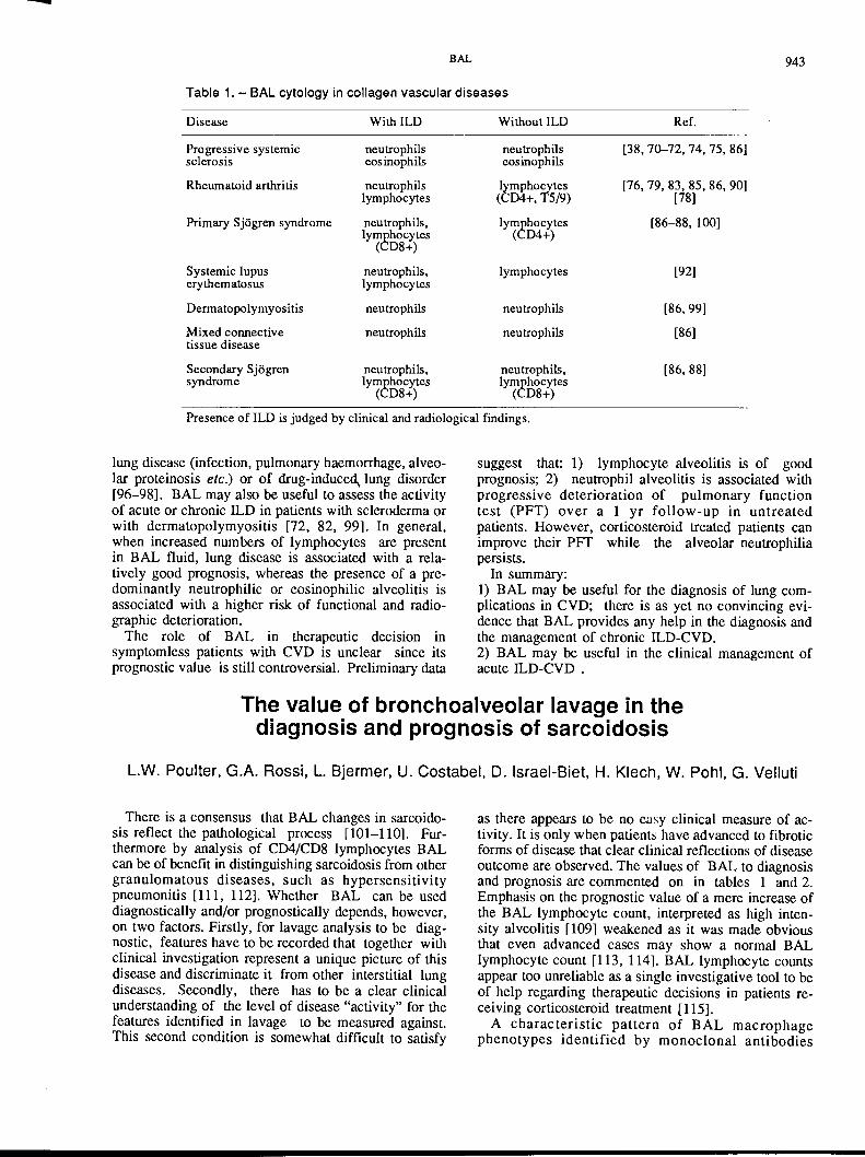
Table 1 . - BAL cytology in collagen vascular diseases
Presence of ILD is judged by clinical and radiological findings .
lung disease (infection, pulmonary haemorrhage, alveo-lar proteinosis etc .) or of drug-induced~ lung disorder[96-98] . BAL may also be useful to assess the activityof acute or chronic ILD in patients with scleroderma orwith dermatopolymyositis [72, 82, 99] . In general,when increased numbers of lymphocytes are presentin BAL fluid, lung disease is associated with a rela-tively good prognosis, whereas the presence of a pre-dominantly neutrophilic or eosinophilic alveolitis isassociated with a higher risk of functional and radio-graphic deterioration .The role of BAL in therapeutic decision in
symptomless patients with CVD is unclear since itsprognostic value is still controversial . Preliminary data
There is a consensus that BAL changes in sarcoido-sis reflect the pathological process [101-110] . Fur-thermore by analysis of CD4/CD8 lymphocytes BALcan be of benefit in distinguishing sarcoidosis from othergranulomatous diseases, such as hypersensitivitypneumonitis [111, 112] . Whether BAL can be useddiagnostically and/or prognostically depends, however,on two factors . Firstly, for lavage analysis to be diag-nostic, features have to be recorded that together withclinical investigation represent a unique picture of thisdisease and discriminate it from other interstitial lungdiseases . Secondly, there has to be a clear clinicalunderstanding of the level of disease "activity" for thefeatures identified in lavage to be measured against .This second condition is somewhat difficult to satisfy
The value of bronchoalveolar lavage in thediagnosis and prognosis of sarcoidosis
943
suggest that: 1) lymphocyte alveolitis is of goodprognosis ; 2) neutrophil alveolitis is associated withprogressive deterioration of pulmonary functiontest (PFT) over a 1 yr follow-up in untreatedpatients . However, corticosteroid treated patients canimprove their PFT while the alveolar neutrophiliapersists .
In summary :1) BAL may be useful for the diagnosis of lung com-plications in CVD; there is as yet no convincing evi-dence that BAL provides any help in the diagnosis andthe management of chronic ILD-CVD.2) BAL may be useful in the clinical management ofacute ILD-CVD .
L.W. Poulter, G .A. Rossi, L . Bjermer, U . Costabel, D . Israel-Biet, H . Klech, W. Pohl, G. Velluti
as there appears to be no easy clinical measure of ac-tivity . It is only when patients have advanced to fibroticforms of disease that clear clinical reflections of diseaseoutcome are observed . The values of BAL to diagnosisand prognosis are commented on in tables 1 and 2 .Emphasis on the prognostic value of a mere increase ofthe BAL lymphocyte count, interpreted as high inten-sity alveolitis [109] weakened as it was made obviousthat even advanced cases may show a normal BALlymphocyte count [113, 114] . BAL lymphocyte countsappear too unreliable as a single investigative tool to beof help regarding therapeutic decisions in patients re-ceiving corticosteroid treatment [115] .A characteristic pattern of BAL macrophage
phenotypes identified by monoclonal antibodies
Disease With ILD Without ILD Ref.
Progressive systemic neutrophils neutrophils [38, 70-72, 74, 75, 86]sclerosis eosinophils eosinophilsRheumatoid arthritis neutrophils lymphocytes [76,79,83,85,86,901
lymphocytes (CD4+, T5/9) [78]
Primary Sj6gren syndrome neutrophils, lymphocytes [86-88, 100]lymphocytes (CD4+)(CD8+)
Systemic lupus neutrophils, lymphocytes [92]erythematosus lymphocytes
Dermatopolymyositis neutrophils neutrophils [86, 99]Mixed connective neutrophils neutrophils [86]tissue disease
Secondary Sj6gren neutrophils, neutrophils, [86, 88]syndrome lymphocytes lymphocytes
(CD8+) (CD8+)
946
(BOOP), human immune deficiency virus (HIV) infectedpatients and drug induced pneumonitis.
It is worth mentioning that the presence of very highpercentages of lymphocytes in association with increasesin mast cells >1% might represent a diagnostic indica-tor of EAA [22] . Of course, this combination is onlyof value in cases which are currently, or have beenrecently, exposed to antigen since mast cells return tothe normal range within one to three months after re-moval from exposure .
The pattern of alveolitis in EAA during the follow-up
Although it is difficult to precisely separate patientson the basis of antigen exposure and, thus, cor-rectly subdivide EAA cases into strictly defined groups,a distinction needs to be made between patients whocontinue to be exposed to antigens and patients whohad been removed from the antigenic exposure .Concerning those patients whocontinue to be exposed
to antigens, several authors have shown a decrease(percentage or absolute) of lymphocytes during the fol-low-up [137, 138] while other authors have demonstratedthat the increase of the total number of lymphocyteswas a persistent feature in EAApatients still exposed torelevant antigens [139] . With regard to immunologicalsurface markers, a recovery of the CD4/CD8 ratio hasbeen evidenced during the follow-up only in thosepatients who had been removed from further antigenexposure [138, 140], thus suggesting that the immuno-logical abnormalities in these patients progress towardsnormal . Note that the behaviour of the CD4/CD8 ratiois not consistent in all cases . A recovery of the CD4/CD8 ratio was not found in subjects still exposed torelevant antigens [141] .As far as clinical management is concerned, studies
performed on this topic have indicated that there is nocorrelation between radiographic changes, pulmonaryfunction, BAL findings or levels of precipitating anti-bodies and different phases of the disease [141-144].
This chapter aims to review the clinical use of BALin patients with interstitial lung disease (ILD) asso-ciated with occupational or environmental exposure toinorganic dust and minerals . Excluded from this paperare occupational asthma and ILD due to inhalation oforganic dusts (extrinsic allergic alveolitis).
Indications for performing a BAL in ILD associ-ated with inorganic dust exposure are: 1) the exclusion
H. KLECH AND C . HUITER
Asymptomatic EAA patients
Although in asymptomatic EAA patients the increaseof lymphocytes (mostly CD8+ cells) with respect tocontrols is less prominent, the data are qualitativelysimilar to those observed in symptomatic patients [112,127, 143] . Data indicating that an alveolitis similar tothat observed in EAA patients develops in asympto-matic patients raises the question of how, when andwhy clinical features become apparent. The answer,however, still remains inconclusive .
Analysis of humoral constituents of BAL
The analysis of humoral constituents of BAL doesnot significantly improve the diagnosis of patients withEAA, as compared to the great value of the BAL cytol-ogy and immunocytology in the clinical assessment ofthis disease. However, the evaluation of hyaluronateand type III procollagen peptide concentrations in BALmight be useful in monitoring the disease [60, 145] .
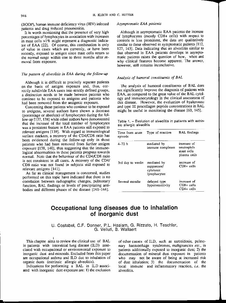
Table 1 . - Evolution of alveolitis in patients with extrin-sic allergic alveolitis
Occupational lung diseases due to inhalationof inorganic dust
U . Costabel, C.F . Donner, P.L . Haslam, G. Rizzato, H . Teschler,G . Velluti, B. Wallaert
of other causes of ILD, such as sarcoidosis, pulmo-nary haemorrhage syndromes, malignancies etc ., inpatients additionally exposed to inorganic dust ; 2) thedocumentation of mineral dust exposure in patientswho may not be aware of being at increased riskof dust inhalation ; 3) the documentation of thelocal immune and inflammatory reaction, i .e . thealveolitis .
Time from acuteepisode
Type of reaction BAL findings
4-72 h mediated by increase ofimmune complexes neutrophils
mast cellsplasma cells
3rd day to weeks mediated by increase ofsuppressor/ CD8+ cellscytotoxiclymphocytes
Several months delayed type increase ofhypersensitivity CD8+ cells
CD4+ cells
BAL
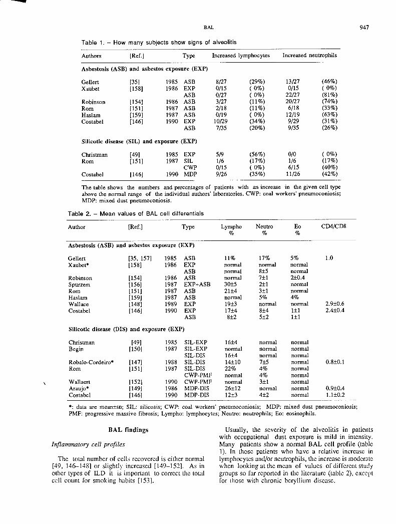
Table 1 . - How many subjects show signs of alveolitis
The table shows the numbers and percentages of patients with an increase in the given cell typeabove the normal range of the individual authors' laboratories . CWP: coal workers' pneumoconiosis ;MDP : mixed dust pneumoconiosis .
Inflammatory cell profiles
BAL findings
The total number of cells recovered is either normal[49, 146-148] or slightly increased [149-152] . As inother types of ILD it is important to correct the totalcell count for smoking habits [153] .
* : data are mean±sD; SIL : silicosis ; CWP : coal workers' pneumoconiosis ; MDP : mixed dust pneumoconiosis ;PMF : progressive massive fibrosis ; Lympho : lymphocytes ; Neutro : neutrophils ; Eo : eosinophils .
947
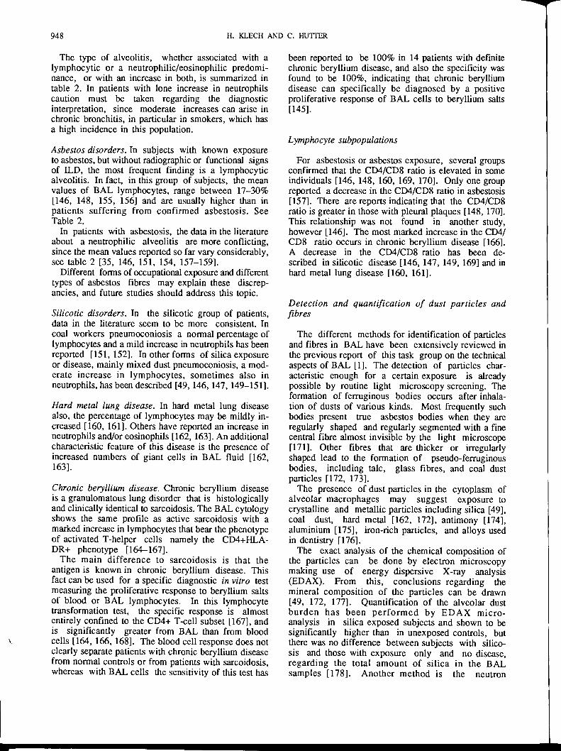
Usually, the severity of the alveolitis in patientswith occupational dust exposure is mild in intensity .Many patients show a normal BAL cell profile (table1) . In those patients who have a relative increase inlymphocytes and/or neutrophils, the increase is moderatewhen looking at the mean of values of different studygroups so far reported in the literature (table 2), exceptfor those with chronic beryllium disease .
Table 2. - Mean values of BAL cell differentials
Author [Ref.] Type Lympho Neutro Eo CD4/CD8
Asbestosis (ASB) and asbestos exposure (EXP)
Gellert [35, 157] 1985 ASB 11% 17% 5% 1.0Xaubet* [158] 1986 EXP normal normal normal
ASB normal 8±5 normalRobinson [154] 1986 ASB normal 7±1 2±0.4Spurzem [156] 1987 EXP+ASB 30±5 2±1 normalRom [151] 1987 ASB 21±4 3±1 normalHaslam [159] 1987 ASB normal 5% 4%Wallace [148] 1989 EXP 19±3 normal normal 2.9±0.6Costabel [146] 1990 EXP 17±4 8±4 1±1 2.4±0.4
ASB 8±2 5±2 1±1
Silicotic disease (DIS) and exposure (EXP)
Christman [49] 1985 SIL-EXP 16±4 normal normalBegin [150] 1987 SIL-EXP normal normal normal
SIL-DIS 16±4 normal normalRobalo-Cordeiro* [147] 1988 SIL-DIS 14±10 7±5 normal 0.8±0 .1Rom [151] 1987 SIL-DIS 22% 4% normal
CWP-PMF normal 4% normalWallaert [152] 1990 CWP-PMF normal 3±1 normalAraujo* [149] 1986 MDP-DIS 26±12 normal normal 0.9±0.4Costabel [146] 1990 MDP-DIS 12±3 4±2 normal 1.1±0.2
Authors [Ref.] Type Increased lymphocytes Increased neutrophils
Asbestosis (ASB) and asbestos exposure (EXP)
Gellert [35] 1985 ASB 8/27 (29%) 13/27 (46%)Xaubet [158] 1986 EXP 0/15 (0%) 0/15 (0%)
ASB 0/27 ( 0%) 22/27 (81%)Robinson [154] 1986 ASB 3/27 (11%) 20/27 (74%)Rom [151] 1987 ASB 2/18 (11%) 6/18 (33%)Haslam [159] 1987 ASB 0/19 (0%) 12/19 (63%)Costabel [146] 1990 EXP 10/29 (34%) 9/29 (31%)
ASB 7/35 (20%) 9/35 (26%)
Silicotic disease (SIL) and exposure (EXP)
Christman [491 1985 EXP 5/9 (56%) 0/0 (0%)Rom [151] 1987 SIL 1/6 (17%) 1/6 (17%)
CWP 0/15 ( 0%) 6/15 (40%)Costabel [146] 1990 MDP 9/26 (35%) 11/26 (42%)
948
The type of alveolitis, whether associated with alymphocytec or a neutrophilic/eosinophilic predomi-nance, or with an increase in both, is summarized intable 2. In patients with lone increase in neutrophilscaution must be taken regarding the diagnosticinterpretation, since moderate increases can arise inchronic bronchitis, in particular in smokers, which hasa high incidence in this population.
Asbestos disorders . In subjects with known exposureto asbestos, but without radiographic or functional signsof ILD, the most frequent finding is a lymphocytecalveolitis . In fact, in this group of subjects, the meanvalues of BAL lymphocytes, range between 17-30%[146, 148, 155, 156] and are usually higher than inpatients suffering from confirmed asbestosis . SeeTable 2.
In patients with asbestosis, the data in the literatureabout a neutrophilic alveolitis are more conflicting,since the mean values reported so far vary considerably,see table 2 [35, 146, 151, 154, 157-159] .
Different forms of occupational exposure and differenttypes of asbestos fibres may explain these discrep-ancies, and future studies should address this topic.
Silicotic disorders . In the silicotic group of patients,data in the literature seem to be more consistent . Incoal workers pneumoconiosis a normal percentage oflymphocytes and a mild increase in neutrophils has beenreported [151, 152] . In other forms of silica exposureor disease, mainly mixed dust pneumoconiosis, a mod-erate increase in lymphocytes, sometimes also inneutrophils, has been described [49, 146, 147, 149-151] .
Hard metal lung disease . In hard metal lung diseasealso, the percentage of lymphocytes may be mildly in-creased [160, 161] . Others have reported an increase inneutrophils and/or eosinophils [162, 163] . An additionalcharacteristic feature of this disease is the presence ofincreased numbers of giant cells in BAL fluid [162,163] .
Chronic beryllium disease . Chronic beryllium diseaseis a granulomatous lung disorder that is histologicallyand clinically identical to sarcoidosis. The BAL cytologyshows the same profile as active sarcoidosis with amarked increase in lymphocytes that bear the phenotypeof activated T-helper cells namely the CD4+HLA-DR+ phenotype [164-167].The main difference to sarcoidosis is that the
antigen is known in chronic beryllium disease. Thisfact can be used for a specific diagnostic in vitro testmeasuring the proliferative response to beryllium saltsof blood or BAL lymphocytes. In this lymphocytetransformation test, the specific response is almostentirely confined to the CD4+ T-cell subset [167], andis significantly greater from BAL than from bloodcells [164, 166, 168] . The blood cell response does notclearly separate patients with chronic beryllium diseasefrom normal controls or from patients with sarcoidosis,whereas with BAL cells the sensitivity of this test has
H. KLECH AND C . HU=R
been reported to be 100% in 14 patients with definitechronic beryllium disease, and also the specificity wasfound to be 100%, indicating that chronic berylliumdisease can specifically be diagnosed by a positiveproliferative response of BAL cells to beryllium salts[145] .
Lymphocyte subpopulations
For asbestosis or asbestos exposure, several groupsconfirmed that the CD4/CD8 ratio is elevated in someindividuals [146, 148, 160, 169, 170] . Only one groupreported a decrease in the CD4/CD8 ratio in asbestosis[157]. There are reports indicating that the CD4/CD8ratio is greater in those with pleural plaques [148, 170] .This relationship was not found in another study,however [146] . The most marked increase in the CD4/CD8 ratio occurs in chronic beryllium disease [166] .A decrease in the CD4/CD8 ratio has been de-scribed in silicotic disease [146, 147, 149, 169] and inhard metal lung disease [160, 161] .
Detection and quantification of dust particles andfibres
The different methods for identification of particlesand fibres in BAL have been extensively reviewed inthe previous report of this task group on the technicalaspects of BAL [1] . The detection of particles char-acteristic enough for a certain exposure
is alreadypossible by routine light
microscopy screening. Theformation of ferruginous bodies occurs after inhala-tion of dusts of various kinds. Most frequently suchbodies present true asbestos bodies when they areregularly shaped and regularly segmented with a finecentral fibre almost invisible by the light microscope[171] . Other fibres that are thicker or irregularlyshaped lead to the formation of
pseudo-ferruginousbodies, including talc, glass fibres, and coal dustparticles [172, 173] .The presence of dust particles in the cytoplasm of
alveolar macrophages may suggest exposure tocrystalline and metallic particles including silica [49],coal dust, hard metal [162, 172], antimony [174],aluminium [175], iron-rich particles, and alloys usedin dentistry [176] .The exact analysis of the chemical composition of
the particles can be done by electron microscopymaking use of energy dispersive X-ray analysis(EDAX). From this, conclusions regarding themineral composition of the particles can be drawn[49, 172, 177] . Quantification of the alveolar dustburden has been performed by EDAX micro-analysis in silica exposed subjects and shown to besignificantly higher than in unexposed controls, butthere was no difference between subjects with silico-sis and those with exposure only and no disease,regarding the total amount of silica in the BALsamples [178] . Another method is the neutron
activation analysis, which is especially useful for thedetection of trace metals in the cell-free BAL fluid,showing high concentrations of tungsten (W), tanta-lum (Ta) and cobalt (Co) in hard metal lung disease[163].The quantification of asbestos bodies is best done
by filtration of 5-15 ml fresh BAL fluid, cellsincluded, onto millipore filters, and counting thenumber of asbestos bodies [179] . Uncoated asbestos fi-bres can only be counted by electron microscopy [177],but this is, so far, without clinical value .Asbestos body counts correlate with the type of
asbestos related disorder being higher in those withbenign pleural disease or malignant mesothelioma[179]. Asbestos body counts in BAL correlate closelywith concentrations of asbestos bodies in lung tissueobtained by biopsy or at autopsy. A BAL count ofmore than one asbestos body per ml is highly indicativeof a lung concentration exceeding 1,000 asbestosbodies per g dry tissue [180, 181] . Only seven percentof non-asbestos exposed white collar workers haveasbestos bodies at concentrations >I-ml-1 BAL fluid[179]. In general, demonstration of dust in the lungsis an indication of exposure but is no evidence ofdisease . On the other hand, a minority of patientswith definite asbestos exposure and disease may have
BAL
no detectable asbestos bodies in their BAL fluid [179] .Demonstration of dust in BAL is especially useful inpatients with ILD or pleural disease who have pre-viously unknown or uncertain exposure to asbestos orother dusts.
Value of BAL for clinical diagnosis and management
The demonstration of dust in BAL fluid or cells isindicative for exposure, but is no evidence of disease.There is currently no known BAL level of particlesabove which development of disease is inevitable .ILD has to be proven by routine clinical methods likechest radiography, computerized tomographic (CT)scanning and lung function test.There is no clinical value of differential cell counts
in ILD due to occupational dust exposure, except forchronic beryllium disease.For the management of patients with known ILD
due to dust exposure, BAL is currently of no provenvalue, except for chronic beryllium disease and for therecognition of the co-existence of another disorder ofdifferent cause, such as sarcoidosis, hypersensitivitypneumonitis, haemorrhage syndrome and others[182] .
The clinical role of BAL in pulmonary histiocytosis XC. Danel, D . Israel-Biet, U . Costabel, G.A . Rossi, B . Wallaert
Pulmonary histiocytosis X (PHX) is a rare chronicgranulomatous disorder involving cells of themonophagocytic system . The diagnostic feature of thisdisease is the finding of Langerhans cells (LC) whichreact with the monoclonal antibody CD1 (OKT6) andwhich contain characteristic cytoplasmic organelles[183, 184] . After its introduction as a new means ofstudying peripheral lung and alveolar cell populations,BAL has rapidly proved useful in the diagnosis of PHX[185] .
Diagnostic value of BAL in PIIX
Several studies have shown the major value of BALin the diagnosis of PHX [185, 186] . The total cellcount is usually increased. HANcE et al . have reportedthat 90% of their PHX patients were smokers [186] . Itis well known that the total cell recovery is usuallyhigher in smokers than in nonsmokers . Besides, thenonsmoking patients with PHX have a normal alveolarcell count. The differential cell count shows a highpercentage of alveolar macrophages (AM), a slight in-crease of neutrophils and eosinophils [185] . On electronmicroscopy, a significant percentage ofLangerhans cells(LC) display highly specific pentalaminar structures ofconstant width, with a tennis racket shape at one end[183, 185] . As this ultrastructural analysis is time con-suming, a more rapid and sensitive technique has been
949
developed using monoclonal antibodies to LC (CD1positive cells) [184] . For some other authors, the find-ing of PS 100 BAL positive cells could ensure thediagnosis of PHX. However, this antibody is far lessspecific of LC than CD1 and its use is therefore notrecommended.The actual value of BAL and in particular the
presence of LC in the diagnosis of PHX is difficultto assess . Some authors have reported a mean of 5%CD1 positive cells in the BAL of patients with PBX,while in other interstitial lung diseases, less than 3% ofthe total cells were found to be CD1 positive [184] .
In fact, recent studies have shown that LC are nor-mally present in the lower respiratory tract and in lungparenchyma of normal subjects, particularly in smokers[186, 187] . Alteration of this epithelium seems to be animportant stimulus in attracting LC to the lung [130],and cigarette smoking is known to produce such epithe-lial abnormalities in the lower respiratory tract . Besides,cigarette smoke actually increases the number of LCfound in BAL fluid [186] .
Furthermore, LC have been found in the lung ofpatients with diseases other than PHX, in fibroticlung disorders, benign inflammatory conditionsor bronchoalveolar carcinoma for instance [65,167,168] .Therefore, as the mere presence of LC in BAL isnot pathognomonic of PHX, particularly in smokingpatients, a percentage of at least 5% of CD1 labelledalveolar cells is required to confirm the diagnosis.
950
On the other hand, with PHX having a patchydistribution, a localized BAL can miss the diagnosis,as well as a transbronchial biopsy . Confirmation by anopen lung biopsy is therefore advisable .
Conclusions
There are strong arguments to support the usefulnessof lavage cell analysis in the diagnosis of pulmonary
The clinical role of BAL in eosinophilic lung diseasesC. Danel, D . Israel-Biet, U . Costabel, G.A . Rossi, B . Wallaert
Eosinophilic infiltrates in the lung can be encoun-tered in a great variety of disorders such as asthma,eosinophilic pneumonia, allergic bronchopulmonaryaspergillosis or Churg and Strauss vasculitis . In thischapter we will concentrate on eosinophilic pneumoniaranging from the acute but mild and remitting Loeffler'ssyndrome to the severe chronic eosinophilic pneumo-nia. As these diseases can be life-threatening butremarkably reversible under corticosteroid therapy, arapid diagnosis is of major importance . Since no alveo-lar eosinophilia is ever observed in normal controls,any increase in the percentage of eosinophils in BALargues for a pathological process. In any type ofeosinophilic lung (EL), acute or chronic, BAL alwaysdisplays a high alveolar eosinophilia, whether or notassociated with a blood eosinophilia [190-193].
Besides its diagnostic value, BAL has also givenclues to the pathogenesis of eosinophilic lung injury .Indeed, eosinophils secrete not only neutral proteasesand oxygen radicals but also a major basic protein (MBP)and a cationic protein (ECP) known to be able to induceacute lung damage and pulmonary fibrosis [194] .Finally, BAL is also valuable in EL for the clinicalfollow-up of patients under treatment [195].
Diagnostic value of BAL in eosinophilic lung
As, in these disorders, eosinophils are largelylocated in air spaces, the diagnostic yield of BAL isvery high, usually making more invasive techniques(open lung biopsy or transbronchial biopsy) unneces-sary . The analysis of BAL and blood should be
The clinical role of BAL in alveolar proteinosis
C . Danel, D . Israel-Biet, U . Costabel, G.A . Rossi, B . Wallaert
Pulmonary alveolar proteinosis (PAP) is a rare disor-der characterized by accumulation of periodic-acid-Schiff(PAS)-positive phospholipidic material in the alveolarspaces [196] . PAP can be idiopathic or secondary tovarious conditions, such as immunosuppression, ma-lignant haematological disorders, silicosis or, more
H. KLECH AND C . HUTI'ER
histiocytosis X. However, false negative resultscan be related to the patchy distribution or to thestage of the disease . False positive results havealso beenreported in heavy smokers or in bronchoalveolar carci-nomas, for instance . This highlights the fact that BALdata should be interpreted carefully in the context ofclinical and radiological data. One requires at least 5%of LC in BAL to confirm the diagnosis . This eithergives sufficient diagnostic clues or else points to thenecessity of an open lung biopsy .
performed in parallel. The diagnostic value of a highalveolar eosinophilia is all the greater if the level of theblood eosinophilia is normal .
It is usually in eosinophilic pneumonia (EP) that thehighest eosinophilic count is observed [190-193] . Ifthe increase of total recovered cells is not always sig-nificant, the percentage of eosinophils is markedly ab-normal, sometimes increased up to 90% of total cells,associated or not to a few mast cells, and always higherthan the neutrophil count. A proportion of theseeosinophils can undergo necrosis, and fine eosinophilicgranules can be observed in alveolar macrophages.Nevertheless, such a high alveolar eosinophilia canalso be observed in some parasitic disorders or in theChurg-Strauss syndrome [192] . Less pronounced eosi-nophil increases (5-10%) can be found in sarcoidosis,histiocytosis X, drug induced pneumonia, collagenvascular disease, asthma and idiopathic pulmonaryfibrosis [190-192].
Conclusions
In eosinophilic lung diseases (EL), BAL is of greatvalue not only for the diagnosis and the follow-up ofpatients treated, but also for the study of theirpathogenesis . EL is one of the diseases in whichBAL can give enough clues to the diagnosis to avoid,in many cases, an open lung biopsy . The highest eosi-nophil counts ever seen in BAL fluid are observed here,ranging from 20-90% of the cells . These results aremost useful when the X-ray findings are atypical andperipheral eosinophilia absent.
rarely, diffuse interstitial lung diseases [53, 196, 197] .As the clinical and radiological presentations are not
specific, PAP can remain misdiagnosed . SegmentalBAL appears to be essential in management of thisdisease for diagnosis, follow-up, and therapeuticpurposes [197].
Diagnostic value of BAL in PAP
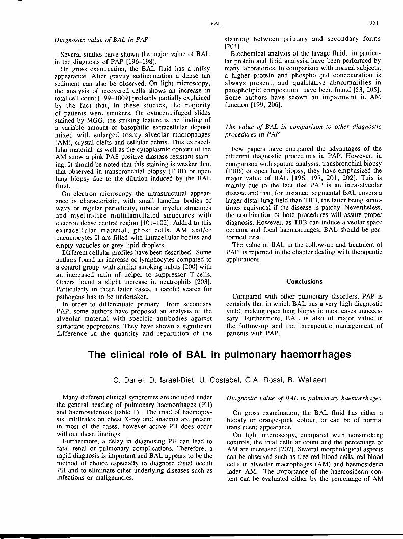
Several studies have shown the major value of BALin the diagnosis of PAP [196-198] .On gross examination, the BAL fluid has a milky
appearance . After gravity sedimentation a dense tansediment can also be observed . On light microscopy,the analysis of recovered cells shows an increase intotal cell count [ 199-1009] probably partially explainedby the fact that, in these studies, the majorityof patients were smokers. On cytocentrifuged slidesstained by MGG, the striking feature is the finding ofa variable amount of basophilic extracellular depositmixed with enlarged foamy alveolar macrophages(AM), crystal clefts and cellular debris . This extracel-lular material as well as the cytoplasmic content of theAM show a pink PAS positive diastase resistant stain-ing. It should be noted that this staining is weaker thanthat observed in transbronchial biopsy (TBB) or openlung biopsy due to the dilution induced by the BALfluid.On electron microscopy the ultrastructural appear-
ance is characteristic, with small lamellar bodies ofwavy or regular periodicity, tubular myelin structuresand myelin-like multilamellated structures withelectron dense central region [101-102] . Added to thisextracellular material, ghost cells, AM and/orpneumocytes II are filled with intracellular bodies andempty vacuoles or grey lipid droplets .
Different cellular profiles have been described . Someauthors found an increase of lymphocytes compared toa control group with similar smoking habits [200] withan increased ratio of helper to suppressor T-cells.Others found a slight increase in neutrophils [203] .Particularly in these latter cases, a careful search forpathogens has to be undertaken .
In order to differentiate primary from secondaryPAP, some authors have proposed an analysis of thealveolar material with specific antibodies againstsurfactant apoproteins. They have shown a significantdifference in the quantity and repartition of the
The clinical role of BAL in pulmonary haemorrhages
C . Danel, D. Israel-Biet, U . Costabel, G .A. Rossi, B . Wallaert
Many different clinical syndromes are included underthe general heading of pulmonary haemorrhages (PH)and haemosiderosis (table 1) . The triad of haemopty-sis, infiltrates on chest X-ray and anaemia are presentin most of the cases, however active PH does occurwithout these findings .
Furthermore, a delay in diagnosing PH can lead tofatal renal or pulmonary complications. Therefore, arapid diagnosis is important and BAL appears to be themethod of choice especially to diagnose distal occultPH and to eliminate other underlying diseases such asinfections or malignancies .
BAL
Conclusions
95 1
staining between primary and secondary forms[204] .Biochemical analysis of the lavage fluid, in particu-
lar protein and lipid analysis, have been performed bymany laboratories . In comparison with normal subjects,a higher protein and phospholipid concentration isalways present, and qualitative abnormalities inphospholipid composition have been found [53, 2051 .Some authors have shown an impairment in AMfunction [199, 2061 .
The value of BAL in comparison to other diagnosticprocedures in PAP
Few papers have compared the advantages of thedifferent diagnostic procedures in PAP. However, incomparison with sputum analysis, transbronchial biopsy(TBB) or open lung biopsy, they have emphasized themajor value of BAL [196, 197, 201, 202] . This ismainly due to the fact that PAP is an intra-alveolardisease and that, for instance, segmental BAL covers alarger distal lung field than TBB, the latter being some-times equivocal if the disease is patchy . Nevertheless,the combination of both procedures will assure properdiagnosis. However, as TBB can induce alveolar spaceoedema and focal haemorrhages, BAL should be per-formed first.The value of BAL in the follow-up and treatment of
PAP is reported in the chapter dealing with therapeuticapplications
Compared with other pulmonary disorders, PAP iscertainly that in which BAL has a very high diagnosticyield, making open lung biopsy in most cases unneces-sary . Furthermore, BAL is also of major value inthe follow-up and the therapeutic management ofpatients with PAP.
Diagnostic value of BAL in pulmonary haemorrhages
On gross examination, the BAL fluid has either abloody or orange-pink colour, or can be of normaltranslucent appearance .On light microscopy, compared with nonsmoking
controls, the total cellular count and the percentage ofAM are increased [207] . Several morphological aspectscan be observed such as free red blood cells, red bloodcells in alveolar macrophages (AM) and haemosiderinladen AM. The importance of the haemosiderin con-tent can be evaluated either by the percentage of AM
952
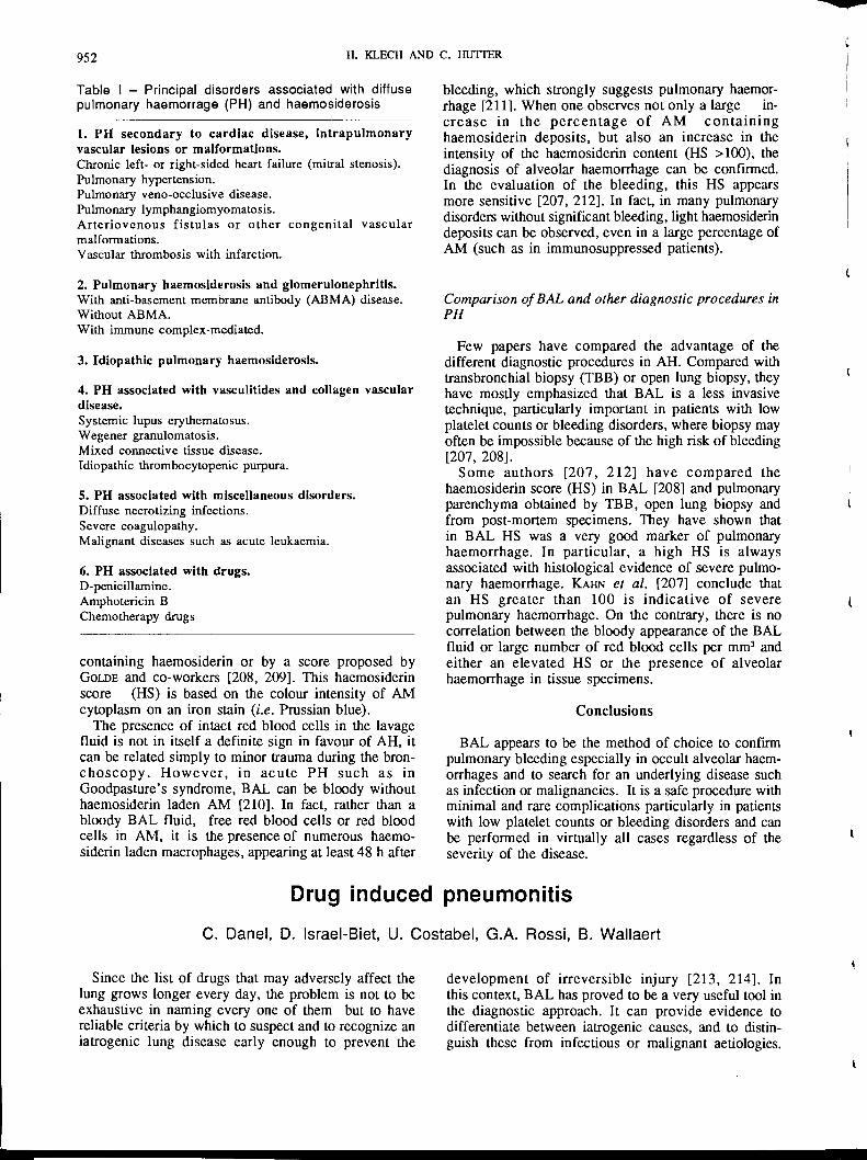
Table I - Principal disorders associated with diffusepulmonary haemorrage (PH) and haemosiderosis
1 . PH secondary to cardiac disease, intrapulmonaryvascular lesions or malformations .Chronic left- or right-sided heart failure (mitral stenosis) .Pulmonary hypertension.Pulmonary veno-occlusive disease.Pulmonary lymphangiomyomatosis .Arteriovenous fistulas or other congenital vascularmalformations .Vascular thrombosis with infarction.
2 . Pulmonary haemosiderosis and glomerulonephritis .With anti-basement membrane antibody (ABMA) disease.Without ABMA.With immune complex-mediated .
3 . Idiopathic pulmonary haemosiderosis.
4 . PH associated with vasculitides and collagen vasculardisease.Systemic lupus erythematosus .Wegener granulomatosis .Mixed connective tissue disease .Idiopathic thrombocytopenic purpura .
5 . PH associated with miscellaneous disorders.Diffuse necrotizing infections.Severe coagulopathy.Malignant diseases such as acute leukaemia .
6 . PH associated with drugs.D-penicillamine .Amphotericin BChemotherapy drugs
containing haemosiderin or by a score proposed byGOLDE and co-workers [208, 209] . This haemosiderinscore (HS) is based on the colour intensity of AMcytoplasm on an iron stain (i .e . Prussian blue) .The presence of intact red blood cells in the lavage
fluid is not in itself a definite sign in favour of AH, itcan be related simply to minor trauma during the bron-choscopy . However, in acute PH such as inGoodpasture's syndrome, BAL can be bloody withouthaemosiderin laden AM [210] . In fact, rather than abloody BAL fluid, free red blood cells or red bloodcells in AM, it is the presence of numerous haemo-siderin laden macrophages, appearing at least 48 h after
Since the list of drugs that may adversely affect thelung grows longer every day, the problem is not to beexhaustive in naming every one of them but to havereliable criteria by which to suspect and to recognize aniatrogenic lung disease early enough to prevent the
H. KLECH AND C. HUTMR
Drug induced pneumonitis
bleeding, which strongly suggests pulmonary haemor-rhage [211] . When one observes not only a large in-crease in the percentage of AM containinghaemosiderin deposits, but also an increase in theintensity of the haemosiderin content (HS >100), thediagnosis of alveolar haemorrhage can be confirmed .In the evaluation of the bleeding, this HS appearsmore sensitive [207, 212] . In fact, in many pulmonarydisorders without significant bleeding, light haemosiderindeposits can be observed, even in a large percentage ofAM (such as in immunosuppressed patients) .
Comparison ofBAL and other diagnostic procedures inPH
Few papers have compared the advantage of thedifferent diagnostic procedures in AH. Compared withtransbronchial biopsy (TBB) or open lung biopsy, theyhave mostly emphasized that BAL is a less invasivetechnique, particularly important in patients with lowplatelet counts or bleeding disorders, where biopsy mayoften be impossible because of the high risk of bleeding[207, 2081 .Some authors [207, 212] have compared the
haemosiderin score (HS) in BAL [208] and pulmonaryparenchyma obtained by TBB, open lung biopsy andfrom post-mortem specimens . They have shown thatin BAL HS was a very good marker of pulmonaryhaemorrhage . In particular, a high HS is alwaysassociated with histological evidence of severe pulmo-nary haemorrhage. KAHN et al . [207] conclude thatan HS greater than 100 is indicative of severepulmonary haemorrhage. On the contrary, there is nocorrelation between the bloody appearance of the BALfluid or large number of red blood cells per mm' andeither an elevated HS or the presence of alveolarhaemorrhage in tissue specimens .
Conclusions
BAL appears to be the method of choice to confirmpulmonary bleeding especially in occult alveolar haem-orrhages and to search for an underlying disease suchas infection or malignancies . It is a safe procedure withminimal and rare complications particularly in patientswith low platelet counts or bleeding disorders and canbe performed in virtually all cases regardless of theseverity of the disease.
C. Danel, D. Israel-Biet, U . Costabel, G .A. Rossi, B. Wallaert
development of irreversible injury [213, 214] . Inthis context, BAL has proved to be a very useful tool inthe diagnostic approach . It can provide evidence todifferentiate between iatrogenic causes, and to distin-guish these from infectious or malignant aetiologies .
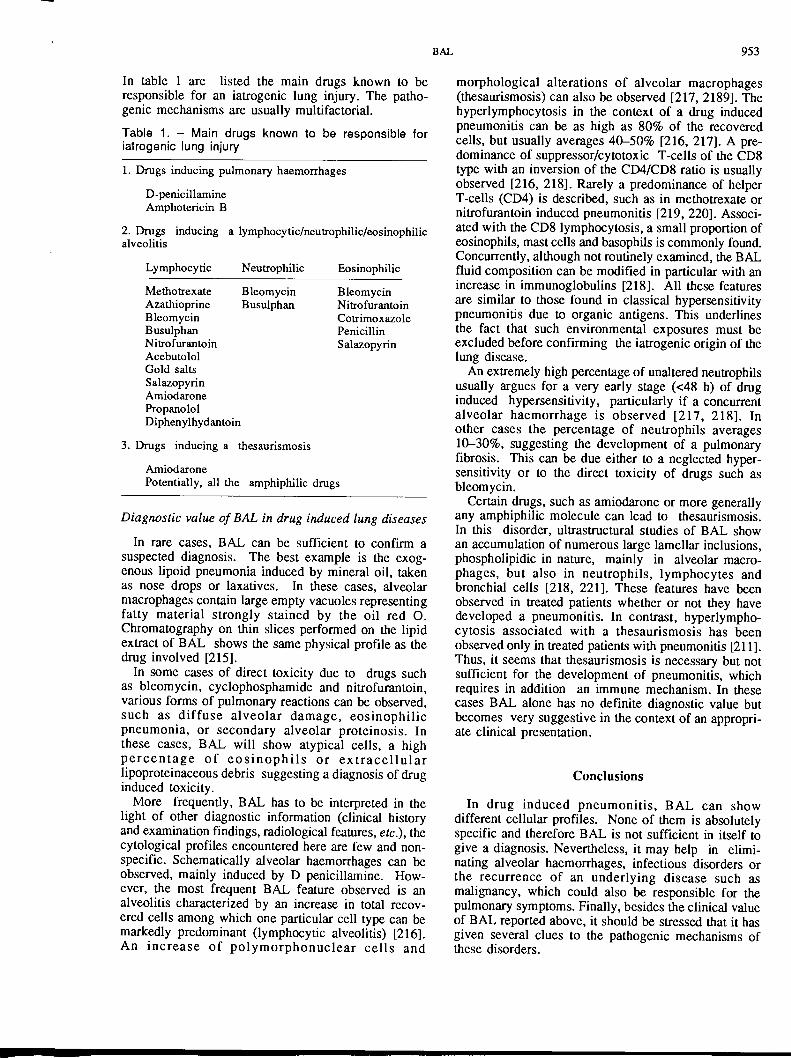
In table 1 are listed the main drugs known to beresponsible for an iatrogenic lung injury . The patho-genic mechanisms are usually multifactorial .Table 1 . - Main drugs known to be responsible foriatrogenic lung injury
1 . Drugs inducing pulmonary haemorrhages
3. Drugs inducing a thesaurismosisAmiodaronePotentially, all the amphiphilic drugs
Diagnostic value ofBAL in drug induced lung diseases
In rare cases, BAL can be sufficient to confirm asuspected diagnosis. The best example is the exog-enous lipoid pneumonia induced by mineral oil, takenas nose drops or laxatives . In these cases, alveolarmacrophages contain large empty vacuoles representingfatty material strongly stained by the oil red O .Chromatography on thin slices performed on the lipidextract of BAL shows the same physical profile as thedrug involved [215] .
In some cases of direct toxicity due to drugs suchas bleomycin, cyclophosphamide and nitrofurantoin,various forms of pulmonary reactions can be observed,such as diffuse alveolar damage, eosinophilicpneumonia, or secondary alveolar proteinosis . Inthese cases, BAL will show atypical cells, a highpercentage of eosinophils or extracellularlipoproteinaceous debris suggesting a diagnosis of druginduced toxicity .More frequently, BAL has to be interpreted in the
light of other diagnostic information (clinical historyand examination findings, radiological features, etc .), thecytological profiles encountered here are few and non-specific . Schematically alveolar haemorrhages can beobserved, mainly induced by D penicillamine . How-ever, the most frequent BAL feature observed is analveolitis characterized by an increase in total recov-ered cells among which one particular cell type can bemarkedly predominant (lymphocytic alveolitis) [216] .An increase of polymorphonuclear cells and
BAL
morphological alterations of alveolar macrophages(thesaurismosis) can also be observed [217, 2189]. Thehyperlymphocytosis in the context of a drug inducedpneumonitis can be as high as 80% of the recoveredcells, but usually averages 40-50% [216, 217] . A pre-dominance of suppressor/cytotoxic T-cells of the CD8type with an inversion of the CD4/CD8 ratio is usuallyobserved [216, 218] . Rarely a predominance of helperT-cells (CD4) is described, such as in methotrexate ornitrofurantoin induced pneumonitis [219, 220] . Associ-ated with the CD8 lymphocytosis, a small proportion ofeosinophils, mast cells and basophils is commonly found.Concurrently, although not routinely examined, the BALfluid composition can be modified in particular with anincrease in immunoglobulins [218] . All these featuresare similar to those found in classical hypersensitivitypneumonitis due to organic antigens . This underlinesthe fact that such environmental exposures must beexcluded before confirming the iatrogenic origin of thelung disease.An extremely high percentage of unaltered neutrophils
usually argues for a very early stage (<48 h) of druginduced hypersensitivity, particularly if a concurrentalveolar haemorrhage is observed [217, 218] . Inother cases the percentage of neutrophils averages10-30%, suggesting the development of a pulmonaryfibrosis . This can be due either to a neglected hyper-sensitivity or to the direct toxicity of drugs such asbleomycin .
Certain drugs, such as amiodarone or more generallyany amphiphilic molecule can lead to thesaurismosis .In this disorder, ultrastructural studies of BAL showan accumulation of numerous large lamellar inclusions,phospholipidic in nature, mainly in alveolar macro-phages, but also in neutrophils, lymphocytes andbronchial cells [218, 221] . These features have beenobserved in treated patients whether or not they havedeveloped a pneumonitis. In contrast, hyperlympho-cytosis associated with a thesaurismosis has beenobserved only in treated patients with pneumonitis [211].Thus, it seems that thesaurismosis is necessary but notsufficient for the development of pneumonitis, whichrequires in addition an immune mechanism . In thesecases BAL alone has no definite diagnostic value butbecomes very suggestive in the context of an appropri-ate clinical presentation .
Conclusions
953
In drug induced pneumonitis, BAL can showdifferent cellular profiles . None of them is absolutelyspecific and therefore BAL is not sufficient in itself togive a diagnosis. Nevertheless, it may help in elimi-nating alveolar haemorrhages, infectious disorders orthe recurrence of an underlying disease such asmalignancy, which could also be responsible for thepulmonary symptoms. Finally, besides the clinical valueof BAL reported above, it should be stressed that it hasgiven several clues to the pathogenic mechanisms ofthese disorders .
D-penicillamineAmphotericin B
2. Drugs inducing aalveolitis
lymphocytic/neutrophilic/eosinophilic
Lymphocytic Neutrophilic EosinophilicMethotrexate Bleomycin BleomycinAzathioprine Busulphan NitrofurantoinBleomycin CotrimoxazoleBusulphan PenicillinNitrofurantoin SalazopyrinAcebutololGold saltsSalazopyrinAmiodaronePropanololDiphenylhydantoin
954
The clinical use of BAL in patients with pulmonary infections
M. Rust, C. Albera, L. Carratu, C . Danel, D . Israel-Biet, H . Klech, S .I . Rennard,A.J .A . Robalo-Cordeiro, G . Semenzato, G . Velluti, H . Worth
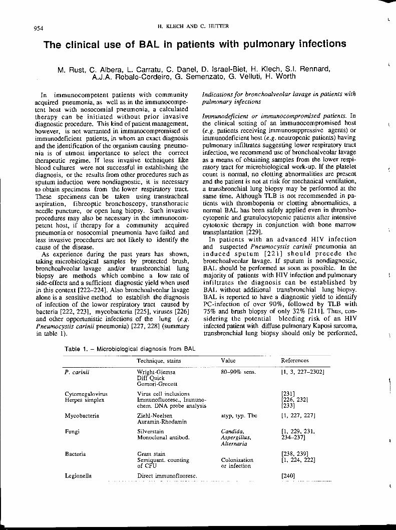
In immunocompetent patients with communityacquired pneumonia, as well as in the immunocompe-tent host with nosocomial pneumonia, a calculatedtherapy can be initiated without prior invasivediagnostic procedure . This kind of patient management,however, is not warranted in immunocompromised orimmunodeficient patients, in whom an exact diagnosisand the identification of the organism causing pneumo-nia is of utmost importance to select the correcttherapeutic regime. If less invasive techniques likeblood cultures were not successful in establishing thediagnosis, or the results from other procedures such assputum induction were nondiagnostic, it is necessaryto obtain specimens from the lower respiratory tract .These specimens can be taken using transtrachealaspiration, fibreoptic bronchoscopy, transthoracicneedle puncture, or open lung biopsy . Such invasiveprocedures may also be necessary in the immunocom-petent host, if therapy for a community acquiredpneumonia or nosocomial pneumonia have failed andless invasive procedures are not likely to identify thecause of the disease.As experience during the past years has shown,
taking microbiological samples by protected brush,bronchoalveolar lavage and/or transbronchial lungbiopsy are methods which combine a low rate ofside-effects and a sufficient diagnostic yield when usedin this context [222-224] . Also bronchoalveolar lavagealone is a sensitive method to establish the diagnosisof infection of the lower respiratory tract caused bybacteria [222, 223], mycobacteria [225], viruses [226]and other opportunistic infections of the lung (e .g .Pneumocystis carinii pneumonia) [227, 228] (summaryin table 1) .
H . KLECH AND C. HUTI'ER
Table 1 . - Microbiological diagnosis from BAL
Technique, stainsP. carinii
Wright-GiemsaDiff QuickGomori-Grocott
Cytomegalovirus
Virus cell inclusionsHerpes simplex
Immunofluoresc ., Immuno-chem. DNA probe analysis
Mycobacteria Ziehl-NeelsenAuramin-Rhodamin
Fungi
SilverstainMonoclonal antibod.
Bacteria
Gram stainSemiquant. countingof CFU
Legionella
Direct immunofluoresc .
Indications for bronchoalveolar lavage in patients withpulmonary infections
Immunodeficient or immunocompromised patients . Inthe clinical setting of an immunocompromised host(e.g . patients receiving immunosuppressive agents) orimmunodeficient host (e.g . neutropenic patients) havingpulmonary infiltrates suggesting lower respiratory tractinfection, we recommend use of bronchoalveolar lavageas a means of obtaining samples from the lower respi-ratory tract for microbiological work-up. If the plateletcount is normal, no clotting abnormalities are presentand the patient is not at risk for mechanical ventilation,a transbronchial lung biopsy may be performed at thesame time . Although TLB is not recommended in pa-tients with thrombopenia or clotting abnormalities, anormal BAL has been safely applied even in thrombo-cytopenic and granulocytopenic patients after intensivecytotoxic therapy in conjunction with bone marrowtransplantation [229] .In patients with an advanced HIV infection
and suspected Pneumocystis carinii pneumonia aninduced sputum [221] should precede thebronchoalveolar lavage . If sputum is nondiagnostic,BAL should be performed as soon as possible . In themajority of patients with HIV infection and pulmonaryinfiltrates the diagnosis can be established byBAL without additional transbronchial lung biopsy .BAL is reported to have a diagnostic yield to identifyPC-infection of over 90%, followed by TLB with75% and brush biopsy of only 32% [211] . Thus, con-sidering the potential bleeding risk of an HIVinfected patient with diffuse pulmonary Kaposi sarcoma,transbronchial lung biopsy should only be performed,
Value References80-90% sens . [1, 3, 227-23021
[231][226, 232][233]
atyp, typ. Tbc [1, 227, 227]
Candida, [1, 229, 231,Aspergillus, 234-237]Alternaria
[238, 239]Colonization [1, 224, 222]or infection
[240]
if prior investigations including BAL werenondiagnostic.
Immunocompetent patients . Bronchoalveolar lavage hasbeen successfully used in this clinical setting also, inparticular in patients suggestive for nosocomial pneu-monia by help of Gram stains and bacterial cultures ;semiquantitative counting of bacteria helps to differen-tiate between colonization and infection [222, 224, 239,239] . Legionella infections can be detected either bydirect immunofluorescence technique [240] or by bac-terial culture .
Technique of bronchoalveolar lavage
Bronchoalveolar lavage is performed duringfibreoptic bronchoscopy as described previously [1] .Although some local anaesthesia may be necessary toperform this procedure, the anaesthetic should not beinstilled directly into the segment to be lavaged, as itmay inhibit bacterial growth in the culture .Bronchoalveolar lavage should be performed in asegment which has been shown to be infiltrated on chestradiograph or from which purulent secretion is dis-charged during bronchoscopy . In adult patients avolume of 50-100 ml of saline should be used in thisclinical setting. For the interpretation of laboratory re-sults from BAL it may be helpful to obtain specimensfrom the oral cavity and hypopharynx at the time ofthe BAL. Supplemental oxygen should be given duringthe entire procedure and for at least 1 h after thebronchoscopy .As immunocompromised patients with a pneumonia
are at risk to develop respiratory failure, prior to BALan arterial blood gas analysis should confirm that thepatient is not at risk to develop respiratory distressduring or after bronchoalveolar lavage . If arterialoxygen tension (Pao2) despite supplemental oxygen is<65 mmHg bronchoalveolar lavage should be per-formed with care, reducing the volume of saline tobe instilled. As the Pao2 may drop substantially afterbronchoalveolar lavage, adequate preparations haveto be taken so that the patient can be intubated andventilated if necessary . During the procedure vital signs,oxygen saturation and cardiac rhythm should be moni-tored continuously .
Work-up of specimens obtained by bronchoalveolarlavage
Specimens obtained from immunocompromised orimmunodeficient patients should be processed assoon as possible, thus avoiding further contaminationor missing such agents as anaerobic bacteria .
BAL
Bronchoalveolar lavage fluid should be worked up forbacterial, fungal, opportunistic and viral infections. Inaddition the specimens should be examined by acytopathologist to exclude a malignancy . The techniquesused for these purposes are described in the technicalrecommendations and guidelines for BAL. In summaryBAL fluid should be stained and cultured quantitativelyfor bacteria [224] using appropriate media, stained andcultured for mycobacteria including mycobacteria otherthan M. tuberculosis (MOTT) and for fungi. APneumocystis carinii infection should be ruled out byappropriate stains (Wright-Giemsa, silver stain, toluid-ine blue or monoclonal antibodies) . Viral infectionsshould be excluded using antibodies, viral cultures andDNA/RNA-probe analysis . If necessary electronmicroscopy enables a rapid differentiation of virus inbronchoalveolar lavage fluid.
In patients with HIV infection and diffuse pulmonaryinfiltrates a cell differential on a bronchoalveolar lav-age slide may help to establish the diagnosis of lym-phocytic interstitial pneumonia . Results from stainingwith appropriate antibodies and the demonstration ofHIV in material from BAL may indicate the presenceof nonspecific interstitial pneumonitis [242]
Interpretation of laboratory results
Results from BAL of immunocompromised orimmunodeficient patients should be evaluated with care,considering the underlying disease, the history, theimmunological status and the clinical features . Inparticular, the presence of cytomegalovirus (CMV) asshown by cultures or DNA-probe does not alwaysindicate a clinically relevant infection. In caseof detection of fungi or bacteria the clinician hasto decide whether there is an infection, which shouldbe treated, or a mere colonization . Quantitativecultures [224] may help to distinguish these twoconditions.
Conclusions
955
BAL is the method of choice in diagnosis of oppor-tunistic infections (bacteria, viruses, fungi, protozoa) ofthe lower respiratory tract in particular in immunodefi-cient or immunocompromised patients . Highestdiagnostic yield is reported in the diagnosis of P . cariniipneumonia (>90%), which in many cases obviates theneed of a lung biopsy . BAL can even be performed inpatients with underlying respiratory insufficiency or inthrombocytopenic patients provided appropriate safetymeasures and selection of patients are undertaken .In patients with bacterial infections BAL may contrib-ute to discrimination between bacterial colonization ortrue parenchymatous infection .
95 6
S .I . Rennard, C . Albera, L. Carratu, W. Bauer, H . Eckert, J . Linder, M. Pirozynski, A.J.A .Robalo-Cordeiro, C . Sanguinetti, G. Semenzato, N . Secen, I . Striz, H . Teschler, G . Velluti
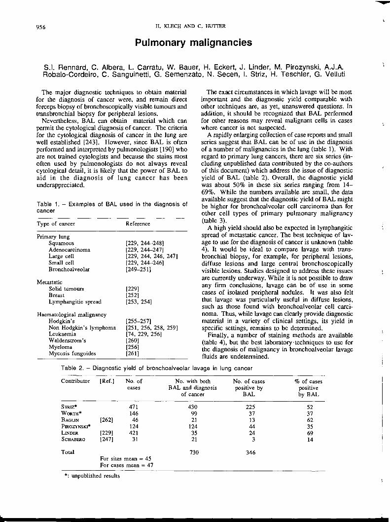
The major diagnostic techniques to obtain materialfor the diagnosis of cancer were, and remain directforceps biopsy of bronchoscopically visible tumours andtransbronchial biopsy for peripheral lesions .Nevertheless, BAL can obtain material which can
permit the cytological diagnosis of cancer. The criteriafor the cytological diagnosis of cancer in the lung arewell established [243]. However, since BAL is oftenperformed and interpreted by pulmonologists [190] whoare not trained cytologists and because the stains mostoften used by pulmonologists do not always revealcytological detail, it is likely that the power of BAL toaid in the diagnosis of lung cancer has beenunderappreciated .
Table 1 . - Examples of BAL used in the diagnosis ofcancer
Table 2. - Diagnostic
Pulmonary malignancies
* : unpublished results
H. KLECH AND C . HUITER
yield of bronchoalveolar
For sites mean = 45For cases mean = 47
The exact circumstances in which lavage will be mostimportant and the diagnostic yield comparable withother techniques are, as yet, unanswered questions . Inaddition, it should be recognized that BAL performedfor other reasons may reveal malignant cells in caseswhere cancer is not suspected.Arapidly enlarging collection of case reports and small
series suggest that BAL can be of use in the diagnosisof anumber of malignancies in the lung (table 1) . Withregard to primary lung cancers, there are six series (in-cluding unpublished data contributed by the co-authorsof this document) which address the issue of diagnosticyield of BAL (table 2) . Overall, the diagnostic yieldwas about 50% in these six series ranging from 14-69%. While the numbers available are small, the dataavailable suggest that the diagnostic yield of BAL mightbe higher for bronchoalveolar cell carcinoma than forother cell types of primary pulmonary malignancy(table 3) .A high yield should also be expected in lymphangitic
spread of metastatic cancer . The best technique of lav-age to use for the diagnosis of cancer is unknown (table4) . It would be ideal to compare lavage with trans-bronchial biopsy, for example, for peripheral lesions,diffuse lesions and large central bronchoscopicallyvisible lesions. Studies designed to address these issuesare currently underway . While it is not possible to drawany firm conclusions, lavage can be of use in somecases of isolated peripheral nodules. It was also feltthat lavage was particularly useful in diffuse lesions,such as those found with bronchoalveolar cell carci-noma. Thus, while lavage can clearly provide diagnosticmaterial in a variety of clinical settings, its yield inspecific settings, remains to be determined .
Finally, a number of staining methods are available(table 4), but the best laboratory -techniques to use forthe diagnosis of malignancy in bronchoalveolar lavagefluids are undetermined.
lavage in lung cancer
Type of cancer Reference
Primary lungSquamous [229, 244-248]Adenocarcinoma [229, 244-247]Large cell [229, 244, 246, 247]Small cell [229, 244-246]Bronchoalveolar [249-251]
MetastaticSolid tumours [229]Breast [252]Lymphangitic spread [253, 254]
Haematological malignancyHodgkin's [255-257]Non Hodgkin's lymphoma [251, 256, 258, 259]Leukaemia [74, 229, 256]Waldenstrom's [260]Myeloma [256]Mycosis fungoides [2611
Contributor [Ref.] No . ofcases
No . with bothBAL and diagnosis
of cancer
No . of casespositive byBAL
% of casespositiveby BAL
STRrz* 471 430 225 52WORTH* 146 99 37 37BAGLIN [262] 46 21 13 62PB2OZYNSKI* 124 124 44 35UNDER [229] 421 35 24 69SCHABERG [247] 31 21 3 14
Total 730 346
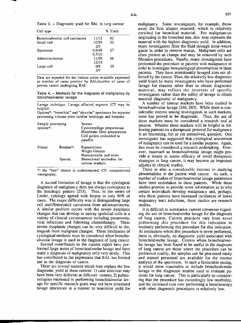
Table 3. - Diagnostic yield for BAL in lung cancer
Data are reported for the various series available expressedas number of cases positive by BAL/number of cases ofproven cancer undergoing BAL
Table 4. - Methods for the diagnosis of malignancy bybronchoalveolar lavage
Lavage technique : Lavage affected segment (CT may behelpful)Options* : "bronchial" and "alveolar" specimens for separateprocessing volume prior to/after brushings and biopsies
Sample processing
Smearsoptions* :
Cytocentrifuge preparationsMembrane filter preparationsCell pellets embedded inparaffin
Stains : Routine* : PapanicolaouWright-GiemsaHaematoxylin and eosin
Special:
Monoclonal antibodies fortumour markers
* : the "best" choice is undetermined ; CT: computerizedtomography .
A second limitation of lavage is that the cytologicaldiagnosis of malignancy does not always correspond tothe histologic pattern [253] . Thus, in the series ofLinder, cytology agreed with biopsy in only 80% ofcases . The major difficulty was in distinguishing largecell undifferentiated carcinoma from adenocarcinoma.A similar problem occurs with the severe dysplasticchanges that can develop in airway epithelial cells in avariety of clinical circumstances including pneumonia,viral infections and following chemotherapy . Thesesevere dysplastic changes can be very difficult to dis-tinguish from malignant changes. These limitations ofcytological methods must be considered when broncho-alveolar lavage is used in the diagnosis of lung cancer.
Several contributors to the current report have per-formed large series of bronchoalveolar lavage and havemade a diagnosis of malignancy only very rarely . Thishas contributed to the impression that BAL has limiteduse in the diagnosis of cancer .There are several reasons which may explain the low
diagnostic yield at these centres : 1) case selection mayhave been very different at different centres ; 2) pulmo-nologists interested in performing bronchoalveolar lav-age for specific research goals may not have processedlavage specimens in a manner to maximize yield for
BAL 957
malignancy . Some investigators, for example, throwaway the first aliquot returned, which is relativelyenriched for bronchial material . For malignanciesoriginating in the bronchial tree, this may represent thematerial with the highest diagnostic yield . In addition,many investigators filter the fluid through loose-weavegauze in order to remove mucus. Malignant cells areoften present as clumps and may be removed by suchfiltration procedures . Finally, many investigators haveperformed the procedure in patients with malignancy inorder to investigate immunological abnormalities in thesepatients . They have intentionally lavaged sites not af-fected by the cancer. Thus, the relatively low diagnosticyield found by many investigators who have performedlavage for reasons other than to obtain diagnosticmaterial, may reflect the interests of specificinvestigators rather than the utility of lavage to obtainmaterial diagnostic of malignancy .A number of tumour markers have been studied in
bronchoalveolar lavage [246, 263] . While there is con-siderable interest among investigators in such markers,none has proved to be diagnostic . Thus, the use ofthese markers must be considered a research tool atpresent . Whether these markers will be helpful in fol-lowing patients on a therapeutic protocol for malignancyis an interesting, but as yet unresolved, question. Oneinvestigator has suggested that cytological assessmentof malignancy can be used for a similar purpose. Again,this must be considered a research undertaking. How-ever, inasmuch as bronchoalveolar lavage might pro-vide a means to assess efficacy of novel therapeuticstrategies in lung cancer, it may become an importantadjunct in clinical studies .There is also a considerable interest in studying
abnormalities in the patient with cancer. As such, anumber of studies of bronchoalveolar lavage parametershave been undertaken in these patients . While thesestudies promise to provide some information as to whycertain individuals develop malignancy and, perhaps,why these patients have increased incidences of lowerrespiratory tract infections, these studies are researchstudies .
It is difficult to summarize current consensus regard-ing the use of bronchoalveolar lavage for the diagnosisof lung cancer . Current practices vary from neverperforming this procedure for this indication toroutinely performing this procedure for this indication .At institutions where this procedure is never performed,there is, obviously, no diagnostic yield associated withbronchoalveolar lavage. Centres where bronchoalveo-lar lavage has been found to be useful in the diagnosisof lung cancer are those where the procedure can beperformed readily, the samples can be processed easilyand trained personnel are available for the routineanalysis of the specimens . In such a favourable setting,it would seem reasonable to include bronchoalveolarlavage in the diagnostic routine used to evaluate pa-tients for lung cancer. This is particularly so consider-ing that the procedure has exceedingly low morbidity,and the increased cost over performing a bronchoscopywith other diagnostic procedures is relatively low .
Cell type % Yield
Bronchoalveolar cell carcinoma 11/12 92Small cell 10/35 32
2/3Squamous 0.9/49 27
0.7/10Adenocarcinoma 11/20 66
12/15Large cell 0/5 25
3/7
958 H . KLECH AND C . HU7TER
Bronchial asthmaL.M . Fabbri, V . De Rose, Ph . Godard, G .A . Rossi
In the past few years fibreoptic bronchoscopyand bronchoalveolar lavage fluid analysis have beenextended to subjects with asthma and they areincreasingly used to study airway cell profile and fluidcomponents, as well as to study the characteristics ofthe harvested cells in vitro [198, 264, 265] . Initialstudies were performed in stable asthmatics who werefree of bronchospasm, and suggested that broncho- al-veolar lavage could be safely performed as a re-search tool in carefully selected,
asymptomaticasthmatic subjects .
The guidelines provided by twointernational committees set up to evaluate the use ofbronchoalveolar lavage and record its application,recognized and established the safety of the procedurein those selected patients [1, 266] . Many studieshave been carried out without major complicationson stable asthmatics before and afterbronchoprovocation with allergens or occupationalagents, and an international workshop on the use ofbronchoalveolar lavage in asthma established the cri-teria to perform this procedure safely during the courseof asthmatic responses to asthmogenic stimuli, as aresearch tool [266] .The general conclusion from the literature is that
the bronchoalveolar lavage technique is safe in asthma,and that as long as reasonable guidelines are chosenfor the selection of patients, the mortality is zero andthe morbidity is very low. However, special careshould be exercised in asthmatic patients with markedbronchial responsiveness, and supplemental oxygendelivery and electrocardiographic (ECG) monitoring isstrongly advised in patients with severe underlyingdiseases or in any critical conditions [1, 198, 266] .Additional criteria are provided to select patientsto undergo bronchoalveolar lavage following aerosolor local bronchoprovocation [266] .
Clinical application ofbronchoalveolar lavage in asthma
The analysis of cells, mediators, proteins and en-zymes obtained from the alveolar spaces and the invitro study of cells recovered from the respiratorytract can help to elucidate pathogenic mechanismsin asthma . In stable, mild asthmatics no distinctivecellular profile is diagnostic although eosinophils,neutrophils, epithelial cells, metachromatic cells andlymphocytes may be increased [198, 264, 265] . Thereseems to be no difference between the cellular profileof atopic and non-atopic stable asthmatics .The major limitation of the standard technique of
BAL is its intrinsic low sensitivity due to the fact thatlarge volumes of fluid are instilled both in the airwaysand in the alveoli . To obtain true bronchial lavage byusing lower volumes of fluid, new techniques have been
recently developed to lavage isolated airway segmentsemploying either a double balloon bronchoscope ora double balloon tipped catheter inserted through adouble lumen bronchoscope (see following sections) .This technique is extremly promising both because itis specific for the airways and because it alreadyallowed the consistent recovery of cells and mediatorsbefore and after bronchoprovocation from the air-ways of asthmatic subjects, and some of the results seemto be specific for asthmatic airways [267-269] .The lack of specifity of bronchoalveolar lavage cell
profile in asthma would discourage any clinicalapplication of this procedure, especially since thediagnosis and monitoring of the activity of the diseaseseem to be accomplished effectively by using objec-tive functional parameters, such as the measurement ofairway responsiveness to nonspecific stimuli and theassessment of the spontaneous diurnal variability ofairflow obstruction. Few patients with current activeasthma have been evaluated . In most of the studiescarried out in asthmatics the level of airwayhyperresponsiveness, when it was measured, variedfrom mild to moderate, the subjects were defined asasymptomatic, and there was no attempt to evaluatethe activity of the disease by using more
than onefunctional parameter (i .e .
the spontaneous variabilityof the airflow obstruction in addition to the level ofnonspecific airway responsiveness) . Thus, the resultsof bronchoalveolar lavage analysis in those patientsmay not be relevant to asthma but just to well-controlled asthmatics.
In formulating a reasonable position about the clini-cal use of bronchoalveolar lavage and its analysis inasthmatic subjects, one must acknowledge that it isstill an experimental procedure, that needs furtherassessment and it must continue to be included as partof clinical research protocols. It may be proved to beclinically helpful in the evaluation of pulmonaryinfiltrates in asthmatics [264] .Bronchoalveolar lavage has also been considered for
the therapy of status asthmaticus or life-threateningasthma attacks [198, 270] . The technique used fortherapeutic lavages was not in fact bronchoalveolarlavage but just
segmental
washings, both becausethe procedure was not standardized and because thefluid was not analysed . Because of the limited expe-rience and the lack of carefully designed clinicaltrials, the therapeutic segmental washings in patientswith asthma must still be considered experimental innature and performed in selected patients by well-trainedphysicians with an extensive experience in this field(see chapter : Therapeutic applications of BAL) .
Onefurther promising application of bronchoalveolarlavage in asthma may be the assessment of thecellular response to antiasthma therapy [271, 272] .
Conclusions
In agreement with the recent state-of-the-art paperand review articles, we believe that there is noindication at present for the use of bronchoalveolar
Chronic bronchitis and emphysema
E. Pozzi, V . De Rose, S.I . Rennard, L.M. Fabbri
Despite the widespread use of bronchoalveolarlavage (BAL) in several lung diseases, only a fewstudies have evaluated its usefulness in patients withchronic bronchitis and/or emphysema .
Bronchoalveolar lavage in the diagnosis of chronicbronchitis and emphysema
Chronic bronchitis is defined by the presence ofsymptoms, and emphysema by the presence ofpathological enlargements of airspace with destructivechanges in the walls, thus bronchoalveolar lavage hasno application in the definition of the diagnosis ofeither disease. In addition, there is no indication atpresent for the use of such a procedure in clinical workfor staging or monitoring the course of chronic bronchi-tis and emphysema because of the lack of specificity ofthe findings from bronchoalveolar lavage fluid analysis .Due to various degrees of severity of obstruction greatcare should be taken when lavaging these patients (seechapter on Side-effects and Safety of BAL).
Findings in bronchoalveolar savage fluidfrom chronicbronchitis and emphysema
Asthma, chronic bronchitis and emphysema aregrouped under the terminology of chronic obstructivepulmonary disease (COPD) . At present, very little isknown about the biochemical and cellular changes thatoccur in BAL in each stage of chronic bronchitis andemphysema, and it can be summarized in the followingpoints : with the exception of smokers who have beenwell characterized and who are likely to have smallairways disease, there is little information about bron-choalveolar lavage findings in subjects with obstructionof the small airways (small airways disease) and insubjects with simple chronic bronchitis .
In patients with COPD the recovered fluid is reducedto 10-40% of that instilled [273-276] and the broncho-alveolar lavage fluid contains an increased number ofneutrophils as well as bronchial lavage fluid [274-277] . Bronchoalveolar neutrophilia is not specific forCOPD, since it is present in smokers without COPD,patients with cystic fibrosis, and in some interstitial lung
BAL 959
lavage in clinical practice for the diagnosis, staging,monitoring or therapy of bronchial asthma. The onlyindication that may prove to be clinically helpful isthe presence of pulmonary infiltrates in asthmatics,particularly for the diagnosis of allergic broncho-pulmonary aspergillosis .
diseases [101, 1981 . In BAL from patients with emphy-sema and alphas-PI, deficiency there is a severe neu-trophilia (77 .8%±18 .4 of the differential count),suggesting high elastase burden in the alveolar liningfluid and reduced concentrations of alphas-PI, whereasthe concentration of alphaz-macroglobulin andantileucoproteases is normal [275].
Use ofbronchoalveolar lavage in the therapy ofchronicbronchitis and emphysema
At present, bronchial lavage has a limited role in thetherapy of chronic bronchitis and emphysema. It maybe used in some selected cases for removal of abundantsecretions .
In the future it could provide a useful method ofassessment of the effect of therapy. For example,according to the hypothesis that lung destruction inCOPD is primarily mediated by a protease/antiproteaseimbalance in the lower respiratory tract, the preventionof structural changes leading to severe functionalimpairment might be obtained by enhancing theantiprotease screen of the respiratory tract. Severalpharmacological approaches have been investigated :1) genetically engineered mutants of alpha,-AT and low-molecular weight elastase inhibitors ; and 2) alphas-ATthat may be administrered in sufficient quantities byinfusion to replete deficient patients . BAL might beused to evaluate the efficacy of this therapy, to verifywhether adequate enzyme concentrations are reached inalveolar lining fluid [278, 279] .
Conclusions
In conclusion, in agreement with recent reviewarticles [101, 1981, we believe that there is no indica-tion at present for the use of bronchoalveolar lavagefor the diagnosis, staging or monitoring of chronicbronchitis and emphysema because of the lack ofspecificity of the findings from bronchoalveolar lavagefluid analysis . However, bronchoalveolar lavage frompatients with mild or moderate airflow obstruction canbe safely accomplished for the investigation ofthe mechanisms involved in the development of thedisease.
960
Therapeutic applications of BALC. Danel, D . Israel-Biet, U . Costabel, L.M. Fabbri, H . Klech
Although BAL had been used for therapeutic pur-poses prior to its use as a diagnostic procedure and thevalue of BAL in the exploration and management ofsome interstitial lung diseases is now well established,its place in therapy is controversially reported . As earlyas 1963, RAMmz et al. [280] were the first to performa whole lung lavage (WLL) using a large volume offluid in patients with pulmonary alveolar proteinosis.Since then, this technique has been proposed to removeany alveolar filling material in conditions such asalveolar proteinosis [196, 281], alveolar microlithiasis[282], acute silicosis [283], or accidental inhalation ofradioactive particles [289, 205] . Its use has also beenproposed in obstructive lung diseases [286] to removethe mucus secretions accumulated in the bronchial treeas in asthma [287, 288] or in cystic fibrosis [289, 290] .This lavage differs from the segmental BAL currentlyused for diagnostic or research purposes in that it isperformed under general anaesthesia, and uses a muchlarger fluid volume . The actual procedure varies slightlyfrom one centre to another and has not yet been stand-ardized [196, 291] . WLL is a safe procedure as shownby the absence of chronic side-effects over periods aslong as 25 yrs in patients treated for pulmonary alveolarproteinosis (PAP) [192] . On the other hand, its efficacyis known to be dependent on the type of disorder inwhich it is performed.We will briefly review the main pathological condi-
tions in which WLL is currently performed .
BAL in alveolar proteinosis
The benefit of therapeutic WLL is now well demon-strated in this disease. First proposed by RAMIREz et al .[280], the technique has been slightly modified over theyears. When the diagnosis of primary PAP is established,the decision to perform a therapeutic bronchopulmo-nary lavage should be based upon the patient's exercisetolerance and on his symptomatology, because sponta-neous remission is always possible . When indicated,the performance of a WLL requires an experienced staffand considerable back-up facilities [196] . The first fluidsamples to be recovered have a milky aspect whichclears up progressively during the lavage. This treatmentalways improves the patient's symptoms [196, 281] .Some authors have shown a significant improvement ofalveolar macrophage (AM) function after therapeuticWLL, demonstrating that the effect in AM function inPAP is reversible . Furthermore, this treatment could alsoreduce the rate of secondary infections [196, 281] . Al-though idiopathic forms of PAP are always improvedby WLL, the periodicity of the need for therapeuticBAL varies widely from one patient to another, de-pending on the individual course of the disease.
In case the clinical symptoms do not dramaticallyimprove after a whole lung lavage, a clinical and patho-
H . KLECH AND C. HUTCER
logical search should be made for an associated condi-tion ; an open lung biopsy is then required to eliminate,for instance, acute silicosis, infections and/or malignancy[283, 293] .
BAL in asthma
Mucus plugs are known to contribute to the severehypoxaemia in patients with status asthmaticus due tolarge ventilatory defect. These plugs can be removedby suction through a bronchoscope after the instillationof saline or acetylcysteine [287, 288] . However, thisprocedure was thought to have a high risk/benefit ratio .Some investigators have markedly improved the benefitof this technique by limiting the indications andthrough technical modifications . Clinical benefitis likely if tenacious mucus plugging or tracheobron-chial casts are present. Nevertheless, despite thisstudy [288], it seems that WLL in patients withsevere asthma must still be considered as experimentalin nature and performed in selected patients, by well-trained physicians with an extensive experience inthis field and only in the context of an intensive careunit.
BAL in pneumoconiosis
It is well known that inhaled inorganic dust damagesthe lung by inducing an inflammatory reaction that pro-gressively leads to fibrosis . WLL has been proposed inorder to remove the irritating dust before this irrevers-ible damage occurs especially in the acute form ofsilicosis [294] . The lavage fluid is usually striking withits black or brown colour and numerous alveolar mac-rophages containing dust particles . It seems that theprocedure results in rapid symptomatic improvement butwithout modification of the pulmonary function or theprognosis [294] .
BAL in inhalation of radioactive particles
The benefit of WLL in human contamination isnot yet clearly defined [284, 285] . Experimentalstudies on dogs and baboons have been carried outover the last twenty years to determine the efficacyof WLL in the removal of such particles . It seemsthat, although the longer the radioactive materialis present in the lung, the greater the dose delivered,WLL should not be performed in the early stagesof contamination since it can prevent the usual physi-ological clearance of inhaled particles from the upperrespiratory tract. WLL seems to be indicated inlevels of contamination inducing acute effects, while itsvalue in patients with lesser exposure is not clearlyestablished.
Other therapeutic applications of BAL
WLL has been proposed in the treatment of someother pulmonary disorders such as alveolar microlithi-asis or exogenous lipoidosis, with some clinical butwithout any objective functional or radiologicalimprovement [282] .
In cystic fibrosis (CF), the benefit of WLL is alsodifficult to evaluate . It was expected that periodicalrepeated WLL could, if not arrest, at least slow downthe progressive deterioration of lung function caused bythe accumulation of bronchial secretions [289, 290] .Some authors have proposed WLL using anti-fungal drugs as a local treatment of aspergillosis,
1 .
Klech H, Pohl W et al . - Technical recommendationsand guidelines for bronchoalveolar lavage (BAL) . Report ofthe European Society of Pneumology Task Group on BAL.Eur Respir J, 1989, 2, 561-585 .2 .
Petro W, Linder O Kaspar P. - BronchoalveolarLavage Durchfukrung and Sickerbeit . Atemw Lungenkrkh,1989, 15, 573-577 .3 .
Rust M, Stellenwert der bronchoalveolaeren lavage inder pneumologischen Diagnostik bei Patienten mit HIViinfection . Atemw Lungenkrkh, 1989, 11, 614-618 .4 .
Wardlan AJ, Collins JV, Kay AB. - Mechanisms inasthma using the technique of bronchoalveolar lavage . IntArehs Allergy Appl Immun, 1987, 82, 518-525 .5 .
Rankin JA, Synder PE, Schacter EN, Matthay RA . -Bronchoalveolar lavage. HS Safety in subjects with mildasthma . Chest, 1984, 85, 723-728 .6 .
Summary and recommendations of a workshop on theinvestigative use of fiberoptic bronchoscopy andbronchoalveolar lavage in asthmatics . Am Rev Resp Dis, 1985,132, 180-182 .7 . Cole P, Turton C, Lanyon H, Collins J . -Bronchoalveolar lavage for the preparation of free lung cells :technique and complications. Br J Dis Chest, 1980, 74, 273-278 .8 .
Pingleton AK, Harrison GF, Stechschulte DJ, WesseliusLJ, Kerby GR, Ruth WE. - Effect of location pH and tem-perature of instillate in bronchoalveolar lavage in normalvolunteers . Am Rev Respir Dis, 1983, 128, 1035-1037 .9 .
Strumpf IJ, Feld MK, Cornelius MJ, Keogh BA, Crys-tal RG . - Safety of fiberoptic bronchoalveolar lavage inevaluation of interstitial lung disease . Chest, 1981, 80, 268-271 .10 .
Reynolds HJ, Newball HH. - Analysis of proteins andrespiratory cells obtained from human lungs by bronchiallavage . J Lab Clin Med, 1974, 84, 559-573 .11 . Dhillon DP, Haslam PL, Townsend PJ, Primett Z,Collins JV, Turner-Warwick M. - Bronchoalveolar lavage inpatient with interstitial lung diseases: Side effects and factorsaffecting fluid recovery . EurJ Respir Dis, 1986, 68, 342-350.12 . Ettensohn DB, Jankowski MJ, Duncan PG, Lalor PA.- Bronchoalveolar lavage in the normal volunteer subject . 2)Safety and results of repeated BAL, and use in the assess-ment of intrasubject variability . Chest, 1988, 94, 281-285 .13 .
Ancic P, Diaz P, Galleguillos F . - Pulmonary functionchanges after bronchoalveolar lavage in asthmatic patients.Br J Dis Chest, 1984, 78, 261-163 .
BAL
References
96 1
a frequent complication of CF [290]. This requires fur-ther investigation .
Conclusions
The therapeutic value of BAL is now perfectlyestablished in alveolar proteinosis, which remains theonly definite indication of this procedure. In otherlung disorders, this technique still has a risk/benefitratio which does not argue for its use in routineclinical practice . Its indication should be discussedfor each patient and performed by an experiencedstaff in the context of an intensive care unit.
14 .
Bums DM, Shure D, Francoz R Kalafer M, Harrell J,Witztum K, Moser KM. - The physiological consequences ofsaline lobar lavage in healthy human adults . Am Rev RespirDis, 1983, 127, 695-701 .15 . Kelly C, Hendrick D, Walters H . - The effecton bronchoalveolar lavage on bronchial responsivenessin patients with airflow obstruction . Chest, 1988, 93,325-328 .16 .
Lin CC, Wu JL, Huang WC. - Pulmonary function innormal subjects after bronchoalveolar lavage . Chest, 1988, 5,1049-1053 .17 . Pirozynski M, Sliwinski P, Zielinski J . - Effect ofdifferent volume of BAL fluid on arterial oxgyen saturation.Eur Respir J, 1988, 1, 943-947 .18 . Tilles TS, Goldenheim PD, Ginns LC, Hales CA . -Pulmonary function in normal subjects and patients withsarcoidosis after bronchoalveolar lavage . Chest, 1986, 89,244-248 .19 .
Klech H, Pohl W. - Unpublished data.20 . Kirby JG, O'Bryne PM, Hargreave FE. -Bronchoalveolar lavage does not alter airways responsive-ness in asthmatic subjects . Am Rev Resp Dis, 1987, 135, 554-556 .21 . Klech H, Koehn H, Pohl W, Schenk E, Losch S,Mostbeck A, Kummer F . - Diagnostic standardbronchoalveolar lavage does not alter regional or global 99-Tc-DTPA lung clearance . Eur Respir J 1988a, 1, Suppl 2,279s .22 .
Haslam PL . - Bronchoalveolar Lavage . Scan Resp Med,1984, 6, 55-70 .23 .
Daniele RP, Elias JA, Epstein PE, Rossman MD. -Bronchoalveolar lavage: role in the pathogenesis, diagnosisand management of interstitial lung disease . Ann Intern Med,1985, 102, 93-108 .24 .
Crystal RG, Reynolds HY, Kalica AR . - Broncho-alveolar lavage . Chest, 1986, 89, 122-131 .25 . Stack BHR, Choo-Kang YFJ, Heard BE. - Theprognosis of cryptogenic fibrosing alveolitis . Thorax, 1972,27, 535-542 .26 .
Carrington CB, Gaensler EA, Coutu RE, Fitzgerald MX,Gupta RG . - Natural history and treated course of usual anddesquamative
interstitial pneumonia. N Engl J Med, 1978,298, 801-811 .27 . Turner-Warwick M, Burrows B, Johnson A . -Cryptogenic fibrosing alveolitis : clinical features and theirinfluence on survival . Thorax, 1980, 35, 171-180 .
962
28 . Turner-Warwick M, Burrows B, Johnson A . -Cryptogenic fibrosing alveolitis : response to corticosteroidtreatment and its effect on survival . Thorax, 1980, 35,593-599 .29 . Livingstone JL, Lewis JG, Reid L, JeffersonKE . - Diffuse interstitial pulmonary fibrosis . A clinical,radiological and pathological study on 45 patients . Q JMed,1964, 233, 71-102 .30 .
Liebow AA, Steer A, Billingsley JG . - Desquamativeinterstitial pneumonia . Am J Med, 1965, 39, 369-404 .31 .
Wright PH, Heard BE, Steel SJ, Turner-Warwick M . -Cryptogenic fibrosing alveolitis: assessment by gradedtrephine lung biopsy histology compared with clinical,radiographic, and physiological features . BrJ Dis Chest, 1981,75, 61-70 .32 .
Weinberger SE, Kelman JA, Elson NA, Young RC Jr,Reynolds HY, Fulmer JD, Crystal RG . - Bronchoalveolarlavage in interstitial lung disease. Ann Intern Med, 1978, 89,459-466 .33 .
Reynolds HY, Fulmer JD, Kazmierowski JA, RobertsWC, Frank MM, Crystal RG . - Analysis of cellular andprotein content of bronchoalveolar lavage fluid from patientswith idiopathic pulmonary fibrosis and chronic hypersensi-tivity pneumonitis . J Clin Invest, 1977, 59, 165-175 .34 .
Haslam PL, Turton CWG, Lukoszek A, Salsbury AJ,Dewar A, Collins JV, Turner-Warwick M . - Bronchoalveo-lar lavage fluid cell counts in cryptogenic fibrosing alveolitisand their relation to therapy . Thorax, 1980, 35, 328-339 .35 .
Gellert AR, Langford JA, Winter RID, UthayakumarS, Sinha G, Rudd RM . - Asbestosis : assessment bybronchoalveolar lavage and measurement of pulmonary epi-thelial permeability . Thorax, 1985, 40, 508-514 .36 .
Sykes SE, Morgan A, Moores SR, Holmes A, DavisonW. - Dose-dependent effects in the subacute response of therat lung to quartz . Exp Lung Res, 1983, 5, 227-244 .37 .
Hallgren R, Bjermer L, Lungren R, Venge P . - Theeosinophil component of the alveolitis in idiopathicpulmonary fibrosis . Am Rev Respir Dis, 1989, 139, 373-377 .38 .
Pesci A, Bertorelli G, Manganelli P, Ambanelli U . -Bronchoalveolar lavage analysis of interstitial lung disease inCREST syndrome . Clin Exp Rheumatol, 1986, 4, 121-124 .39 . Velay B, Pages J, Cordier JF, Brume J . -Hypereosinophilia in bronchoalveolar lavage . Diagnosticvalue and correlations with blood eosinophilia. Rev MalRespir, 1987, 4, 257-260 .40.
Haslam PL, Dewar A, Turner-Warwick M . - Lavageeosinophils and histamine . In: Cellular Biology of the Lung .G . Cumming, G . Bonsignore eds, Plenum Publishing, 1982,pp . 77-87 .41 .
De Vuyst P, Jedwab J, Dumortier P, Vandermoten G,van de Weyer R, Yernault JC . - Asbestos bodies inbronchoalveolar lavage . Am Rev Respir Dis, 1982, 126,972-976 .42 .
Haslam PL, Coutts II, Watling AF, Cromwell O, duBois RM, Townsend PJ, Collins JV, Turner-Warwick M. -Bronchoalveolar lavage features associated with radiographicevidence of fibrosis in pulmonary sarcoidosis . Sarcoidosis,1981, 209-215 .43 .
Roth C, Huchon GJ, Arnoux A, Stanislas-Lequern G,Marsac JH, Chretien J . - Bronchoalveolar cells in advancedpulmonary sarcoidosis . Am Rev Respir Dis, 1981, 124, 9-12 .44 .
Lin YH, Haslam PL, Turner-Warwick M. - Chronicpulmonary sarcoidosis : relationship between lung lavage cellcounts, chest radiograph, and results of standard lung func-tion tests . Thorax, 1985, 40, 501-507 .45 .
Rudd RM, Haslam PL, Turner-Warwick M. - Cryp-togenic fibrosing alveolitis - relationship of pulmonary
H. KLECH AND C. FRITTER
physiology and bronchoalveolar lavage to response to treat-ment and prognosis . Am Rev Respir Dis, 1981, 124, 1-8 .46 .
Watters LC, Schwarz MI, Cherniack RM, Waldrom JA,Dunn TL, Stanford RE, King TE. - Idiopathic pulmonaryfibrosis . Pretreatment bronchoalveolar lavage cellularconstituents and their relationships with lung histopathologyand clinical response to therapy . Am Rev Respir Dis, 1987,153, 696-704 .47 .
Shindoh Y, Shimura S, Tomioka M, Aikawa T, SasakiH, Takishima T . - Cellular analysis in bronchoalveolarlavage fluids in infiltrative and fibrotic stages of idiopathicpulmonary fibrosis . Tohoku J Exp Med, 1986, 149, 47-60 .48 .
Gellert AR, Langford JA, Uthayakumar S, Rudd RM.- Bronchoalveolar lavage and clearance of 99m-Tc-DTPAin asbestos workers without evidence of asbestosis . Br JDisChest, 1985, 79, 251-257 .49 .
Christman JW, Emerson RJ, Graham WGB, Davis GS .- Mineral dust and cell recovery from the bronchoalveolarlavage of healthy Vermont granite workers. Am Rev RespirDis, 1985, 132, 393-399 .50 . Nagai S, Fujimura N, Hirata T, Izumi T . -Differentiation between idiopathic pulmonary fibrosis andinterstitial pneumonia associated with collagen vasculardiseases by comparison of the ratio of OKT4+ cells andOKT8+ cells in BAL T-lymphocytes . EurJRespir Dis, 1985,67, 1-9 .51 .
Pesci A, Bertorelli G, Manganelli P. - Differentiationbetween idiopathic pulmonary fibrosis and interstitialpneumonia associated with collagen vascular diseases bycomparison of the ratio of OKT4+ cells and OKT8+ inBALF T-lymphocytes (letter) . Eur J Respir Dis, 1986, 68,155-156 .52 .
Takahashi H, Nukiwa T, Matsuoka R, Danbara T, NatoriH, Arai T, Kira S . - Carcinoembryonic antigen in broncho-alveolar lavage fluid in patients with idiopathic pulmonaryfibrosis. Jpn J Med, 1985, 24, 236-243 .53 . Haslam PL, Hughes DA, Dewar A, Pantin CF . -Lipoprotein macroaggregates in bronchoalveolar lavage fluidfrom patients with diffuse interstitial lung disease : compari-son with idiopathic alveolar lipoproteinosis . Thorax, 1988, 43,140-146 .54 .
Bitterman PB, Rennard SI, Keogh BA, Wewers MD,Adelberg S, Crystal RG . - Familial idiopathic pulmonaryfibrosis . Evidence of lung inflammation in unaffected familymembers . N Engl J Med, 1986, 314, 1343-1347 .55 . Keogh B, Line B, Rust M, Hunninghake G,Meier-Sydow J, Crystal RG . - Clinical staging of patientswith idiopathic pulmonary fibrosis. Am Rev Respir Dis, 1981,123, 89 .56 . Peterson MW, Monick M, Hunninghake GW . -Prognostic role of eosinophils in pulmonary fibrosis . Chest,1987, 92, 51-56 .57 .
Libby DM. - The eosinophil in idiopathic pulmonaryfibrosis . Chest, 1987, 92, 7-8 .58 .
Turner-Warwick M, Haslam PL . - The value of serialbronchoalveolar lavages in assessing the clinical progressof patients with cryptogenic fibrosing alveolitis . Am RevRespir Dis, 1987, 135, 26-34 .59 .
Cantin AM, North SL, Fells GA, Hubbard RC, CrystalRG . - Oxidant-mediated epithelial cell injury in idiopathicpulmonary fibrosis . J Clin Invest, 1987, 79, 1665-1673 .60 .
Bjermer L, Lundgren R, Hallgren R . - Hyaluronan andtype III procollagen peptide concentrations in bronchoalveo-lar lavage fluid in idiopathic pulmonary fibrosis . Thorax, 1989,44, 126-131 .61 .
Haslam PL, Cromwell O, Dewar A, Turner-WarwickM. - Evidence of increased histamine levels in lung lavage
fluids from patients with cryptogenic fibrosing alveolitis . ClinExp Immunol, 1981, 44, 587-593 .62 . Schmidt M, Brugger E, Heinrich J. - Proteolyticactivites in bronchoalveolar lavage fluid of interstitial lungdiseases : correlation to stage and prognosis . Respiration, 1987,52, 115-121 .63 .
Bitterman PB, Wewers MD, Rennard SI, Adelberg S,Crystal RG . - Modulation of alveolar macrophage-drivenfibroblast proliferation by alternative macrophage mediators.J Clin Invest, 1986, 77, 700-708.64 .
Rennard SI, Bitterman PB, Ozaki T, Rom WN, CrystalRG. - Colchicine suppresses the release of fibroblast growthfactors from alveolar macrophages in vitro. Am Rev RespirDis, 1988, 137, 181-185.65 . O'Donnell K, Keogh B, Cantin A, Crystal RG . -Pharmacologic suppression of the neutrophil component ofthe alveolitis in idiopathic pulmonary fibrosis . Am Rev RespirDis, 1987, 136, 288-292.66 . Watters LC, King TE, Chemiack RM, Waldron JA,Stanford RE, Willcox ML, Christopher KL, Schwarz MI . -Bronchoalveolar lavage fluid neutrophils increase aftercorticosteroid therapy in smokers with idiopathic pulmonaryfibrosis . Am Rev Respir Dis, 1986, 133, 104-109.67 . Hughes DA, Haslam PL . - Changes inphosphatidylglycerol in bronchoalveolar lavage fluids frompatients with cryptogenic fibrosing alveolitis . Chest, 1989, 95,82-89.68 . Robinson PC, Watters LC, King TE, MasonRJ . - Idiopathic pulmonary fibrosis . Abnormalities in bron-choalveolar lavage fluid phospholipids . Am Rev Respir Dis,1988, 137, 585-591 .69 .
Haslam P, Turton C, Heard B, Wkoszek A, Collins J,Salibury A, Turner-Warwick M. - Bronchoalveolar lavage inpulmonary fibrosis : composition of cells obtained with lungbiopsy and clinical features . Thorax, 1980, 35, 9-18 .70.
Konig G, Luderschmidt C, Hammer C, Adelmann-GrillBC, Braun-Falco O, Fruhmann G . - Lung involvement inscleroderma . Chest, 1984, 85, 318-324.71 . Silver RM, Metcalf JF, Stanley JH, LeroyEC . - Intertitial lung disease in scleroderma - analysisby bronchoalveolar lavage . Arthritis Rheum, 1984, 127,1254-1262.72 .
Kallenberg CGM, Jansen HM, Elema D, The TH . -Steroid-responsive interstitial pulmonary disease in systemicsclerosis . Monitoring by bronchoalveolar lavage . Chest, 1984,86, 489-492.73 .
Jansen HM, Schutte AJH, Elema JD, Giessen MVD,Peset Reig R, Leeuwen MAV, Sluiter HJ, The TH . - Localimmune complexes and inflammatory response in patientswith chronic interstitial pulmonary disorders associated withcollagen vascular diseases . Clin Exp Immunol, 1984, 56,311-320.74 . Rossi GA, Bitterman PB, Rennard SL, Ferrans VJ,Crystal RG . - Evidence for chronic inflammation as acomponent of the interstitial lung disease associated withprogressive systemic sclerosis . Am Rev Respir Dis, 1985, 131,612-617.75 .
Edelson JD, Hyland RH, Ramsoen M, Chamberlain DW,Kortan P, Meindok HO, Klein MH, Braude AC, Lee P, RebuckAS . - Lung inflammation in scleroderma : clinical, radio-graphic, physiologic and cytopathological features . JRheumatol, 1985, 12, 957-963.76 .
Garcia JGN, Parhami N, Killam D, Garcia PL, KeoghBA . -Bronchoalveolar lavage fluid evaluation in rheumatoidarthritis. Am Rev Respir Dis, 1986, 133, 450-454.77 . Owens GR, I Paradis IL, Gryzan S, Medsger TA,Follansbee WP, Klein HA, Dauber JA . - Role of inflamma-
BAL 963
tion in the lung disease of systemic sclerosis : comparisonwith idiopathic pulmonary fibrosis . J Lab Clin Med, 1986, 107,253-260.78 .
Tishler M, Grief J, Freman E, Yaron M, Topilsky M.- Bronchoalveolar lavage a sensitive tool for early diagnosisof pulmonary involvement in rheumatoid arthritis . JRheumatol, 1986, 13, 547-550.79 .
Balbi B, Cosulich E, Risso A, Sacco O, Balzano E,Rossi GA. - The interstitial lung disease associated withrheumatoid arthritis : evidence for imbalance of helper T-lymphocyte subpopulations at sites of disease activity . BullEur Physiopathol Respir, 1987, 23, 241-247.80 .
Harrison NK, Glanville AR, Strickland B, Haslam PL,Corrin B, Addis BJ, Lawrence R, Millar AB, Black CM,Turner-Warwick M. - Pulmonary involvement in systemicsclerosis : the detection of early changes by thin section CTscan, bronchoalveolar lavage and 9mTc-DTPA clearance .Respir Med, 1989, 83, 403-414.81 . Idell S, Carcia JGN, Gonzalez K, McLarty J, FairDS . - Fibrinopeptide A reactive and proagulant activity inbronchoalveolar lavage: Relationship to rheumatoid intersti-tial lung disease. J Rheumatol, 1989, 16, 592-598.82 . Greene NB, Solinger AM, Baughmann RP . -Patients with collagen vascular disease and dyspnea. The valueof gallium scanning and bronchoalveolar lavage in predictingresponse to steroid therapy and clinical outcome. Chest, 1987,91, 698-703 .83 .
Garcia JGN, James HL, Zinkgraf S, Perlman BA, KeoghMB. - Lower respiratory tract abnormalities in rheumatoidinterstitial lung disease . Potential role of neutrophil in lunginjury . Am Rev Respir Dis, 1987, 136, 811-817 .84 . Weiland JE, Garcia JGN, Davis WB, Gadek JA . -Neutrophil collagenase in rheumatoid interstitial lungdisease . J Appl Physiol, 1987, 62, 628-633 .85 .
Herer B, DE Castelbajac D, Israel-Biet D, Venet A,Huchon G, Chretien J. - Le lavage broncho-alveolaire dansles formes pulmonaires de la polyarthrite rhumatoide . AnnMedInterne, 1988, 139, 310-314.86 . Wallaert B, Hatron PY, Grosbois JM, Tonnel AB,Devulder B, Voisin C. - Subclinical pulmonary involvementin collagen vascular diseases assessed by bronchoalveolarlavage : relationship between alveolitis and subsequentchanges in lung function . Am Rev Respir Dis, 1986, 133,574-580.87 .
Hatron PY, Wallaert B, Gosset D, Tonnel AB, VoisinC, Devulder B . - Subclinical lung inflammation in Sjogrensyndrome. Correlation with clinical and biologicalcharacteristics of the disease . Arthritis Rheum, 1987, 30,1226-1231 .88 .
Wallaert B, Prin L, Hatron PY, Tonnel AB, Voisin C.- Lymphocyte subpopulations in bronchoalveolar lavage inSjogren's syndrome . Evidence for an expansion of cytotoxic/suppressor subset in patients with alveolar neutrophilia . Chest,1987, 92, 1025-1031.89 .
Wallaert B, Bart F, AerLS C, Ouaissi A, Hatron PY,Tonnel AB, Voisin C . - Evidence for activated alveolarmacrophage as a component of subclinical inflammatoryalveolitis in collagen-vascular diseases . Thorax, 1988, 43,24-30.90 .
Perez TH, Farre JM, Gosset Ph, Wallaert B, DuquesnoyB, Voisin C, Delcambre B, Tonnel AB . - Subclinical alveo-lar inflammation in rheumatoid arthritis : superoxide anion,neutrophil chemotactic activity and fibronectin generation byalveolar macrophages . Eur Respir J, 1989, 2, 7-13 .91 .
Perez Th, Gosset Ph, Farre JM, Duquesnoy B, WallaertB, Tonnel AB . - Production spontance de tumor necrosisfactor par les macrophages alveolaires au cours de la
964
polyarthrite rhumatoide. Rev MalRespir, 1989, Suppl. 1,R18 .92 . Wallaert B, Aerts C, Bart F, Hatron PY, Dracon M,Tonnel AB, Voisin C . - Alveolar macrophage dysfunction insystemic lupus erythematosus . Am Rev Respir Dis, 1987, 136,293-297 .93 .
Wallaert B, Dugas M, Perez Th, Hatron PY, Gosset D,Ramon Ph, Aerts C, Tonnel AB, Voisin C . - Alveolar mac-rophage dysfunctions in collagen vascular diseases . LocalImmunity, 1988, 4, 79-95 .94 .
Martinot JB, Wallaert B, Hatron PY, Francis C, VoisinC, Sibille Y . - Clinical and subclinical alveolitis in collagenvascular diseases . Contribution of alpha-2-macroglobulinlevels in BAL fluid . Eur Respir J, 1989, 2, 437-443 .95 . Sibille Y, Martinot JB, Polomski LL, Wallaert B,Demusis M, Rankin JA, Voisin C, Gee JBL . - Phagocytesenzymes in bronchoalveolar lavage from patients withpulmonary sarcoidosis and collagen vascular disorders . AmRev Respir Dis, 1989, 139, A192 .96 .
Akoun G, Mayaud C, Touboul J, Denis M, Milleron B,Perrot J . - Use of bronchoalveolar lavage in the evaluation ofmethotrexate lung disease . Thorax, 1987, 42, 652-655 .97 .
Vivet Ph, Ameille J, Capron F, Leclerc P, Dessirier JL,Rochemaure J . - Pneumopathie interstitielle diffuse au coursd'un traitement par les sels d'or . AnnMed Interne, 1984, 135,54-58 .98 . Garcia GN, Munim A, Nugent KM, Bishop M,Hoie-Garcia P, Parhami N, Keogh BA. - Alveolar macro-phages gold retention in rheumatoid arthritis . J Rheumatol,1987, 14, 435-438 .99 .
Hatron PY, Wallaert B, -Fourrier JL, Fournier E,Gosselin B, Devulder B. - Dermato-polymyosite et fibrosepulmonaire associees a un syndrome de Gougerot-Sjogren .Etude de 3 observations . Rev Med Interne, 1985, 6, 97-104 .100 . Ziza JM, Kaplan G, Salomon C, Kahn MF. - Fibrosespulmonaires graves revelatrices d'un syndrome de GougerotSjogren primitif. Ann Med Interne, 1986, 137, 46-50 .101 . Rossi GA. - Bronchoalveolar lavage in theinvestigation of disorders of the lower respiratory tract. EurJ Respir Dis, 1986, 69, 293-315 .102. Venet A, Sandron D, Israel-Biet D. - Broncho-alveolar lavage in interstitial lung diseases . Bull EurPhysiopathol Respir, 1985, 21, 465-476 .103 . Crystal RG, Roberts WC, Hunninghake GW etal. - Pulmonary sarcoidosis : a disease characterized andperpetuated by activated lung T-lymphoctyes . Ann Intern Med,1981, 94, 73 .104 . Venet A . - Immunology of sarcoidosis . AnnMed Intern(Paris), 1984, 135, 113 .105 . Robinson B, Crystal RG . - Gamma interferon isspontaneously released by alveolar macrophages and lungT-lymphocytes in patients with pulmonary sarcoidosis . J ClinInvest, 1985, 75, 1488 .106 . Campbell D, Janossy G, DuBois RM, Poulter LW. -Immunocompetent cells in bronchoalveolar lavage reflect thecell populations in transbronchial biopsies in pulmonarysarcoidosis . Am Rev Respir Dis, 1985, 132, 1300 .107 . Spiteri MA, Clarke SW, Poulter LW. - Phenotypicand functional changes in alveolar macrophages contributeto the pathogenesis of pulmonary sarcoidosis . Clin ExpImmunol, 1988, 74, 359 .108 . Campbell DA, Poulter LW, Du Bois RM. - Phenotypicanalysis of BAL cells from patients with intersititial lungdiseases . Thorax, 1986, 41, 429 .109 . Keogh BA, Hunninghake GW, Line BR et al . - Thealveolitis of pulmonary sarcoidosis - evaluation of naturalhistory and alveolitis dependent changes in lung function .Am Rev Respir Dis, 1983, 128, 256.
H. KLECH AND C. HUTfER
110. Semenzato G, Chilosi M, Ossi E et al . - Broncho-alveolar lavage and lung histology : comparative analysis ofinflammatory and immunocompetent cells in patients withsarcoidosis and hypersensitivity pneumonitis . Am Rev RespirDis, 1985, 132, 400 .111 . Hunninghake GW, Crystal RG. - Pulmonary sar-coidosis- a disorder mediated by excess helper T-lymphocyteactivity at sites of disease activity . NEngl JMed, 1981, 305,419-434 .112 . Leatherman JW, Michael AF, Schwartz BA, HoidalJR . - Lung T-cells in hypersensitivity pneumonitis . AnnIntern Med, 1984, 100, 390-392 .113 . Israel-Biet D, Venet A, Chretien J . - Persistent highalveolar lymphocytosis as a predictive criterion of chronicpulmonary sarcoidosis . Ann NY Acad Sci, 1986, 465,395-406 .114 . Klech H. - Clinical risk assessment in sarcoidosis .Outlook . In : Sarcoidosis and other granulomatous disorders .C . Grassi, G . Rizzato, E . Pozzi eds, 1988, pp. 461-482 .115 . Turner-Warwick M, McAllister W, Lawrence R, BrittenA, Haslam PL. - Corticosteroid treatment in pulmonarysarcoidosis : do serial lavage lymphocyte counts, serum angi-otensin converting enzyme measurements and gallium-67scans help managment? Thorax, 1986, 41, 903-913 .116 . Ainslie G, du Bois RM, Poulter LW. - Relationshipbetween lavage immunocytology and clinical status in sar-coidosis . In : Sarcoidosis and Other Granulomatous DisordersEds . Grassi C, Rizzato G, Pozzi E . Elsevier Amsterdam, 1988,p.171 .117 . Ainslie G, du Bois RM, Poulter LW. - Relationbetween immunocytological features of bronchoalveolarlavage fluid and clinical indices in sarcoidosis . Thorax, 1989,44, 501 .118 . Perrin-Fayolle M, Pacheco Y, Harf R et al . -Angiotensin converting enzyme in bronchoalveolar lavagefluid in pulmonary sarcoidosis . Thorax, 1981, 34, 790 .119 . Bjermer L, Thurell M, Hallgren R . - Procollagen Illpeptide in bronchoalveolar lavage fluid. Lab Invest, 1986, 55,654 .120 . Bjermer L, Engstrom-Laurent A, Thunell M, HallgrenR . - Hyaluronic acid in bronchoalveolar lavage fluid inpatients with sarcoidosis : relationship to lavage mast cells .Thorax, 1987, 42, 933 .121 . Costabel U, Zaiss A, Wagner DJ et al. - Value ofbronchoalveolar lavage lymphocyte subpopulations for thediagnosis of sarcoidosis . In : Sarcoidosis and othergranulomatus disorders . C . Grassi, G . Rizzato, E . Pozzi eds,Excerpta Medica Amsterdam, 1988, p . 429 .122 . Costabel U, Bross KJ, Guzman J et al . - Predictivevalue of bronchoalveolar lavage T-cell subsets for the courseof pulmonary s arcoidosis . Ann NY Acad Sci, 1986, 465,418-426 .123 . Costabel U, Bross KJ, Ruhle KH et al . - Prognose andT-Lymphozyten-Subpopulationen in der bronchoalveolaerenlavage (BAL) bei pulmonaler Sarkoidose-eine dreijahrigeVerlaufsstudie . Prax Klin Pneumol, 1987, 41, 854-855 .124. Bjermer L, Rosenhall L, Hallgren R . -The predictivevalue of bronchoalveolar lavage cell analysis in sarcoidosis .Thorax, 1988, 43, 4, 284-288 .125 . Flint KC, Lenny KBP, Hudspith BN, Brostoff J, PearceFL, Geraint-James D, McJohnson N . - Bronchoalveolarmast cells in sarcoidosis increased numbers and accentuationof mediator release . Thorax, 1986, 41, 94-99 .126 . Costabel U, Bross KJ, Marxen J, Matthys H . - Tlymphocytosis in bronchoalveolar lavage fluid of hypersensi-tivity pneumonitis . Chest, 1984, 85, 514-518 .127 . Semenzato G, Agostini C, Zambello R, Trentin L,
Chilosi M, Pizzolo G, Marcer G, Cipriani A. - Lung T-cellsin hypersensitivity pneumonitis : phenotypic and functionalanalyses . J Immunol, 1986, 137, 1164-1172 .128 . Fournier E, Tonnel AB, Gosset Ph, Wallaert B, AmeisenJC, Voisen C . - Early neutrophil alveolitis after antigeninhalatioi in hypersensitivity pneumonitis . Chest, 1985, 88,563-566 .129 . Haslam PL . - Bronchoalveolar lavage in extrinsicallergic alveolitis . Eur J Respir Dis, 1987, 71 (Suppl .154), 120 .130 . Soler P, Valeyre D, Georges R, Battesti JP, Basset F,Hance A . - The role of epithelial abnormalities inrecruiting Langerhans cells to the lower respiratory tract. AmRev Respir Dis, 1986, 133, A243 .131 . Laviolette M, Cormier Y, Leblanc P, Soler P, HanceAJ . - Bronchoalveolar lavage (BAL) in farmer's lung (FL) :significance of mast cells . Am Rev Respir Dis, 1989, 139,A189 .132 . Bjermer L, Engstrom-Laurent A, Lundgren R,Rosenthall L, Hallgren R. - Bronchoalveolar mastocytosisin farmer's lung is related to the disease activity . Arch InternMed, 1988, 148, 1362-1365 .133 . Costabel U, Bross KJ, Guzman J, Matthys H. -Plasmazellen and Lymphozytensubpopulationen in derbronchoalveolaeren Lavage bei exogen-allergischer Alveoli-tis . Prax klin Pneumol, 1985, 39, 925-926 .134 . Costabel U, Bross KJ, Ruble KH, Lohr GW, MatthysH . - la-like antigens on T-cells and their subpopulations inpulmonary sarcoidosis and hypersensitivity pneumonitis :analysis of bronchoalveolar and blood lymphocytes . Am RevRespir Dis, 1985, 131, 337-342.135 . Semenzato G, Trentin L, Zambello R, Agostini C,Masciarelli M, Cipriani A, Marcer G . - Different types ofcytotoxic lymphocytes are involved in the cytolyticmechanisms taking place in the lung of patients withhypersensitivity pneumonitis . Am Rev Respir Dis, 1988, 137,70-74 .136 . Semenzato G, Agostini C . - Editorial . Humanretroviruses and lung involvement . Am Rev Respir Dis, 1989,139, 1317-1322 .137 . Costabel U, Guzman J, Seyboth S, Ruhle KH, MatthysH . - Serial analysis of lung lymphocytes and T-cell subsetsduring the course of hypersensitivity pneumonitis . Am RevRespir Dis, 1987, 135, 372A .138 . Trentin L, Marcer G, Chilosi M, Zambello R, AgostiniC, Masciarelli M, Bizzotto R, Gemignani C, Cipriani A,Di Vittorio G, Semenzato G . - Longitudinal study ofalveolitis in hypersensitivity pneumonitis patients : animmunological evaluation . J Allergy Clin Immunol, 1988, 82,577-585 .139 . Cormier Y, Belanger J, Laviolette M. - Prognostic sig-nificance of bronchoalveolar lymphocytosis in farmer's lung .Am Rev Respir Dis, 1987, 135, 692-695 .140 . Yoshizawa Y, Ohdama S, Tanoue M, TanakaM, Ohtsuka M, Uetake K, Hasegawa S . - Analysis ofbronchoalveolar lavage cells and fluids in patients withhypersensitivity pneumonitis : possible role of chemotacticfactors in the pathogenesis of the disease . Int Arch AllergyAppl Immunol, 1986, 80, 376-382.141 .
Cormier Y, Belanger J, Leblanc P, Herbert J, LavioletteM. - Lymphocyte subpopulations in extrinsic allergic alveo-litis . Ann NY Acad Sci, 1986, 465, 370-377 .142 . Herbert J, Beaudoin J, Laviolette M, Beaudoin R,Belanger J, Cormier Y. - Absence of correlation between thedegree of alveolitis and antibody levels of Micropolysporafaeni. Clin Exp Immunol, 1985, 60, 572-578 .143 . Cormier Y, Belanger J, Laviolette M. - Persistent
BAL 965
bronchoalveolar lymphocytosis in asymptomatic farmers . AmRev Respir Dis, 1986, 133, 843-847 .144 . Monkare S, Haahtela T . - Farmer's lung : a five yearfollow-up of eighty six patients . Clin Allergy, 1987, 17,143-151 .145 . Teschler H, Schmidt B, Zwang B, Ziesche R, MatthysH, Konietzko N, Costabel U. - Procollagen-III-peptide levelsin BAL fluid of patients with hypersensitivity pneumonitis .Am Rev Respir Dis, 1989, 139, A189 .146 . Costabel U, Teschler H. - Inflammation and immunereactions in interstitial lung disease (ILD) associated withinorganic dust exposure . Eur Respir J, 1989, 190, 3, 363-364 .147 . Robalo-Cordeiro AJA, Leite ACP, Rosa MAS, LimaMAM, Cordeiro CR, Gaspar E, Pego MA. - A alveolite dasilicose . Via Pneumologica, 1988, 2, 113-125 .148 . Wallace JM, Oishi JS, Barbers RG, Batra P, AberleDR . - Bronchoalveolar lavage cell and lymphocytephenotype profiles in healthy asbestos exposed shipyardworkers . Am Rev Respir Dis, 1989, 139, 33-38 .149 . Araujo A, Alfarroba E, Freitas e Costa M. - Therole of monoclonal antibodies in the study of chronicinflammatory respiratory diseases induced by dust inhalation .Eur J Respir Dis, 1986, 69 (Suppl . 146), 203-210.150 . Begin R, Cantin AM, Boileau RD, Bisson GY. -Spectrum of alveolitis in quartz-exposed human subjects .Chest, 1987, 92, 1061-1067 .151 . Rom WN, Bitterman PB, Rennard SI, Cantin A, Crys-tal RG . - Characterization of the lower respiratory tract in-flammation of nonsmoking individuals with interstitial lungdisease associated with chronic inhalation of inorganic dusts .Am Rev Respir Dis, 1987, 136, 1429-1434.152 . Wallaert B, Lassalle Ph, Fortin F, Aerts C, Bart F,Fournier E, Voisin C . - Superoxide anion generation byalveolar inflammatory cells in simple pneumoconiosis andprogressive massive fibrosis of non smoking coal workers .Am Rev Respir Dis, 1989, 1990, 141, 129-133 .153 . Costabel U, Bross KJ, Reuter Ch, Ruble KH, MatthysH . - Alterations in immunoregulatory T-cell subsets in ciga-rette smokers . Chest, 1986, 90, 39-44 .154 . Robinson BWS, Rose AH, James A, Whitaker D, MuskAW. - Alveolitis of pulmonary asbestosis . Chest, 1986, 90,396-402 .155 . Begin R, Martel M, Desmarais Y, Drapeau G, BoileauR, Rola-Pleszczynski M, Masse S . - Fibronectin andprocollagen 3 levels in bronchoalveolar lavage ofasbestos-exposed human subjects and sheep . Chest, 1986, 89,237-243 .156 . Spurzem JR, Saltini C, Rom W, Winchester RJ,Crystal RG . - Mechanisms of macrophage accumulation inthe lungs of asbestos exposed subjects . Am Rev Respir Dis,1987, 136, 276-280 .157 . Gellert AR, Macey MG, Uthayakumar S, Newland AC,Rudd RM. - Lymphocyte subpopulations in bronchoalveolarlavage fluid in asbestos workers . Am Rev Respir Dis, 1985,132, 824-828 .158 . Xaubet A, Rodriguez-Roisin R, Bombi JA, Marin A,Roca J, Agusti-Vidal A. - Correlation of bronchoalveolarlavage and clinical and functional findings in asbestosis . AmRev Respir Dis, 1986, 133, 848-854 .159 . Haslam PL, Dewar A, Butchers P, Primett ZS, Newman-Taylor A, Turner-Warwick M. - Mast cells, atypicallymphocytes and neutrophils in bronchoalveolar lavage inextrinsic allergic alveolitis . Am Rev Respir Dis, 1987, 135,35-47 .160 . Forni A, Ortisi E, Rivolta G, Chiappino G . -Bronchoalveolar lavage T-lymphocyte subpopulations in
966
occupational and non-occupational lung diseases . In : NewFrontiers in Cytology. K . Goerttler, G.E . Feichter, S . Witteds, Springer, Berlin-Heidelberg-New York, 1988, pp .440-445 .161 . Massaglia GM, Avolio G, Barberis S, Cacciabue M,Galietti F, Giorgis GE, Miravalle C . - Pneumoconiosiscaused by hard metals . A case series . In: Sarcoidosis and othergranulomatous disorders . C . Grassi, G . Rizzato, E . Pozzi eds,Elsevier Science Publishers, Amsterdam, 1988, pp . 709-710 .162 . Davison AG, Haslam PL, Corrin B, Coutts II, DewarA, Riding WD, Studdy PR, Newman-Taylor AJ . - Interstitallung disease and asthma in hard metal workers :bronchoalveolar lavage, ultrastructural and analyticalfindings and results of bronchial provocation tests . Thorax,1983, 38, 119-128 .163 . Rizzato G, Lo Cicero S, Barberis M, Torre M, PietraR, Sabbioni E . - Trace of metal exposure in hard metal lungdisease . Chest, 1986, 90, 101-106 .164 . Epstein PE, Dauber JH, Rossman MD, Daniele RP . -Bronchoalveolar lavage in a patient with chronic berylliosis :evidence for hypersensitivity pneumonitis . Ann Intern Med,1982, 97, 213-216 .165 . Cullen MR, Kominsky JR, Rossman MD, CherniackMG, Rankin JA, Balmes JR, Kern JA, Daniele RP, Palmer L,Naegel GP, McManus K, Cruz R . - Chronic berylliumdisease in a precious metal refinery . Am Rev Respir Dis, 1987,135, 201-208 .166 . Rossman MD, Kern JA, Elias JA, Cullen MR, EpsteinPE, Preuss OP, Markham TN, Daniele RP . - Proliferativeresponse of bronchoalveolar lymphocytes to beryllium . AnnIntern Med, 1988, 108, 687-693 .167 . Saltini C, Winestock K, Kirby M, Pinkston P, CrystalRG. - Maintenance of alveolitis in patients with chronicberyllium disease by beryllium-specific helper T cells .NEnglJ Med, 1989, 320, 1103-1109 .168 . Buhl R, Bargon J, Rust M, Kronenberger H, BergmannL, Meier-Sydow J . - Lymphozytentransformationstest mitZellen der bronchoalveolaeren Lavage and des peripherenBlutes bei chronischer Berylliose . Prax klin Pneumol, 1987,41,860-861 .169 . Costabel U, Bross KJ, Huck E, Guzman J, Matthys H .- Lung and blood lymphocyte subsets in asbestosis and inmixed dust pneumoconiosis . Chest, 1987, 91, 110-112.170 . Sprince NL, Oliver LC, McLoud TC, Morris TA, TillesDS, Eisen EA, Ginns LC. - T-lymphocyte subsets inbronchoalveolar lavage and peripheral blood in asbestosworkers : correlations with exposure and pleural placques .Chest, 1987, 91, 309 .171 . Churg A, Warnock ML, Green N . - Analysis of thecores of ferruginous (asbestos) bodies from the general popu-lation . H . True asbestos bodies and pseudoasbestos bodies .Lab Invest, 1979, 40, 31-38 .172. Johnson NF, Haslam PL, Dewar A, Newman-Taylor A,Turner-Warwick M. - Identification of inorganic dust particlesin bronchoalveolar lavage macrophages by energy dispersiveX-ray microanalysis . Arch Environ Health, 1986, 3, 133-144 .173 . De Vuyst P, Dumortier P, Leophonte P, Vende WeyerR, Yernault JC . - Minerological analysis of bronchoalveolarlavage in talc pneumoconiosis . EurJ Respir Dis, 1987, 70,150-159 .174 . Schmitz-Schumann M, Costabel U, Guzman J,Matthys H . - Lungenfibrose nach protrahierterAntimonstaubexposition? Atemw-Lungenkrkh, 1987, 13(Suppl .), S108-S111.175. De Vuyst P, Dumortier P, Rickaert F, Vande Weyer R,Lenclud C, Yernault JC . - Occupational lung fibrosis in analuminium polisher . Eur J Respir Dis, 1986, 68, 131-140 .
H . KLECH AND C . HUTFER
176 . De Vuyst P, Vande Weyer R, De Coster A, MarchandiseFX, Dumortier P, Ketelbant P, Jedwab J, Yernault JC . - Dentaltechnicians pneumoconiosis . A report of two cases . Am RevRespir Dis, 1986, 133, 316 .177 . Gellert AR, Kitajewska JY, Uthayakumar S, KirkhamJB, Rudd RM. - Asbestos fibres in bronchoalveolar lavagefluid from asbestos workers : examination by electron micro-scopy. Br J Ind Med, 1986, 43, 170-176 .178 . Lusuardi M, Capelli A, Braghiroli A, Donner CF, VellutiG . - Clearance of silica particles from pulmonary alveoli inman : influence of smoking and time interval from the latestexposure . In : Advances in pneumology and cardiology .D . Olivieri, A. Cuomo, E. Marangio, A . Pesci eds, MonduzziEditore, Bologna, 1988, pp . 107-111 .179 . De Vuyst P, Dumortier P, Moulin E, Yourassowsky N,Yernault JC . - Diagnostic value of asbestos bodies in bron-choalveolar lavage fluid . Am Rev Respir Dis, 1987, 136, 1219-1224 .180. De Vuyst P, Dumortier P, Moulin E, Yourassowsky N,Roomans P, de Francquen P, Yernault JC . - Asbestos bodiesin bronchoalveolar lavage reflect lung asbestos bodies con-centration . Eur Respir J, 1988, 1, 362-367 .181 . Sebastien P, Armstrong B, Monchaux G, Bignon J . -Asbestos bodies in bronchoalveolar lavage fluid and in lungparenchyma. Am Rev Respir Dis, 1988, 137, 75-78 .182 . Voisin C, Gosselin B, Ramon Ph, Wallaert B, Aerts C,Lenoir L . - Le lavage bronchoalveolaire dans lapneumoconiose des mineurs de carbon . Aspects cytologiques .Rev Fr Mal Respir, 1983, 11, 455-466 .183 . Basset F, Corrin B, Spencer H, Lacronique J, Roth C,Soler P, Battesti JP, Georges R, Chretien J . - Pulmonaryhistioeytosis X . Am Rev Respir Dis, 1978, 118, 811-814 .184 . Chollet S, Soler P, Dournovo P, Richard MS, FerransV, Basset F . - Diagnosis of histioeytosis-X byimmunodetection of Langerhans cells in bronchoalveolarlavage fluid . Am J Pathol, 1984, 115, 225-232 .185 . Basset F, Soler P, Jaurand MC, Bignon J . -Ultrastructural examination of BAL for diagnosis of pulmo-nary histioeytosis X; preliminary report on four cases .Thorax, 1977, 32, 303-306 .186 . Hance A, Basset F, Saumon G, Danel C, Valeyre D,Battesti JP, Chretien J, Georges R . - Smoking and interstitiallung disease : the effect of cigarette smoking on the incidenceof pulmonary histioeytosis X and sarcoidosis . AnnNY AcadSci, 1986, 465, 643-656 .187 . Casolaru M, Bernaudin JF, Saltini C, Ferrans VJ,Crystal R . - Accumulation of Langerhans cells on theepithelial surface of the lower respiratory tract in normalsubjects in association with cigarette smoking . Am Rev RespirDis, 1988, 137, 406-411 .188 . Hammar S, Bockus D, Remington F, Bartha M . -The widespread distribution of Langerhans cells in pathologictissues : an ultrastructural and immunohistochemical study .Hum Pathol, 1986, 17, 894-905 .189 . Kawanami O, Basset F, Ferrans VJ, Soler P, Crystal R .- Pulmonary Langerhans cells in patients with fibrotic lungdisorders . Lab Invest, 1981, 44, 227-233 .190. Grandordy B, Hubert J, Marsac J, Chretien J . -Relationship between alveolar eosinophils (AE) and bloodeosinophils in bronchopuhnonary diseases . Am Rev Respir Dis,1983, 127, 142 .191 . Lieske TR, Sunderranjan EV, Passamonte PM. -Bronchoalveolar lavage and technetium-99m glucoheptonateimaging in chronic eosinophilic pneumonia . Chest, 1984, 85,282-284 .192 . Schmidt B, Teschler H, Kroegel C, Konietzko N,Matthys H, Costabel U . - Bronchoalveolar cell profiles in
Wegeners granuomatosis (WG), chronic eosinophilic pneu-monia (CEP), and Churg-Strauss syndrome (CSS) . Eur RespirJ, 1989, 2(Suppl 8), 6415 .193 . Pesci A, Bertorelli G, Manganelli P, Mori PA, StrinatiF, Marangio E, Olivieri D . - Bronchoalveolar lavage inchronic eosinophilic pneumonia . Respiration, 1988, 54,16-22 .194 . Prin L, Capron M, Gosset P, Wallaert B, Kusnierz JP,Bletry O, Tonnel AB, Capron A . - Eosinophilic lungdisease : Immunologic studies of blood and alveolareosinophils . Clin Exp Immunol, 1986, 63, 249-257 .195 . Dejaegher P, Demedts M. - Bronchoalveolar lavage ineosinophilic pneumonia before and during corticosteroidtherapy . Am Rev Respir Dis, 1984, 129, 629-632 .196 . Claypool W, Rogers R, Matuschak G. - Update on theclinical diagnosis, management and pathogenesis of
pul-monary alveolar proteinosis . Chest, 1984, 85, 550-558 .197 . Martin R, Coalson J, Rogers R, Horton F, Manous L .- Pulmonary alveolar proteinosis : the diagnosis by segmentallavage . Am Rev Respir Dis, 1980, 121, 819-825 .198 . Reynolds HY. - State of the art : bronchoalveolarlavage . Am Rev Respir Dis, 1987, 135, 250-263 .199 . Hoffman R, Dauber J, Rogers R . - Improvement inalveolar macrophage migration after therapeutic whole lunglavage in pulmonary alveolar proteinosis. Am Rev Respir Dis,1989, 139, 1030-1032 .200 . Teschler H, Ziesche R, Matthys D, Greschuchna D,Konietzko N, Costabel U. - Evidence of alveolar lymphocyteactivation in alveolar proteinosis . Eur Respir J, 1988, 1 (Suppl .2), 285s .201 . Costello JF, Moriaty DC, Branthwaite MA, Turner-Warwick M, Corrin B . - Diagnosis and management ofalveolar proteinosis . The role of electron microscopy . Thorax,1975, 30, 121-132 .202 . Gilmore L, Talley F, Hook G. - Classification andmorphometric quantification of insoluble material from thelung of patients with alveolar proteinosis . Am J Pathol, 1988,133, 252-264 .203 . Case records of the Massachusetts General Hospital . NEngl J Med, 1988, 318 (18), 1186-1194 .204 . Singh G, Katyal S, Bedrossian C, Rogers R . -Pulmonary alveolar proteinosis : staining for surfactantapoprotein in alveolar proteinosis and in conditions simulat-ing it. Chest, 1983, 83, 82-86 .205 . Kogishi K, Kurozumi M, Fujita Y, Murayama T, KuzeF, Siuzuki Y . - Isolation and partial characterisation ofhuman low molecular weight protein associated with pulmo-nary surfactant. Am Rev Respir Dis, 1988, 137, 1426-1431 .206 . Nugent K, Pesanti E . - Macrophage function inpulmonary alveolar proteinosis . Am Rev Respir Dis, 1983, 127,780-781 .207 . Kahn F, Jones J, England D . - Diagnosis of pulmonaryhemorrhage in the immunocompromised host . Am Rev RespirDis, 1987, 136, 155-160 .208 . Golde D, Drew L, Klein H, Finley T, Cline M. - Oc-cult pulmonary hemorrhage in leukaemia . Br Med J, 1975,2, 166-168 .209 . Drew L, Finley T, Golde D . - Diagnostic lavage andoccult pulmonary hemorrhage in thrombocytopenicimmunocompromised patients . Am Rev Respir Dis, 1977, 116,215-221 .210 . Holdsworth S, Boyce N, Thomson NM, Atkins RC. -The clinical spectrum of acute glomerulonephritis and lunghemorrhage (Goodpasture's syndrome) . Q J Med, 1985, 216,75-86 .211 . Sherman J, Winnie G, Thomassen MJ, Abdul-Karim F,Boat T . - Time course of hemosiderin production and
clearance by human pulmonary macrophages . Chest, 1984, 86,409-411 .212. Danel C, Lebourgeois M, De Blic J, Scheinmann P,Nezelof C . - Approche anatomoclinique de Fhemosiderosepulmonaire idiopathique : a propos de 12 cas . Arch Anat CytolPathol, 1989, 37, 160-165 .213 . Cooper J, White D, Matthay R . - Drug-inducedpulmonary disease . Part l : Cytotoxic drugs . Am Rev RespirDis, 1986, 133, 221-340 .214. Cooper J, White D, Matthay R . - Drug-inducedpulmonary disease . Part 2 : Noncytotoxic drugs . Am Rev RespirDis, 1986, 133, 488-505 .215 . Dougay G, Levade T, Caratero A, Salvayre R, LangueD, Carles P. - Paraffinose alveolaire: etude cytologique etbiochimique du liquide de lavage bronchiolo-alveolaire . RevMal Respir, 1985, 2, 231-237 .216 . Akoun G, Mayaud C, Milleron B, Perrot J . - Drugrelated pneumonitis and drug induced hypersensibilitypneumonitis . Lancet, 1984, 1, 1362 .217 . Salmeron S, Brochard L, Rain B, Herve P, Benot F,Simonneau G, Duroux P . - Early neutrophil alveolitis afterrechallenge in drug induced alveolitis . Thorax, 1988, 43, 647-648 .218 . Israel-Biet D, Venet A, Caubarrere I, Bonan G, DanelC, Chretien J . - Bronchoalveolar lavage in amiodaronepneumonitis . Cellular a bnormalities and their relevance topathogenesis . Chest, 1987, 91, 214-221 .219 . White D, Rankin J, Stover D, Gellene R, Gupta S . -Methotrexate pneumonitis - Bronchoalveolar lavagefindings suggest an immunological disorder. Am Rev RespirDis, 1989, 139, 18-21 .220 . Akoun G, Mayaud C, Toubol Y, Denis M Millcrou O,Perrot J . - Use of bronchoalveolar lavage in the evaluationof methotrexate lung disease . Thorax, 1987, 42, 652-655 .221 . Danel C, Israel-Biet D, Venet A, Caubarrere I, ChretienJ. - Ultrastructural comparison of bronchoalveolar lavage(BAL) in patients under amiodarone with or without pulmo-nary symptoms . Eur Respir J, 1988, 1 Suppl . 2), 254s .222 . Worth H, Schmitz KF, Krech T, Hermeler H, HorschR, Mueller F, Hoffmann K, Breuer HW. - Erregernachweis in der bronchoalveolaeren Lavagefluessigkeit beiPatienten mit Pneumonien . Prax Klin Pneumol, 1988, 42,106-108 .223 . Baughman RP, Thorpe JE, Staneck J, Rashkin M, FramePT . - Use of the protected specimen brush in patients withendotracheal or tracheostomy tubes . Chest, 1987, 91, 233-236 .224 . Kahn FW, Jones JM . - Diagnosing bacterial respira-tory infection by bronchoalveolar lavage . J Infect Dis, 1987,155, 862-869.225 . De Garcia J, Curull V, Vidal R, Riba A, Orriols R,Martin N, Morell F. - Diagnostic value of bronchoalveolarlavage in suspected pulmonary tuberculosis . Chest, 1988, 93,329-332 .226 . Cordonnier C, Escudier E, Nicolas JC, Fleury J,Deforges L, Ingrand D, Bricout F, Bernaudin JF . -Evaluation of three assays on alveolar lavage fluid in thediagnosis of cytomegalovirus pneumonitis after bone mar-row transplantation . J Infect Dis, 1987, 155, 495-500.227 . Stover DE, White DA, Romano PA, Gellene RA . -Diagnosis of pulmonary disease in acquired immunedeficiency syndrome (AIDS) . Role of bronchoscopy andbronchoalveolar lavage . Am Rev Respir Dis, 1984, 130, 659-662 .228 . Broaddus C, Dake MD, Stilbarg MS, Blumenfeld W,Hadley WK, Golden JA, Hopewell PC . - Bronchoalveolarlavage and transbronchial biopsy for the diagnosis of
968
pulmonary infections in the acquired immunodeficiencysyndrome . Ann Intern Med, 1985, 102, 747-752.229. Linder J, Rennard S . - Bronchoalveolar Lavage .ASCP Press, 1988 .230. Rust M. - Pulmonale Infektionen bei Patienten mitAIDS . Praxis Klin Pneumol, 1988, 42, 689-692.231 . Linder J, Vaughan WP, Armitage JO, Ghafouri MA,Hurkmann D, Mroczek E, Miller N, Rennard SI . -Cytopathology of opportunistic infection in bronchoalveolarlavage . Am J Clin Pathol, 1987, 88, 421-428.232. Cleaves CA, Woid AB, Smith TF . - Monoclonal an-tibody for rapid laboratory detection of cytomegalovirus in-fections : characterization and diagnostic application. MayoClin Proc, 1985, 577-585 .233. Hilbourne LH, Neilberg RK, Cheng L, Lewin KJ . -Direct in situ hybridization for rapid detection of cytome-galovirus in bronchoalveolar lavage . Am J Clin Pathol, 1987,87, 86-89.234. Andrews CP, Weiner MH. - Aspergillus antigen de-tection in bronchoalveolar lavage fluid from patients withinvasive aspergillosis and aspergillomas. Am J Med, 1982, 73,372-380.235 . Hopkin JM, Young JA, Turney H, et al . - Rapiddiagnosis of obscure pneumonia in immunosuppressed renalpatients by cytology of alveolar lavage fluid. Lancet, 1983,2, 229-301 .236. Kahn FW, Jones JM, England DM. - The role ofbronchoalveolar lavage in the diagnosis of invasive pulmo-nary aspergillus . Am J Clin Pathol, 1986, 86, 518-523.237. Radio SJ, Rennard SI, Ghafouri MA, Linder J. - Thecytomorphology of Alternaria in bronchoalveolar lavagespecimens. Acta Cytol, 1987, 31, 243-248.238. Chastre J, Fagon JY, Soler P, Bornet M, Domart Y,Trouillet JL, Gilbert C, Hance AJ . - Diagnosis of nosocomialbacterial pneumonia in intubated patients undergoing ventila-tion : comparison of the usefulness of bronchoalveolarlavage and the protected specimen brush. Am J Med, 1988,85, 499-506.239. Johanson WG, Seidenfeld JJ, Gomez P, de los SantosR, Coalson JJ . - Bacterial diagnosis of nosocomial pneumo-nia following prolonged mechanical ventilation . Am Rev RespDis, 1988, 137, 259-264.240. Blackmon JA, Chandler FW, Cherry WB, et al. -Legionellosis. Am J Pathol, 1981, 103, 429-465 .241. Bigby TD, Margolskee D, Curtis JL, Michael PF,Sheppard D, Hadley WK, Hopewell PC . - The usefulness ofinduced sputum in the diagnosis of Pneumocystis cariniipneumonia in patients with the acquired immunodeficiencysyndrome . Am Rev Respir Dis, 1986, 133, 515-518 .242. Suffredini AF, Ognibene FP, Lack EE, Simmons JT,Brenner M, Gill VJ, Lane HC, Fausti AS, Parrillo JE, MasurH, Shelhamer JH . - Nonspecific interstitial pneumonitis : acommon cause of pulmonary disease in the acquired immu-nodeficiency syndrome . Ann Intern Med, 1987, 107, 7-13 .243. Johnson WW, Frable WJ . - The cytopathology ofthe respiratory tract: a review . Am J Pathol, 1976, 84, 372-424.244. Linder J, Radio SJ, Robbins RA, Ghafouri M, RennardSI . - Bronchoalveolar lavage in the cytologic diagnosis ofcarcinoma of the lung . Acta Cytol, 1987, 31, 796-801 .245. Korfhage L, Broghamer WL, Richardson ME, ParkerJE, Gilkey CM. -Pulmonary cytology in the post-therapeuticmonitoring of patients with bronchogenic carcinoma. ActaCytol, 351-355.246. Wiesner B, Knoll P, Jager J, Kessler G. - Value ofbronchoalveolar lavage for the diagnosis of adenocarcinomaof the lung . Erkr Atmungsorgane, 167, 1-2, 158-162.
H . KLECH AND C . HUTI'ER
247. Schaberg T, Hennig H, Rahn W, Preussler H,Loddenkemper R. - Stellenwert der bronchoalveolaereLavage (BAL) in der Diagnostik von Tumorerkrankugen derLunge. Atemw Lungenkrkh, 1989, 15, 636-640.248. Ghafouri MA, Rasmussen JK, Sears K, Clayton M, ErtlRF, Robbins RA, Rennard SI. - Use of sequentialbronchoalveolar lavage to enrich for "bronchial" and"alveolar" material . Clin Res, 1985, 33, 464A .249. Springmeyer SC, Hackman R, Carlson JJ, McClellanJE . - Bronchiole-alveolar cell carcinoma diagnosed bybronchoalveolar lavage . Chest, 1983, 83, 278-279.250. Sestini P, Rottoli L, Gotti G, Miracco C, Luzi P. -Bronchoalveolar lavage diagnosis of bronchiole-alveolarcarcinoma. Eur J Respir Dis, 1985, 66, 55-58 .251 . Costabel U, Bross KJ, Guzman J, Matthys H. -Bronchoalveolar lavage in patients with pulmonary infiltratescaused by malignancies . Atemw Lungenkrkh, 1987, 13, Suppl1, S79-882.252. Radio SJ, Rennard ST, Kessinger A, Vaughan WP,Linder J. - Breast carcinoma in bronchoalveolar lavage .Arch Pathol Lab Med, 1989, 113, 333-336.253. Fedullo AJ, Ettensohn DB. - Bronchoalveolar lavagein lymphangitic spread of adenocarcinoma to the lung . Chest,1985, 87, 129-131 .254. Levy H, Horak DA, Lewis MI . - The value ofbronchial washings and bronchoalveolar lavage in thediagnosis of lymphangitic carcinomatosis . Chest, 1988, 94,1028-1030.255. Morales FM, Matthews JI. - Diagnosis of parenchymalHodgkin's disease using bronchoalveolar lavage . Chest, 1987,91, 785-787.256. Myers JL, Fulmer JD . - Bronchoalveolar lavage inthe diagnosis of pulmonary lymphomas. Chest, 1987, 91,642-643.257 . Wisecarver J, Ness MJ, Rennard SI, Armitage JO,Linder J. - Bronchoalveolar lavage in the assessment ofpulmonary Hodgkin's disease . Acta Cytol, (In Press) .258 . Davis WB, Gadek JE. - Detection of pulmonarylymphoma by bronchoalveolar lavage . Chest, 1987, 91,787-790.259. Costabel U, Bross KJ, Matthys H. - Diagnosis ofbronchoalveolar lavage of cause of pulmonary infiltratesin haematological malignancies . Br Med J, 1985, 290,1041 .260. Kobayashi H, Ii K, Hizawa K, Maeda T. - Twocases of pulmonary Waldenstrom's macroglobulinemia.Chest, 1985, 88, 297-299.261 . Miller KS, Sahn SA . - Mycosis fungoides presentingas ARDS and diagnosed by bronchoalveolar lavage . Chest,1986, 89, 312-314.262. Baglin JY, Danel C, Carnot F, Lacronique J, Jaubert F,Chretien J. - Interest of bronchoalveolar lavage (BAL) inthe diagnosis of lung tumors with normal fiberopticexamination . Proceedings International Conference onBronchoalveolar Lavage, 1984 .263 . Robalo-Cordeiro AJA, Rosa MS, Moreira MS,Loureiro MC, Leite ACP, De Almeida JRG, Gaspar E, GarcaoMF. - Carcinoma Bronquico. Coimbra Med, 1987, 8,121-132.264. Godard P, Bousquet J, Lebel P, Michel FB . - Lelavage bronchoalveolaire chez l'asthmatique. Bull EurPhysiopathol Respir, 1987, 23, 73-83.265. Michel FB, Godard Ph, Bousquet J. - Usefulness ofbronchoalveolar lavage in asthmatics. The right clinicalpractice . Int Arch Allergy Appl Immunol, 1989, 88,101-107.266. Bernstein EL, Boushey AH, Cherniack RM et al .
- Summary and recommendation of a workshop on theinvestigative use of fiberoptic bronchoscopy andbronehoalveolar lavage in asthmatics . Am Rev Respir Dis,1985, 132, 180-182.267. Metzger WJ, Zavala D, Richerson HB et al . -Local allergen challenge and bronchoalveolar lavage inasthmatic lung . Am Rev Respir Dis, 1987, 135, 433-440.268. Gravelyn TR, Pan PM, Eschenbacher WL. -Mediator release in an isolated airway segment in subjectswith asthma. Am Rev Respir Dis, 1988, 137, 641-646.269. Zehr BB, Casale TB, Wood BS et al. - Use ofsegmental lavage to obtain relevant mediators from thelungs of asthmatics and control subjects . Chest, 1989, 95,1059-1063 .270. Danel C, Israel-Biet D, Costabel U, Fabbri L, KlechH. - Therapeutic applications of BAL. SEP BAL TaskGroup Report, in press.271 . Diaz P, Galleguillos F, Gonzales C et al . -Bronchoalveolar lavage in asthma . J Allergy Clin Immunol,1984, 74, 41-48272. Boschetto P, Fabbri LM, Zocca E et al . -Prednisone inhibits late asthmatic reactions and airwayinflammation induced by toluene diisocyanate in sensi-tized subjects . J Allergy Clin Immunol, 1987, 80, 261-267 .273. Finley TN, Swenson EW, Curran WS, Huber GL,Ladman AL - Bronchopulmonary lavage in normalsubjects and patients with obstructive lung diseases . Ann InternMed, 1977, 66, 651-658.274. Martin TR, Raghu G, Maunder RJ, Springmeyer SC. -The effects of chronic bronchitis and chronic airflowobstruction on lung cell population recovered by broncho-alveolar lavage . Am Rev Respir Dis, 1985, 132, 254-260.275 . Morrison HM, Kramps JA, Burnett D, Stockley RA. -Lung lavage fluid from patients with alpha-1-proteinaseinhibitor deficiency or chronic obstructive bronchitis,antielastase function and cell profile . Clin Sci, 1987, 72,373-381.267. Thompson AB, Daughton D, Robbins RA, GhafouriMA, Oehlerking M, Rennard SI . - Intraluminal airwayinflammation in chronic bronchitis : characterization andcorrelation with clinical parameters . Am Rev Respir Dis, 1989,140,1527-1537 .277. Hunninghake GW, Gadek JE, Kawanami O, FerransNJ, Crystal RG. - Inflammatory and immune processes inthe human lung in health and disease : evaluation by broncho-alveolar lavage . Am J Pathol, 1979, 97, 149-178 .278 . Gadek JE, Klein HG, Holland PV, Crystal RG . -Replacement therapy of alpha- 1-anti trypsin deficiency .Reversal of protease/antiprotease imbalance within thealveolar structures of PIZ subjects . J Clin Invest, 1981, 68,1158-1165 .279. Wewers MD, Casolaro A, Sellers SE et al . -Replacement therapy for alpha- l-antitrypsin deficiency
BAL 969
associated with emphysema. N Engl J Med, 1987, 316,1055-1062.280. Ramirez RJ, Schultz RB, Dutton RE . - Pulmonaryalveolar proteinosis . A new technique and rationale fortreatment. Arch Intern Med, 1963, 112, 419-431 .281 . Rogers R, Levin D, Gray B, Mosely L. - Physiologiceffects of bronchopulmonary lavage in alveolar proteinosis .Am Rev Respir Dis, 1978, 118, 255-264.282. Palombini B, Da Silva Porto N, Camargo J. -Bronchopulmonary lavage in alveolar microlithiasis . Chest,1983, 80, 242-243.283. Lavandier M, Belleau R, Desmeules M, BenazeraGA. - Echec des lavages pulmonaires massifs dans lessilicoproteinoses alveolaires . Rev Pneumol Clin, 1989, 45,42-43.284. Muggenburg BA, Felicetti S, Silbaugh S. - Removalof inhaled radioactive particles by lung lavage . A review .Health Phys, 1977, 33, 213-220.285. Nolibe D, Metivier H, Masse R, Chretien J. -Benefits and risks of bronchopulmonary lavage : a review .Radiation Protection Dosimetry, 1989, 26, 337-343 .286. Rogers RM, Braunstein MS, Shuman JF . - Role ofbronchopulmonary lavage in the treatment of respiratoryfailure : a review . Chest, 1972, 62, 955-1065 .287. Helm WH, Barran KM, Mukerjee SC . - Bronchiallavage in asthma and bronchitis . Ann Allergy, 1972, 30,518-523.288. Klech H, Pohl W, Koehn H, Kummer F. - Indicationfor therapeutic bronchial lavage in refractory statusasthmaticus with mucus plugging . Atemwegs Lungenkrkrh1990, Suppl 1, 17-19.289. Kylstra J, Rauch D, Hall K, Spock A. - Volume-controlled lung lavage in the treatment of asthma,bronchiectasis and mucoviscidosis . Am Rev Respir Dis, 1971,103, 651-665.290. Bacculard A, Khiati M, Briand M, Grimfeld A,Tournier . - Aspergillose et mucoviscidose. Interat de1'apport local d'antifungique par lavage bronchoalveolaire.Arch Fr Pediatr, 1983, 40, 109-112.291 . Selecky P, Wasserman K, Benfield J, Lippman M. -The clinical and physiological effect of whole-lung lavage inpulmonary alveolar proteinosis : a ten years experience . AnnThorac Surg, 1977, 24, 451-460.292. Green D, Criner G. - Twenty five years follow-up ofpatient treated with lung lavage for pulmonary alveolarproteinosis . N Engl J Med, 1987, 317, 839-840.293 . Dusser D, Danel C, Huchon G, Chretien J. -Proteinose alveolaire avec insuffisance respiratoire persistanteapres LBA therapeutique. Rev Pneumol Clin, 1988, 44,48-53 .294. Masson G, Abraham J, Hoffman L, Cole S, LippmannM, Wasserman K. - Treatment of mixed-dust pneumoconio-sis with whole lung lavage . Am Rev Respir Dis, 1982, 126,1102-1107.
Eur Respir J1990, 3, 970-973
The aims of the meeting were to clarify the condi-tions and mechanisms involved in respiratory musclefatigue and to critically assess relevant therapeuticstrategies .A . de Troyer (Belgium) reviewed the action and
co-ordination of respiratory muscles . Rib cage andabdominal muscles function in parallel, during quietbreathing, to achieve uniform expansion . The dome ofthe diaphragm descends during inspiration by contrac-tion of the vertical muscle fibres . This action helpsthe lower ribs to swing outwards and upwards .
In patients with cervical cord transection at C5, whobreathe only with the diaphragm, the abdomen expandsduring tidal ventilation with the lower part of the ribcage . The upper part of the cage is sucked in . Inthe normal person, during normal tidal breathing theremust be some action on the upper rib cage to preventit from being drawn inwards . Electromyographicrecordings demonstrate activity of the scalene muscles,which are not therefore solely accessory muscle .
Intercostal muscles are also important . There arethree groups, the parasternals, the external intercostalsand the levator costae . They are all active during tidalinspiration . In a series of dog experiments, involvingselective section of the nerve supply to the three groupsof muscles, the parasternals were determined to be byfar the most important accounting for 80% of themovement of the upper rib cage . The muscles of tidalbreathing are, therefore, the diaphragm, the scalenes andthe parasternals, a view that many traditional physi-ologists might find surprising .The amount of activity varies with body position .
Parasternal inspiratory shortening is reduced in theupright standing position compared to the supine pos-ture, but still plays a role in elevating the ribs despitethe increased loading . The question of whether expiratorymuscles contract during diaphragmatic tidal breathingwas answered negatively .M . Green (UK) reviewed respiratory muscle function
and fatigue with an overall view that demonstrated theremarkable difficulty in proving the presence and extentof respiratory muscle fatigue in normal and pathologi-cal man. Symptoms and signs such as impaired
Respiratory muscle fatigue
The International Meeting on Respiratory Muscle Fatigue was held in Florence (Italy)
on March 8th-9th, 1990 with the sponsorship of 3rd Clinica Medica Institute(Florence University), Pro-Juventute Don Carlo Gnocchi Foundation (Florence)
and S.E.P .C .R . Rehabilitation Working Group
G. Scano (Italy)
CONFERENCE REPORT
cough, breathlessness, orthopnoea, repeated infection,weakness and wasting of intercostal muscles, paradoxicaland chest wall movement are all highly nonspecific .Equally, vital capacity and other simple respiratoryfunction tests such as lung volumes and carbonmonoxide transfer coefficient (Kco) are nonspecific,although in certain circumstances vital capacity maybe useful if the clinical situation and previous historyare well known . Maximal inspiratory pressures (Pimaxand maximal expiratory pressures are more specificbut suffer the problem of a very wide range of normalvalues . Pimax can be as low as 30-50 cmH20 whenvalues are mostly >100 cmH20. Pressures of <80cmH20 in males, <60 cmH20 in females, stand a highchance of being associated with respiratory muscleweakness . Other tests using gastric and oesophagealballoons measure transdiaphragmatic pressure (Pdi) andelectrodes measure diaphragmatic electromyography(EMG) . M. Green (UK) supported his use of the "Snifftest" for measuring diaphragmatic twitches as one ofthe best available measures of respiratory musclefunction .To evaluate nerve function, the phrenic nerve is
stimulated percutaneously in the neck, and thetransdiaphragmatic pressures measured. Conductiontime of the phrenic nerve, also measured, is normally<10.5 ms from the stimulus artifact to the muscle actionpotential (MAP) .Respiratory muscle fatigue is more difficult to
evaluate . The following techniques, not available foruse in the clinical setting, are being studied : a)frequency-force curve of the diaphragm to measure lowfrequency fatigue (LFF) and high frequency fatigue(HFF) ; b) power spectrum electromyogram of the dia-phragm or intercostal muscle to calculate the H/L ratio ;and c) relaxation rate of a single diaphragmatic sniff orphrenic twitch particularly in Intensive Care Units .Low frequency stimulation is very unpleasant for bothnormal subjects and patients, and HFF is difficult tointerpret in a clinical setting . The relationship betweenH/L ratio and LFF is still unclear . The tests are not easyto use and thus precise recognition and quantitation offatigue changes remain elusive .
C. Roussos (Greece) discussed the physiology ofrespiratory muscle fatigue . Fatigue was describedphysiologically as a progressive disorder starting withnormal 100% muscle performance, leading into phasesof weakening muscle, and rapid terminal decline toexhaustion and cessation of function . Respiratoryfailure may be due to fatigue of central respiratorycontrol mechanisms, or peripheral respiratory musclefatigue which depends upon a balance between theenergy demand for the work of breathing and the energyavailable through oxygen supply, blood flow and energysubstrate . The balance of fatigue is markedly shifted bythe lung volume at which the respiratory muscles areexpected to function .
Increasing lung volume increases energy demands .Respiratory failure is either acute or chronic in onset.Acute failure passes through a stage of normal breathingto tachypnoea, than bradypnoea and finally outputfailure . In chronic respiratory failure, the processtakes several years, with tachypnoea and low tidalvolume being the most extended stages . The concept ofrespiratory muscle fatigue in chronic obstructive airwaysdisease and chronic muscle disorders has been promotedby various authors . It gave rise to a number of studieswhich looked at external negative pressure ventilationas a means of achieving respiratory muscle rest .Indicators such as dyspnoea, maximal inspiratory andexpiratory pressures, diaphragmatic EMG and Pdi weremeasured . The results were unconvincing in termsof both physiological and clinical benefit.A . Grassino (Canada) discussed approaches to therapy
involving a number of basic principles . Firstly, todeal with toxicity from such sources as sepsis, acidosisand heart failure . Ensure good nutrition and considerdrugs such as theophyllines, (32agonsits, oxygen anddigitalis to enhance muscle function . Studies by Aubier(France) and J Moxham (UK) using aminophyllineproduced conflicting results for respiratory muscle fa-tigue, probably because clinical conditions, patients, andtechniques differed . The effect of aminophylline is notdramatic, but (32agonists are promising . They may im-prove respiratory muscle endurance and/or recovery timefrom fatigue . When the diaphragm is forced intostrenuous activity, hypoxia or a combination of hypoxiaand hypercapnia may impair contractile force . In hisseries of 300 chronic obstructive pulmonary disease(COPD) patients, an airways resistance maximalinspiratory pressure (Raw/MIP) ratio of 0.3-0.4 relatedto CO2 retention . When chronic CO2 retaining patientsenter a state of acute respiratory failure, a decision hasto be taken as to whether the respiratory muscles areoverloaded and liable to failure or, whether ventilation/perfusion (V/Q) mismatching is the major explanationof deteriorating blood gases . Both causes may be presentand it is difficult to apportion responsibility . If respira-tory muscles do not provide sufficient ventilation andthere are strong suspicions of fatigue, they need to berested . Respiratory muscle rest, although theoreticallyof value, remains a contentious issue . If a ventilator isused to support respiration suitable operating pressuresare selected and the patient must not fight the system .
RESPIRATORY MUSCLE FATIGUE 97 1
High positive expiratory pressures shorten the diaphragmand reduce transdiaphragmatic pressures . The iron lungis a good means of treating comatose patients . Bloodgases are corrected but excessive hyperventilation willcreate weaning problems. It may be necessary to con-trol the treatment using EMG and Pdi measurements .Improving hypoxia and reducing hypercapnia aregenerally thought to be worthwhile .For long-term prevention of respiratory muscle
fatigue there are theoretical benefits in training the forceand endurance of respiratory muscles and diaphragmbut such suggestions are by no means of proven value .
S . Cibella (Italy) discussed the applicability of EMGspectral analysis to respiratory pathophysiology andthe detection of respiratory muscle fatigue . Manyindices, derived from EMG spectral analysis have beenused to detect fatigue, but central frequency (cf) has thelowest coefficient of variability . Unfortunately, someexperimental and biological variables time-elapsedfrom the beginning of the contraction and length ofthe EMG portion under analysis can determine changesin the EMG power spectrum and central frequencyindependently of fatigue . Variations in fibre lengthoccur continuously in the diaphragm due to changesin lung inflation and abdominothoracic configuration .The influence of these variables on cf obtained fromthe diaphragmatic EMG demonstrates thatabdominothoracic configuration has an importantinfluence on cf at least at the lowest levels of lunginflation .P. Howard (UK) discussed the use of external negative
pressure ventilation (ENPV) particulary in patients withobstructive airways disease and respiratory failure . Itis not clear from the literature what is to be expected ofexternal negative pressure ventilation . There are twoviews, firstly to relieve respiratory muscle fatigue andsecondly to improve blood gases . The two features werenot necessarily associated . A review of the literaturepointed to the remarkable difficulties of measuringrespiratory muscle fatigue in COPD patients . Highlyinconsistent results are obtained but probably the mostwidely used test is diaphragmatic EMG.
It is important to capture the ventilatory rhythm ofpatients whatever system of external negative pressureventilation is selected, Poncho, pneumowrap or cuirass .In COPD, the pressures need to be negative of the orderof 20-25 cmH20 at a ventilatory rate one or two breathsper minute above the patient's resting frequency . Noclear benefit to respiratory muscle fatigue or improve-ment of the sensation of dyspnoea has been observed .The large Montreal study, which attempted to relievedyspnoea in severe bronchitics using ENPV, apparentlyproduced negative results . The machines were foundto be uncomfortable and intolerable beyond one or twohours of use .As a means of improvement of blood gases, more
consistent results are obtained . Short periods of day-time or nocturnal ventilation in bronchitics might beuseful, but consistent improvement is more often seenin neuromuscular respiratory failure. Care has to betaken when using external negative pressure ventilation
972
in hypoxic bronchitics to avoid exacerbation ofobstructive sleep apnoea .D. Rodenstein (Belgium) reviewed the use of the new
technique of ventilatory support, i .e . nasal intermittentpositive pressure ventilation . He stressed that adetailed evaluation of individual patients, whetherthey suffer from COPD or neuromuscular ventilatoryfailure, is essential . This necessitates not only respira-tory function measurements and blood gases but alsomeasures of maximal inspiratory and expiratorypressures and tests of diaphragmatic function . The useof the various types of assisted ventilation should bejudged against the results of these indicators . In thesleep apnoea syndromes, continuous positive airwaypressure (CPAP) is undoubtedly the best form of therapy .In restrictive lung disease with neuromuscular failure,assisted ventilation with cuirass might give the bestresults . In COPD, the situation is much more difficult .Patients do not easily accept cuirass ventilation . Inter-mittent nasal pressure ventilation has been applied withbenefit in acute exacerbations and in one or two patientsover a longer-term but the major difficulty is still to getpatients to accept assisted ventilation . Long-termdomicilliary oxygen therapy should always be tried firstand may well be the only option for many patients .A . Rossi (Italy) summarized the treatment of
ventilatory pump failure stressing the need to optimizedrug therapy and relieve, as far as possible, systemicdisease and reversible respiratory disorder. Inchronic obstructive airways disease, the importance ofPEEP during ventilatory support in acute ventilatoryfailure was stressed for flow-limited patients . ExternalPEEP must be less than intrinsic PEEP . The managementof carbon dioxide retention, the use of mechanicalventilation and the problems of weaning, reduction ofventilatory load by relief of bronchoconstriction, treat-ment of infection and thick mucus and the addition ofextrinsic PEEP to flow-limited patients were allreviewed . In his view, the use of both aminophyllineand adrenergic agonists were associated with a signifi-cant increase in maximum transdiaphragmatic pressurein COPD patients . The improvement was not fullyexplained by changes in lung volume . Other mecha-nisms, such as a direct action on the muscle fibre or alocal increased blood flow to the diaphragm, needconsideration . Selective approaches titrating therapeuticoptions to the patient particularly in COPD were stressed .
It is clear from the last three papers that no easytherapeutic solution to the problem of respiratoryfailure in COPD yet exists .N . Pride (UK) discussed respiratory muscles in
neurological disorders such as traumatic quadriplegia,motor neurone disease, old poliomyelitis and dissemi-nated sclerosis, neuromuscular disorders such asmyasthenia gravis and muscle disease such as dystrophiamyotonica and the dystrophies . When the onlyabnormality of the respiratory system is known to berespiratory muscle or neurological disease then vitalcapacity is a reasonable indicator of prognosis . Totallung capacity and functional residual capacity (FRC)are diminished and the pattern of breathing is one of
G . SCANO
rapid shallow breaths . In all muscle disorders thereis marked inspiratory muscle weakness with MIPgenerally less than -40 cmH20. There is a dispute as towhether one should measure the pressure during acough or a sniff. In these disorders supportive ventilationhas generally given good results on blood gasimprovement, the non-invasive method of nasalpositive pressure ventilation and cuirass negativepressure ventilation being far more successful than inCOPD. There was less certainty about improvementof respiratory muscle weakness . In early stages ofthe disease, disproportionate muscle weakness associatedwith respiratory failure, given a period of dailyventilation may lead to a successful prolongation oflife .C . Donner (Italy) discussed respiratory muscle man-
agement in rehabilitation . In obstructive airways disease,hypoxia and mechanical distortion of the thoracic cagegive rise to increased ventilatory need which may beassociated with dyspnoea and possible muscle fatigue .Any method of increasing ventilation by the patientinvolves more energy expenditure . A number ofoptions are available for rehabilitation to try to bringthe balance between energy need and supply closer.Measures of improving hypoxaemia by treatment ofbronchoconstriction and infection and supplementaloxygen should be undertaken . Diaphragmatic training,whole-body exercise and specific inspiratorymuscle training are available . Whole-body training hadlittle effect on respiratory muscles . Specific inspiratorymuscle training has more potential but care has to betaken to ensure that the necessary inspiratory musclepressures are achieved during the training programme .Studies are still proceeding . Thus, for the treatment ofrespiratory failure external negative pressure and nasalventilation are options . It was still not clear whether inthe longer-term these are any better than tracheostomyand intermittent positive pressure ventilation . In allcases blood gases improve. Long-term domiciliary oxy-gen therapy (LTOT) is the first method of treatmentand when this fails the other options are available .Tracheostomy and IPPV should not necessarily berejected at this stage .Problems of nutrition lead to negative energy balance
and muscle wasting in COPD. It may be necessary toincrease calorific intake to 30% above baseline levels .This is difficult due to loss of appetite and intoleranceof normal food intake. There is clearly a need for furtherresearch in this area .R . Sergysels (Belgium) reviewed the application of
physiotherapy to rehabilitation, questioning whetheralteration of breathing pattern could usefully improveenergy conservation, relieve muscle fatigue and achieveless dyspnoea . Several techniques of breath training hadbeen suggested to include low frequency breathing,abdomino-diaphragmatic breathing and body positionalchange . There was no evidence, despite many claims inthe literature, that any of the directed breathing tech-niques could lead to a permanent change of breathingpattern. Many of the low frequency types of breathingand abdomino-diaphragmatic breathing which change
breathing pattern by increasing inspiratory time as afraction of total breath duration (Ti/Ttot) may inducefatigue in patients with severe airflow obstruction .The only body position that significantly decreases
FRC in normal subjects and COPD patients is thesupine position . Few patients describe a decrease ofbreathlessness when so positioned and prefer to adopt aforward sitting position . In the latter position, anincrease in FRC occurs in normal subject and in someCOPD patients making it difficult to explain the reliefof dyspnoea. The effect of body position on dyspnoearemains unexplained. These techniques should not beincluded in rehabilitation programmes until furtherinformation is available .
RESPIRATORY MUSCLE FATIGUE 973
In the free papers, maximum transdiaphragmaticpressures were studied in normal persons . It was feltpossible to produce diaphragmatic fatigue after vigorousexercise but recovery was quick and certainly within30 min during resting conditions . Maximum Pdi felldue to changes of gastric pressure . It was not clear whythis should be so . Diaphragmatic EMG measured inrelation to respiratory fatigue and neurorespiratorydrive was found to indicate respiratory drive moreaccurately than mouth occlusion pressure (P,,.,) in COPD.Two papers reviewed the treatment of acute andchronic ventilatory failure in COPD with nasalintermittent positive pressure ventilation ; benefit wasclaimed .
Eur Respir J1990, 3, 974-976
BAL and asthma
From alveoli back to bronchi : new perspectives ofbronchoalveolar lavage
An International Workshop was held at the Medical Centre of Rehabilitation,Veruno (NO), Italy, April 19th-21st, 1990 . Organized by Clinica Del
Lavoro Foundation, Pavia, Italy . Sponsored by S.E .P.C.R . Rehabilitation WorkingGroup and Associazione Italiana Pneumologie Ospedalieri (AIPO), Italy .
The Bronchoalveolar Lavage Conference 1989investigated the possible uses of the technique andapplications were define in obstructive airways disease,the diagnosis of pulmonary infection, particularly inimmunocompromised patients, the investigation ofmalignancy and lung toxicity . This year, the discussionswere focused more on asthma, bronchitis and cancer .This restriction is testimony to the increasing scientificapplication of the technique evident during the two dayproceedings .
Inflammation of the airways has long been acceptedas a fundamental part of the asthma mechanism, but tothose not closely involved with bronchoalveolar lavage(BAL) continuing inflammation of small airways hasnot been an appreciated feature of bronchitis . Thisinflammation contrasts with the clinical manifestationsof occasional attacks of green sputum and pyrexia whichare quickly controlled with antibiotics . In a numberof patients a more sinister long-term inflammation ofthe bronchioles continues, causing progressive tissuedestruction . It is here that the contrasts with bronchialasthma are marked, where despire years of floridinflammation few patients proceed to destructive andpermanent airway change . In bronchitis a proportion ofhospital patients seem to undergo a progressivedecline, as judged by spirometry, which is given nomore than temporary relief by current therapeuticapplications . FLErcHER et al . (1976) [1] studied earlybronchitis in postmen and found it to be largely relatedto smoking, and to subside after cessation of the habit .This suggests that bronchial inflammation in bronchitis,like that in asthma, need not necessarily lead to destruc-tive disease . What is it that triggers airway destructionin bronchitis and to a lesser extent in bronchial asthma?We know of a number of trigger factors, viral infection,perhaps also bacterial infection, tobacco smoke insusceptible persons and damaging fume of an
C. Donner (Italy), P . Howard (UK)
CONFERENCE REPORT
overwhelming nature . The Bhopal disaster in India is agood example of the latter and there must be others .
Therefore, airway inflammation was appropriatelyconsidered in more detail . In the first paper by Foresi(Italy), remarkable concordance was observed betweencells produced by bronchoalveolar lavage (BAL) andcells in biopsy specimens found in the wall of inflamedbronchi . It has always been worrying thatbronchoalveolar lavage does not reliably reflect thehappenings in the adjacent airway . Reassurance wasevident in this paper . Bronchial hyperresponsiveness isalso associated with an inflammatory reaction, butit must be different from that associated withbronchial asthma as the asthmatic expression has stillnot occurred .Many technical problems remain in the BAL
procedure . Walters (UK) and Klech (Austria) remindedus of the difficulties of dilution, the treatment ofcells and the sequence and volume of lavages . Whilst itis accepted that many studies employ techniquesdeveloped in individual departments a plea wasmade for adherence to the standardization of techniquesalready printed through the auspices of the SEPBronchoalveolar Lavage Working Party [2] . This willmake it easier to compare the work of differentdepartments .The importance of high dose aerosolized steroid
treatment of bronchial asthma was emphasized byWalters (UK). Interestingly, the good clinical responsewas not mirrored by a concurrent reduction of inflam-matory cells and other measurements in BAL. Therewere questions as to the relevance of the florid manifes-tations of airway inflammation in bronchial asthma tothe associated bronchoconstriction .BAL in bronchial asthma is associated with increas-
ing numbers of neutrophils, eosinophils, lymphocytesand macrophages . There seem to be different patterns,different sorts of asthma and differences at variousstage of the disease . Godard (France) felt that themacrophage was the central cell to which the inductionand activities of the other cells relate . This was not
universally agreed, others pointed to the respectiveimportance of the other cells . G. Rossi (Italy) examinedinflammatory cells and their activity in small airwaysand alveoli . He questioned whether low aliquot BALtruly reflects airway pathology alone. However, the ideathat inflammation at the two sites, bronchi and alveoli,might be different was well accepted . He suggested anearly release of eosinophils with production of specificimmunoglobulin E (IgE) as a first phase in the asth-matic reaction .
Chronic bronchitis
Jeffery (UK) outlined the pathological differences inthe airway of the asthmatic and the bronchitic . Heconsidered morphological, microscopic and cellularchanges. There were marked differences betweenbronchitis in asthma and chronic bronchitis in terms ofairway size, mucosal disruption, smooth muscle hyper-trophy, characteristics of mucus and the pattern ofinflammatory cells. Such differences must point todifferences in consequence of the inflammatoryreaction .A number of papers looked at the components of
airway destruction in bronchitis . Costabel (FDR)described a suppression of immune response ofalveolar macrophages in smokers and pointed toincreased levels of oxidized methionine in bronchiticswith airway obstruction, noting no differences in thismeasurement between smokers and nonsmokers in theabsence of airways obstruction. This was interesting,as it suggested that some specific change had occurredin those patients reacting adversely to the smokinghabit.
In subsequent papers the mechanisms of oxidant-antioxidant balance, protease-antiprotease balance anddeficiency of surfactant were considered . All may con-tribute to progressive airways destruction. Oxidants wereclassified as either endogenous or exogenous in origin .Therapeutic intervention might stimulate, respectively,the patients endogenous antioxidant sources or addexogenous therapy by means of tablets or aerosols,preferably the latter .
Similar discussion reviewed the concept of excessalveolar protease, which was thought to contribute todestructive emphysema. The principle consequence isa deficiency of alpha,-antitrypsin (a,-AT) in epitheliallining fluid, allowing digestive destruction ofsmall airways and alveolar walls. In a masterlyexposition, the work of the National Institute of Healthin Bethesda, Maryland, on alphal-antitrypsin substitu-tion therapy was detailed by Hubbard (USA) .Intravenous weekly or monthly injections of humana,-AT or inhalation of recombinant a,-AT throughaerosols were considered . The prospects for the thera-peutic use of recombinant a,-AT are increasing . It ispossible to achieve adequate blood levels and, therefore,to replace a,-AT deficiency in patients with congenitaldefects and cystic fibrosis, the latter having an over-whelming neutrophil load with consequent protease
NEW PERSPECTIVES OF BRONCHOALVEOLAR LAVAGE
excess in small airways and alveoli. It has not, as yet,been possible to translate a,-AT replacement toundoubted evidence of clinical benefit, but hopes arehigh for current trials . These are being conducted incongenital a,-AT cystic fibrosis and chronic obstruc-tive pulmonary disease .
Lusuardi (Italy) reviewed the role of surfactant onmaintenance of airway integrity, airway defence, con-trol of small airway infection and inhibition of tissuedestruction . Two therapeutic applications should beconsidered : replacement of lost surfactant in chronicobstructive pulmonary disease (COPD) and relatedconditions and stimulation of natural surfactantproduction by drugs such as Ambroxol . In the firststudies of Ambroxol, no clear ability to increase thephospholipid pool in the lung was realized but these areearly investigations . Both the dose and the dosageschedules might be incorrect, and work must continuein this important area .
BAL and cancer
975
Three aspects of this topic were considered: detectionof cancer with a view to prevention, investigation ofcarcinogenic mechanisms, and monitoring of therapythrough adjustment of manipulation of the function ofrecovered BAL cells .
Izzotti (Italy) described a fascinating investigationof the attachment of carcinogens to nuclear materialof pulmonary alveolar macrophages (PAM). It ispossible to detect benzpyrene attached to deoxyribonu-cleic acid (DNA) in smokers. This is an excitingdiscovery as it suggests detection of cancer pronepersons might be possible, opening the way to preven-tive chemotherapy . Questions were raised as tohow long the carcinogen might stay attached to thenuclear material of PAM's why some heavy smokerssucceeded in avoiding such DNA attachment, andthe mechanism by which PAM carcinogen affectedtumour formation in neighbouring epithelial orglandular cells .A discussion of the importance of epithelial metapla-
sia as a pre-cancerous lesion in the lung followed .Metaplasia was found to be associated with airwayinflammation from a wide range of trigger factors.Airway inflammation has carcinogenic potential.Cellular function associated with this type of airwayinflammation appeared to be slightly different fromthat associated with bronchial asthma and the destruc-tive lesions of bronchitis . The consequences of thefunctions of inflammatory cells will differ quite markedlyfrom one situation of inflammation to another. BALhas a major potential to examine the function ofindividual cells and provide a new horizon for themethod . Investigation of these functions is less depend-ent on the details of technique necessary for countingcells.The diagnostic value of BAL was reviewed . In con-
junction with skilled cytology it is now possible toachieve 60-70% positive diagnostic yield for lung
976
cancer . Further work needs to be undertaken to definethe precise use of BAL in cancer diagnosis . When bron-choscopy reveals an obvious tumour in the main air-ways for which biopsy is performed or an obviousperipheral lesion in which it is easy to place a needle,BAL is less important . For diffuse lesions of doubtfulaetiology there is an important and developing applica-tion .More detailed examinations of lymphocyte and
macrophage function yielded complex and disparatefindings . It is difficult, as yet, to identify a clearchannel through which these studies might contribute tothe therapy or diagnosis of cancer except to re-emphasizethat cancer associated cells are functionally differentfrom those in the inflammation associated with bron-chitis and asthma . An investigation of tumour necrosisfactor, produced by pulmonary alveolar macrophages,showed this substance to be reduced by prostaglandinE2. Prostaglandin EZ was found to be in high concentra-tion in a number of lung cancers. Perhaps the tumourcells can turn-off one of their destructive opponents.Investigation of cellular function, by use of BAL, aftercancer chemotherapy or cancer immunotherapy hasstarted.
C. DONNER, P . HOWARD
Conclusion
In summary, this Conference showed that the scien-tific aspects of BAL are being focused on aspects ofinflammation in asthma, bronchitis and cancer .Inflammation is clearly not a uniform condition andcellular function within it is variable. We are remindedthat twenty years ago, at the birth of modernimmunology, lymphocyte function remained an enigmaas all of the cells looked alike. Measurement of subsetfunction is now common-place . The same process, itwould seem, will develop for inflammatory cells . BALis ideally placed to be the modern tool of this newscience.
References
1 .
Fletcher CM, Peto R, Tinker C, Spiezer FE . - In : Thenatural history of chronic bronchitis and emphysema. OxfordUniversity Press, New York, 1976.2.
SEP Bonchoalveolar Lavage Working Party Technicalrecommendations and guidelines for bronchoalveolar lavage(BAL). H. Klech, W. Pohl eds, Eur Respir J, 1989, 2, 561-585.