Embed Size (px)
Citation preview
Clinical Policy Bulletin: Lung Cancer Screening
Revised February 2015
Number: 0380
Policy
I. Aetna considers annual low-dose computed tomography (LDCT) scanning, also known as spiral CT or helical CT scanning, medically necessary for current or former smokers ages 55 to 80 years with a 30 pack-year or more smoking history and, if a former smoker, has quit within the past 15 years. Aetna considers LDCT experimental and investigational as a screening test for all other indications.
II. Aetna considers computer-aided detection for chest radiographs
experimental and investigational for screening or diagnosis of lung cancer and for all other indications. There is presently inadequate evidence in the medical literature that population-based mass lung cancer screening with computer-aided detection for chest radiographs will contribute substantially to the detection of smaller cancers, or decreases mortality.
III. Aetna considers positron emission tomography (PET) experimental and
investigational for lung cancer screening because its effectiveness for this indication has not been established.
Background
Spiral Computed Tomography Scanning:
Studies have shown that standard chest x-ray screening even when combined with sputum cytology does not decrease lung cancer mortality. Computed tomography (CT) is more sensitive in detecting parenchymal opacities than plain chest radiography; however, the expense, time, and radiation dose has prohibited CT from being considered of use as a screening modality. The latest generation of low-dose CT (LDCT) scanners (also known as spiral CT or helical CT) has the ability to scan the entire thorax in approximately 15 seconds, and the radiation dose used has been reduced to a level equivalent to mammography. Studies
Lung Cancer Screening Page 2 of 25
http://qawww.aetna.com/cpb/medical/data/300_399/0380_draft.html 03/25/2015
have demonstrated that spiral CT can detect small nodules in the lung that are otherwise poorly visible on chest X-ray.
The U.S. Preventive Services Task Force (Moyer, 2014) recommends annual screening for lung cancer with low-dose computed tomography (LDCT) in adults aged 55 to 80 years who have a 30 pack-year smoking history and currently smoke or have quit within the past 15 years. The Task Force stated that screening should be discontinued once a person has not smoked for 15 years or develops a health problem that substantially limits life expectancy or the ability or willingness to have curative lung surgery.
The U.S. Preventive Services Task Force found adequate evidence that annual screening for lung cancer with LDCT in current and former smokers ages 55 to 80 years who have significant cumulative tobacco smoke exposure can prevent a substantial number of lung cancer deaths. THE USPSTF stated that direct evidence from the National Lung Screening Trial (NSLT, 2011), a large, well- conducted randomized, controlled trial (RCT) provides moderate certainty of the benefit of lung cancer screening with LDCT in this population. .
A study published in the New England Journal of Medicine (NEJM) by the International Early Lung Cancer Action Program Investigators (2006) screened 31,567 asymptomatic persons at risk for lung cancer using low-dose CT from 1993 through 2005, and from 1994 through 2005, 27,456 repeated screenings were performed 7 to 18 months after the previous screening. These investigators estimated the 10-year lung-cancer-specific survival rate among participants with clinical stage I lung cancer that was detected on CT screening and diagnosed by biopsy, regardless of the type of treatment received, and among those who underwent surgical resection of clinical stage I cancer within 1 month. A pathology panel reviewed the surgical specimens obtained from participants who underwent resection. Screening resulted in a diagnosis of lung cancer in 484 participants. Of these participants, 412 (85 %) had clinical stage I lung cancer, and the estimated 10-year survival rate was 88 % in this subgroup (95 % confidence interval [CI]: 84 to 91). Among the 302 participants with clinical stage I cancer who underwent surgical resection within 1 month after diagnosis, the survival rate was 92 % (95 % CI: 88 to 95). The 8 participants with clinical stage I cancer who did not receive treatment died within 5 years after diagnosis. The authors concluded that annual spiral CT screening can detect lung cancer that is curable.
The editorial (Unger, 2006) that accompanied the NEJM study noted that "[a] troublesome problem in screening for lung cancer is the definition of a "high-risk" population -- the population that could best benefit from lung cancer screening. The I-ELCAP study included more than 31,000 subjects who were at risk for lung cancer because they had a history of cigarette smoking or a history of occupational exposure (e.g., to asbestos, beryllium, uranium, or radon), or they had never smoked but had been exposed to second-hand smoke with or without a family history of lung cancer. The study was a systematic case-control observational study, not the gold-standard randomized trial .... The I-ELCAP study has considerable merit, but important questions remain. It is possible that without consideration of tumor biology, biases such as lead time and overdiagnosis could have been introduced in the final analysis of mortality. In the short run, chest CT scans alone do not reveal the differences between tumors and growing
Lung Cancer Screening Page 3 of 25
http://qawww.aetna.com/cpb/medical/data/300_399/0380_draft.html 03/25/2015
granulomatous lesions. Moreover, centrally located tumors or tumors located in the airway are not readily detectable by means of CT scanning. The question of cost-effectiveness remains unanswered."
Unger (2006) stated that "[w]e are making solid progress in combining CT scanning with sputum analysis, fluorescence bronchoscopy, and analysis of pulmonary fluids, exhaled gases, and blood by genomic, proteomic, and immunologic methods. Routine clinical applications of these methods, however, are not available. These technological wonders require extensive validation and proof that markers alone or in combination are sufficiently specific for the detection and diagnosis of lung cancer." An expert panel at the Radiological Society of North America's annual meeting (2006) did not endorse CT screening for lung cancer.
A recent study (Bach et al, 2007) reported that screening current or former smokers for lung cancer with CT increases the rate of diagnosis and treatment, but does not reduce the risk of advanced lung cancer or death from lung cancer. These findings imply that the additional small cancers detected by CT screening are unlikely to grow rapidly enough to significantly affect lung cancer mortality overall. These investigators analyzed data on 3,246 asymptomatic current or former smokers who were screened for lung cancer beginning in 1998. Participants received annual CT scans with comprehensive evaluation and treatment of detected nodules. Using a prediction model, these researchers examined the effect of CT screening on individuals by comparing the frequency of lung cancer detection, resection, advanced lung cancer cases, and deaths from lung cancer with what would have occurred in the absence of screening.
At a median follow-up of 3.9 years, there were 144 individuals diagnosed with lung cancer compared with 44.5 expected cases. There were 109 individuals who had a lung resection compared with 10.9 expected cases. There was no evidence of a decline in the number of diagnoses of advanced lung cancers (42 individuals compared with 33.4 expected cases) or deaths from lung cancer (38 deaths due to lung cancer observed and 38.8 expected). The authors stated that early detection and additional treatment did not save lives but did subject patients to invasive and possibly unnecessary treatments. The finding of a 10-fold increase in lung cancer surgeries resulting from screening underscores one of the potential public health consequences of CT screening. They noted that "if the majority of excess early cancers found through screening are unlikely to progress rapidly to a point where they cause clinically significant disease or death, then the thoracic surgeries performed to remove them may be insufficiently beneficial to justify the resulting morbidities. Until more conclusive data are available, asymptomatic individuals should not be screened outside of clinical research studies that have a reasonable likelihood of further clarifying the potential benefits and risks." This is in agreement with Black et al (2007) who stated that there is currently insufficient evidence that CT screening is clinically effective in reducing mortality from lung cancer.
These new findings are in contrast to the 2006 NEJM study, which concluded that CT screening could prevent 80 % of lung cancer deaths. The authors of that study had argued that a large RCT of CT screening be stopped, because the effectiveness of the method had already been proven. However, the authors of
Lung Cancer Screening Page 4 of 25
http://qawww.aetna.com/cpb/medical/data/300_399/0380_draft.html 03/25/2015
the current study disagree, stating, "we believe this method is not proven and should not be used broadly until a definitive randomized trial has been completed. That's in progress and will not be finished until 2009."
In an editorial that accompanied the study by Bach et al, Black and Baron (2007) stated that these findings present a stark contrast to those of the I-ELCAP study (International Early Lung Cancer Action Program Investigators, 2006) published 6 months earlier. The I-ELCAP investigators concluded from their findings that CT screening in populations at risk for lung cancer could prevent 80 % of lung cancer deaths. Black and Baron (2007) noted that because of the presence of a simulated control group, the measurement of mortality, and the completeness of the outcome assessment, the study by Bach et al more directly addresses the population effect of CT screening than does the ELCAP study.
An assessment by the California Technology Assessment Forum (CTAF, 2007) concluded that spiral CT for lung cancer screening did not meet CTAF's assessment criteria. The CTAF found: "None of the studies -- even the most recent, large, international study -- were designed to account for potential biases such as lead-time and length-time biases, and so cannot offer firm evidence that the ability of LDCT [low dose spiral computerized tomography] to detect small, early-stage cancers actually leads to decreased mortality. The one study which does compare mortality rates to a historical control did not find any survival advantage for those screened with LDCT. The risks of screening (radiation exposure, follow-up non-invasive and invasive procedures, anxiety) are potentially great, particularly if the benefits of screening are unproven. Thus, use of LDCT screening cannot yet be recommended outside of the investigational setting."
Infante et al (2008) stated that despite the high survival rates reported for screening-detected cases, the potential of screening of high-risk subjects for reducing lung cancer mortality is still unproven. These researchers herewith presented the baseline results of a randomized trial comparing screening for lung cancer with annual spiral computed tomography (CT) versus a yearly clinical review. Male subjects, 60 to 74 years old, and smokers of 20+ pack-years were enrolled. All subjects received a baseline medical examination, chest X-rays (CXR) and sputum cytology upon accrual. Participants randomized in the spiral CT group received a spiral CT scan at baseline, then yearly for the following 4 years. For controls, a yearly clinical examination was scheduled for the following 4 years. A total of 2,472 subjects were randomized (1,276 spiral CT arm, 1,196 controls). Age, smoking exposure and co-morbid conditions were similar in the 2 groups. In the spiral CT group, 28 lung cancers were detected, 13 of which were visible in the baseline chest X-rays (overall prevalence 2.2 %). A total of 16 out of 28 tumors (57 %) were stage I, and 19 (68 %) were resectable. In the control group, 8 cases were detected by the baseline chest X-rays (prevalence rate 0.67 %), 4 (50 %) were stage I, and 6 (75 %) were resectable. The authors concluded that baseline lung cancer detection rate in the spiral CT arm was higher than in most published studies. The stage I detection rate was increased 4-fold by spiral CT versus chest X-rays. However, more tumors in an advanced stage were also detected by CT. The high resection rate of screening-detected patients suggests a possible increase in cure rate. However, longer follow-up is needed for definitive conclusions. Furthermore, Smith and Berg (2008) stated that although screening
Lung Cancer Screening Page 5 of 25
http://qawww.aetna.com/cpb/medical/data/300_399/0380_draft.html 03/25/2015
with helical CT is currently under investigation in RCTs, observational studies have not shown evidence that it can detect lung cancer that is curable.
Infante et al (2009) explored the effect of screening with low-dose spiral CT (LDCT) on lung cancer mortality. Secondary endpoints are incidence, stage at diagnosis, and resectability. Male subjects, aged 60 to 75 years, smokers of 20 or more pack-years, were randomized to screening with LDCT or control groups. All participants underwent a baseline, once-only chest X-ray and sputum cytology examination. Screening-arm subjects had LDCT upon accrual to be repeated every year for 4 years, whereas controls had a yearly medical examination only. A total of 2,811 subjects were randomized and 2,472 were enrolled (LDCT = 1,276; control = 1,196). After a median follow-up of 33 months, lung cancer was detected in 60 (4.7 %) patients receiving LDCT and 34 (2.8 %) control subjects (p = 0.016). Resectability rates were similar in both groups. More patients with stage I disease were detected by LDCT (54 % versus 34 %; p = 0.06) and fewer cases were detected in the screening arm due to intercurrent symptoms. However, the number of advanced lung cancer cases was the same as in the control arm. Twenty patients in the LDCT group (1.6 %) and 20 controls (1.7 %) died of lung cancer, whereas 26 and 25 died of other causes, respectively. The authors concluded that the mortality benefit from lung cancer screening by LDCT might be far smaller than anticipated.
Pastorino (2010) stated that lung cancer is the primary cause of cancer mortality in developed countries. First diagnosis only when disease has already reached the metastatic phase is the main reason for failure in treatment. In this regard, although low-dose spiral CT has proven to be effective in the early detection of lung cancer (providing both higher resectability and higher long-term survival rates), the capacity of annual CT screening to reduce lung cancer mortality in heavy smokers has yet to be demonstrated. Numerous ongoing large-scale RCTs are under way in high-risk individuals with different study designs. The initial results should be available within the next 2 years.
The National Lung Screening Trial Research Team (2011) noted that the National Lung Screening Trial (NLST) is a randomized multi-center study comparing low- dose helical CT with chest radiography in the screening of older current and former heavy smokers for early detection of lung cancer, which is the leading cause of cancer-related death in the United States. Five-year survival rates approach 70 % with surgical resection of stage IA disease; however, more than 75 % of individuals have incurable locally advanced or metastatic disease, the latter having a 5-year survival of less than 5 %. It is plausible that treatment should be more effective and the likelihood of death decreased if asymptomatic lung cancer is detected through screening early enough in its pre-clinical phase. For these reasons, there is intense interest and intuitive appeal in lung cancer screening with low-dose CT. The use of survival as the determinant of screening effectiveness is, however, confounded by the well-described biases of lead time, length, and over- diagnosis. Despite previous attempts, no test has been shown to reduce lung cancer mortality, an end point that circumvents screening biases and provides a definitive measure of benefit when assessed in a RCT that enables comparison of mortality rates between screened individuals and a control group that does not undergo the screening intervention of interest. The NLST is such a trial.
Lung Cancer Screening Page 6 of 25
http://qawww.aetna.com/cpb/medical/data/300_399/0380_draft.html 03/25/2015
Jett and Midthun (2011) noted that screening for lung cancer is not currently recommended, even in persons at high-risk for this condition. Most patients with lung cancer present with symptomatic disease that is usually at an incurable, advanced stage. The recently reported NLST showed a 20 % decrease in deaths from lung cancer in high-risk persons undergoing screening with LDCT of the chest compared with chest radiography. The high-risk group included in the trial comprised asymptomatic persons aged 55 to 74 years, with smoking history of at least 30 pack-years. Screening with LDCT detected more cases of early-stage lung cancer and fewer cases of advanced-stage cancer, confirming that screening has shifted the stage of cancer at diagnosis and provides more persons with the opportunity for curative treatment. Although CT screening has risks and limitations, the 20 % decrease in deaths is the single most dramatic decrease ever reported for deaths from lung cancer, with the possible exception of smoking cessation. The authors stated that physicians should offer CT screening for lung cancer to patients who fit the high-risk profile defined in the NLST.
In contrast, Silvestri (2011) stated that after the publication of the NLST results, physicians will be faced with whether to begin ordering LDCT of the chest to screen for lung cancer in patients with a history of tobacco use. Despite the encouraging reduction in deaths observed by using LDCT in the NLST study population, recommending adoption of lung cancer screening in general practice is premature. Lessons learned from prostate and breast cancer screening should remind us that the reductions in deaths expected with screening are unfortunately not as readily achievable as initially believed. Furthermore, the potential harms of false-positive findings on chest CT are very real. The morbidity and even mortality associated with invasive diagnostic testing and surgical resection due to false- and true-positive findings on CT are likely to increase when the approach taken in the NLST is applied in non-specialty care settings and among the population at highest risk, namely, those with smoking-related co-morbid conditions. Although the NLST results are perhaps encouraging, they do not tell us enough that we can be sure that patients who undergo LDCT in an attempt to find early-stage lung cancer will have more benefit than harm.
In a position statement by the United Kingdom Lung Screen (UKLS) investigators following the NLST report, Field et al (2011) described the remaining questions that need to be answered by further research and to comment on the use of CT screening in the UK outside a clinical trial. The detailed design process of the UKLS protocol and international discussions were used to identify the research questions that remain to be answered and to inform those who may choose to consider offering CT screening, before these questions are answered. A series of research imperatives have been identified and these investigators advised that CT screening should be part of the ongoing clinical trial in the UK, currently in the pilot phase (UKLS). United Kingdom Lung Screen is randomizing 4,000 individuals for the pilot and a total of 32,000 for the main study. The authors concluded that there remain unresolved issues with respect to CT screening for lung cancer. These include its feasibility, psychosocial and cost-effectiveness in the UK, harmonization of CT acquisition techniques, management of suspicious screening findings, the choice of screening frequency and the selection of an appropriate risk group for the intervention.
Lung Cancer Screening Page 7 of 25
http://qawww.aetna.com/cpb/medical/data/300_399/0380_draft.html 03/25/2015
In an editorial accompanying NSLT, Sox (2011) commented: "Policymakers should wait for cost-effectiveness analyses of the NLST data, further follow-up data to determine the amount of overdiagnosis in the NLST, and, perhaps, identification of biologic markers of cancers that do not progress. Modeling should provide estimates of the effect of longer periods of annual screening and the effect of better adherence to screening and diagnostic evaluation. Systematic reviews that include other, smaller lung-cancer screening trials will provide an overview of the entire body of evidence. Finally, it may be possible to define subgroups of smokers who are at higher or lower risk for lung cancer and tailor the screening strategy accordingly."
Saghir et al (2012) noted that the effects of LDCT screening on disease stage shift, mortality and over-diagnosis are unclear. These researchers reported lung cancer findings and mortality rates at the end of screening in the Danish Lung Cancer Screening Trial. A total of 4,104 men and women, healthy heavy smokers/former smokers were randomized to 5 annual LDCT screenings or no screening. Two experienced chest radiologists read all CT scans and registered the location, size and morphology of nodules. Nodules between 5 and 15 mm without benign characteristics were re-scanned after 3 months. Growing nodules (greater than 25 % volume increase and/or volume doubling time less than 400 days) and nodules greater than 15 mm were referred for diagnostic work-up. In the control group, lung cancers were diagnosed and treated outside the study by the usual clinical practice. Participation rates were high in both groups (screening: 95.5 %; control: 93.0 %; p < 0.001). Lung cancer detection rate was 0.83 % at baseline and mean annual detection rate was 0.67 % at incidence rounds (p = 0.535). More lung cancers were diagnosed in the screening group (69 versus 24, p < 0.001), and more were low-stage (48 versus 21 stage I-IIB non-small cell lung cancer (NSCLC) and limited stage small cell lung cancer (SCLC), p = 0.002), whereas frequencies of high-stage lung cancer were the same (21 versus 16 stage IIIA-IV NSCLC and extensive stage SCLC, p = 0.509). At the end of screening, 61 patients died in the screening group and 42 in the control group (p = 0.059); 15 and 11 died of lung cancer, respectively (p = 0.428). The authors concluded that CT screening for lung cancer brings forward early disease, and at this point no stage shift or reduction in mortality was observed. More lung cancers were diagnosed in the screening group, indicating some degree of over-diagnosis and need for longer follow-up.
Bach and colleagues (2012) conducted a systematic review of the evidence regarding the benefits and harms of lung cancer screening using LDCT. A multi- society collaborative initiative (involving the American Cancer Society, American College of Chest Physicians, American Society of Clinical Oncology, and National Comprehensive Cancer Network) was undertaken to create the foundation for development of an evidence-based clinical guideline. MEDLINE (Ovid: January 1996 to April 2012), EMBASE (Ovid: January 1996 to April 2012), and the Cochrane Library (April 2012) were used for data selection. Of 591 citations identified and reviewed, 8 randomized trials and 13 cohort studies of LDCT screening met criteria for inclusion. Primary outcomes were lung cancer mortality and all-cause mortality, and secondary outcomes included nodule detection, invasive procedures, follow-up tests, and smoking cessation. Critical appraisal using pre-defined criteria was conducted on individual studies and the overall body
Lung Cancer Screening Page 8 of 25
http://qawww.aetna.com/cpb/medical/data/300_399/0380_draft.html 03/25/2015
of evidence. Differences in data extracted by reviewers were adjudicated by consensus. Three randomized studies provided evidence on the effect of LDCT screening on lung cancer mortality, of which the National Lung Screening Trial was the most informative, demonstrating that among 53,454 participants enrolled, screening resulted in significantly fewer lung cancer deaths (356 versus 443 deaths; lung cancer-specific mortality, 274 versus 309 events per 100,000 person- years for LDCT and control groups, respectively; relative risk, 0.80; 95 % CI: 0.73 to 0.93; absolute risk reduction, 0.33 %; p = 0.004). The other 2 smaller studies showed no such benefit. In terms of potential harms of LDCT screening, across all trials and cohorts, approximately 20 % of individuals in each round of screening had positive results requiring some degree of follow-up, while approximately 1 % had lung cancer. There was marked heterogeneity in this finding and in the frequency of follow-up investigations, biopsies, and percentage of surgical procedures performed in patients with benign lesions. Major complications in those with benign conditions were rare. The authors concluded that low-dose computed tomography screening may benefit individuals at an increased risk for lung cancer, but uncertainty exists about the potential harms of screening and the generalizability of results. The authors stated that “Screening a population of individuals at a substantially elevated risk of lung cancer most likely could be performed in a manner such that the benefits that accrue to a few individuals outweigh the harms that many will experience. However, there are substantial uncertainties regarding how to translate that conclusion into clinical practice”.
Goulart et al (2012) noted that a recent randomized trial showed that LDCT screening reduces lung cancer mortality. Using data from the 2009 National Health Interview Survey, CMS, and the NLST, the authors performed an economic analysis of LDCT screening that includes a budget impact model, an estimate of additional costs per lung cancer death avoided attributed to screening, and a literature search of cost-effectiveness analyses of LDCT screening. They conducted a 1-way sensitivity analysis, reporting expenditures in 2011 U.S. dollars, and took the health care payer and patient perspectives. Low-dose CT screening will add $1.3 to $2.0 billion in annual national health care expenditures for screening uptake rates of 50 % to 75 %, respectively. However, LDCT screening will avoid up to 8,100 premature lung cancer deaths at a 75 % screening rate. The prevalence of smokers who qualify for screening, screening uptake rates, and cost of LDCT scan were the most influential parameters on health care expenditures. The additional cost of screening to avoid 1 lung cancer death is $240,000. Previous cost-effectiveness analyses have not conclusively shown that LDCT is cost-effective. The authors stated that LDCT screening may add substantially to the national health care expenditures. Although LDCT screening can avoid more than 8,000 lung cancer deaths per year, a cost- effectiveness analysis of the NLST will be critical to determine the value of this intervention and to guide decisions about its adoption.
Pinsky and Berg (2012) noted that the major NLST eligibility criteria were age 55 to 74 years, a 30 + pack year smoking history and current smoking status or having quit in the last 15 years. These investigators utilized data from SEER (Surveillance, Epidemiology and End Results), the U.S. Census and the National Health Interview Survey, as well as 2 statistical models of lung cancer risk, to estimate the proportion of the total U.S. population and of those currently diagnosed with lung cancer that would be covered by the NLST and other
Lung Cancer Screening Page 9 of 25
http://qawww.aetna.com/cpb/medical/data/300_399/0380_draft.html 03/25/2015
suggested eligibility criteria. For the NLST criteria, 26.7 % of lung cancers and 6.2 % of the population (over 40) were covered. A criterion of ever smokers aged 50 to 79 years would cover 68 % of the cancers while screening 30 % of the (over 40) population. To extend recommended screening beyond the NLST eligibility criteria, 2 questions are key. First, can the 20 % mortality reduction observed in NLST be extrapolated to populations at moderately lower risk? Second, given that such an extrapolation is valid, what background incidence rate is high enough for the balance between the benefits and harms of screening to be favorable? The authors stated that further research on these questions is needed.
In a case-control and prospective cohort study, Raji and associates (2012) evaluated the discrimination of the Liverpool Lung Project (LLP) risk model and demonstrated its predicted benefit for stratifying patients for CT screening by using data from 3 independent studies from Europe and North America. Participants in the European Early Lung Cancer (EUELC) and Harvard case-control studies and the LLP population-based prospective cohort (LLPC) study were included in this analysis. Main outcome measure was 5-year absolute risks for lung cancer predicted by the LLP model. The LLP risk model had good discrimination in both the Harvard (area under the receiver-operating characteristic curve [AUC], 0.76 [95 % CI: 0.75 to 0.78]) and the LLPC (AUC, 0.82 [CI, 0.80 to 0.85]) studies and modest discrimination in the EUELC (AUC, 0.67 [CI, 0.64 to 0.69]) study. The decision utility analysis, which incorporated the harms and benefit of using a risk model to make clinical decisions, indicated that the LLP risk model performed better than smoking duration or family history alone in stratifying high-risk patients for lung cancer CT screening. The authors concluded that validation of the LLP risk model in 3 independent external data sets demonstrated good discrimination and evidence of predicted benefits for stratifying patients for lung cancer CT screening. Moreover, they stated that further studies are needed to prospectively evaluate model performance and evaluate the optimal population risk thresholds for initiating lung cancer screening.
Pastorino et al (2012) stated that the efficacy and cost-effectiveness of LDCT screening in heavy smokers is currently under evaluation worldwide. These researchers’ screening program started with a pilot study on 1,035 volunteers in Milan in 2000 and was followed-up in 2005 by a randomized trial comparing annual or biennial LDCT with observation, named Multicentric Italian Lung Detection (MILD), which included 4,099 participants, 1,723 randomized to the control group, 1,186 to biennial LDCT screening, and 1,190 to annual LDCT screening. Follow-up was stopped in November 2011, with 9,901 person-years for the pilot study and 17,621 person-years for MILD. A total of 49 lung cancers were detected by LDCT (20 in biennial and 29 in the annual arm), of which 17 were identified at baseline examination; 63 % were of stage I and 84 % were surgically resectable. Stage distribution and resection rates were similar in the 2 LDCT arms. The cumulative 5-year lung cancer incidence rate was 311/100,000 in the control group, 457 in the biennial, and 620 in the annual LDCT group (p = 0.036); lung cancer mortality rates were 109, 109, and 216/100,000 (p = 0.21), and total mortality rates were 310, 363, and 558/100,000, respectively (p = 0.13). Total mortality in the pilot study was similar to that observed in the annual LDCT arm at 5 years. The authors concluded that there was no evidence of a protective effect of annual or biennial LDCT screening. Furthermore, a meta-analysis of the 4
Lung Cancer Screening Page 10 of 25
http://qawww.aetna.com/cpb/medical/data/300_399/0380_draft.html 03/25/2015
published randomized trials showed similar overall mortality in the LDCT arms compared with the control arm.
Ma and colleagues (2013) provided an estimate of the annual number of lung cancer deaths that can be averted by screening, assuming the screening regimens adopted in the NLST are fully implemented in the United States. The annual number of lung cancer deaths that can be averted by screening was estimated as a product of the screening effect, the U.S. population size (obtained from the 2010 US Census data), the prevalence of screening eligibility (estimated using the 2010 National Health Interview Survey [NHIS] data), and the lung cancer mortality rates among screening-eligible populations (estimated using the NHIS data from 2000 to 2004 and the third National Health and Nutrition Examination Survey linked mortality files). Analyses were performed separately by sex, age, and smoking status, with Poisson regression analysis used for mortality rate estimation. Uncertainty of the estimates of the number of avertable lung cancer deaths was quantified by simulation. Approximately 8.6 million Americans (95 % CI: 8.0 to 9.2 million), including 5.2 million men (95 % CI: 4.8 to 5.7 million) and 3.4 million women (95 % CI: 3.0 to 3.8 million), were eligible for lung cancer screening in 2010. If the screening regimen adopted in the NLST was fully implemented among these screening-eligible U.S. populations, a total of 12,250 (95 % CI: 10,170 to 15,671) lung cancer deaths (8,990 deaths in men and 3,260 deaths in women) would be averted each year. The authors concluded that the data from the current study indicate that LDCT screening could potentially avert approximately 12,000 lung cancer deaths per year in the U.S. Moreover, they stated that further studies are needed to estimate the number of avertable lung cancer deaths and the cost-effectiveness of LDCT screening under different scenarios of risk, various screening frequencies, and various screening uptake rates.
Aberle et al (2013) stated that the major harms of LDCT are radiation exposure, high false-positive rates, and the potential for over-diagnosis.
The American Cancer Society’s guidelines on “Lung cancer screening” (Wender et al, 2013) provided the following recommendations:
Clinicians should ascertain the smoking status and smoking history of their patients aged 55 years to 74 years. Clinicians with access to high-volume, high- quality lung cancer screening and treatment centers should initiate a discussion about lung cancer screening with patients aged 55 years to 74 years who have at least a 30-pack-year smoking history, currently smoke, or have quit within the past 15 years, and who are in relatively good health. Core elements of this discussion should include the following benefits, uncertainties, and harms of screening:
Benefit: Screening with low-dose computed tomography (LDCT) has been shown to substantially reduce the risk of dying from lung cancer. Limitations: LDCT will not detect all lung cancers or all lung cancers early, and not all patients who have a lung cancer detected by LDCT will avoid death from lung cancer. Harms: There is a significant chance of a false-positive result, which will require additional periodic testing and, in some instances, an invasive procedure to determine whether or not an abnormality is lung cancer or some non-lung cancer-related incidental finding. Fewer than 1 in 1,000
Lung Cancer Screening Page 11 of 25
http://qawww.aetna.com/cpb/medical/data/300_399/0380_draft.html 03/25/2015
patients with a false-positive result experience a major complication resulting from a diagnostic work-up. Death within 60 days of a diagnostic evaluation has been documented, but is rare and most often occurs in patients with lung cancer. Chest x-rays (CXR) should not be used for cancer screening.
The American College of Chest Physicians’ clinical practice guidelines on “Screening for lung cancer: Diagnosis and management of lung cancer” (Detterbeck et al, 2013) provided the following recommendations:
In patients at risk for developing lung cancer, screening for lung cancer with CXR once or at regular intervals is not recommended (Grade 1A). In patients at risk for developing lung cancer, screening for lung cancer with sputum cytology at regular intervals is not suggested (Grade 2B). For smokers and former smokers who are age 55 to 74 and who have smoked for 30 pack-years or more and either continue to smoke or have quit within the past 15 years, annual screening with LDCT should be offered over both annual screening with CXR or no screening, but only in settings that can deliver the comprehensive care provided to National Lung Screening Trial (NLST) participants (Grade 2B). (Note: The most effective duration or frequency of screening is not known). For individuals who have accumulated fewer than 30 pack-years of smoking or are either younger than age 55 or older than 74, or individuals who quit smoking more than 15 years ago, and for individuals with severe co-morbidities that would preclude potentially curative treatment and/or limit life expectancy, computed tomography (CT) screening should not be performed (Grade 2C).
Computer-Aided Detection for Chest Radiographs:
Computer-aided detection (CAD) has become one of the principal research areas in medical imaging and diagnostic radiology. It can be defined as diagnoses rendered by radiologists who utilize the output from computerized algorithm analyses of medical images as a second opinion in detecting lesions and in making diagnostic decisions. Presently, there are 2 diseases for which the United States Food and Drug Administration has given pre-market approval: (i) detection of breast cancer (adjunct to mammography), and (ii) detection of signs consistent with lung cancer on chest radiographs. Current CAD schemes for the latter include nodule detection, interstitial disease detection, temporal subtraction, differential diagnosis of interstitial disease, and distinction between benign and malignant pulmonary nodules.
Available data on the use of CAD for detecting lung cancer appear to come mainly from one group of investigators (Abe, Doi, Kakeda, Shiraishi, and Suzuki). Their findings need to be further tested in clinical settings.
Coppini et al (2003) described a neural-network-based system for the CAD of lung nodules in chest radiograms. Images from the public Japanese Society of Radiological Technology (JSRT) database, including 247 radiograms, were used to build and test the system. These researchers performed a further test by using a second private database with 65 radiograms collected and annotated at the Radiology Department of the University of Florence. Both data sets included
Lung Cancer Screening Page 12 of 25
http://qawww.aetna.com/cpb/medical/data/300_399/0380_draft.html 03/25/2015
nodule and non-nodule radiographs. The use of a public data set along with independent testing with a different image set made the comparison with other systems easier and allowed a deeper understanding of system behavior. For the JSRT database, the authors observed that by varying sensitivity from 60 to 75 % the number of false alarms per image lies in the range 4 to 10, while accuracy is in the range 95.7 to 98.0 %. When the second data set was used, comparable results were obtained. These investigators concluded that observed system performances support the undertaking of system validation in clinical settings.
Sharsishi et al (2003) examined the effect of a high sensitivity in CAD for lung nodules in chest radiographs when extremely subtle cases were presented to radiologists. The chest radiographs used in this study consisted of 36 normal images and 54 abnormal images containing solitary lung nodules, of which 25 were extremely subtle and 29 were very subtle. Receiver operating characteristic (ROC) analysis for detecting lung nodules was performed with and without CAD. The levels of CAD output were simulated with a hypothetical ideal performance of 100 % sensitivity, but with 3 or 4 false positives per image. Six radiologists participated in an observer study in which cases were interpreted first without and then with the use of CAD. The average A(z) values for radiologists without and with CAD were 0.682 and 0.808, respectively. The performance of radiologists was improved significantly when high sensitivity was used (p = 0.0003). However, the radiologists were not able to recognize some extremely subtle nodules (5 of 54 nodules by all radiologists), even with the correct CAD output; these nodules were then considered as non-actionable. None of 306 computer-false positives was incorrectly regarded as a nodule by all radiologists, but 63 false positives were incorrectly identified by 1 or more radiologists. These investigators concluded that the accuracy of radiologists in the detection of some extremely subtle solitary pulmonary nodules can be improved significantly when the sensitivity of a CAD scheme can be made to be at an extremely high level. However, all of the 6 radiologists failed to identify some nodules (about 10 %), even with the correct output of the CAD.
Kakeda et al (2004) assessed the usefulness of a new commercially available CAD system with an automated method of detecting nodules due to lung cancers on chest radiograph. For patients with cancer, 45 cases with solitary lung nodules up to 25 mm in diameter (nodule size range, 8 to 25 mm in diameter; mean, 18 mm; median, 20 mm) were used. For healthy patients, 45 cases were selected on the basis of confirmation on chest CT. All chest radiographs were obtained with a computed radiography system. The CAD output images were produced with a newly developed CAD system, which consisted of an image server including CAD software called EpiSight/XR. Eight radiologists (4 board-certified radiologists and 4 radiology residents) participated in observer performance studies and interpreted both the original radiographs and CAD output images using a sequential testing method. The observers' performance was evaluated with ROC analysis. The average area under the curve value increased significantly from 0.924 without to 0.986 with CAD output images. Individually, the use of CAD output images was more beneficial to radiology residents than to board-certified radiologists. The authors concluded that this CAD system for digital chest radiographs can assist radiologists and has the potential to improve the detection of lung nodules due to lung cancer.
Lung Cancer Screening Page 13 of 25
http://qawww.aetna.com/cpb/medical/data/300_399/0380_draft.html 03/25/2015
Suzuki et al (2005) developed a technique that uses a multiple massive-training artificial neural network (multi-MTANN) to reduce the number of false-positive results in a CAD scheme for detecting nodules in chest radiographs. These investigators found that use of the trained multi-MTANN eliminated 68.3 % of false -positive findings with a reduction of 1 true-positive result. The false-positive rate of the original CAD scheme was improved from 4.5 to 1.4 false positives per image, at an overall sensitivity of 81.3 %, suggesting that this technique reduced the false-positive rate of the CAD scheme for lung nodule detection on chest radiographs, while maintaining a level of sensitivity.
Doi (2005) stated that because CAD can be applied to all imaging modalities, all body parts, and all kinds of examinations, it is likely that CAD will have a major impact on medical imaging and diagnostic radiology in the 21st century.
Li et al (2008) retrospectively examined the sensitivity of and number of false- positive marks made by a commercially available CAD system for identifying lung cancers previously missed on chest radiographs by radiologists, with histopathological results as the reference standard. A CAD nodule detection program was applied to 34 postero-anterior digital chest radiographs obtained in 34 patients (13 women, 21 men; mean age of 69 years). All 34 radiographs showed a nodular lung cancer that was apparent in retrospect but had not been mentioned in the report. Two radiologists identified these radiologist-missed cancers on the chest radiographs and graded them for visibility, location, subtlety (extremely subtle to extremely obvious on a 10-point scale), and actionability (actionable or not actionable according to whether the radiologists probably would have recommended follow-up if the nodule had been detected). The CAD results were analyzed to determine the numbers of cancers and false-positive nodules marked and to correlate the CAD results with the nodule grades for subtlety and actionability. The chi-2 test or Fisher exact test for independence was used to compare CAD sensitivity between the very subtle (grade 1 to 3) and relatively obvious (grade greater than 3) cancers and between the actionable and not actionable cancers. The CAD program had an overall sensitivity of 35 % (12 of 34 cancers), identifying 7 (30 %) of 23 very subtle and 5 (45 %) of 11 relatively obvious radiologist-missed cancers (p = 0.21) and detecting 2 (25 %) of 8 missed not actionable and 10 (38 %) of 26 missed actionable cancers (p = 0.33). The CAD program made an average of 5.9 false-positive marks per radiograph.
White and associates (2009) examined the ability of a CAD system to detect lung cancer overlooked at initial interpretation by the radiologist. In patients with lung cancer diagnosed from 1995 to 2006 at 2 institutions, each chest radiograph obtained prior to tumor discovery was evaluated by 2 radiologists for an overlooked lesion. The size and location of the nodules were documented and graded for subtlety (grades 1 to 4, 1 = very subtle). Each radiograph with a missed lesion was analyzed by a commercial CAD system, as was the follow-up image at diagnosis. An age-matched and sex-matched control group was used to assess CAD false-positive rates. Missed lung cancer was found in 89 patients (age range of 51 to 86 years; mean age of 65 years; 9 women, 80 men) on 114 radiographs. Lesion size ranged from 0.4 to 5.5 cm (mean of 1.8 cm). Lesions were most commonly peripheral (n = 63, 71 %) and in upper lobes (n = 67, 75 %). Lesion subtlety score was 1, 2, 3, or 4 on 43, 49, 17, and 5 radiographs,
Lung Cancer Screening Page 14 of 25
http://qawww.aetna.com/cpb/medical/data/300_399/0380_draft.html 03/25/2015
respectively. Computer-aided detection identified 53 (47 %) and 46 (52 %) undetected lesions on a per-image and per-patient basis, respectively. The average size of lesions detected with CAD was 1.73 cm compared with 1.85 cm for lesions that were undetected (p = 0.47). A significant difference (p = 0.017) was found in the average subtlety score between detected lesions (score = 2.06) and undetected lesions (score = 1.68). An average of 3.9 false-positive results occurred per radiograph; an average of 2.4 false-positive results occurred per radiograph for the control group. The authors concluded that CAD has the potential to detect approximately 50 % of the lesions overlooked by human readers at chest radiography.
Yanagawa and co-workers (2009) assessed the performance of a commercially available CAD system in the detection of pulmonary nodules with or without ground-glass opacity (GGO) using 64-detector-row CT compared to visual interpretation. Computed tomographic examinations were performed on 48 patients with existing or suspicious pulmonary nodules on chest radiography. Three radiologists independently reported the location and pattern (GGO, solid, or part solid) of each nodule candidate on CT scans, assigned each a confidence score, and then analyzed all scans using the CAD system. A reference standard was established by a consensus panel of different radiologists, who found 229 non -calcified nodules with diameters greater than or equal to 4 mm. True-positive and false-positive results and confidence levels were used to generate jackknife alternative free-response receiver-operating characteristic plots. The sensitivity of GGO for 3 radiologists (60 % to 80 %) was significantly higher than that for the CAD system (21%) (McNemar's test, p < 0.0001). For overall and solid nodules, the figure-of-merit values without and with the CAD system were significantly different (p = 0.005 to 0.04) on jackknife alternative free-response receiver- operating characteristic analysis. For GGO and part-solid nodules, the figure-of- merit values with the CAD system were greater than those without the CAD system, indicating no significant differences. The authors concluded that radiologists are significantly superior to this CAD system in the detection of GGO, but the CAD system can still play a complementary role in detecting nodules with or without GGO.
De Boo and colleagues (2009) stated that detection of focal pulmonary lesions is limited by quantum and anatomical noise and highly influenced by variable perception capacity of the reader. Multiple studies have proven that lesions -- missed at time of primary interpretation -- were visible on the chest radiographs in retrospect. Computer-aided diagnosis schemes do not alter the anatomical noise but aim at decreasing the intrinsic limitations and variations of human perception by alerting the reader to suspicious areas in a chest radiograph when used as a "second reader". Multiple studies have shown that the detection performance can be improved using CAD especially for less experienced readers at a variable amount of decreased specificity. There seem to be a substantial learning process for both, experienced and inexperienced readers, to be able to optimally differentiate between false-positive and true-positive lesions and to build up sufficient trust in the capabilities of these systems to be able to use them at their full advantage. Studies so far focused on stand-alone performance of the CAD schemes to reveal the magnitude of potential impact or on retrospective evaluation of CAD as a second reader for selected study groups. The authors stated that more research is needed to evaluate the performance of these systems in
Lung Cancer Screening Page 15 of 25
http://qawww.aetna.com/cpb/medical/data/300_399/0380_draft.html 03/25/2015
clinical routine and to examine the trade-off between performance increase in terms of increased sensitivity and decreased inter-reader variability and loss of specificity and secondary indicated follow-up examinations for further diagnostic work-up.
Way and colleagues (2010) assessed the effect of CAD on radiologists' estimates of the likelihood of malignancy of lung nodules on CT imaging. A total of 256 lung nodules (124 malignant and 132 benign) were retrospectively collected from the thoracic CT scans of 152 patients. An automated CAD system was developed to characterize and provide malignancy ratings for lung nodules on CT volumetric images. An observer study was conducted using ROC analysis to evaluate the effect of CAD on radiologists' characterization of lung nodules. Six fellowship- trained thoracic radiologists served as readers. The readers rated the likelihood of malignancy on a scale of 0 % to 100 % and recommended appropriate action first without CAD and then with CAD. The observer ratings were analyzed using the Dorfman-Berbaum-Metz multi-reader, multi-case method. The CAD system achieved a test area under the ROC curve (A(z)) of 0.857 +/- 0.023 using the perimeter, 2 nodule radii measures, 2 texture features, and 2 gradient field features. All 6 radiologists obtained improved performance with CAD. The average A(z) of the radiologists improved significantly (p < 0.01) from 0.833 (range of 0.817 to 0.847) to 0.853 (range of 0.834 to 0.887). The authors concluded that CAD has the potential to increase radiologists' accuracy in assessing the likelihood of malignancy of lung nodules on CT imaging.
de Hoop et al (2010) evaluated how CAD affects reader performance in detecting early lung cancer on chest radiographs. In this ethics committee-approved study, 46 individuals with 49 CT-detected and histologically proved lung cancers and 65 patients without nodules at CT were retrospectively included. All subjects participated in a lung cancer screening trial. Chest radiographs were obtained within 2 months following screening CT. Four radiology residents and 2 experienced radiologists were asked to identify and localize potential cancers on the chest radiographs, first without and subsequently with the use of CAD software. A figure of merit was calculated by using free-response ROC analysis. Tumor diameter ranged from 5.1 to 50.7 mm (median of 11.8 mm). Fifty-one % (22 of 49) of lesions were subtle and detected by 2 or fewer readers. Stand-alone CAD sensitivity was 61 %, with an average of 2.4 false-positive annotations per chest radiograph. Average sensitivity was 63 % for radiologists at 0.23 false- positive annotations per chest radiograph and 49 % for residents at 0.45 false- positive annotations per chest radiograph. Figure of merit did not change significantly for any of the observers after using CAD. Computer-aided detection marked between 5 and 16 cancers that were initially missed by the readers. These correctly CAD-depicted lesions were rejected by radiologists in 92 % of cases and by residents in 77 % of cases. The authors concluded that the sensitivity of CAD in identifying lung cancers depicted with CT screening was similar to that of experienced radiologists. However, CAD did not improve cancer detection because, especially for subtle lesions, observers were unable to sufficiently differentiate true-positive from false-positive annotations.
The American College of Radiology's Appropriateness Criteria® screening for pulmonary metastases (Mohammed et al, 2010) stated that "[c]omputer-aided detection (CAD) for pulmonary metastatic disease has been adapted to chest CT
Lung Cancer Screening Page 16 of 25
http://qawww.aetna.com/cpb/medical/data/300_399/0380_draft.html 03/25/2015
from applications from mammography. Although these programs are in their developmental phases, it has been suggested that CAD can be used as a second look after the radiologist has completed reviewing the study. Nevertheless, these programs require more development and currently can only be used when there is limited breathing artifact and stable lung expansion. A recent study demonstrated that CAD detected 82.4 % of known pulmonary nodules under ideal conditions. CAD is still in the experimental phase and currently has limited use in evaluating patients with pulmonary metastatic disease".
Mazzone et al (2013) stated that the sensitivity of CT-based lung cancer screening for the detection of early lung cancer is balanced by the high number of benign lung nodules identified, the unknown consequences of radiation from the test, and the potential costs of a CT-based screening program. Computer-aided detection chest radiography may improve the sensitivity of standard chest radiography while minimizing the risks of CT-based screening. Study subjects were age 40 to 75 years with 10+ pack-years of smoking and/or an additional risk for developing lung cancer. Subjects were randomized to receive a PA view chest radiograph or placebo control (went through the process of being imaged but were not imaged). Images were reviewed first without then with the assistance of CAD. Actionable nodules were reported and additional evaluation was tracked. The primary outcome was the rate of developing symptomatic advanced stage lung cancer. A total of 1,424 subjects were enrolled; 710 received a CAD chest radiograph, 29 of whom were found to have an actionable lung nodule on prevalence screening. Of the 15 subjects who had a chest CT performed for additional evaluation, a lung nodule was confirmed in 4, 2 of which represented lung cancer. Both of the cancers were seen by the radiologist unaided and were identified by the CAD chest radiograph. The cumulative incidence of symptomatic advanced lung cancer was 0.42 cases per 100 person-years in the control arm; there were no events in the screening arm. The authors concluded that further evaluation is needed to determine if CAD chest radiography has a role as a lung cancer screening tool.
In summary, while CAD for chest radiographs may be potentially useful in screening lung cancer, its clinical value needs to be established by RCTs.
Positron Emission Tomography (PET):
Chien et al (2013) stated that although LDCT is a recommended modality for lung cancer screening in high-risk populations, the role of other modalities, such as [(18)F]fluorodeoxyglucose-positron emission tomography (PET), is unclear. These investigators conducted a systematic review to describe the role of PET in lung cancer screening. A systematic review was conducted by reviewing primary studies focusing on PET screening for lung cancer until July 2012. Two independent reviewers identified studies that were compatible for inclusion/exclusion criteria. The analysis was restricted to English and included studies published since 2000. A descriptive analysis was used to summarize the results, and the pooled diagnostic performance of selective PET screening was calculated by weighted average using individual sample sizes. Among the identified studies (n = 3,497), 12 studies were included for analysis. None of the studies evaluated the effectiveness of primary PET screening specific to lung cancer. Eight studies focused on primary PET screening for all types of cancer;
Lung Cancer Screening Page 17 of 25
http://qawww.aetna.com/cpb/medical/data/300_399/0380_draft.html 03/25/2015
the detection rates of lung cancer were low. Four studies reported evidence of lung cancer screening programs with selective PET, in which the estimated pooled sensitivity and specificity was 83 % and 91 %, respectively. The authors concluded that the role of primary PET screening for lung cancer remains unknown. However, PET has high sensitivity and specificity as a selective screening modality. Moreover, they stated that further studies must be conducted to evaluate the use of PET or PET/CT screening for high-risk populations, preferably using randomized trials or prospective registration.
In a Cochrane review, Manser and colleagues (2013) examined if screening for lung cancer, using regular sputum examinations, CXR or CT scanning of the chest, reduces lung cancer mortality. These investigators searched electronic databases: the Cochrane Central Register of Controlled Trials (CENTRAL) (The Cochrane Library 2012, Issue 5), MEDLINE (1966 to 2012), PREMEDLINE and EMBASE (to 2012) and bibliographies. They also hand-searched the journal Lung Cancer (to 2000) and contacted experts in the field to identify published and unpublished trials. Controlled trials of screening for lung cancer using sputum examinations, CXR or chest CT were included in this analysis. These researchers performed an intention-to-screen analysis. Where there was significant statistical heterogeneity, they reported risk ratios (RRs) using the random-effects model. For other outcomes they used the fixed-effect model. These investigators included 9 trials in the review (8 RCTs and 1 controlled trial) with a total of 453,965 subjects. In one large study that included both smokers and non-smokers comparing
annual CXR screening with usual care there was no reduction in lung cancer mortality (RR 0.99, 95 % CI: 0.91 to 1.07). In a meta-analysis of studies comparing different frequencies of CXR screening, frequent screening with CXR was associated with an 11 % relative increase in mortality from lung cancer compared with less frequent screening (RR 1.11, 95 % CI: 1.00 to 1.23); however several of the trials included in this meta-analysis had potential methodological weaknesses. These researchers observed a non-statistically significant trend to reduced mortality from lung cancer when screening with CXR and sputum cytology was compared with CXR alone (RR 0.88, 95 % CI: 0.74 to 1.03). There was one large methodologically rigorous trial in high-risk smokers and ex-smokers (those aged 55 to 74 years with greater than or equal to 30 pack-years of smoking and who quit less than or equal to 15 years prior to entry if ex-smokers) comparing annual LDCT screening with annual CXR screening; in this study the relative risk of death from lung cancer was significantly reduced in the LD CT group (RR 0.80, 95 % CI: 0.70 to 0.92). The authors concluded that the current evidence does not support screening for lung cancer with CXR or sputum cytology. Annual LD CT screening is associated with a reduction in lung cancer mortality in high-risk smokers; but further data are needed on the cost-effectiveness of screening and the relative harms and benefits of screening across a range of different risk groups and settings. This review does not mention the use of PET as a screening tool.
Furthermore, the American Cancer Society’s guidelines on “Lung cancer screening” (Wender et al, 2013), the American College of Chest Physicians’ clinical practice guidelines on “Screening for lung cancer: Diagnosis and management of lung cancer” (Detterbeck et al, 2013), as well as the National Comprehensive Cancer Network’s clinical practice guideline on “Non-small cell lung cancer” (Version 3.2014) do not mention the use of PET as a screening tool.
Lung Cancer Screening Page 18 of 25
http://qawww.aetna.com/cpb/medical/data/300_399/0380_draft.html 03/25/2015
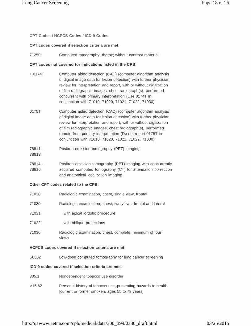
CPT Codes / HCPCS Codes / ICD-9 Codes
CPT codes covered if selection criteria are met:
71250 Computed tomography, thorax; without contrast material
CPT codes not covered for indications listed in the CPB:
+ 0174T Computer aided detection (CAD) (computer algorithm analysis of digital image data for lesion detection) with further physician review for interpretation and report, with or without digitization of film radiographic images, chest radiograph(s), performed concurrent with primary interpretation (Use 0174T in conjunction with 71010, 71020, 71021, 71022, 71030)
0175T Computer aided detection (CAD) (computer algorithm analysis of digital image data for lesion detection) with further physician review for interpretation and report, with or without digitization of film radiographic images, chest radiograph(s), performed remote from primary interpretation (Do not report 0175T in conjunction with 71010, 71020, 71021, 71022, 71030)
78811 - 78813
Positron emission tomography (PET) imaging
78814 - 78816
Positron emission tomography (PET) imaging with concurrently acquired computed tomography (CT) for attenuation correction and anatomical localization imaging
Other CPT codes related to the CPB:
71010 Radiologic examination, chest, single view, frontal
71020 Radiologic examination, chest, two views, frontal and lateral
71021 with apical lordotic procedure
71022 with oblique projections
71030 Radiologic examination, chest, complete, minimum of four
views
HCPCS codes covered if selection criteria are met:
S8032 Low-dose computed tomography for lung cancer screening
ICD-9 codes covered if selection criteria are met:
305.1 Nondependent tobacco use disorder
V15.82 Personal history of tobacco use, presenting hazards to health [current or former smokers ages 55 to 79 years]
Lung Cancer Screening Page 19 of 25
http://qawww.aetna.com/cpb/medical/data/300_399/0380_draft.html 03/25/2015
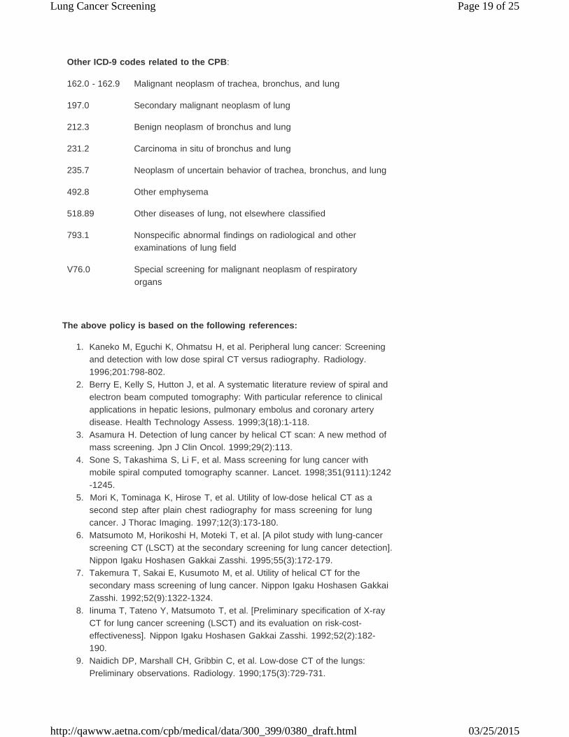
Other ICD-9 codes related to the CPB:
162.0 - 162.9 Malignant neoplasm of trachea, bronchus, and lung
197.0 Secondary malignant neoplasm of lung
212.3 Benign neoplasm of bronchus and lung
231.2 Carcinoma in situ of bronchus and lung
235.7 Neoplasm of uncertain behavior of trachea, bronchus, and lung
492.8 Other emphysema
518.89 Other diseases of lung, not elsewhere classified
793.1 Nonspecific abnormal findings on radiological and other examinations of lung field
V76.0 Special screening for malignant neoplasm of respiratory
organs
The above policy is based on the following references:
1. Kaneko M, Eguchi K, Ohmatsu H, et al. Peripheral lung cancer: Screening and detection with low dose spiral CT versus radiography. Radiology. 1996;201:798-802.
2. Berry E, Kelly S, Hutton J, et al. A systematic literature review of spiral and electron beam computed tomography: With particular reference to clinical applications in hepatic lesions, pulmonary embolus and coronary artery disease. Health Technology Assess. 1999;3(18):1-118.
3. Asamura H. Detection of lung cancer by helical CT scan: A new method of mass screening. Jpn J Clin Oncol. 1999;29(2):113.
4. Sone S, Takashima S, Li F, et al. Mass screening for lung cancer with mobile spiral computed tomography scanner. Lancet. 1998;351(9111):1242 -1245.
5. Mori K, Tominaga K, Hirose T, et al. Utility of low-dose helical CT as a second step after plain chest radiography for mass screening for lung cancer. J Thorac Imaging. 1997;12(3):173-180.
6. Matsumoto M, Horikoshi H, Moteki T, et al. [A pilot study with lung-cancer screening CT (LSCT) at the secondary screening for lung cancer detection]. Nippon Igaku Hoshasen Gakkai Zasshi. 1995;55(3):172-179.
7. Takemura T, Sakai E, Kusumoto M, et al. Utility of helical CT for the secondary mass screening of lung cancer. Nippon Igaku Hoshasen Gakkai Zasshi. 1992;52(9):1322-1324.
8. Iinuma T, Tateno Y, Matsumoto T, et al. [Preliminary specification of X-ray CT for lung cancer screening (LSCT) and its evaluation on risk-cost- effectiveness]. Nippon Igaku Hoshasen Gakkai Zasshi. 1992;52(2):182- 190.
9. Naidich DP, Marshall CH, Gribbin C, et al. Low-dose CT of the lungs: Preliminary observations. Radiology. 1990;175(3):729-731.
Lung Cancer Screening Page 20 of 25
http://qawww.aetna.com/cpb/medical/data/300_399/0380_draft.html 03/25/2015
10.
11.
12.
13.
14.
15.
16.
17.
18.
19.
20.
21.
22.
23.
24.
25.
van Klaveren RJ, Habbema JDF, Pedersen JH, et al. Lung cancer screening by low-dose spiral computed tomography. Eur Respir J. 2001;18 (5):857-866. Jain P, Arroliga AC. Spiral CT for lung cancer screening: Is it ready for prime time? Cleve Clin J Med. 2001;68(1):74-81. Ellis JR, Gleeson FV. New concepts in lung cancer screening. Curr Opin Pulm Med. 2002;8(4):270-274. Manser RL, Irving LB, Stone C, et al. Screening for lung cancer. Cochrane Database Syst Rev. 2004;(1):CD001991. State of Minnesota Department of Health, Health Technology Advisory Committee. Helical computed tomography (CT) for lung cancer screening for asymptomatic patients. technology Assessment. St. Paul, MN: Health Technology Advisory Committee; 2000. Institute for Clinical Systems Improvement (ICSI). Computed tomography screening for lung cancer. ICSI Technology Assessment. Bloomington, MN: ICSI; 2001. Swedish Council on Technology Assessment in Health Care (SBU). Computed tomography in screening for lung cancer - early assessment briefs (ALERT). Stockholm, Sweden: SBU; 2002. Remy-Jardin M, Remy J, Wattinne L, Giraud F. Central pulmonary thromboembolism: Diagnosis with spiral volumetric CT with the single- breath-hold technique -- comparison with pulmonary angiography. Radiology. 1992;185(2):381-387. Bettmann MA, Boxt LM, Gomes AS, et al. Acute chest pain -- suspected pulmonary embolism. American College of Radiology. ACR Appropriateness Criteria. 2000;215(Suppl):15-21. American College of Emergency Physicians Clinical Policies Committee; Clinical Policies Committee Subcommittee on Suspected Pulmonary Embolism. Clinical policy: Critical issues in the evaluation and management of adult patients presenting with suspected pulmonary embolism. Ann Emerg Med. 2003;41(2):257-270. Henschke CI, Yankelevitz DF, McCauley DI, et al. Guidelines for the use of spiral computed tomography in screening for lung cancer. Eur Respir J Suppl. 2003;39:45s-51s. Mulshine JL. Screening for lung cancer: In pursuit of pre-metastatic disease. Nat Rev Cancer. 2003;3(1):65-73. Truong MT, Munden RF. Lung cancer screening. Curr Oncol Rep. 2003;5 (4):309-312. Manser RL, Irving LB, Byrnes G, et al. Screening for lung cancer: A systematic review and meta-analysis of controlled trials. Thorax. 2003;58 (9):784-789. Canadian Coordinating Office for Health Technology Assessment (CCOHTA). Multi-slice/helical computed tomography for lung cancer screening. Issues in Emerging Health Technologies. Issue 48. Ottawa, ON: CCOHTA; June 2003. U.S. Preventive Services Task Force. Lung cancer screening: Recommendation statement. Ann Intern Med. 2004;140(9):738- 739. Available at: http://www.preventiveservices.ahrq.gov/. Accessed June 9, 2005.
Lung Cancer Screening Page 21 of 25
http://qawww.aetna.com/cpb/medical/data/300_399/0380_draft.html 03/25/2015
26.
27.
28.
29.
30.
31.
32.
33.
34.
35.
36.
37.
38.
39.
40.
41.
Bepler G, Goodridge Carney D, et al. A systematic review and lessons learned from early lung cancer detection trials using low-dose computed tomography of the chest. Cancer Control. 2003;10(4):306-314. Humphrey LL, Johnson M, Teutsch S. Lung cancer screening: An update for the U.S. Preventive Services Task Force. Preventive Services Task Force Systematic Evidence Review No. 31. Rockville, MD: Agency for Healthcare Research and Quality (AHRQ); 2004. Palda VA, Van Spall HGC. Screening for lung cancer: Updated recommendations from the Canadian Task Force on Preventive Health Care. London, ON: Canadian Task Force on Preventive Health Care (CTFPHC); August 2003. Swedish Council on Technology Assessment in Health Care (SBU). Computed tomography in screening for lung cancer. Version 2.0 - early assessment briefs (Alert). Stockholm, Sweden: SBU; 2003. Armstrong P, Husband JE, Holemans JA. Population screening for lung cancer. Hosp Med. 2004;65(7):404-411. Diederich S, Wormanns D. Impact of low-dose CT on lung cancer screening. Lung Cancer. 2004;45 Suppl 2:S13-S19. Manser RL, Irving LB, de Campo MP, et al. Overview of observational studies of low-dose helical computed tomography screening for lung cancer. Respirology. 2005;10(1):97-104. Gohagan JK, Marcus PM, Fagerstrom RM, et al. Final results of the Lung Screening Study, a randomized feasibility study of spiral CT versus chest X -ray screening for lung cancer. Lung Cancer. 2005;47(1):9-15. Jett JR. Limitations of screening for lung cancer with low-dose spiral computed tomography. Clin Cancer Res. 2005;11(13 Pt 2):4988s-4992s. Wardwell NR, Massion PP. Novel strategies for the early detection and prevention of lung cancer. Semin Oncol. 2005;32(3):259-268. Novello S, Fava C, Borasio P, et al. Three-year findings of an early lung cancer detection feasibility study with low-dose spiral computed tomography in heavy smokers. Ann Oncol. 2005;16(10):1662-1666. Black C, Bagust A, Boland A., et al. The clinical effectiveness and cost- effectiveness of computed tomography screening for lung cancer: Systematic reviews. Health Technol Assess. 2006;10(3):1–106. Foerster V, Murtagh J, Lentle BC, et al. CT and MRI for selected clinical disorders: A systematic review of clinical systematic reviews. Technology Report Issue 59. Ottawa, ON: Canadian Agency for Drugs and Technologies in Health (CADTH); 2005. Murtagh J, Warburton R N, Foerster V, et al. CT and MRI for selected clinical disorders: A systematic review of economic evaluations. Technology Report Issue 68. Ottawa, ON: Canadian Agency for Drugs and Technologies in Health (CADTH); 2006. International Early Lung Cancer Action Program Investigators; Henschke CI, Yankelevitz DF, Libby DM, et al. Survival of patients with stage I lung cancer detected on CT screening. N Engl J Med. 2006;355(17):1763-1771. Unger M. A pause, progress, and reassessment in lung cancer screening. N Engl J Med. 2006;355(17):1822-1824. No authors listed. Expert panel does not yet endorse CT screening for lung cancer. Health Techology Trends. 2007;19(2):4-5.
Lung Cancer Screening Page 22 of 25
http://qawww.aetna.com/cpb/medical/data/300_399/0380_draft.html 03/25/2015
42.
43.
44.
45.
46.
47.
48.
49.
50.
51.
52.
53.
54.
55.
56.
57.
58.
Bach PB, Jett JR, Pastorino U, et al. Computed tomography screening and lung cancer outcomes. JAMA. 2007;297(9):953-961. Black C, de Verteuil R, Walker S, et al. Population screening for lung cancer using computed tomography, is there evidence of clinical effectiveness? A systematic review of the literature. Thorax. 2007;62(2):131 -138. Black WC, Baron JA. CT screening for lung cancer: Spiraling into confusion? JAMA. 2007;297(9):995-997. California Technology Assessment Forum (CTAF). Low dose spiral computerized tomography for lung cancer screening. A Technology Assessment. San Francisco, CA: CTAF; February 28, 2007. Available at: http://ctaf.org/content/general/detail/687. Accessed May 24, 2007. Alberts WM. Diagnosis and management of lung cancer executive summary. ACCP evidence-based clinical practice guidelines (2nd Edition). Chest. 2007;132(3 Suppl):1S-19S. Available at: http://www.chestjournal.org/cgi/content/full/132/3_suppl/1S. Accessed April 29, 2008. Yau G, Lock M, Rodrigues G. Systematic review of baseline low-dose CT lung cancer screening. Lung Cancer. 2007;58(2):161-170. Infante M, Lutman FR, Cavuto S, et al; DANTE Study Group. Lung cancer screening with spiral CT: Baseline results of the randomized DANTE trial. Lung Cancer. 2008;59(3):355-363. Smith JJ, Berg CD. Lung cancer screening: Promise and pitfalls. Semin Oncol Nurs. 2008;24(1):9-15. Infante M, Cavuto S, Lutman FR, et al; DANTE Study Group. A randomized study of lung cancer screening with spiral computed tomography: Three- year results from the DANTE trial. Am J Respir Crit Care Med. 2009;180 (5):445-453. Pastorino U. Lung cancer screening. Br J Cancer. 2010;102(12):1681- 1686. Ellery B, Mundy L, Hiller J. Screening for lung cancer utilising computed tomography (CT). Health Technology Prioritising Summary Update. Canberra, ACT: Adelaide Health Technology Assessment; March 2010. National Lung Screening Trial Research Team, Aberle DR, Berg CD, Black WC, et al. The National Lung Screening Trial: Overview and study design. Radiology. 2011;258(1):243-253. Sox HC. Better evidence about screening for lung cancer. N Engl J Med. 2011;365(5):455-457. National Lung Screening Trial Research Team, Aberle DR, Adams AM, Berg CD, et al. Reduced lung-cancer mortality with low-dose computed tomographic screening. N Engl J Med. 2011;365(5):395-409. Jett JR, Midthun DE. Screening for lung cancer: For patients at increased risk for lung cancer, it works. Ann Intern Med. 2011;155(8):540-542. Silvestri G. Screening for lung cancer: It works, but does it really work? Ann Intern Med. 2011;155(8):537-539. Field JK, Baldwin D, Brain K, et al; UKLS Team. CT screening for lung cancer in the UK: Position statement by UKLS investigators following the NLST report. Thorax. 2011;66(8):736-737.
Lung Cancer Screening Page 23 of 25
http://qawww.aetna.com/cpb/medical/data/300_399/0380_draft.html 03/25/2015
59.
60.
61.
62.
63.
64.
65.
66.
67.
68.
69.
Saghir Z, Dirksen A, Ashraf H, et al. CT screening for lung cancer brings forward early disease. The randomised Danish Lung Cancer Screening Trial: status after five annual screening rounds with low-dose CT. Thorax. 2012;67(4):296-301. Bach PB, Mirkin JN, Oliver TK, et al. Benefits and harms of CT screening for lung cancer: A systematic review. JAMA. 2012;307(22):2418-2429. Goulart BH, Bensink ME, Mummy DG, Ramsey SD. Lung cancer screening with low-dose computed tomography: Costs, national expenditures, and cost-effectiveness. J Natl Compr Canc Netw. 2012;10(2):267-275. Pinsky PF, Berg CD. Applying the National Lung Screening Trial eligibility criteria to the US population: What percent of the population and of incident lung cancers would be covered? J Med Screen. 2012;19(3):154-156. Raji OY, Duffy SW, Agbaje OF, et al. Predictive accuracy of the Liverpool Lung Project risk model for stratifying patients for computed tomography screening for lung cancer: A case-control and cohort validation study. Ann Intern Med. 2012;157(4):242-250. Pastorino U, Rossi M, Rosato V, et al. Annual or biennial CT screening versus observation in heavy smokers: 5-year results of the MILD trial. Eur J Cancer Prev. 2012;21(3):308-315. Aberle DR, Abtin F, Brown K. Computed tomography screening for lung cancer: Has it finally arrived? Implications of the national lung screening trial. J Clin Oncol. 2013;31(8):1002-1008. Ma J, Ward EM, Smith R, Jemal A. Annual number of lung cancer deaths potentially avertable by screening in the United States. Cancer. 2013;119 (7):1381-1385. Wender R, Fontham ET, Barrera E Jr, et al. American Cancer Society lung cancer screening guidelines. CA Cancer J Clin. 2013;63(2):106-117. Detterbeck FC, Mazzone PJ, Naidich DP, Bach PB. Screening for lung cancer: Diagnosis and management of lung cancer, 3rd ed: American College of Chest Physicians evidence-based clinical practice guidelines. Chest. 2013;143(5 Suppl):e78S-e92S. Moyer VA; U.S. Preventive Services Task Force. Screening for lung cancer: U.S. Preventive Services Task Force recommendation statement. Ann Intern Med. 2014;160(5):330-338.
Computer-Aided Detection for Chest Radiographs:
1. Coppini G, Diciotti S, Falchini M, Neural networks for computer-aided
diagnosis: Detection of lung nodules in chest radiograms. IEEE Trans Inf Technol Biomed. 2003;7(4):344-357.
2. Shiraishi J, Abe H, Engelmann R, Doi K. Effect of high sensitivity in a computerized scheme for detecting extremely subtle solitary pulmonary nodules in chest radiographs: Observer performance study. Acad Radiol. 2003;10(11):1302-1311.
3. Kakeda S, Moriya J, Sato H, et al. Improved detection of lung nodules on chest radiographs using a commercial computer-aided diagnosis system. AJR Am J Roentgenol. 2004;182(2):505-510.
4. Freedman M. Improved small volume lung cancer detection with computer- aided detection: Database characteristics and imaging of response to breast cancer risk reduction strategies. Ann N Y Acad Sci. 2004;1020:175- 189.
Lung Cancer Screening Page 24 of 25
http://qawww.aetna.com/cpb/medical/data/300_399/0380_draft.html 03/25/2015
5.
6.
7.
8.
9.
10.
11.
12.
13.
14.
15.
16.
Abe H, Macmahon H, Shiraishi J, et al. Computer-aided diagnosis in chest radiology. Semin Ultrasound CT MR. 2004;25(5):432-437. Suzuki K, Shiraishi J, Abe H, et al. False-positive reduction in computer- aided diagnostic scheme for detecting nodules in chest radiographs by means of massive training artificial neural network. Acad Radiol. 2005;12 (2):191-201. Doi K. Current status and future potential of computer-aided diagnosis in medical imaging. Br J Radiol. 2005;78 Spec No 1:S3-S19. Shiraishi J, Li F, Doi K. et al. Computer-aided diagnosis for improved detection of lung nodules by use of posterior-anterior and lateral chest radiographs. Acad Radiol. 2007;14(1):28-37. Li F, Engelmann R, Metz CE, et al. Lung cancers missed on chest radiographs: Results obtained with a commercial computer-aided detection program. Radiology. 2008;246(1):273-280. White CS, Flukinger T, Jeudy J, Chen JJ. Use of a computer-aided detection system to detect missed lung cancer at chest radiography. Radiology. 2009;252(1):273-281. Yanagawa M, Honda O, Yoshida S, et al. Commercially available computer -aided detection system for pulmonary nodules on thin-section images using 64 detectors-row CT: Preliminary study of 48 cases. Acad Radiol. 2009;16(8):924-933. De Boo DW, Prokop M, Uffmann M, et al. Computer-aided detection (CAD) of lung nodules and small tumours on chest radiographs. Eur J Radiol. 2009;72(2):218-225. Way T, Chan HP, Hadjiiski L, et al. Computer-aided diagnosis of lung nodules on CT scans: ROC study of its effect on radiologists' performance. Acad Radiol. 2010;17(3):323-332. de Hoop B, De Boo DW, Gietema HA, et al. Computer-aided detection of lung cancer on chest radiographs: Effect on observer performance. Radiology. 2010;257(2):532-540. Mohammed TH, Chowdhry A, Reddy GP, etal; Expert Panel on Thoracic Imaging. ACR Appropriateness Criteria® screening for pulmonary metastases. [online publication]. Reston, VA: American College of Radiology (ACR); 2010. Mazzone PJ, Obuchowski N, Phillips M, et al. Lung cancer screening with computer aided detection chest radiography: Design and results of a randomized, controlled trial. PLoS One. 2013;8(3):e59650.
Positron Emission Tomography (PET):
1. Chien CR, Liang JA, Chen JH, et al. [(18)F]Fluorodeoxyglucose-positron
emission tomography screening for lung cancer: A systematic review and meta-analysis. Cancer Imaging. 2013;13(4):458-465.
2. Manser R, Lethaby A, Irving LB, et al. Screening for lung cancer. Cochrane Database Syst Rev. 2013;6:CD001991.
3. National Comprehensive Cancer Network. Clinical practice guideline: Non- small cell lung cancer. Version 3.2014. NCCN: Fort Washington, PA.
Lung Cancer Screening Page 25 of 25
http://qawww.aetna.com/cpb/medical/data/300_399/0380_draft.html 03/25/2015
Copyright Aetna Inc. All rights reserved. Clinical Policy Bulletins are developed by Aetna to assist in
administering plan benefits and constitute neither offers of coverage nor medical advice. This Clinical Policy
Bulletin contains only a partial, general description of plan or program benefits and does not constitute a
contract. Aetna does not provide health care services and, therefore, cannot guarantee any results or
outcomes. Participating providers are independent contractors in private practice and are neither
employees nor agents of Aetna or its affiliates. Treating providers are solely responsible for medical advice
and treatment of members. This Clinical Policy Bulletin may be updated and therefore is subject to change.
CPT only copyright 2008 American Medical Association. All Rights Reserved.


![Effects of Curative-Intent Lung Cancer Therapy on ... · I-IIIA disease [1] and eligible to undergo curative-intent therapy through a combination of lung cancer resection surgery](https://img.pdfslide.net/doc/110x75/5f1063207e708231d448dbed/effects-of-curative-intent-lung-cancer-therapy-on-i-iiia-disease-1-and-eligible.jpg)