Embed Size (px)
Citation preview
Single Photon Emission Computed Tomography (SPECT) Page 1 of 29
05/21/2015
Clinical Policy Bulletin: Single Photon Emission Computed Tomography (SPECT)
Number: 0376
Policy
I. Non-Cardiac Indications: Aetna considers single photon emission computed tomography (SPECT) medically necessary for any of the following indications:
A. Assessment of osteomyelitis, to distinguish bone from soft tissue
infection; or B. Detection of spondylolysis and stress fractures not visible from x-ray;
or C. Diagnosing and assessing hemangiomas of the liver; or D. Diagnosing pulmonary embolism (by means of SPECT
ventilation/perfusion scintigraphy); or E. Differentiation of necrotic tissue from tumor of the brain; or F. Distinguishing Parkinson's disease from essential tremor; or G. Imaging of parathyroids in parathyroid disease; or H. Localization of abscess, for suspected or known localized infection
or inflammatory process; or I. Lymphoma, to distinguish tumor from necrosis; or J. Neuroendocrine tumors, diagnosis and staging; or K. Pre-surgical ictal detection of seizure focus in members with
epilepsy (in place of positron emission tomography (PET)).
II. Aetna considers SPECT experimental and investigational for all other non- cardiac indications, including any of the following, because its diagnostic value has not been established in the peer-reviewed medical literature in these situations:
Single Photon Emission Computed Tomography (SPECT) Page 2 of 29
05/21/2015
A. Diagnosis or assessment of members with attention deficit/hyperactivity disorder (CPB 0426 - Attention Deficit/Hyperactivity Disorder); or
B. Diagnosis or assessment of members with autism (CPB 0648 - Pervasive Developmental Disorders); or
C. Diagnosis or assessment of members with personality disorders (e.g., aggressive and violent behaviors, anti-social personality disorder including psychopathy, schizotypal personality disorder, as well as borderline personality disorder); or
D. Diagnosis or assessment of members with schizophrenia; or E. Diagnosis or assessment of stroke members; or F. Differential diagnosis of Parkinson's disease from other Parkinsonian
syndromes; or G. Evaluation of members with endoleak; or H. Evaluation of members with generalized pain or insomnia; or I. Evaluation of members with head trauma; or J. Initial or differential diagnosis of members with suspected dementia
(e.g., Alzheimer's disease, dementia with Lewy bodies, frontotemporal dementia, and vascular dementia); or
K. Multiple sclerosis; or L. Pre-surgical evaluation of members undergoing lung volume
reduction surgery; or M. Prosthetic graft infection; or N. Scanning of internal carotid artery during temporary balloon
occlusion; or O. Vasculitis.
III. Cardiac Indications: Aetna considers SPECT medically necessary for the following indications for members who do not meet any of the exclusion criteria below:
A. Assessing myocardial viability before referral for myocardial
revascularization procedures; or B. Diagnosis of coronary artery disease (CAD) in members with an
uninterpretable resting electrocardiogram (ECG) and restricted exercise tolerance.
IV. Aetna considers SPECT experimental and investigational for all other
cardiac indications because its effectiveness for indications other than the ones listed above has not been established.
Exclusion criteria: Aetna considers SPECT myocardial perfusion imaging experimental and investigational for the following indications for which the study is considered “inappropriate” according to appropriateness criteria from the American College of Cardiology (ACC) (Brindis et al, 2005):
A. As a routine screening evaluation after a percutaneous transluminal
coronary angioplasty (PTCA) with or without stenting or coronary
Single Photon Emission Computed Tomography (SPECT) Page 3 of 29
05/21/2015
artery bypass surgery (CABG) prior to discharge from the acute care setting; or
B. As a routine screening evaluation after a re-vascularization procedure (PTCA with stenting or CABG) at an interval of less than 2 years from the procedure if there is no worsening in the members symptomatology and if the member had symptoms prior to the intervention, and there is no history of congestive heart failure. Note: If there is a history of congestive heart failure and the member is status post re-vascularization, repeat nuclear imaging as frequently as annually may be medically necessary; or
C. Assessment of vulnerable plaque; or D. Evaluation of a member with an acute coronary event and
hemodynamic instability, shock, or mechanical complications of the event; or
E. In the setting of acute chest pain or equivalent symptoms with a high likelihood of being acute coronary syndrome, when there has been a diagnosis of acute myocardial infarction, in the immediate post- thombolytic period, or when there is a high pre-test likelihood of significant coronary disease as demonstrated by marked ST segment elevation on the ECG; or
F. Prior to high-risk+ surgery when the member is asymptomatic and there was a normal cardiac catheterization, coronary intervention (PTCA, stenting, CABG), or normal nuclear stress test less than 1 year before the surgical date; or
G. Prior to intermediate-risk+ non-cardiac surgery if the member is capable of, and has no contraindication to standard stress testing**; or
H. Prior to low-risk+ non-cardiac surgery for risk assessment; or I. Re-evaluation of members without chest pain or equivalent
symptoms, without known coronary disease, at high-risk for coronary disease (based upon the Framingham score greater than 10)*, who have an initial negative radionuclear imaging study, when it has been less than 2 years since the last radionuclear study; or
J. Re-evaluation of members without chest pain or equivalent symptoms or with stable symptoms, with known coronary disease as determined by prior abnormal catheterization or SPECT cardiac study (but without prior infarction), when it has been less than 1 year since the last radionuclear study. Note: if the member has worsening symptoms or if the member had silent ischemia, more frequent imaging or other diagnostic testing or interventions may be medically necessary; or
K. Screening of members with chest pain or chest pain equivalent symptoms when there is a low probability of coronary disease (Framingham score less than 10)*, no history of diabetes, and there are no impediments or contraindications to non-nuclear stress testing**; or
L. Screening of members without chest pain or equivalent symptoms when there is a low probability of coronary disease (Framingham score less than 10)* and no history of diabetes; or
Single Photon Emission Computed Tomography (SPECT) Page 4 of 29
05/21/2015
* A tool for calculating Framingham score is available from the National Heart Lung and Blood Institute at the following website: http://hp2010.nhlbihin.net/atpIII/calculator.asp?usertype=prof.
** According to ACC guidelines, the following are impediments or contraindications to non-nuclear stress testing:
A. A history of physical impairments that would preclude physically
performing the exercise portion of a stress test; or B. A history of prior proven ischemic cardiac events; or C. Proven CAD by past SPECT or coronary catheterization; or D. The member's ECG would prevent interpretation of a standard
stress testing by being “uninterpretable” during the test, i.e., left bundle branch block (LBBB), paced rhythm, Wolf Parkinson White syndrome, left ventricular hypertrophy (LVH) with ST segment depression, or digoxin use.
+ Surgical risk categories.
High-risk surgery (risk of cardiac death or myocardial infarction (MI) greater than 5 %): emergent major operations (particularly in the elderly), aortic and peripheral vascular surgery, prolonged surgical procedures associated with large fluid shifts and/or blood loss. Intermediate-risk surgery (risk of cardiac death or MI 1 % to 5 %): carotid endarterectomy, head and neck surgery, surgery of the chest or abdomen, orthopedic surgery, prostate surgery. Low-risk surgery (cardiac death or MI less than 1 %): endoscopic procedures, superficial procedures, cataract surgery, breast surgery.
Source: ACC (2005).
V. Aetna considers myocardial sympathetic innervation imaging, with or
without SPECT, experimental and investigational because its effectiveness has not been established.
VI. Aetna considers SPECT-CT fusion medically necessary for parathyroid
imaging in persons with an enlarged parathyroid gland, parathyroid hyperplasia or suspected parathyroid adenoma or carcinoma, and laboratory evidence of hyperparathyroidism (parathyroid hormone greater than 55 pg/ml and serum calcium greater than 10.2 mg/dL).
Aetna considers SPECT-CT fusion imaging as experimental and investigational for other indications because of insufficient evidence of its effectiveness.
VII. Aetna considers freehand SPECT/ultrasonography (US) fusion imaging in
persons with thyroid disease experimental and investigational because of insufficient evidence of its effectiveness.
Background
Single Photon Emission Computed Tomography (SPECT) Page 5 of 29
05/21/2015
Single photon emission computed tomography (SPECT) is a nuclear medicine technique that uses radiopharmaceuticals, a rotating camera (single or multiple- head), and a computer to produce images representing slices through the body in different planes. Single photon emission computed tomography images are functional in nature rather than being purely anatomical such as ultrasound, computed tomography (CT), and magnetic resonance imaging (MRI).
SPECT for Myocardial Perfusion Imaging
Single photon emission computed tomography has been applied to the heart for myocardial perfusion imaging. It is an effective non-invasive diagnostic technology when evaluating patients for clinically significant coronary artery disease (CAD) in the following circumstances: for diagnosing CAD in patients with an abnormal resting electrocardiogram (ECG) and restricted exercise tolerance; or assessing myocardial viability before referral for myocardial re-vascularization.
Single photon emission computed tomography has a far greater sensitivity for detecting silent ischemia than stress ECG. Pooled data of exercise SPECT studies in over 1,000 patients revealed a 90 % overall sensitivity for detecting CAD. In assessing myocardial viability, studies indicate that, even in patients with severe irreversible thallium defects on standard exercise-redistribution imaging, thallium re-injection provides information regarding myocardial viability that is comparable to that provided by positron emission tomography (PET).
SPECT for Detection of Neurological Diseases
Single photon emission computed tomography examines cerebral function by documenting regional blood flow and metabolism; SPECT of the brain is a useful alternative to PET for the pre-surgical ictal detection of seizure focus. Effective surgical treatment of patients with intractable epilepsy is dependent on accurate localization of the epileptic focus and precise delineation of the epileptogenic region. It is generally agreed that this requires EEG recording with electrocorticography. This testing is designed to identify the source or sources of the seizures, as well as an assessment of brain function. The search for non- invasive and cost-effective means of seizure localization has been an important area of research.
Seizures are associated with dramatic increases in blood flow, localized in partial seizures and global during generalized seizures. Ictal SPECT uses the physiologic increase in regional cerebral blood blow during seizures to potentially localize the epileptogenic region. Ictal studies, although more difficult logistically, are reported to have a sensitivity of 81 to 93 %. (Interictal SPECT demonstrates hypo-perfusion and is reported to have a sensitivity ranging from 40 to 75 %).
Other imaging modalities help in localizing abnormal epileptogenic tissue. Quantitative MRI has a sensitivity of 80 to 90 % for the lateralization of temporal lobe epilepsy. However, there are instances where ictal SPECT has identified lesions not detected by MRI. Depth electrocorticography and intra-operative electrocorticography, though accurate in many forms of epilepsy, are both highly invasive. In cortical developmental disorders, these EEG techniques often fail to
Single Photon Emission Computed Tomography (SPECT) Page 6 of 29
05/21/2015
localize the epileptogenic area. Single photon emission computed tomography imaging may offer a safe and accurate alternative.
Ictal SPECT testing requires that seizure activity be provoked through reduction of anti-epileptic medications. Video EEG monitoring may also be performed. Due to the nature of ictal SPECT, it should be performed in a hospital setting.
Studies of SPECT in the inter-ictal or post-ictal detection of seizure focus, determination of seizure subtypes and monitoring of drug therapy for epilepsy have not established its efficacy for these indications.
Clinical studies indicate that SPECT is more accurate at detecting acute ischemia than CT scan. By 8 hours after infarction, about 20 % of CT scans will be positive but nearly 90 % of SPECT scans will be. Beyond 72 hours, the sensitivities of CT and SPECT are nearly identical. In addition, the defects noted on SPECT are frequently larger than those noted on CT studies.
Among the treatments of acute ischemic stroke, the use of tissue plasminogen activator (t-PA) has been shown to reduce neurologic impairment if administered within 3 hours of onset. This is administered when CT is negative for hemorrhage. Single photon emission computed tomography can not detect hemorrhage in order to rule out use of thrombolysis. Therefore, while SPECT is more sensitive than CT in detecting ischemia, it has not been shown to be useful in detecting ischemia early enough to play a role in the use of thrombolysis. Studies using SPECT in the presence of cerebro-vascular accident (CVA) have timed its performance at less than 6 hours to less than 14 days from the time of onset.
Studies have also reported on SPECT's value with regard to the assessment of prognosis after stroke, determination of stroke type, monitoring therapies used post-stroke, diagnosis of vasospasm following subarachnoid hemorrhage and in the diagnosis/prognosis regarding transient ischemic attacks (TIAs). Single photon emission computed tomography may prove to be helpful in these applications in the future.
Single photon emission computed tomography has proven useful in distinguishing recurrent brain tumor from radiation necrosis. Radiation necrosis needs to be distinguished from recurrent brain tumor to determine whether chemotherapy is necessary. Radiation necrosis is more common in brain tumor patients receiving local boost irradiation. Radiation necrosis and brain tumor may have similar clinical signs and symptoms, and are not reliably distinguished clinically.
Single photon emission computed tomography has been studied in the diagnosis of patients with Alzheimer's disease AD). At present, the definitive diagnosis of AD can only be made on pathologic examination of brain tissue. Alzheimer's disease is largely a diagnosis of exclusion. The diagnostic evaluation of the demented patient is therefore aimed largely at identifying other potentially treatable causes of cognitive impairment. A technology assessment of SPECT for dementia and AD prepared by the Institute for Clinical Effectiveness and Health Policy (Ferrante, 2004) concluded: "SPECT has not clearly demonstrated its usefulness in assessing patients with dementia, and it has no precise indications for diagnosis, evaluation of prognosis or monitoring response to treatment."
Single Photon Emission Computed Tomography (SPECT) Page 7 of 29
05/21/2015
In AD, SPECT shows decreased perfusion in the association cortex of the parietal lobe and the posterior temporal regions. Frontal association cortex is predominantly affected in some cases, but usually is not involved until late in the course of the disease. The occipital lobes are less involved and the paracentral cortex is spared. Although SPECT studies have been reported in over 500 patients with AD, in most cases the diagnosis was made clinically and only in 53 patients it was confirmed by autopsy. Controlled studies of SPECT in AD have shown a sensitivity of this procedure to vary from 50 % to 95 %. The best results have been reported in the more recent studies, using higher resolution equipment. In the 2001 practice parameter on the diagnosis of dementia, the American Academy of Neurology does not recommend SPECT for routine use in either initial or differential diagnosis of dementia (Knopman et al, 2001).
Neuroimaging markers are increasingly important in the early diagnosis of the dementias, not only to detect the rare tumor, but also to differentiate the various types of neurodegenerative dementias. The potential of MRI and PET as surrogates for disease progression for therapeutic trials is being examined in a large multi-center trial. However, SPECT has been reported to show specific findings in both fronto-temporal dementia (FTD) and AD and is much more available and less expensive than PET. McNeil et al (2007) evaluated the diagnostic accuracy of technetium-99–labeled hexamethyl propylene amine oxime SPECT images obtained at initial evaluation in 25 pathologically confirmed cases of FTD and 31 pathologically confirmed cases of AD. A nuclear medicine physician, blinded to the clinical findings, rated the images. Except in the case of 1 FTD patient, for whom neuropsychological data were unavailable, the clinical and neuropsychological evaluation was more accurate than SPECT alone in predicting the histological diagnosis.
In a longitudinal, prospective study, Devanand and colleagues (2010) examined the utility of SPECT to predict conversion from mild cognitive impairment (MCI) to AD. A total of 127 patients with MCI and 59 healthy comparison subjects followed- up for 1 to 9 years were included in this study. Diagnostic evaluation, neuropsychological tests, social/cognitive function, olfactory identification, apolipoprotein E genotype, MRI, and brain Tc hexamethyl-propylene-aminoxime SPECT scan with visual ratings, and region of interest (ROI) analyses were done. Visual ratings of SPECT temporal and parietal blood flow did not distinguish eventual MCI converters to AD (n = 31) from non-converters (n = 96), but the global rating predicted conversion (41.9 % sensitivity and 82.3 % specificity, Fisher's exact test p = 0.013). Blood flow in each ROI was not predictive, but when dichotomized at the median value of the patients with MCI, low flow increased the hazard of conversion to AD for parietal (hazard ratio [HR]: 2.96, 95 % confidence interval [CI]: 1.16 to 7.53, p = 0.023) and medial temporal regions (HR: 3.12, 95 % CI: 1.14 to 8.56, p = 0.027). In the 3-year follow-up sample, low parietal (p < 0.05) and medial temporal (p < 0.01) flow predicted conversion to AD, with or without controlling for age, Mini-Mental State Examination, and apolipoprotein E ε4 genotype. These measures lost significance when other strong predictors were included in logistic regression analyses: verbal memory, social/cognitive functioning, olfactory identification deficits, hippocampal, and entorhinal cortex volumes. The authors concluded that SPECT visual ratings showed limited utility in predicting MCI conversion to AD. The modest predictive
Single Photon Emission Computed Tomography (SPECT) Page 8 of 29
05/21/2015
utility of quantified low parietal and medial temporal flow using SPECT may decrease when other stronger predictors are available.
Single photon emission computed tomography has also been reported to be used in neoplasms (grading of gliomas), HIV encephalopathy, head trauma, Huntington's chorea, persistent vegetative states and in diagnosing brain death. Based upon the current evidence, SPECT has not proven to be established in any of these clinical situations. More study is needed in these areas.
The Food and Drug Administration (FDA) has approved 4 radiopharmaceuticals for imaging of the brain: I-123 isopropyliodoamphetamine (IMP, Spectamine); Tc- 99m HMPAO (hexamethyl propylamine oxime, Ceretec); Tc-99m ECD (ethyl cysteinate dimer, Neurolite), and thallium 201 diethyldithiocarbamate (T1-DDC).
SPECT for the Liver
Hemangiomas are the most common benign lesion of the liver. It is important to differentiate these lesions from others that may be malignant. Because of the risk of hemorrhage inherent in a percutaneous biopsy of liver hemangiomas, non- invasive modalities have been used for differentiation. Hepatic hemangiomas are so vascular that their blood pool is easily differentiated from other solid hepatic masses by nuclear scanning with labeled red blood cells (RBCs). The labeled RBCs are injected intravenously and resolution is markedly improved with SPECT. Review articles and published studies support SPECT as an appropriate diagnostic modality to differentiate hepatic lesions as hemangiomas.
SPECT for Neuroendocrine Tumors
Carcinoids and other neuroendocrine tumors have somatostatin receptors; therefore, they can be imaged with somatostatin analogs (octreotide, pentetreotide) tagged with an appropriate radioisotope (Khan and Jones, 2005). Single photon emission computed tomography and subtraction techniques improve detection. An assessment from the Australian Medical Services Advisory Committee stated that SPECT is often performed in conjunction with antibody imaging (Octreoscan) of neuroendocrine tumors, usually at 24 and occasionally at 48 hours after the injection. The assessment explained that SPECT is able to differentiate more easily between areas of pathological uptake and physiological uptake in the abdomen. The assessment explained that SPECT can also help to discriminate between mesenteric and bone lesions. The assessment noted that extra-planar images may be obtained from areas of specific interest, using longer exposure time for more easily interpreted imaging.
SPECT for Spondylolysis and Stress Fractures
The role of SPECT of the spine has changed in recent years with the wide availability of MRI and especially contrast-enhanced MRI. Bone scanning with SPECT of the spine may allow for visualization of lesions related to stress fractures or stress reactions in the spine such as spondylolysis. Single photon emission computed tomography has been accepted as extremely sensitive in detecting incipient spondylolysis and stress/insufficiency fractures (Manaster et al, 2005). Single photon emission computed tomography shows stress fractures days to weeks earlier than radiographs in many instances, and differentiate between
Single Photon Emission Computed Tomography (SPECT) Page 9 of 29
05/21/2015
osseous and soft tissue injury as well. However, in most cases bone scans lack specificity and supplemental imaging may be necessary for conclusive diagnosis or to avoid false positives. Single photon emission computed tomography continues to be used in back pain, especially in children and adolescents, to detect incipient spondylolysis that may not be detected by conventional imaging. Because of the sensitivity of SPECT scans, 80 % of all fractures show an abnormality 24 hours post injury and 95 % at 72 hours. A normal scan generally excludes the diagnosis of stress/insufficiency fracture, and the patient may return to normal activity.
SPECT for the Internal Carotid Artery
Internal carotid artery temporary balloon occlusion (TBO) test is a well-established part of the preoperative evaluation of patients with aneurysm or tumor involving the neck or skull base in whom arterial sacrifice or prolonged temporary occlusion is considered a possible part of the surgical or endovascular therapy. Temporary balloon occlusion is performed in conjunction with cerebral blood flow analysis to identify those patients who will not tolerate permanent carotid occlusion. Traditionally, xenon-enhanced CT has been used during TBO to detect signs of ischemia; however, SPECT has recently been reported as a method to detect focal hypo-perfusion during test occlusion. Several small studies in the published literature report preliminary results that SPECT scanning during TBO is a safe and effective method to assess cerebral blood flow. However, it is premature to recommend SPECT over the conventional xenon-enhanced CT.
SPECT for the Diagnosis/Assessment of Attention Deficit/Hyperactivity Disorder
Functional neuroimaging such as PET and SPECT has been used to study patients with attention deficit/hyperactivity disorder (ADHD). Although some studies have shown differences in brain structure or function comparing children with and without ADHD, these findings do not differentiate reliably between children with and without this disorder (i.e., although group means may differ significantly, the overlap in findings among children with and without ADHD results in high rates of false-positives and false-negatives). As a result, SPECT should not be used as a screening or diagnostic tool for children with ADHD. The American Academy of Pediatrics' Practice Guideline on “Diagnosis and Evaluation of the Child with Attention-Deficit/Hyperactivity Disorder” does not recommend neuroimaging studies in the diagnosis of ADHD. An evidence review by McGough and Barkley (2004) stated that there are insufficient scientific data to justify use of laboratory assessment measures, including neuropsychological tests and brain imaging, in diagnosing adult ADHD.
SPECT for the Diagnosis/Assessment of Autism
The American Academy of Neurology (2000) stated that there is no evidence to support a role of neuroimaging modalities such as SPECT in the clinical diagnosis of autism.
SPECT for the Diagnosis of Pulmonary Embolism
In a prospective, observational study, Miles and colleagues (2009) compared SPECT ventilation/perfusion (V/P) scintigraphy with multi-slice CT pulmonary
Single Photon Emission Computed Tomography (SPECT) Page 10 of 29
05/21/2015
angiography (CTPA) in the diagnosis of pulmonary embolism (PE). A total of 100 patients who were 50 years of age or older were recruited; 79 underwent both diagnostic 16-detector CTPA, and planar and SPECT V/P scintigraphy. The agreement between the CTPA and the SPECT V/P scintigraphy for the diagnosis of PE was calculated. The sensitivity and specificity of blinded SPECT scintigraphy reporting was calculated against a reference diagnosis made by a panel of respiratory physicians that was provided with CTPA and planar V/P scintigraphy reports, clinical information, and 3-month follow-up data. The observed percentage of agreement between SPECT V/P scintigraphy and CTPA data for the diagnosis of PE was 95 %. When calculated against the respiratory physicians' reference diagnosis, SPECT V/P scintigraphy had a sensitivity of 83 % and a specificity of 98 %. The authors concluded that these findings indicated that SPECT V/P scintigraphy is a viable alternative to CTPA for the diagnosis of PE and has potential advantages in that it was feasible in more patients and had fewer contraindications; lower radiation dose; and, arguably, fewer non-diagnostic findings than CTPA.
Gutte et al (2010) evaluated the diagnostic performance of 3-dimensional V/P SPECT in comparison with planar V/P scintigraphy. Consecutive patients suspected of acute PE were referred to a V/P SPECT, as the first-line imaging procedure. Patients with positive D-dimer (greater than 0.5 mg/L) or after clinical assessment with a Wells score of more than 2 were included and had a V/P SPECT, low-dose CT, planar V/P scintigraphy, and pulmonary multi-detector CTPA performed the same day. Ventilation studies were performed using Krypton (Kr). Patient follow-up was at least 6 months. A total of 36 patient studies were available for analysis, of which 11 (31 %) had PE. V/P SPECT had a sensitivity of 100 % and a specificity of 87 %. Planar V/P scintigraphy had a sensitivity of 64 % and a specificity of 72 %. The authors concluded that V/P SPECT has a superior diagnostic performance compared with planar V/P scintigraphy and should be preferred when diagnosing PE.
The European Association of Nuclear Medicine (EANM)'s practice guidelines on ventilation/perfusion scintigraphy (Bajc et al, 2009a) stated that PE can only be diagnosed with imaging techniques, which in practice is performed using V/P scintigraphy or multi-detector computed tomography of the pulmonary arteries (MDCT). The basic principle for the diagnosis of PE based upon V/P scintigraphy is to recognize lung segments or subsegments without perfusion but preserved ventilation, i.e., mismatch. Ventilation studies are in general performed after inhalation of Kr-labelled or technetium-labelled aerosol of diethylene triamine pentaacetic acid (DTPA) or Technegas. Perfusion studies are performed after intravenous injection of macro-aggregated human albumin. Radiation exposure using documented isotope doses is 1.2 to 2 mSv. Planar and tomographic techniques (Planar V/P and SPECT V/P) are analyzed. Single photon emission computed tomography V/P has higher sensitivity and specificity than Planar V/P. The interpretation of either Planar V/P or SPECT V/P should follow holistic principles rather than obsolete probabilistic rules. Pulmonary embolism should be reported when mis-match of more than 1 subsegment is found. For the diagnosis of chronic PE, V/P scintigraphy is of value. The additional diagnostic yield from V/P scintigraphy includes chronic obstructive lung disease (COPD), heart failure and pneumonia. Single photon emission computed tomography V/P is strongly
Single Photon Emission Computed Tomography (SPECT) Page 11 of 29
05/21/2015
preferred to Planar V/P as the former permits the accurate diagnosis of PE even in the presence of co-morbid diseases such as COPD and pneumonia.
The EANM's practice guidelines on VP scintigraphy also noted that to reduce the costs, the risks associated with false-negative and false-positive diagnoses, and unnecessary radiation exposure, pre-imaging assessment of clinical probability is recommended. Diagnostic accuracy is approximately equal for MDCT and Planar V/P and better for SPECT V/P, which is feasible in about 99 % of patients, while MDCT is often contraindicated. As MDCT is more readily available, access to both techniques is vital for the diagnosis of PE. Single photon emission computed tomography V/P gives an effective radiation dose of 1.2 to 2 mSv. For SPECT V/P, the effective dose is about 35 % to 40 % and the absorbed dose to the female breast 4 % of the dose from MDCT performed with a dose-saving protocol. Thus, SPECT V/P is recommended as a first-line procedure in patients with suspected PE. It is particularly favored in young patients, especially females, during pregnancy, and for follow-up and research (Bajc et al, 2009b).
Other Indications
Single photon emission computed tomography has proven useful in distinguishing lymphoma from necrosis in the chest and abdomen. It is also useful in localizing abscesses, and distinguishing abscess from other infectious or inflammatory processes. Single photon emission computed tomography is also useful in osteomyelitis in distinguishing inflammation of soft tissue from bone.
Guidelines on parathyroid scintigraphy from the Society of Nuclear Medicine (Greenspan et al, 2004) state that there is a developing consensus that SPECT imaging is useful, because, when used in conjunction with planar imaging, SPECT provides increased sensitivity and more precise anatomic localization. They note that this is particularly true in detecting both primary and recurrent hyperparathyroidism resulting from ectopic adenomas. In the mediastinum, accurate localization may assist in directing the surgical approach, such as median sternotomy versus left or right thoracotomy. The Parathyroid Task Group of the EANM (Hindie et al, 2009) stated that the use of SPECT/CT has a major role for obtaining anatomical details on ectopic foci. However, its use as a routine procedure before target surgery is still investigational. Preliminary data suggest that SPECT/CT has lower sensitivity in the neck area compared to pinhole imaging.
In a review on neuroimaging in psychopathy (including anti-social personality disorder and violent behavior), Pridmore et al (2005) noted that functional neuroimaging strongly suggests dysfunction of particular frontal and temporal lobe structures in subjects with psychopathy. However, there are difficulties in selecting homogeneous index cases and appropriate control groups. These investigators stated that further studies are needed.
An assessment of SPECT for schizophrenia by the Institute for Clinical Effectiveness and Health Policy (Pichon Riviere et al, 2004) found that SPECT has identified increases in dopaminergic activity in the striated body and other areas of the brain during the episodes of schizophrenia exacerbation. Single photon emission computed tomography has also been able to identify decreases in frontal cortex uptake that are associated with negative symptoms of schizophrenia. The
Single Photon Emission Computed Tomography (SPECT) Page 12 of 29
05/21/2015
investigators, however, were unable to find substantial evidence for the role of SPECT scans in therapeutic decision-making in schizophrenia. They concluded that the use of SPECT scan in schizophrenia remains investigational.
In the recent practice parameter on the diagnosis and prognosis of new onset Parkinson's disease (PD) (an evidence-based review) by the American Academy of Neurology (AAN), Suchowersky et al (2006) stated that SPECT scanning may not be useful in differentiating PD from other parkinsonian syndromes.
In a meta-analysis of the literature on diagnostic accuracy of SPECT in parkinsonian syndromes, Vlaar and colleagues (2007) concluded that SPECT with pre-synaptic radiotracers is relatively accurate to differentiate patients with PD in an early phase from normalcy, patients with PD from those with essential tremor, and PD from vascular parkinsonism. The accuracy of SPECT with both pre- synaptic and post-synaptic tracers to differentiate between PD and atypical parkinsonian syndrome is relatively low.
The American College of Radiology's "Appropriateness Criteria® dementia and movement disorders" (Wippold et al, 2010) stated that "[a] diminution of the width of the pars compacta on MRI has been described in PD patients compared to controls, with overlap between groups. This diminished width probably reflects selective neuronal loss of the pars compacta. Other authors have found a normal appearance of the substantia nigra on T2-weighted images in a majority of PD patients. More recently, PET and SPECT tracer studies exploring the presynaptic nigrostriatal terminal function and the postsynaptic dopamine receptors have attempted to classify the various Parkinson syndromes although much of this work remains investigational".
van der Vaart et al (2008) described the current applications of PET and SPECT as a diagnostic tool for vascular disease as relevant to vascular surgeons. These researchers noted that PET and SPECT may be used to assess plaque vulnerability, biology of aneurysm progression, prosthetic graft infection, and vasculitis. Moreover, the authors stated that considerable further information will be needed to define whether and where PET or SPECT will fit in a clinical strategy. The necessary validation studies represent an exciting challenge for the future but also may require the development of inter-disciplinary imaging groups to integrate expertise and optimize nuclear diagnostic potential.
A report by the New Zealand Health Services Assessment Collaboration (Smartt & Campbell-Page, 2009) of combined CT and SPECT (SPECT-CT) scanning in oncology concluded that SPECT-CT imaging in oncology requires further assessment. The report stated that SPECT-CT imaging is being explored in a wide range of cancers for a variety of purposes, and , that there is some evidence of an evolving role in specific areas such as lymph node assessment and mapping and the identification of bone metastases. There is also a growing body of literature comparing the effectiveness and roles of SPECT-CT and PET-CT imaging in oncology which may be expected to mature in the next few years. "However, the quality of the evolving evidence and the cost-effectiveness of combined CT and nuclear imaging systems have yet to be fully assessed." The report stated that there are a number of outstanding questions relating to the clinical and cost- effectiveness of SPECT-CT in oncology. In particular there is a need to compare the clinical utility of hybrid scanners using low performance X-ray scanners with
Single Photon Emission Computed Tomography (SPECT) Page 13 of 29
05/21/2015
the newer multislice machines to assess the additional benefits of the new generation scanners in clinical practice. The report stated that there is also a need to assess the impact of SPECT-CT on patients’ outcomes and management in specific indications where SPECT imaging/ planar scintigraphy are standard practice e.g. functional imaging of bone (bone scans). Other research needs identified in the report include: 1) research on the cost-effectiveness of SPECT-CT imaging for specific purposes such as lymph-node mapping and sentinel node identification; 2) research to assess the impact of SPECT-CT on patients’ outcomes and management in specific indications where SPECT imaging/ planar scintigraphy are standard practice, e.g. functional imaging of bone (bone scans).
The American College of Radiology’s clinical guideline on “Head trauma” (ACR, 2012) stated that “Advanced imaging techniques (perfusion CT, perfusion MRI, SPECT, and PET) have utility in better understanding selected head-injured patients but are not considered routine clinical practice at this time”.
Jamadar et al (1999) stated that ventilation/perfusion scans with single-photon emission computed tomography (SPECT) were reviewed to determine their usefulness in the evaluation of lung volume reduction surgery (LVRS) candidates, and as a predictor of outcome after surgery. A total of 50 consecutive planar ventilation (99mTc-DTPA aerosol) and perfusion (99mTc-MAA) scans with perfusion SPET of patients evaluated for LVRS were retrospectively reviewed. Technical quality and the severity and extent of radiotracer defects in the upper and lower halves of the lungs were scored from visual inspection of planar scans and SPET data separately. An emphysema index (EI) (extent x severity) for the upper and lower halves of the lung, and an EI ratio for upper to lower lung were calculated for both planar and SPET scans. The ratios were compared with post- LVRS outcomes, 3, 6 and 12 months after surgery. All perfusion and SPET images were technically adequate. Forty-six percent of ventilation scans were not technically adequate due to central airway tracer deposition. Severity, extent, EI scores and EI ratios between perfusion and SPET were in good agreement (r = 0.52 to 0.68). The mean perfusion EI ratio was significantly different between the 30 patients undergoing bi-apical LVRS and the 17 patients excluded from LVRS (3.3 +/- 1.8 versus 1.2 +/- 0.7; p < 0.0001), in keeping with the anatomic distribution of emphysema by which patients were selected for surgery by computed tomography (CT). The perfusion EI ratio correlated moderately with the change in FEV1 at 3 months (r = 0.37, p = 0.04), 6 months (r = 0.36, p = 0.05), and 12 months (r = 0.42, p = 0.03), and the transition dyspnea index at 6 months (r = 0.48, p = 0.014) after LVRS. The authors concluded that patients selected to undergo LVRS have more severe and extensive apical perfusion deficits than patients not selected for LVRS, based on CT determination. Moreover, they stated that SPECT after aerosol V/Q imaging does not add significantly to planar perfusion scans. Aerosol DTPA ventilation scans are not consistently useful. Perfusion lung scanning may be useful in selecting patients with successful outcomes after LVRS.
Inmai and colleagues (2000) noted that 99mTc-Technegas (Tcgas) SPECT is useful for evaluating the patency of the airway and highly sensitive in detecting regional pulmonary function in pulmonary emphysema. The aim of this study was to evaluate regional ventilation impairment by this method pre- and post- thoracoscopic LVRS in patients with pulmonary emphysema. There were 11
Single Photon Emission Computed Tomography (SPECT) Page 14 of 29
05/21/2015
patients with pulmonary emphysema. The mean age of patients was 64.1 years. All patients were males. LVRS was performed bilaterally in 8 patients and unilaterally in 3 patients. Post-inhalation of Tcgas in the sitting position, the subjects were placed in the supine position and SPECT was performed. Distribution of Tcgas on axial images was classified into 4 types: (i) homogeneous, (ii) inhomogeneous, (iii) hot spot, and (iv) defect. Three slices of axial SPECT images, the upper, middle and lower fields were selected, and changes in deposition patterns post LVRS were scored (Tcgas score). Post-LVRS, dyspnea on exertion and pulmonary function tests were improved. Pre-LVRS, inhomogeneous distribution, hot spots and defects were observed in all patients. Post-LVRS, improvement in distribution was obtained not only in the surgical field and other fields, but also in the contralateral lung of unilaterally operated patients. In 5 patients some fields showed deterioration. The Tcgas score correlated with improvements in FEV1.0, FEV1.0 % and % FEV1.0. The authors concluded that Tcgas SPECT is useful for evaluating changes in regional pulmonary function post -LVRS.
Also, an UpToDate review on “Lung volume reduction surgery in COPD” (Martinez, 2014) does not mention the use of SPECT for pre-surgical evaluation.
Nakai et al (2014) reported the case of an 84-year old woman who presented with persistent type II endoleak with sac expansion from 57 mm to 75 mm during 4- year follow-up after endovascular abdominal aortic aneurysm repair. The patient underwent trans-abdominal embolization with coils and N-butyl cyanoacrylate/ethiodized oil mixture (2.5 ml). Because of the anticipated embolization artifacts on follow-up computed tomography (CT), technetium-99m- labeled human serum albumin diethylenetriamine pentaacetic acid single-photon emission computed tomography ((99m)Tc-HSAD SPECT) was performed before and after the intervention. Peri-graft accumulation on (99m)Tc-HSAD SPECT corresponding to the endoleak disappeared after embolization. The authors noted that CT scan performed 12 months after embolization showed no signs of sac expansion; and they stated that (99m)Tc-HSAD SPECT may be useful for evaluating therapeutic effect after embolization for endoleak.
The American College of Radiology Appropriateness Criteria on “Dementia and movement disorders” (ACR, 2014) stated that “An evidence-based review performed by the AAN concluded that SPECT imaging cannot be recommended for either the initial or the differential diagnosis of suspected dementia because it has not demonstrated superiority to clinical criteria. Also, compared with PET, SPECT has a lower diagnostic accuracy and is inferior in its ability to separate healthy controls from patients with true dementia”.
Freesmeyer et al (2014) reported an initial experience regarding the feasibility and applicability of quasi-integrated freehand SPECT/ultrasonography (US) fusion imaging in patients with thyroid disease. Local ethics committee approval was obtained, and 34 patients were examined after giving written informed consent. After intravenous application of 75 MBq of technetium 99m pertechnetate, freehand 3-D SPECT was performed. Data were reconstructed and transferred to a US system. The combination of 2 independent positioning systems enabled real -time fusion of metabolic and morphologic information during US examination.
Single Photon Emission Computed Tomography (SPECT) Page 15 of 29
05/21/2015
Quality of automatic co-registration was evaluated visually, and deviation was determined by measuring the distance between the center of tracer distribution and the center of the US correlate. All examinations were technically successful. For 18 of 34 examinations, the automatic co-registration and image fusion exhibited very good agreement, with no deviation. Only minor limitations in fusion offset occurred in 16 patients (mean offset ± standard deviation, 0.67 cm ± 0.3; range of 0.2 to 1.0 cm). SPECT artifacts occurred even in situations of clear thyroid findings (e.g., unifocal autonomy). The authors concluded that the freehand SPECT/US fusion concept proved feasible and applicable; however, technical improvements are needed.
Ryken et al (2014) determined which imaging techniques most accurately differentiate true tumor progression from pseudo-progression or treatment-related changes in patients with previously diagnosed glioblastoma. The authors stated that the following recommendations apply to adults with previously diagnosed glioblastoma who are suspected of experiencing progression of the neoplastic process:
Recommendation Level II: Magnetic resonance imaging (MRI) with and without gadolinium enhancement is recommended as the imaging surveillance method to detect the progression of previously diagnosed glioblastoma. Recommendation Level II: Magnetic resonance spectroscopy (MRS) is recommended as a diagnostic method to differentiate true tumor progression from treatment-related imaging changes or pseudo-progression in patients with suspected progressive glioblastoma. Recommendation Level III: The routine use of positron emission tomography (PET) to identify progression of glioblastoma is not recommended. Recommendation Level III: Single-photon emission computed tomography (SPECT) imaging is recommended as a diagnostic method to differentiate true tumor progression from treatment-related imaging changes or pseudo- progression in patients with suspected progressive glioblastoma.
The National Comprehensive Cancer Network’s clinical practice guideline on “Central nervous system cancers” (Version 2.2014) does not mention SPECT as a management tool. Furthermore, an UpToDate review on “Management of recurrent high-grade gliomas” (Batchelor et al, 2015) does not mention SPECT as a management tool.
CPT Codes / HCPCS Codes / ICD-9 Codes
Noncardiac indications:
CPT codes covered if selection criteria are met:
78071 Parathyroid planar imaging (including subtraction, when performed); with tomographic (SPECT)
Single Photon Emission Computed Tomography (SPECT) Page 16 of 29
05/21/2015
78072 Parathyroid planar imaging (including subtraction, when performed); with tomographic (SPECT), and concurrently acquired computed tomography (CT) for anatomical localization
78205 Liver imaging (SPECT)
78206 with vascular flow
78320 Bone and/or joint imaging; tomographic (SPECT)
78607 Brain imaging, complete study; tomographic (SPECT)
78647 Cerebrospinal fluid flow, imaging (not including introduction of
material); tomographic (SPECT)
78803 Radiopharmaceutical localization of tumor or distribution of radiopharmaceutical agent(s); tomographic (SPECT)
78807 Radiopharmaceutical localization of inflammatory process;
tomographic (SPECT)
Other CPT codes related to the CPB:
78608 - 78609
Brain imaging, positron emission tomography (PET)
HCPCS codes covered if selection criteria are met:
A9584 Iodine 1-123 ioflupane, diagnostic, per study does, up to 5
millicuries
ICD-9 codes covered if selection criteria are met:
151.0 - 151.9 Malignant neoplasm of stomach [carcinoid or neuroendocrine tumors]
157.0 - 157.9 Malignant neoplasm of pancreas [VIPoma, Islet cell tumors]
162.0-162.9 Malignant neoplasm of trachea, bronchus and lung [carcinoid]
191.0 - 191.9 Malignant neoplasm of brain [differentiation of necrotic tissue
from tumor]
192.1 Malignant neoplasm of cerebral meninges [meningioma]
194.0 Malignant neoplasm of adrenal gland [paragangliomas, pheochromocytomas]
194.1 Malignant neoplasm of parathyroid gland
194.3 Malignant neoplasm of pituitary gland and craniopharyngeal
duct
194.6 Malignant neoplasm of aortic body and other paraganglia
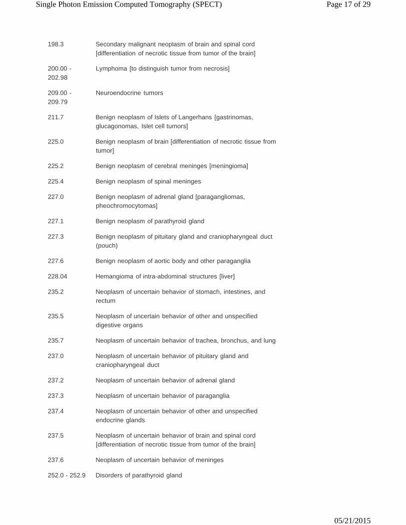
Single Photon Emission Computed Tomography (SPECT) Page 17 of 29
05/21/2015
198.3 Secondary malignant neoplasm of brain and spinal cord [differentiation of necrotic tissue from tumor of the brain]
200.00 - 202.98
Lymphoma [to distinguish tumor from necrosis]
209.00 - 209.79
Neuroendocrine tumors
211.7 Benign neoplasm of Islets of Langerhans [gastrinomas,
glucagonomas, Islet cell tumors]
225.0 Benign neoplasm of brain [differentiation of necrotic tissue from tumor]
225.2 Benign neoplasm of cerebral meninges [meningioma]
225.4 Benign neoplasm of spinal meninges
227.0 Benign neoplasm of adrenal gland [paragangliomas,
pheochromocytomas]
227.1 Benign neoplasm of parathyroid gland
227.3 Benign neoplasm of pituitary gland and craniopharyngeal duct (pouch)
227.6 Benign neoplasm of aortic body and other paraganglia
228.04 Hemangioma of intra-abdominal structures [liver]
235.2 Neoplasm of uncertain behavior of stomach, intestines, and
rectum
235.5 Neoplasm of uncertain behavior of other and unspecified digestive organs
235.7 Neoplasm of uncertain behavior of trachea, bronchus, and lung
237.0 Neoplasm of uncertain behavior of pituitary gland and
craniopharyngeal duct
237.2 Neoplasm of uncertain behavior of adrenal gland
237.3 Neoplasm of uncertain behavior of paraganglia
237.4 Neoplasm of uncertain behavior of other and unspecified endocrine glands
237.5 Neoplasm of uncertain behavior of brain and spinal cord
[differentiation of necrotic tissue from tumor of the brain]
237.6 Neoplasm of uncertain behavior of meninges
252.0 - 252.9 Disorders of parathyroid gland
Single Photon Emission Computed Tomography (SPECT) Page 18 of 29
05/21/2015
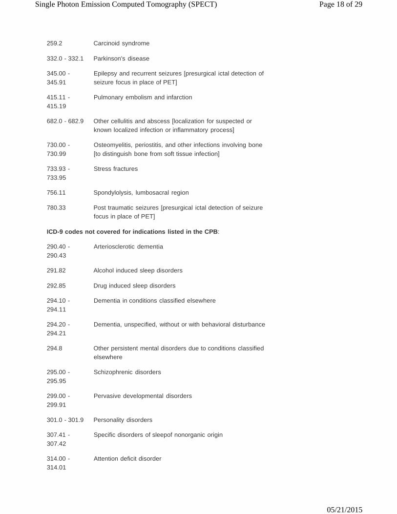
259.2 Carcinoid syndrome
332.0 - 332.1 Parkinson's disease
345.00 - 345.91
Epilepsy and recurrent seizures [presurgical ictal detection of seizure focus in place of PET]
415.11 - 415.19
Pulmonary embolism and infarction
682.0 - 682.9 Other cellulitis and abscess [localization for suspected or
known localized infection or inflammatory process]
730.00 - 730.99
Osteomyelitis, periostitis, and other infections involving bone [to distinguish bone from soft tissue infection]
733.93 - 733.95
Stress fractures
756.11 Spondylolysis, lumbosacral region
780.33 Post traumatic seizures [presurgical ictal detection of seizure
focus in place of PET]
ICD-9 codes not covered for indications listed in the CPB:
290.40 - 290.43
Arteriosclerotic dementia
291.82 Alcohol induced sleep disorders
292.85 Drug induced sleep disorders
294.10 - 294.11
Dementia in conditions classified elsewhere
294.20 - 294.21
Dementia, unspecified, without or with behavioral disturbance
294.8 Other persistent mental disorders due to conditions classified
elsewhere
295.00 - 295.95
Schizophrenic disorders
299.00 - 299.91
Pervasive developmental disorders
301.0 - 301.9 Personality disorders
307.41 - 307.42
Specific disorders of sleepof nonorganic origin
314.00 - 314.01
Attention deficit disorder
Single Photon Emission Computed Tomography (SPECT) Page 19 of 29
05/21/2015
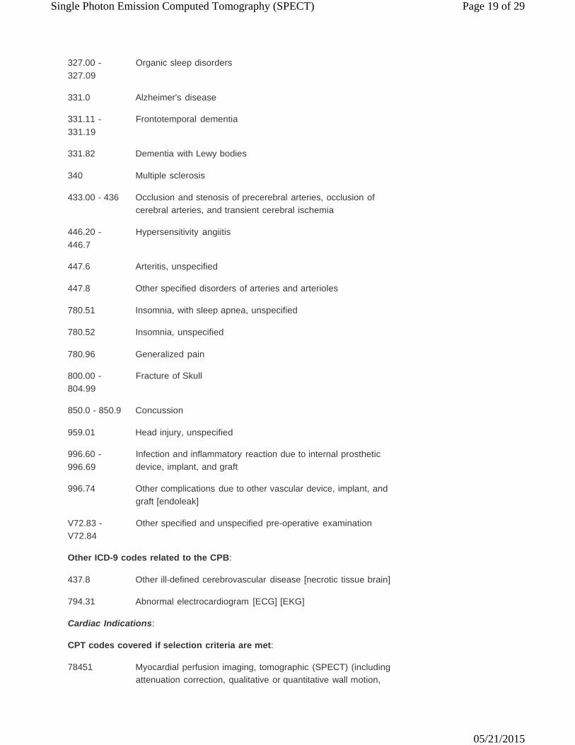
327.00 - 327.09
Organic sleep disorders
331.0 Alzheimer's disease
331.11 - 331.19
Frontotemporal dementia
331.82 Dementia with Lewy bodies
340 Multiple sclerosis
433.00 - 436 Occlusion and stenosis of precerebral arteries, occlusion of
cerebral arteries, and transient cerebral ischemia
446.20 - 446.7
Hypersensitivity angiitis
447.6 Arteritis, unspecified
447.8 Other specified disorders of arteries and arterioles
780.51 Insomnia, with sleep apnea, unspecified
780.52 Insomnia, unspecified
780.96 Generalized pain
800.00 - 804.99
Fracture of Skull
850.0 - 850.9 Concussion
959.01 Head injury, unspecified
996.60 - 996.69
Infection and inflammatory reaction due to internal prosthetic device, implant, and graft
996.74 Other complications due to other vascular device, implant, and
graft [endoleak]
V72.83 - V72.84
Other specified and unspecified pre-operative examination
Other ICD-9 codes related to the CPB:
437.8 Other ill-defined cerebrovascular disease [necrotic tissue brain]
794.31 Abnormal electrocardiogram [ECG] [EKG]
Cardiac Indications:
CPT codes covered if selection criteria are met:
78451 Myocardial perfusion imaging, tomographic (SPECT) (including
attenuation correction, qualitative or quantitative wall motion,
Single Photon Emission Computed Tomography (SPECT) Page 20 of 29
05/21/2015
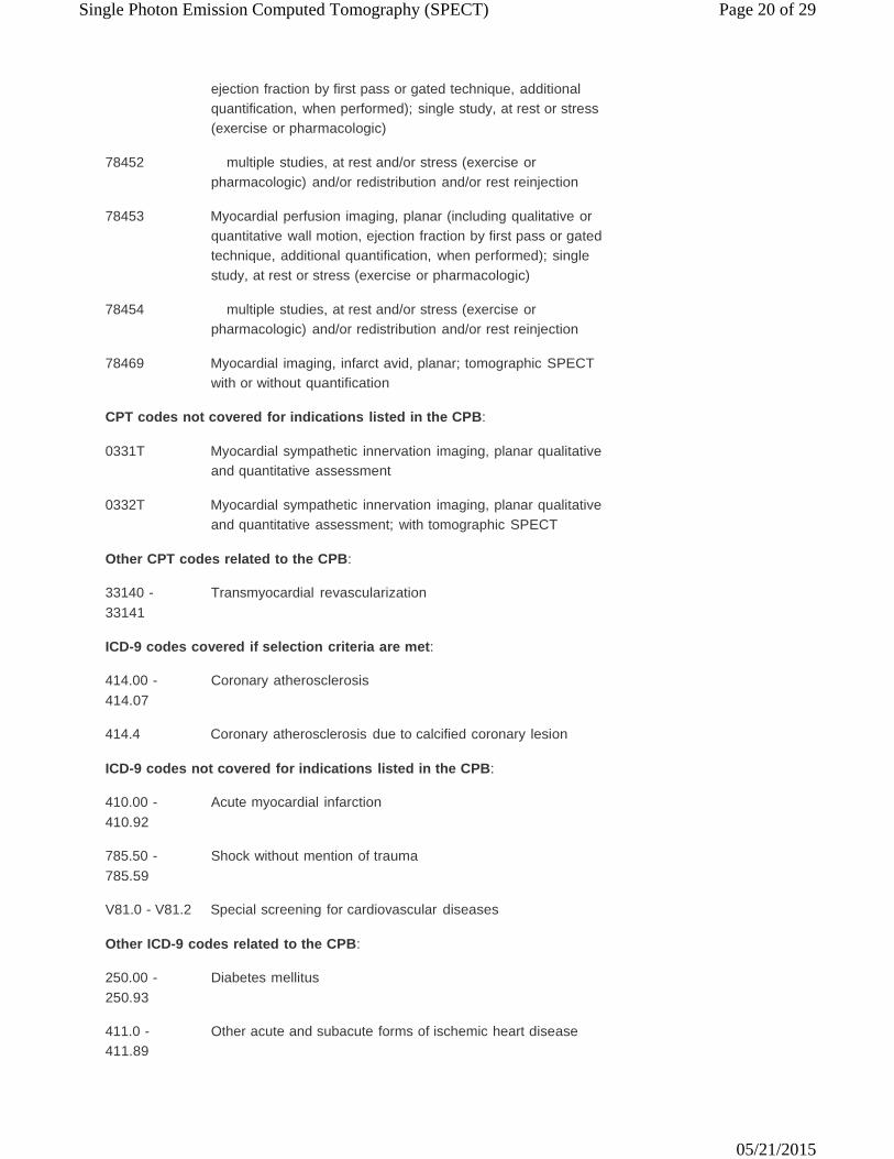
ejection fraction by first pass or gated technique, additional quantification, when performed); single study, at rest or stress (exercise or pharmacologic)
78452 multiple studies, at rest and/or stress (exercise or
pharmacologic) and/or redistribution and/or rest reinjection
78453 Myocardial perfusion imaging, planar (including qualitative or quantitative wall motion, ejection fraction by first pass or gated technique, additional quantification, when performed); single study, at rest or stress (exercise or pharmacologic)
78454 multiple studies, at rest and/or stress (exercise or
pharmacologic) and/or redistribution and/or rest reinjection
78469 Myocardial imaging, infarct avid, planar; tomographic SPECT with or without quantification
CPT codes not covered for indications listed in the CPB:
0331T Myocardial sympathetic innervation imaging, planar qualitative
and quantitative assessment
0332T Myocardial sympathetic innervation imaging, planar qualitative and quantitative assessment; with tomographic SPECT
Other CPT codes related to the CPB:
33140 - 33141
Transmyocardial revascularization
ICD-9 codes covered if selection criteria are met:
414.00 - 414.07
Coronary atherosclerosis
414.4 Coronary atherosclerosis due to calcified coronary lesion
ICD-9 codes not covered for indications listed in the CPB:
410.00 - 410.92
Acute myocardial infarction
785.50 - 785.59
Shock without mention of trauma
V81.0 - V81.2 Special screening for cardiovascular diseases
Other ICD-9 codes related to the CPB:
250.00 - 250.93
Diabetes mellitus
411.0 - 411.89
Other acute and subacute forms of ischemic heart disease
Single Photon Emission Computed Tomography (SPECT) Page 21 of 29
05/21/2015
426.3 Other left bundle branch block
426.7 Anomalous atrioventricular excitation [Wolff-Parkinson-White syndrome
428.0 Congestive heart failure
786.50 - 786.59
Chest pain
V45.01 Cardiac pacemaker status
V45.81 Aortocoronary bypass status
V45.82 Percutaneous transluminal coronary angioplasty status
V72.81 Pre-operative cardiovascular examination
The above policy is based on the following references:
1.
2.
3.
4.
5.
6.
7.
8.
9.
10.
11.
American Academy of Neurology, Therapeutics and Technology Assessment Subcommittee. Assessment: Brain SPECT. Minneapolis, MN: American Academy of Neurology; 1995. Littenberg B, Siegel A, Tosteson AN, Mead T. Clinical efficacy of SPECT bone imaging for low back pain. J Nuclear Med. 1995;36(9):1707-1713. Masdeu JC, Brass LM, Holman BL, et al. Special review: Brain single photon emission tomography. Neurology. 1994;44(10):1970-1977. Limburg M, von Royen EA, Hijdra A, Verbeeten B Jr. rCBF-SPECT in brain infarction: When does it predict outcome? J Nuclear Med. 1991;32(3):382- 387. Markland ON, Anderson AR, Spencer SS. SPECT in epilepsy. J Neuroimaging. 1995;5(Suppl 1):S23-S33. Won JH, Lee JD, Chung TS, et al. Increased contralateral cerebellar uptake of technetium-99m-HMPAO on ictal brain SPECT. J Nucl Med. 1996;3 (37):426-429. Harvey AS, Hopkins IJ, Bowe JM, et al. Frontal lobe epilepsy: Clinical seizure characteristics and localization with ictal technetium-99m-HMPAO SPECT. Neurology. 1993;43(10):1966-1980. Dasheiff RM. A review of interictal cerebral blood flow in the evaluation of patients for epilepsy surgery. Seizure. 1992;1:117-125. Killen AR, Oster G, Colditz GA. An assessment of the role of 123I-N- isopropyliodoamphetamine with single-photon emission computed tomography in the diagnosis of stroke and Alzheimer's disease. Nucl Med Communications. 1989;10:271-284. de Bruine JF, Limburg M, van Royen EA, et al. SPECT brain imaging using 201 diethyldithiocarbamate in acute ischemic stroke. Eur J Nucl Med. 1990;17(5):248-251. De Roo M, Mortelmans L, Devos P, et al. Clinical experience with Tx-99m HM-PAO high resolution SPECT of the brain in patients with cerebrovascular accidents. Eur J Nucl Med. 1989;15:9-15.
Single Photon Emission Computed Tomography (SPECT) Page 22 of 29
05/21/2015
12.
13.
14.
15.
16.
17.
18.
19.
20.
21.
22.
23.
24.
25.
26.
27.
Dierckx RA, Dobbeleir A, Pickut BA, et al. Technetium-99m HMPAO SPECT in acute supratentorial ischemic infarction. Expressing deficits as milliliter of zero perfusion. Eur J Nucl Med. 1995;22(5):427-433. Feldman M, Voth E, Dressler D, et al. 99m Tc-hexamethylpropylene amine oxime SPECT and X-ray CT in acute cerebral ischemia. J Neurol. 1990;237:475-479. Fieschi C, Argentino C, Lenzi Gl, et al. Clinical and instrumental evaluation of patients with ischemic stroke within the first six hours. J Neurological Sci. 1989;91:311-321. Hayman LA, Taber KH, Jhingran SG, et al. Cerebral infarction: Diagnosis and assessment of prognosis by using 123 IMP-SPECT and CT. Am J Nucl Radiol. 1989;10:557-562. Moretti JLM, Tamgac F, Weinmann P, et al. Early and delayed brain SPECT with technetium-99m-ECD and Iodine-123-IMP in subacute strokes. J Nucl Med. 1994;35(9):1444-1449. Yeh SH, Liu RS, Hu HH, et al. Brain SPECT imaging with 99TCM- hexamethylporpyleneamine oxime in the early detection of cerebral infarction: Comparison with transmission computed tomography. Nucl Med Communications. 1986;7:873-888. Giacometti AR, Davis PC, Alazraki NP. Anatomic and physiologic imaging of Alzheimer's disease. Clinics Geriatric Med. 1994;10(2):277-298. Mastin ST, Drane WE, Gilmore RL, et al. Prospective localization of epileptogenic foci: Comparison of PET and SPECT with site of surgery and clinical outcome. Radiology. 1996;199:375-380. Tatum WO, Sperling MR, O'Connor MJ, et al. Interictal single-photon emission computed tomography in partial epilepsy. Accuracy in localization and prediction of outcome. J Neuroimaging. 1995;5(3):142-144. Cross JH, Gordon I, Jackson GD. Children with intractable focal epilepsy: Ictal and interictal 99TcM HMPAO single photon emission computed tomography. Dev Med Child Neuro. 1995;37:673-681. Ho SS, Berkovic SF, Newton MR, et al. Parietal lobe epilepsy: Clinical features and seizure localization by ictal SPECT. Neurology. 1994;44 (12):2277-2284. Grunwald F, Menzel C, Pavics L, et al. Ictal and interictal brain SPECT imaging in epilepsy using technetium-99m-ECD. J Nucl Med. 1995;35 (12):1896-1901. Devous MD, Thisted RA, Morgan GF, et al. SPECT brain imaging in epilepsy: A meta-analysis. J Nucl Med. 1998;39(2):285-293. Hang J, et al. Comparative value of maximal treadmill testing, exercise thallium myocardial perfusion scintigraphy and exercise radionuclide ventriculography for distinguishing high- and low-risk patients soon after myocardial infarction. Am J Cardiol. 1984;(53):1221-1227. Garber AM, Solomon NA. Cost-effectiveness of alternative test strategies for the diagnosis of coronary artery disease. Ann Intern Med. 1999;130 (9):719-728. Cowley D, Corabian P, Hailey D. Functional diagnostic imaging in the assessment of myocardial viability. HTA-16. Edmonton, AB: Alberta Heritage Foundation for Medical Research (AHFMR); 1999.
Single Photon Emission Computed Tomography (SPECT) Page 23 of 29
05/21/2015
28.
29.
30.
31.
32.
33.
34.
35.
36.
37.
38.
39.
40.
41.
42.
43.
44.
45.
46.
Fleischmann KE, Hunink MG, Kuntz KM, et al. Exercise echocardiography or exercise SPECT imaging? A meta analysis of diagnostic test performance. JAMA. 1998;280(10):913-920. Bax JJ, Wijns W, Cornel JH, et al. Accuracy of currently available techniques for prediction of functional recovery after revascularization in patients with left ventricular dysfunction due to coronary artery disease: Comparison of pooled data. J Am Coll Cardiol. 1997;30(6):1451-1460. Berman DS, Kiat H, Van Train KF, et al. Comparison of SPECT using technetium-99m agents and thallium-201 and PET for the assessment of myocardial perfusion and viability. Am J Cardiol. 1990;66(13):72E-79E. Henry TR, Pennell PB. Neuropharmacological imaging in epilepsy with PET and SPECT. Q J Nucl Med. 1998;42(3):199-210. Hatazawa J, Shimosegawa E. Imaging neurochemistry of cerebrovascular disease with PET and SPECT. Q J Nucl Med. 1998;42(3):193-198. Iida H, Eberl S. Quantitative assessment of regional myocardial blood flow with thallium-201 and SPECT. J Nucl Cardiol. 1998;5(3):313-331. Schraml FV, Driver DR, Randolph T, et al. PET versus SPECT for determining myocardial tissue viability using fluorine-18- fluorodeoxyglucose. J Nucl Med Technol. 1997;25(4):272-274. Masterman DL, Mendez MF, Fairbanks LA, Cummings JL. Sensitivity, specificity, and positive predictive value of technetium 99-HMPAO SPECT in discriminating Alzheimer's disease from other dementias. J Geriatr Psychiatry Neurol. 1997;10(1):15-21. Ryding E. SPECT measurements of brain function in dementia: A review. Acta Neurol Scand Suppl. 1996;168:54-58. Treves ST, Connolly LP. Single-photon emission computed tomography (SPECT) in pediatric epilepsy. Neurosurg Clin N Am. 1995;6(3):473-480. Jagust WJ, Johnson KA, Holman BL. SPECT perfusion imaging in the diagnosis of dementia. J Neuroimaging. 1995;5 Suppl 1:S45-S52. Holman BL, Abdel-Dayem H. The clinical role of SPECT in patients with brain tumors. J Neuroimaging. 1995;5 Suppl 1:S34-S39. Masdeu JC, Brass LM. SPECT imaging of stroke. J Neuroimaging. 1995;5 Suppl 1:S14-S22. Spencer SS, Theodore WH, Berkovic SF. Clinical applications: MRI, SPECT, and PET. Magn Reson Imaging. 1995;13(8):1119-1124. Harvey AS, Berkovic SF. Functional neuroimaging with SPECT in children with partial epilepsy. J Child Neurol. 1994;9 Suppl 1:S71-S81. Davis PC, Mirra SS, Alazraki N. The brain in older persons with and without dementia: Findings on MR, PET, and SPECT images. AJR Am J Roentgenol. 1994;162(6):1267-1278. Verani MS. Thallium-201 single-photon emission computed tomography (SPECT) in the assessment of coronary artery disease. Am J Cardiol. 1992;70(14):3E-9E. Rozental JM. Positron emission tomography (PET) and single-photon emission computed tomography (SPECT) of brain tumors. Neurol Clin. 1991;9(20):287-305. Maddahi J, Kiat H, Van Train KF, et al. Myocardial perfusion imaging with technetium-99m sestamibi SPECT in the evaluation of coronary artery disease. Am J Cardiol. 1990;66(13):55E-62E.
Single Photon Emission Computed Tomography (SPECT) Page 24 of 29
05/21/2015
47.
48.
49.
50.
51.
52.
53.
54.
55.
56.
57.
58.
59.
60.
61.
62.
63.
64.
Seabold JE, Nepola JV. Imaging techniques for evaluation of postoperative orthopedic infections. Q J Nucl Med. 1999;43(1):21-28. Growcock GW, Murray RR, Suzley GJ. Tc-99m red blood cell scintigraphy in acute intrahepatic hematoma. Clin Nucl Med. 1994;19(8):752-753. El-Desouki M, Mohamadiyeh M, al-Rashed R, et al. Features of hepatic cavernous hemangioma on planar and SPECT Tc-99m-labeled red blood cell scintigraphy. Clin Nucl Med. 1999;24(8):583-589. Lipman JC, Tumeh SS. The radiology of cavernous hemangioma of the liver. Crit Rev Diagn Imaging. 1990;30(1):1-18. Middleton ML. Scintigraphic evaluation of hepatic mass lesions: Emphasis on hemangioma detection. Semin Nucl Med. 1996;26(1):4-15. Jacobson AF, Teefey SA. Cavernous hemangiomas of the liver. Association of sonographic appearance and results of Tc-99m labeled red blood cell SPECT. Clin Nucl Med. 1994;19(2):96-99. Rubin RA, Lichtenstein GR. Scintigraphic evaluation of liver masses: Cavernous hepatic hemangioma. J Nucl Med. 1993;34(5):849-852. American Academy of Pediatrics. Clinical practice guideline: Diagnosis and evaluation of the child with attention-deficit/hyperactivity disorder. Pediatrics. 2000;105(5):1158-1170. Frank Y, Pavlakis SG. Brain imaging in neurobehavioral disorders. Pediatr Neurol. 2001;25(4):278-287. Filipek PA, Accardo PJ, Ashwal S, et al. Practice parameter: Screening and diagnosis of autism. Report of the Quality Standards Subcommittee of the American Academy of Neurology and the Child Neurology Society. Neurology. 2000;55(4):468-479. Knopman DS, DeKosky ST, Cummings JL, et al. Practice parameter: Diagnosis of dementia (an evidence-based review). Report of the Quality Standards Subcommittee of the American Academy of Neurology. Neurology. 2001;56(9):1143-1153. Wolf H, Jelic V, Gertz HJ, et al. A critical discussion of the role of neuroimaging in mild cognitive impairment. Acta Neurol Scand Suppl. 2003;179:52-76. Patel AD, Iskandrian AE. Role of single photon emission computed tomography imaging in the evaluation of therapy for angina pectoris. Am Heart J. 2003;145(6):952-961. Sokol DK, Edwards-Brown M. Neuroimaging in autistic spectrum disorder (ASD). J Neuroimaging. 2004;14(1):8-15. Lee DS, Jang MJ, Cheon GJ, et al. Comparison of the cost-effectiveness of stress myocardial SPECT and stress echocardiography in suspected coronary artery disease considering the prognostic value of false-negative results. J Nuclear Cardiol. 2002;9(5):515-522. McGough JJ, Barkley RA. Diagnostic controversies in adult attention deficit hyperactivity disorder. Am J Psychiatry. 2004;161(11):1948-1956. Pichon Riviere A, Augustovski F, Cernadas C, et al. Cerebral SPECT in the assessment of patients with schizophrenia [summary]. Report IRR No. 29. Buenos Aires, Argentina:.Institute for Clinical Effectiveness and Health Policy (IECS); 2004. Patel MR, Spertus JA, Brindis RG, et al. ACCF proposed method for evaluating the appropriateness of cardiovascular imaging. J Am Coll Cardiol. 2005;46(8):1606-1613.
Single Photon Emission Computed Tomography (SPECT) Page 25 of 29
05/21/2015
65.
66.
67.
68.
69.
70.
71.
72.
73.
74.
75.
Mowatt G, Brazzelli M, Gemmell H, et al. Systematic review of the prognostic effectiveness of SPECT myocardial perfusion scintigraphy in patients with suspected or known coronary artery disease and following myocardial infarction. Nucl Med Commun. 2005;26(3):217-229. National Institute for Clinical Excellence (NICE). Myocardial perfusion scintigraphy for the diagnosis and management of angina and myocardial infarction. Technology Appraisal Guidance No. 73. London, UK: NICE; 2003. Mowatt G, Vale L, Brazzelli M, et al. Systematic review of the effectiveness and cost-effectiveness, and economic evaluation, of myocardial perfusion scintigraphy for the diagnosis and management of angina and myocardial infarction. Health Technol Asses, 2004;8(30):1-222. Pridmore S, Chambers A, McArthur M. Neuroimaging in psychopathy. Aust N Z J Psychiatry. 2005;39(10):856-865. Suchowersky O, Reich S, Perlmutter J, et al. Practice Parameter: Diagnosis and prognosis of new onset Parkinson disease (an evidence-based review): Report of the Quality Standards Subcommittee of the American Academy of Neurology. Neurology. 2006;66(7):968-975. Brindis RG, Douglas PS, Hendel RC, et al.; American College of Cardiology Foundation Quality Strategic Directions Committee Appropriateness Criteria Working Group; American Society of Nuclear Cardiology; American Heart Association. ACCF/ASNC appropriateness criteria for single-photon emission computed tomography myocardial perfusion imaging (SPECT MPI): A report of the American College of Cardiology Foundation Quality Strategic Directions Committee Appropriateness Criteria Working Group and the American Society of Nuclear Cardiology endorsed by the American Heart Association. J Am Coll Cardiol. 2005;46(8):1587-1605. Matchar DB, Kulasingam SL, Huntington A, et al. Positron emission tomography, single photon emission computed tomography, computed tomography, functional magnetic resonance imaging, and magnetic resonance spectroscopy and for the diagnosis and management of Alzheimer's dementia. Technology Assessment. Prepared by the Duke Evidence-based Practice Center for the Agency for Healthcare Research and Quality (AHRQ) under Contract No. 290-02-0025. Rockville, MD: Agency for Healthcare Research and Quality (AHRQ); April 30, 2004. Whiting P, Gupta R, Burch J, et al. A systematic review of the effectiveness and cost-effectiveness of neuroimaging assessments used to visualise the seizure focus in people with refractory epilepsy being considered for surgery. Health Technol Assess. 2006:10(4):1-250. Schillaci O, Scopinaro F, Angeletti S, et al. SPECT improves accuracy of somatostatin receptor scintigraphy in abdominal carcinoid tumors. J Nuclear Med. 1996;37(9):1452-1456. Perault C, Schvartz C, Wampach H, et al. Thoracic and abdominal SPECT- CT image fusion without external markers in endocrine carcinomas. The Group of Thyroid Tumoral Pathology of Champagne-Ardenne. J Nuclear Med. 1997;38(8):1234-1242. Kälkner KM, Nilsson S, Ylä-Jääski J, et al. Comparison between transveral SPECT, 3-dimensional rendering and 3-dimensional rendering plus clipping in the diagnosis of somatostatin receptor distribution in malignancies using
Single Photon Emission Computed Tomography (SPECT) Page 26 of 29
05/21/2015
76.
77.
78.
79.
80.
81.
82.
83.
84.
85.
86.
87.
88.
89.
111In-DTPA-D-Phe1-octreotide scintigraphy. Cancer Biother Radiopharm. 1997; 2(2):89-99. Westlin JE, Ahlstrom H, Yla-Jaaski J, et al. Three-dimensional OctreoScan111 SPECT of abdominal manifestation of neuroendocrine tumours. Acta Oncol. 1993; 32(2):171-176. Signore A, Procaccini E, Chianelli M, et al. SPECT imaging with 111In- octreotide for the localization of pancreatic insulinoma. Q J Nucl Med. 1995;39(4 Suppl 1):111-112. Medical Services Advisory Committee (MSAC). OctreoScan scintigraphy for gastroentero-pancreatic neuroendocrine tumours. Final Assessment Report. MSAC Application 1003. Canberra, ACT: MSAC; August 1999. Khan AN, Jones C. Lung, carcinoid. eMedicine Radiology Topic 403. Omaha, NE: eMedicine.com; updated January 31, 2005. Anand M, Cowie A. Pancreas, islet cell tumors. eMedicine Radiology Topic 363. Omaha, NE: eMedicine.com; updated March 17, 2004. Ferrante D. SPECT for the diagnosis and assessment of dementia and Alzheimer's disease [summary]. Report ITB No. 14. Buenos Aires, Argentina: Institute for Clinical Effectiveness and Health Policy (IECS); 2004. Manaster BJ, Grossman JW, Dalinka MK, et al; Expert Panel on Musculoskeletal Imaging. Stress/insufficiency fracture, including sacrum, excluding other vertebrae. ACR Appropriateness Criteria. Reston, VA: American College of Radiology (ACR); 2005. Greenspan BS, Brown ML, Dillehay GL, et al. The Society of Nuclear Medicine Procedure Guideline for Parathyroid Scintigraphy. Version 3.0. Reston, VA: Society of Nuclear Medicine; June 2004. Colantonio L, Augustovski F, Pichon Riviere A. Usefulness of SPECT in epilepsy [summary]. Report ITB No.32. Buenos Aires, Argentina: Institute for Clinical Effectiveness and Health Policy (IECS); 2007. Lau J, Kent D, Tatsioni A, et al.; Tufts-New England Medical Center Evidence-based Practice Center. Vulnerable plaques; A brief review of the concept and proposed approaches to diagnosis and treatment. Technology Assessment. Prepared for AHRQ under Contract No. 290-02-0022. Rockville, MD: Agency for Healthcare Research and Quality (AHRQ); January 22, 2004. McNeill R, Sare GM, Manoharan M, et al. Accuracy of single-photon emission computed tomography in differentiating frontotemporal dementia from Alzheimer’s disease. J Neurol Neurosurg Psychiatry. 2007;78(4):350- 355. Vlaar AM, van Kroonenburgh MJ, Kessels AG, Weber WE. Meta-analysis of the literature on diagnostic accuracy of SPECT in parkinsonian syndromes. BMC Neurol. 2007;7:27. Sharples L, Hughes V, Crean A, et al. Cost-effectiveness of functional cardiac testing in the diagnosis and management of coronary artery disease: A randomised controlled trial. The CECaT trial. Health Technol Assess. 2007;11(49):1-136. van der Vaart MG, Meerwaldt R, Slart RH, et al. Application of PET/SPECT imaging in vascular disease. Eur J Vasc Endovasc Surg. 2008;35(5):507- 513.
Single Photon Emission Computed Tomography (SPECT) Page 27 of 29
05/21/2015
90.
91.
92.
93.
94.
95.
96.
97.
98.
99.
100.
101.
102.
Cronin P, Dwamena BA, Kelly AM, Carlos RC. Solitary pulmonary nodules: Meta-analytic comparison of cross-sectional imaging modalities for diagnosis of malignancy. Radiology. 2008;246(3):772-782. Iwata K, Kubota M, Ogasawara K. Comparsion with myocardial perfusion MRI and myocardial perfusion SPECT in the diagnostic performance of coronary artery disease: A meta-analysis. Nippon Hoshasen Gijutsu Gakkai Zasshi. 2008;64(2):251-258. Fujita S, Nagamachi S, Wakamatsu H, et al. Usefulness of triple-phase thallium-201 SPECT in non-small-cell lung cancer (NSCLC): Association with proliferative activity. Ann Nucl Med. 2008;22(10):833-839. Smartt, P. Campbell-Page, R. Combined CT and SPECT (SPECT-CT) scanning in oncology: Horizon scanning report. HSAC Report. Christchurch, NZ: Health Services Assessment Collaboration (HSAC); July 2009;2(13). Hindié E, Ugur O, Fuster D, et al; Parathyroid Task Group of the EANM. 2009 EANM parathyroid guidelines. Eur J Nucl Med Mol Imaging. 2009;36 (7):1201-1216. Miles S, Rogers KM, Thomas P, et al. A comparison of single-photon emission CT lung scintigraphy and CT pulmonary angiography for the diagnosis of pulmonary embolism. Chest. 2009;136(6):1546-1553. Bajc M, Neilly JB, Miniati M, et al; EANM Committee. EANM guidelines for ventilation/perfusion scintigraphy: Part 1. Pulmonary imaging with ventilation/perfusion single photon emission tomography. Eur J Nucl Med Mol Imaging. 2009a;36(8):1356-1370. Bajc M, Neilly JB, Miniati M, et al. EANM guidelines for ventilation/perfusion scintigraphy: Part 2. Algorithms and clinical considerations for diagnosis of pulmonary emboli with V/P(SPECT) and MDCT. Eur J Nucl Med Mol Imaging. 2009b;36(9):1528-1538. Gutte H, Mortensen J, Jensen CV, et al. Comparison of V/Q SPECT and planar V/Q lung scintigraphy in diagnosing acute pulmonary embolism. Nucl Med Commun. 2010;31(1):82-86. Devanand DP, Van Heertum RL, Kegeles LS, et al. (99m)Tc hexamethyl- propylene-aminoxime single-photon emission computed tomography prediction of conversion from mild cognitive impairment to Alzheimer disease. Am J Geriatr Psychiatry. 2010;18(11):959-972. Wippold FJ II, Cornelius RS, Broderick DF, et al; Expert Panel on Neurologic Imaging. ACR Appropriateness Criteria dementia and movement disorders [online publication]. Reston, VA: American College of Radiology (ACR); 2010. Greenwood JP, Maredia N, Younger JF, et al. Cardiovascular magnetic resonance and single-photon emission computed tomography for diagnosis of coronary heart disease (CE-MARC): A prospective trial. Lancet. 2012;379(9814):453-460. Davis PC, Wippold FJ II, Cornelius RS, et al; Expert Panel on Neurologic Imaging. ACR Appropriateness Criteria® head trauma. [online publication]. Reston (VA): American College of Radiology (ACR); 2012. Available at: http://www.guideline.gov/content.aspx? id=37919&search=Single+Photon+Emission+Computed+Tomography+. Accessed March 22, 2013.
Single Photon Emission Computed Tomography (SPECT) Page 28 of 29
05/21/2015
103.
104.
105.
106.
107.
108.
109.
110.
111.
112.
Jamadar DA, Kazerooni EA, Martinez FJ, Wahl RL. Semi-quantitative ventilation/perfusion scintigraphy and single-photon emission tomography for evaluation of lung volume reduction surgery candidates: Description and prediction of clinical outcome. Eur J Nucl Med. 1999;26(7):734-742. Inmai T, Sasaki Y, Shinkai T, et al. Clinical evaluation of 99mTc-Technegas SPECT in thoracoscopic lung volume reduction surgery in patients with pulmonary emphysema. Ann Nucl Med. 2000;14(4):263-269. Rasch H, Falkowski AL, Forrer F, et al. 4D-SPECT/CT in orthopaedics: A new method of combined quantitative volumetric 3D analysis of SPECT/CT tracer uptake and component position measurements in patients after total knee arthroplasty. Skeletal Radiol. 2013;42(9):1215-1223. Martinez FJ. Lung volume reduction surgery in COPD. Last reviewed February 2014. UpToDate Inc. Waltham, MA. Nakai M, Sato H, Ikoma A, et al. The use of technetium-99m-labeled human serum albumin diethylenetriamine pentaacetic acid single-photon emission CT scan in the follow-up of type II endoleak treatment. J Vasc Interv Radiol. 2014;25(3):405-409. Wippold FJ II, Brown DC, Broderick DF, et al; Expert Panel on Neurologic Imaging. ACR Appropriateness Criteria® dementia and movement disorders [online publication]. Reston (VA): American College of Radiology (ACR); 2014. Freesmeyer M, Opfermann T, Winkens T. Hybrid integration of real-time US and freehand SPECT: Proof of concept in patients with thyroid diseases. Radiology. 2014;271(3):856-861. Ryken TC, Aygun N, Morris J, et al. The role of imaging in the management of progressive glioblastoma: A systematic review and evidence-based clinical practice guideline. J Neurooncol. 2014;118(3):435-460. National Comprehensive Cancer Network. Clinical practice guideline: Central nervous system cancers. Version 2.2014. NCCN: Fort Washington, PA. Batchelor T, Shih HA, Carter BS. Management of recurrent high-grade gliomas. UpToDate Inc., Waltham, MA. Last reviewed February 2015.
Copyright Aetna Inc. All rights reserved. Clinical Policy Bulletins are developed by Aetna to assist in
administering plan benefits and constitute neither offers of coverage nor medical advice. This Clinical Policy
Bulletin contains only a partial, general description of plan or program benefits and does not constitute a
contract. Aetna does not provide health care services and, therefore, cannot guarantee any results or
outcomes. Participating providers are independent contractors in private practice and are neither
Single Photon Emission Computed Tomography (SPECT) Page 29 of 29
http://qawww.aetna.com/cpb/medical/data/300_399/0376_draft.html 05/2112015
employees nor agents of Aetna or its affiliates. Treating providers are solely responsible for medical advice
and treatment of members. This Clinical Policy Bulletin may be updated and therefore is subject to change.
CPT only copyright 2008 American Medical Association. All Rights Reserved.