Embed Size (px)
Citation preview
CLINICAL ARTICLEJ Neurosurg 129:805–814, 2018
ABBREVIATIONS EGFR = epidermal growth factor receptor; HER = human epidermal growth factor receptor–2; MPST = malignant peripheral nerve sheath tumor; NF2 = neurofibromatosis Type 2; PDGFR-b = platelet-derived growth factor receptor–b; RTK = receptor tyrosine kinase; SPS = sporadic peripheral schwannoma; TMA = tissue microarray.SUBMITTED December 23, 2015. ACCEPTED February 15, 2017.INCLUDE WHEN CITING Published online September 8, 2017; DOI: 10.3171/2017.2.JNS153004.
Clinicopathological variables of sporadic schwannomas of peripheral nerve in 291 patients and expression of biologically relevant markersEric D. Young, BS,1 Davis Ingram, BS,2,6 William Metcalf-Doetsch, MD,7 Dilshad Khan, MD,8 Ghadah Al Sannaa, MD,3,6 Francois Le Loarer, MD, PhD,9 Alexander J. F. Lazar, MD, PhD,3,6 John Slopis, MD,4 Keila E. Torres, MD, PhD,2,6 Dina Lev, MD,10 Raphael E. Pollock, MD, PhD,11 and Ian E. McCutcheon, MD5
1Department of Cancer Biology, University of Kansas Medical Center, Andover, Kansas; Departments of 2Surgical Oncology, 3Pathology and Laboratory Medicine, 4Neuro-Oncology, and 5Neurosurgery and 6The Sarcoma Research Center, University of Texas MD Anderson Cancer Center, Houston, Texas; 7Department of Neurosurgery, Northwestern University, Chicago, Illinois; 8Department of Internal Medicine, University of Toledo Medical Center, Toledo, Ohio; 9Department of Pathology, Centre Leon Berard, Lyon, France; 10Department of Surgery, Sheba Medical Center, Israel; and 11Department of Surgery, The Ohio State University, Columbus, Ohio
OBJECTIVE While sporadic peripheral schwannomas (SPSs) are generally well treated with surgery, their biology is not well understood. Consequently, treatment options are limited. The aim of this study was to provide a comprehensive de-scription of SPS. The authors describe clinicopathological features and treatment outcomes of patients harboring these tumors, and they assess expression of biomarkers using a clinically annotated tissue microarray. Together, these data give new insight into the biology and management of SPS.METHODS Patients presenting with a primary SPS between 1993 and 2011 (n = 291) were selected from an institu-tional registry to construct a clinical database. All patients underwent follow-up, and short- and long-term outcomes were assessed. Expression of relevant biomarkers was assessed using a new tissue microarray (n = 121).RESULTS SPSs were generally large (mean 5.5 cm) and frequently painful at presentation (55%). Most patients were treated with surgery (80%), the majority of whom experienced complete resolution (52%) or improvement (18%) of their symptoms. Tumors that were completely resected (85%) did not recur. Some patients experienced short-term (16%) and long-term (4%) complications postoperatively. Schwannomas expressed higher levels of platelet-derived growth factor receptor–b (2.1) than malignant peripheral nerve sheath tumors (MPNSTs) (1.5, p = 0.004) and neurofibromas (1.33, p = 0.007). Expression of human epidermal growth factor receptor–2 was greater in SPSs (0.91) than in MPNSTs (0.33, p = 0.002) and neurofibromas (0.33, p = 0.026). Epidermal growth factor receptor was expressed in far fewer SPS cells (10%) than in MPNSTs (58%, p < 0.0001) or neurofibromas (37%, p = 0.007). SPSs more frequently expressed cytoplas-mic survivin (66% of tumor cells) than normal nerve (46% of cells), but SPS expressed nuclear survivin in fewer tumor cells than in MPNSTs (24% and 50%, respectively; p = 0.018).CONCLUSIONS Complete resection is curative for SPS. Left untreated, however, these tumors can cause significant morbidity, and not all patients are candidates for resection. SPSs express a pattern of biomarkers consistent with the dysregulation of the tumor suppressor merlin observed in neurofibromatosis Type 2–associated schwannomas, suggest-ing a shared etiology. This SPS pattern is distinct from that of other tumors of the peripheral nerve sheath.https://thejns.org/doi/abs/10.3171/2017.2.JNS153004KEY WORDS sporadic schwannoma; peripheral nerve; clinical variables; tissue microarray; biomarker expression; oncology
» This article has been updated from its originally published version to correct an abbreviation and Fig. 1. See the corresponding erratum
notice in this issue, p 838. «
J Neurosurg Volume 129 • September 2018 805©AANS 2018, except where prohibited by US copyright law
Unauthenticated | Downloaded 01/27/22 03:08 AM UTC
E. D. Young et al.
J Neurosurg Volume 129 • September 2018806
The intricate histological features of schwanno-mas were first described more than 100 years ago26 and were later found to arise from Schwann
cells of the peripheral nerve sheath.18 Schwannomas oc-cur most frequently as a single, sporadic lesion.13 They are considered benign tumors as demonstrated by their generally low proliferative potential,1 their frequently well-circumscribed gross appearance,29 and the fact that malignant transformation is exceedingly rare.32 However, deep-seated tumors and those intimately associated with critical vascular, neural, and other anatomical structures can cause significant morbidity due to mass effect or erosion of adjacent bone.30 Such situations present chal-lenges in resection,24 which is currently the best therapeu-tic option.17 The slow growth rate of sporadic peripheral schwannomas (SPSs) renders them generally unrespon-sive to conventional chemotherapy,14 suggesting a need for new treatment options, particularly for patients who are not surgical candidates.
Given these challenges, we sought to elucidate the bi-ology of SPS. Loss or dysfunction of the known tumor suppressor gene NF2 on 22q12.225 drives tumorigenesis of schwannomas arising in the hereditary cancer syndrome neurofibromatosis Type 2 (NF2). NF2 encodes the protein merlin, which mediates contact inhibition7 by linking a cell’s physical surroundings and its mitotic activity.33 Mer-lin also restrains cell proliferation by negatively regulating several receptor tyrosine kinases (RTKs), including plate-let-derived growth factor receptor–b (PDGFR-b), human epidermal growth factor receptor–2 (HER-2), and epider-mal growth factor receptor (EGFR).4 Interestingly, loss of heterozygosity in chromosome 22 where merlin is located has been demonstrated in SPS,11 providing evidence for similar pathogenesis in sporadic tumors and their con-genital counterparts. Thus, we hypothesized that if simi-lar mechanisms drive sporadic schwannoma growth, we might be able to detect these expression patterns in tumor samples obtained in humans.
To characterize the biology of these tumors, we con-structed a clinically annotated tissue microarray (TMA) and used it to assess biomarker expression. We also con-structed a clinical database of patients with primary SPS and gathered their clinical presentation, treatment, and outcome information. Together, these new resources pro-vide an informative vignette characterizing the morbidity of SPS both in situ and as sequelae to their resection.
MethodsReview of the Literature
We reviewed tissue-based studies of SPS from the literature containing at least 10 patients with follow-up. From these, only tumors that met the following conditions were included: 1) The sample was diagnosed definitively by histological analysis as a schwannoma (vs malignant schwannoma, malignant peripheral nerve sheath tumor [MPNST], neurofibroma, or other peripheral nerve sheath tumor). 2) The sample was sporadic and did not occur in the context of a congenital neurocutaneous syndrome (e.g., NF2, schwannomatosis). 3) The tumor was peripherally located (vs intracranial, or associated with cranial nerves). The clinicopathological features of these tumors were compared with those in the present series (Table 1).
Clinical DatabaseClinical data were collected and analyzed with the ap-
proval of the institutional review board at the University of Texas MD Anderson Cancer Center. Patients treated for SPS at University of Texas MD Anderson Cancer Center from January 1, 1993, through December 31, 2011, were selected from the pathology archives and institutional tumor registry databases. Patients with a diagnosis of a single schwannoma (neurilemmoma) arising from a pe-ripheral nerve (including spinal nerve roots) were included in the database, as were histological variants such as cel-lular schwannoma, plexiform schwannoma, and ancient
TABLE 1. Literature review of clinicopathological data for patients with sporadic schwannomas arising in the peripheral nerves
Authors & Year No. of Pts Sex Age Range, Yrs* Tumor Size, cm* No. w/ FU FU Range, Yrs* Recurrence (%)
Stout, 1935 46 26 F, 20 M 19–68 (39.3) 1–7.5 (3.2) 23 0.25–14 (2.9) 0 (0.0)Ackerman & Taylor, 1951 19 13 F, 6 M 19–66 (44.6) 3.5–20 (9.3) 16 0.3–8 (3.3) 0 (0.0)†Woodruff et al., 1981 13 6 F, 7 M 25–71 (49.1) 2.7–11 (5.7) 8 0.167–10 (2.58) 0 (0.0)‡Fletcher et al., 1987 16 9 F, 7 M 17–73 (41.9) NA 13 2–32 (15.1) 0 (0.0)§Lodding et al., 1990 29 21 F, 8 M 15–79 (median 55) 2–12 (median 6.0) 28 0.25–19.5 (median 7) 0 (0.0)White et al., 1990 57¶ 35 F, 22 M 1.3–81 (44.0) 1–19.5 (6.2) 49 1–24 (6) 3 (6.1)Agaram et al., 2005 23 14 F, 9 M 0.4–67 (28.8) NA 6 1.1–10.0 (median 2.0) 3 (50.0)Strauss et al., 2011 28 21 F, 7 M NA (median 47) 5–23 (median 9) 28 NA (median 3.25) 0 (0.0)**Voltaggio et al., 2012 51 40 F, 11 M 29–90 (58) 1–10.5 (5.1) 41 1–34.9 (11.5) 0 (0.0)Current study 291 153 F, 138 M 7–89 (49.7) 0.3–26 (5.5) 291 0.1–18.9 (4.0) 0 (0.0)
FU = follow-up; NA = not available, Pts = patients.* Values in parentheses are means unless stated otherwise.† There was progression of residual disease after incomplete excision in 1 patient (5.3%).‡ Residual tumor was observed in 2 patients who received subtotal resection.§ There was progression of residual disease after incomplete excision with need for repeated excisions in 1 patient (6.25%).¶ Series includes 15 patients from Woodruff et al., 1981 and 1983.** No progression of residual disease in 3 subtotally resected tumors.
Unauthenticated | Downloaded 01/27/22 03:08 AM UTC
Clinicopathological features of sporadic peripheral schwannomas
J Neurosurg Volume 129 • September 2018 807
schwannoma. Patients with other benign (e.g., neurofibro-ma and neuroma) and malignant (e.g., MPNST, malignant schwannoma, and psammomatous melanotic schwan-noma) nerve sheath tumors were excluded. Schwannomas arising from the cranial nerves were not included in the data set in order to exclude potential cases of NF2. Only patients with at least 30 days of follow-up were included. Demographic, disease-specific, tumor-specific, and fol-low-up variables were analyzed. Progression of disease was defined as an increase in tumor size as detected by MRI, with both T1-weighted postcontrast and T2-weight-ed images reviewed in all 3 standard planes.
Tissue Microarray ConstructionFrom tumor registry databases, patients with formalin-
fixed, paraffin-embedded schwannoma and control tissues were selected. A total of 188 blocks were selected from 169 patients. Slides were reviewed, and viable areas of tu-mor were delineated by a soft-tissue sarcoma pathologist (F.L.L.). These areas were manually punched in duplicate with a 1.0-mm core and placed into the recipient TMA block; 121 samples from patients with primary SPSs were included. Control tissues included adjacent histologically normal nerves from patients with schwannoma (n = 11). Other control tissues included tumor from patients with NF Type 1 (n = 7); schwannoma (n = 1); glandular malignant schwannoma (n = 1); neurofibroma (n = 2); MPNST (n = 3); sporadic MPNST (n = 2); NF2 (n = 6); schwannomatosis (n = 5); sporadic schwannomas from cranial nerves (n = 8); INI1-negative epithelioid sarcomas (n = 6); INI1-positive, unclassified sarcoma with epithelioid features (n = 1); pig-mented psammomatous schwannoma (n = 1); melanoma from a patient treated for schwannoma (n = 1); and other benign nerve tumors (neuroma [n = 1], spindle cell tumor with schwannian differentiation [n = 1], and benign nerve sheath tumor [n = 2]). Tumors were recurrent at presenta-tion in 7 patients, and tumor status was not available for 1 sample. No tissue sample was included from any tumor that had been irradiated prior to surgery.
ImmunohistochemistryUnstained slides were de-paraffinized using stand-
ard procedures, and antigen retrieval was achieved using Envision FLEX target retrieval solution (PDGFR-b), ci-trate buffer (HER-2, survivin), and proteinase K (EGFR). Antibodies were obtained from Santa Cruz (PDGFR-b, sc-339, 1:50), Thermo Scientific (HER-2, clone e2–4001, 1:300), and Invitrogen (EGFR, clone 31G7, 1:50). Expres-sion of biomarkers was scored by 2 independent observ-ers (A.J.F.L. and G.A.S.). The percentage of tumor cells showing staining and the intensity of staining in the tumor cells on a scale of 0–3, where 0 represents no staining; 1, low staining; 2, moderate staining; and 3, intense stain-ing (see Fig. 2 for results of TMA biomarker expression). Specimens that did not contain sufficient material for scoring were excluded from further analysis.
Statistical AnalysisThe Student 2-tailed t-test was performed to determine
the differences in expression of biomarkers as determined
using immunohistochemistry. Graphs were constructed using R statistical software.
ResultsClinical DatabasePatient Variables
Clinicopathological variables of 291 patients presenting with SPS are summarized in Table 2. Of these, 215 patients were Caucasian (74%), 38 were Hispanic (13%), 19 were Asian or Pacific Islander (7%), 17 were African American (6%), 1 was Native American (0.3%), and race was not specified for 1 patient (0.3%). Diagnosis was made by his-tological examination of the tumor after resection, biopsy, or fine needle aspiration. Diagnosis was made based on findings from presurgical biopsy in 150 samples (51%), fine needle aspiration in 11 samples (4%), incisional biopsy in 1 (0.3%), frozen section in 3 (1%), and postexcision paraffin section in 126 (43%). The chief complaint was not available for 6 patients.
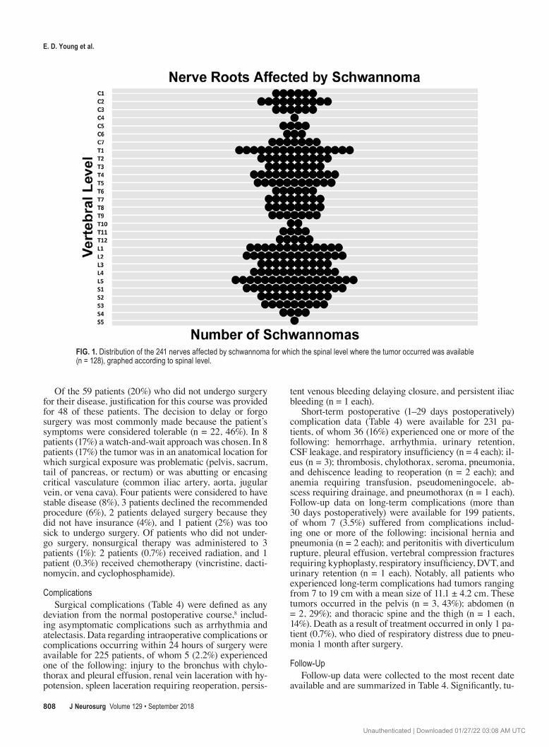
Tumor VariablesTumor size was available for 271 patients and was de-
fined as the maximum dimension reported at radiologi-cal or pathological assessment, as measurements by both methods were unavailable for all patients. Nonvestibular (noncranial nerve) schwannomas located in the head and neck (10% of the total) arose from the cervical nerve roots, sympathetic chain, or subcutaneously. A more detailed description of where tumors arose in these locations and the spinal levels affected by schwannoma are described in Table 3 and Fig. 1, respectively. Erosion of adjacent bone was noted in 17 tumors (6%). Histologically, SPSs were di-vided into the following subclasses: 247 (85%) were iden-tified as conventional schwannomas, 37 (13%) were cellu-lar schwannomas, 6 (2%) showed ancient or degenerative changes, and 1 (0.3%) was a plexiform schwannoma. Of the cellular schwannomas, 12 (32%) occurred in the tho-rax, 11 (30%) in the abdomen/retroperitoneum, 7 (19%) in the extremities, and 7 (17%) in the pelvis.
Treatment VariablesSurgery was the mainstay of treatment, with 232 pa-
tients (80%) undergoing resection. Of these patients, 214 underwent 1 operation (92%), 14 underwent 2 (6%), 2 un-derwent 3 (0.9%), and 2 patients underwent 4 surgeries (0.9%). Blood loss reported as “not significant” or “neg-ligible” was recorded as 0. Notably, tumors smaller than or equal to the median of 4.7 cm had a mean blood loss of 251 ml, whereas tumors larger than the median had a mean blood loss of 733 ml; the difference between the 2 groups was statistically significant (p = 0.00096). As most schwannomas are histologically benign tumors, the extent of resection was reported for 164 (71%) of the 232 patients undergoing surgical extirpation. After resection, surgical margins were either free from tumor (R0) or had tumor present (R1, microscopic tumor present; R2, gross tumor present), as previously defined.20 Tumor removal was done en bloc when the tumor arose from a nonfunctional nerve or one with trivial function, and in a subcapsular fashion when nerve sparing was required for functional preserva-tion.
Unauthenticated | Downloaded 01/27/22 03:08 AM UTC
E. D. Young et al.
J Neurosurg Volume 129 • September 2018808
Of the 59 patients (20%) who did not undergo surgery for their disease, justification for this course was provided for 48 of these patients. The decision to delay or forgo surgery was most commonly made because the patient’s symptoms were considered tolerable (n = 22, 46%). In 8 patients (17%) a watch-and-wait approach was chosen. In 8 patients (17%) the tumor was in an anatomical location for which surgical exposure was problematic (pelvis, sacrum, tail of pancreas, or rectum) or was abutting or encasing critical vasculature (common iliac artery, aorta, jugular vein, or vena cava). Four patients were considered to have stable disease (8%), 3 patients declined the recommended procedure (6%), 2 patients delayed surgery because they did not have insurance (4%), and 1 patient (2%) was too sick to undergo surgery. Of patients who did not under-go surgery, nonsurgical therapy was administered to 3 patients (1%): 2 patients (0.7%) received radiation, and 1 patient (0.3%) received chemotherapy (vincristine, dacti-nomycin, and cyclophosphamide).
ComplicationsSurgical complications (Table 4) were defined as any
deviation from the normal postoperative course,8 includ-ing asymptomatic complications such as arrhythmia and atelectasis. Data regarding intraoperative complications or complications occurring within 24 hours of surgery were available for 225 patients, of whom 5 (2.2%) experienced one of the following: injury to the bronchus with chylo-thorax and pleural effusion, renal vein laceration with hy-potension, spleen laceration requiring reoperation, persis-
tent venous bleeding delaying closure, and persistent iliac bleeding (n = 1 each).
Short-term postoperative (1–29 days postoperatively) complication data (Table 4) were available for 231 pa-tients, of whom 36 (16%) experienced one or more of the following: hemorrhage, arrhythmia, urinary retention, CSF leakage, and respiratory insufficiency (n = 4 each); il-eus (n = 3); thrombosis, chylothorax, seroma, pneumonia, and dehiscence leading to reoperation (n = 2 each); and anemia requiring transfusion, pseudomeningocele, ab-scess requiring drainage, and pneumothorax (n = 1 each). Follow-up data on long-term complications (more than 30 days postoperatively) were available for 199 patients, of whom 7 (3.5%) suffered from complications includ-ing one or more of the following: incisional hernia and pneumonia (n = 2 each); and peritonitis with diverticulum rupture, pleural effusion, vertebral compression fractures requiring kyphoplasty, respiratory insufficiency, DVT, and urinary retention (n = 1 each). Notably, all patients who experienced long-term complications had tumors ranging from 7 to 19 cm with a mean size of 11.1 ± 4.2 cm. These tumors occurred in the pelvis (n = 3, 43%); abdomen (n = 2, 29%); and thoracic spine and the thigh (n = 1 each, 14%). Death as a result of treatment occurred in only 1 pa-tient (0.7%), who died of respiratory distress due to pneu-monia 1 month after surgery.
Follow-UpFollow-up data were collected to the most recent date
available and are summarized in Table 4. Significantly, tu-
FIG. 1. Distribution of the 241 nerves affected by schwannoma for which the spinal level where the tumor occurred was available (n = 128), graphed according to spinal level.
Unauthenticated | Downloaded 01/27/22 03:08 AM UTC
Clinicopathological features of sporadic peripheral schwannomas
J Neurosurg Volume 129 • September 2018 809
mors did not recur in any of the 139 patients who underwent complete, gross-total resection (R0, 125 patients; and R1, 14 patients). Progression of disease occurred in 21 (84%) of 25 patients who received an incomplete (R2, residual gross tumor or debulking) resection. Symptoms were reported at presentation in 184 (79%) of the 232 patients who under-went surgery for schwannoma and in 41 (69%) of the 59 patients who did not undergo surgery. Symptoms persisted without change in 40 patients (98%) without surgery. No patient who forwent surgery experienced complete reso-lution of their symptoms, although one of these patients (2.4%) did report improvement in symptoms.
In symptomatic patients who underwent resection of the schwannoma, the chance and degree of recovery de-pended on the symptom in question. In those with dyses-thesia prior to surgery (n = 10), full absence of dysesthe-sia was achieved in 4 (40%) and partial improvement in 4 (40%), and 3 patients (30%) experienced a change in their symptoms from dysesthesia to pain. For those with par-esthesia before undergoing surgery for their disease (n = 12), the symptoms cleared completely in 5 (42%), partially in 4 (33%), and 3 patients (25%) experienced an increase in pain from either progression of disease or as sequelae of surgery. The time to recovery varied from 10 days to 5 months and occurred more quickly in the paresthesia group than in patients with dysesthesias. Weakness before surgery (n = 9) improved less readily, with 5 patients (56%) reaching normal strength over recovery periods ranging from 1 to 9 months, and 4 (44%) not showing meaningful recovery of strength. Pain was the most common symptom (n = 161) and it cleared completely in 76% of patients for whom follow-up data were available more than 6 months after surgery. In those with incomplete improvement in pain (14%) a plateau was typically reached no later than 6 months after surgery, and the majority who improved did so within 3 months of surgery. No improvement in pain was seen in 8%, and 5% of patients were worse. No sig-nificant differences in outcome were noted by the most common histological variants (Table 5).
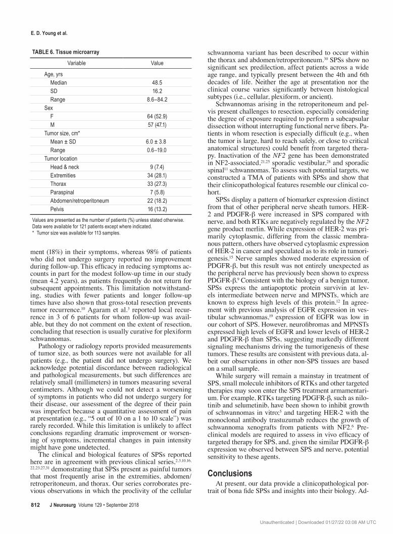
Tissue MicroarrayClinical characteristics of the 121 patients with prima-
ry SPS in the TMA (Table 6) are similar to those in our clinical cohort. Again, the most common site of tumor was the extremities (28%), although in the TMA more tumors
TABLE 2. Demographic, tumor, and treatment variables of patients with SPSs
Variable Value
Age, yrs Median ± SD 50 SD 16.8 Range 7–89Sex F 153 (52.6) M 138 (47.4)Chief complaint Pain 161 (55.3) Paresthesia 14 (4.8) Palpated painless mass 14 (4.8) Dysesthesia 10 (3.4) Weakness/nerve dysfunction 10 (3.4) Cough 8 (2.7) Respiratory symptoms 4 (1.4) Other* 21 (7.2) Asymptomatic, incidental finding 49 (16.8)Tumor size† Mean ± SD 5.5 ± 4.0 Range 0.3–26Tumor location Head & neck 28 (9.6) Extremities 69 (23.7) Thorax 63 (21.6) Paraspinal 33 (11.3) Abdomen/retroperitoneum 64 (22.0) Pelvis 34 (11.7)Surgery Yes 232 (79.7) No 59 (20.3)Complete resection‡ Yes 139 (84.8) No 25 (15.2)Margin status R0 125 (89.9) R1 14 (10.1)Mean op duration ± SD, hrs§ 3.4 ± 3.1Blood loss by vol, ml¶ ≤500 139 (75.1) >500 46 (24.9)Blood loss per op, ml¶ Mean ± SD 533.5 ± 983.7 Median 125.0Intraop complications** Yes 5 (2.2) No 220 (97.8)
CONTINUED IN NEXT COLUMN »
TABLE 2. Demographic, tumor, and treatment variables of patients with SPSs
R0 = gross-total resection with microscopically negative margins; R1 = gross-total resection with microscopically positive margins.Values are presented as the number of patients (%) unless stated otherwise.* Unknown (n = 4); constipation (n = 3); hematuria (n = 3); ataxia (n = 2); and headache, bloody diarrhea, chronic dyspepsia, tonsillitis, weight loss, hemop-tysis, fatigue, flu symptoms, fever (n = 1 each).† Available for 271 patients.‡ Available for 164 patients.§ Available for 155 patients.¶ Available for 185 patients.** Available for 225 patients.
» CONTINUED FROM PREVIOUS COLUMN
Unauthenticated | Downloaded 01/27/22 03:08 AM UTC
E. D. Young et al.
J Neurosurg Volume 129 • September 2018810
originated from the thorax (27%) than the abdomen or ret-roperitoneum (18%); the reverse was true in the clinical cohort.
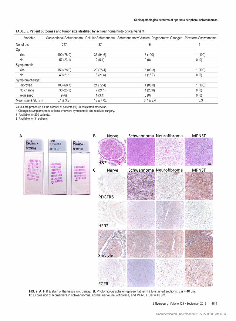
Tissue microarray results are presented in Fig. 2. PDGFR-b was expressed in the cytoplasm of 69% of tu-mor cells at an average intensity of 2.1, which was higher than in MPNSTs and neurofibromas (1.50, p = 0.0039; and 1.33, p = 0.0070, respectively). In nerve, 60% of cells dem-onstrated cytoplasmic staining with an average intensity of 1.8. HER-2 was moderately expressed with an average of
31% of SPS cells staining and an average intensity of 0.91. By comparison, an average of 20% of nerve cells showed HER-2 expression with an intensity of 0.8. HER-2 was expressed at higher levels (0.91) in the cytoplasm of SPS than in MPNSTs and neurofibromas (0.33, p = 0.002; and 0.33, p = 0.026, respectively). EGFR was expressed at low levels, with an average of 10% of tumor cells showing cy-toplasmic staining with an average intensity of 0.68. This was strikingly lower than the percentage of MPNST and neurofibroma cells showing cytoplasmic EGFR expression (58%, p < 0.0001; and 37%, p = 0.007, respectively). Nerve had stronger EGFR staining than schwannomas, with an average of 25% of cells showing cytoplasmic staining and an average intensity of 0.75. Survivin was expressed more highly in SPS than in nerve, with an average of 66% show-ing cytoplasmic staining compared with 46% in nerve (p = 0.057). Nuclear survivin was expressed in fewer sporadic schwannoma cells than in MPNST cells (24% and 50% respectively, p = 0.018).
DiscussionWhile SPSs do not pose the same risks to a patient
as malignant tumors, they can enlarge, with pain being the most common reason for patients to seek treatment. Complete resection of SPS prevents tumor recurrence, but symptoms will not improve if the tumor is left untreated. As difficulty of resection increases with tumor size, delay-ing treatment may lead to excess morbidity. We observed that patients with larger tumors experienced more blood loss during surgery than those with smaller tumors. In ad-dition, the mean tumor size for patients experiencing long-term complications was more than 2 times greater than the overall mean. However, we recognize that a patient’s disease may remain stable indefinitely and that surgery it-self presents the risk of lasting complications, which must be prudently weighed against potential benefits of resec-tion.
Remarkably, 70% of patients undergoing gross-total resection reported complete resolution (52%) or improve-
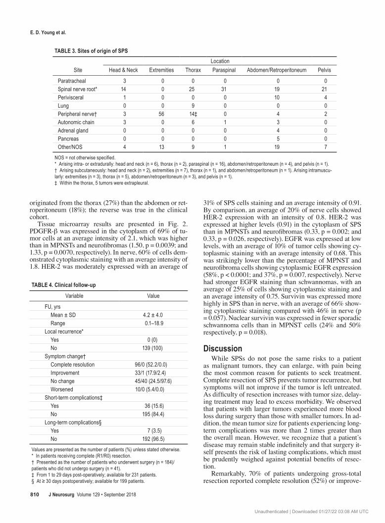
TABLE 4. Clinical follow-up
Variable Value
FU, yrs Mean ± SD 4.2 ± 4.0 Range 0.1–18.9Local recurrence* Yes 0 (0) No 139 (100)Symptom change† Complete resolution 96/0 (52.2/0.0) Improvement 33/1 (17.9/2.4) No change 45/40 (24.5/97.6) Worsened 10/0 (5.4/0.0)Short-term complications‡ Yes 36 (15.6) No 195 (84.4)Long-term complications§ Yes 7 (3.5) No 192 (96.5)
Values are presented as the number of patients (%) unless stated otherwise.* In patients receiving complete (R1/R0) resection.† Presented as the number of patients who underwent surgery (n = 184)/patients who did not undergo surgery (n = 41).‡ From 1 to 29 days post-operatively; available for 231 patients.§ At ≥ 30 days postoperatively; available for 199 patients.
TABLE 3. Sites of origin of SPS
SiteLocation
Head & Neck Extremities Thorax Paraspinal Abdomen/Retroperitoneum Pelvis
Paratracheal 3 0 0 0 0 0Spinal nerve root* 14 0 25 31 19 21Perivisceral 1 0 0 0 10 4Lung 0 0 9 0 0 0Peripheral nerve† 3 56 14‡ 0 4 2Autonomic chain 3 0 6 1 3 0Adrenal gland 0 0 0 0 4 0Pancreas 0 0 0 0 5 0Other/NOS 4 13 9 1 19 7
NOS = not otherwise specified.* Arising intra- or extradurally: head and neck (n = 6), thorax (n = 2), paraspinal (n = 16), abdomen/retroperitoneum (n = 4), and pelvis (n = 1). † Arising subcutaneously: head and neck (n = 2), extremities (n = 7), thorax (n = 1), and abdomen/retroperitoneum (n = 1). Arising intramuscu-larly: extremities (n = 3), thorax (n = 5), abdomen/retroperitoneum (n = 3), and pelvis (n = 1).‡ Within the thorax, 5 tumors were extrapleural.
Unauthenticated | Downloaded 01/27/22 03:08 AM UTC
Clinicopathological features of sporadic peripheral schwannomas
J Neurosurg Volume 129 • September 2018 811
TABLE 5. Patient outcomes and tumor size stratified by schwannoma histological variant
Variable Conventional Schwannoma Cellular Schwannoma Schwannoma w/ Ancient/Degenerative Changes Plexiform Schwannoma
No. of pts 247 37 6 1Op Yes 190 (76.9) 35 (94.6) 6 (100) 1 (100) No 57 (23.1) 2 (5.4) 0 (0) 0 (0)Symptomatic Yes 150 (78.9) 29 (78.4) 5 (83.3) 1 (100) No 40 (21.1) 8 (21.6) 1 (16.7) 0 (0)Symptom change* Improved 103 (68.7) 21 (72.4) 4 (80.0) 1 (100) No change 38 (25.3) 7 (24.1) 1 (20.0) 0 (0) Worsened 9 (6) 1 (3.4) 0 (0) 0 (0)Mean size ± SD, cm 5.1 ± 3.8† 7.8 ± 4.0‡ 6.7 ± 3.4 6.3
Values are presented as the number of patients (%) unless stated otherwise.* Change in symptoms from patients who were symptomatic and received surgery.† Available for 230 patients.‡ Available for 34 patients.
FIG. 2. A: H & E stain of the tissue microarray. B: Photomicrographs of representative H & E–stained sections. Bar = 40 μm. C: Expression of biomarkers in schwannomas, normal nerve, neurofibroma, and MPNST. Bar = 40 μm.
Unauthenticated | Downloaded 01/27/22 03:08 AM UTC
E. D. Young et al.
J Neurosurg Volume 129 • September 2018812
ment (18%) in their symptoms, whereas 98% of patients who did not undergo surgery reported no improvement during follow-up. This efficacy in reducing symptoms ac-counts in part for the modest follow-up time in our study (mean 4.2 years), as patients frequently do not return for subsequent appointments. This limitation notwithstand-ing, studies with fewer patients and longer follow-up times have also shown that gross-total resection prevents tumor recurrence.10 Agaram et al.3 reported local recur-rence in 3 of 6 patients for whom follow-up was avail-able, but they do not comment on the extent of resection, concluding that resection is usually curative for plexiform schwannomas.
Pathology or radiology reports provided measurements of tumor size, as both sources were not available for all patients (e.g., the patient did not undergo surgery). We acknowledge potential discordance between radiological and pathological measurements, but such differences are relatively small (millimeters) in tumors measuring several centimeters. Although we could not detect a worsening of symptoms in patients who did not undergo surgery for their disease, our assessment of the degree of their pain was imperfect because a quantitative assessment of pain at presentation (e.g., “5 out of 10 on a 1 to 10 scale”) was rarely recorded. While this limitation is unlikely to affect conclusions regarding dramatic improvement or worsen-ing of symptoms, incremental changes in pain intensity might have gone undetected.
The clinical and biological features of SPSs reported here are in agreement with previous clinical series,2,3, 10, 16,
22,23,27,31 demonstrating that SPSs present as painful tumors that most frequently arise in the extremities, abdomen/retroperitoneum, and thorax. Our series corroborates pre-vious observations in which the proclivity of the cellular
schwannoma variant has been described to occur within the thorax and abdomen/retroperitoneum.30 SPSs show no significant sex predilection, affect patients across a wide age range, and typically present between the 4th and 6th decades of life. Neither the age at presentation nor the clinical course varies significantly between histological subtypes (i.e., cellular, plexiform, or ancient).
Schwannomas arising in the retroperitoneum and pel-vis present challenges to resection, especially considering the degree of exposure required to perform a subcapsular dissection without interrupting functional nerve fibers. Pa-tients in whom resection is especially difficult (e.g., when the tumor is large, hard to reach safely, or close to critical anatomical structures) could benefit from targeted thera-py. Inactivation of the NF2 gene has been demonstrated in NF2-associated,21,25 sporadic vestibular,28 and sporadic spinal11 schwannomas. To assess such potential targets, we constructed a TMA of patients with SPSs and show that their clinicopathological features resemble our clinical co-hort.
SPSs display a pattern of biomarker expression distinct from that of other peripheral nerve sheath tumors. HER-2 and PDGFR-b were increased in SPS compared with nerve, and both RTKs are negatively regulated by the NF2 gene product merlin. While expression of HER-2 was pri-marily cytoplasmic, differing from the classic membra-nous pattern, others have observed cytoplasmic expression of HER-2 in cancer and speculated as to its role in tumori-genesis.15 Nerve samples showed moderate expression of PDGFR-b, but this result was not entirely unexpected as the peripheral nerve has previously been shown to express PDGFR-b.9 Consistent with the biology of a benign tumor, SPSs express the antiapoptotic protein survivin at lev-els intermediate between nerve and MPNSTs, which are known to express high levels of this protein.12 In agree-ment with previous analysis of EGFR expression in ves-tibular schwannomas,19 expression of EGFR was low in our cohort of SPS. However, neurofibromas and MPNSTs expressed high levels of EGFR and lower levels of HER-2 and PDGFR-b than SPSs, suggesting markedly different signaling mechanisms driving the tumorigenesis of these tumors. These results are consistent with previous data, al-beit our observations in other non-SPS tissues are based on a small sample.
While surgery will remain a mainstay in treatment of SPS, small molecule inhibitors of RTKs and other targeted therapies may soon enter the SPS treatment armamentari-um. For example, RTKs targeting PDGFR-b, such as nilo-tinib and selumetinib, have been shown to inhibit growth of schwannomas in vitro;5 and targeting HER-2 with the monoclonal antibody trastuzumab reduces the growth of schwannoma xenografts from patients with NF2.6 Pre-clinical models are required to assess in vivo efficacy of targeted therapy for SPS, and, given the similar PDGFR-b expression we observed between SPS and nerve, potential sensitivity to these agents.
ConclusionsAt present, our data provide a clinicopathological por-
trait of bona fide SPSs and insights into their biology. Ad-
TABLE 6. Tissue microarray
Variable Value
Age, yrs Median 48.5 SD 16.2 Range 8.6–84.2Sex F 64 (52.9) M 57 (47.1)Tumor size, cm* Mean ± SD 6.0 ± 3.8 Range 0.6–19.0Tumor location Head & neck 9 (7.4) Extremities 34 (28.1) Thorax 33 (27.3) Paraspinal 7 (5.8) Abdomen/retroperitoneum 22 (18.2) Pelvis 16 (13.2)
Values are presented as the number of patients (%) unless stated otherwise. Data were available for 121 patients except where indicated.* Tumor size was available for 113 samples.
Unauthenticated | Downloaded 01/27/22 03:08 AM UTC
Clinicopathological features of sporadic peripheral schwannomas
J Neurosurg Volume 129 • September 2018 813
vancing our understanding of the unique pathogenesis of SPS will help establish new treatment options for patients, which could alleviate morbidity, especially in those with locally advanced or unresectable disease.
AcknowledgmentsWe thank Mr. Paul Cuevas for his assistance in the preparation
of this manuscript. Thanks to Mr. Timothy Marquart, Mr. Blake Ebner, and Mr. Joshua Curry for their insight and critical reading of the manuscript.
Funding was received from the following: Texas Neurofibroma-tosis Foundation (to K. Torres and I. McCutcheon), MD Anderson Physician Scientist Program (to A. J. Lazar), The Sally M. Kings-bury Sarcoma Research Foundation (supporting D. Ingram), and NIH/NCI K08CA160443 (to K. Torres).
References 1. Abe M, Kawase T, Urano M, Mizoguchi Y, Kuroda M,
Kasahara M, et al: Analyses of proliferative potential in schwannomas. Brain Tumor Pathol 17:35–40, 2000
2. Ackerman LV, Taylor FH: Neurogenous tumors within the thorax; a clinicopathological evaluation of forty-eight cases. Cancer 4:669–691, 1951
3. Agaram NP, Prakash S, Antonescu CR: Deep-seated plexiform schwannoma: a pathologic study of 16 cases and comparative analysis with the superficial variety. Am J Surg Pathol 29:1042–1048, 2005
4. Ammoun S, Hanemann CO: Emerging therapeutic targets in schwannomas and other merlin-deficient tumors. Nat Rev Neurol 7:392–399, 2011
5. Ammoun S, Schmid MC, Triner J, Manley P, Hanemann CO: Nilotinib alone or in combination with selumetinib is a drug candidate for neurofibromatosis type 2. Neuro Oncol 13:759–766, 2011
6. Clark JJ, Provenzano M, Diggelmann HR, Xu N, Hansen SS, Hansen MR: The ErbB inhibitors trastuzumab and erlotinib inhibit growth of vestibular schwannoma xenografts in nude mice: a preliminary study. Otol Neurotol 29:846–853, 2008
7. Curto M, McClatchey AI: Nf2/Merlin: a coordinator of receptor signalling and intercellular contact. Br J Cancer 98:256–262, 2008
8. Dindo D, Demartines N, Clavien PA: Classification of surgical complications: a new proposal with evaluation in a cohort of 6336 patients and results of a survey. Ann Surg 240:205–213, 2004
9. Eccleston PA, Funa K, Heldin CH: Expression of platelet-derived growth factor (PDGF) and PDGF alpha- and beta-receptors in the peripheral nervous system: an analysis of sciatic nerve and dorsal root ganglia. Dev Biol 155:459–470, 1993
10. Fletcher CD, Davies SE, McKee PH: Cellular schwannoma: a distinct pseudosarcomatous entity. Histopathology 11:21–35, 1987
11. Fontaine B, Hanson MP, VonSattel JP, Martuza RL, Gusella JF: Loss of chromosome 22 alleles in human sporadic spinal schwannomas. Ann Neurol 29:183–186, 1991
12. Ghadimi MP, Young ED, Belousov R, Zhang Y, Lopez G, Lusby K, et al: Survivin is a viable target for the treatment of malignant peripheral nerve sheath tumors. Clin Cancer Res 18:2545–2557, 2012
13. Gonzalvo A, Fowler A, Cook RJ, Little NS, Wheeler H, McDonald KL, et al: Schwannomatosis, sporadic schwannomatosis, and familial schwannomatosis: a surgical series with long-term follow-up. Clinical article. J Neurosurg 114:756–762, 2011
14. Hanemann CO, Evans DG: News on the genetics,
epidemiology, medical care and translational research of schwannomas. J Neurol 253:1533–1541, 2006
15. Horiguchi S, Hishima T, Hayashi Y, Shiozawa Y, Horiguchi K, Kuroi K, et al: HER-2/neu cytoplasmic staining is correlated with neuroendocrine differentiation in breast carcinoma. J Med Dent Sci 57:155–163, 2010
16. Lodding P, Kindblom LG, Angervall L, Stenman G: Cellular schwannoma. A clinicopathologic study of 29 cases. Virchows Arch A Pathol Anat Histopathol 416:237–248, 1990
17. Makni A, Fetirich F, Mbarek M, Ben Safta Z: Presacral schwannoma. J Visc Surg 149:426–427, 2012
18. Murray MR, Stout AP, Bradley CF: Schwann cell versus fibroblast as the origin of the specific nerve sheath tumor: Observations upon normal nerve sheaths and neurilemomas in vitro. Am J Pathol 16:41–60, 1940
19. Prayson RA, Yoder BJ, Barnett GH: Epidermal growth factor receptor is not amplified in schwannomas. Ann Diagn Pathol 11:326–329, 2007
20. Schwab W, Clasen B, Steinhoff HJ: [New and changed guidelines in the TNM system of head and neck tumors.] HNO 35:112–118, 1987 (Ger)
21. Seizinger BR, Martuza RL, Gusella JF: Loss of genes on chromosome 22 in tumorigenesis of human acoustic neuroma. Nature 322:644–647, 1986
22. Stout AP: The peripheral manifestations of the specific nerve sheath tumor (neurilemoma). Am J Cancer 24:751–780, 1935
23. Strauss DC, Qureshi YA, Hayes AJ, Thomas JM: Management of benign retroperitoneal schwannomas: a single-center experience. Am J Surg 202:194–198, 2011
24. Theodosopoulos T, Stafyla VK, Tsiantoula P, Yiallourou A, Marinis A, Kondi-Pafitis A, et al: Special problems encountering surgical management of large retroperitoneal schwannomas. World J Surg Oncol 6:107, 2008
25. Trofatter JA, MacCollin MM, Rutter JL, Murrell JR, Duyao MP, Parry DM, et al: A novel moesin-, ezrin-, radixin-like gene is a candidate for the neurofibromatosis 2 tumor suppressor. Cell 75:826, 1993
26. Verocay J: Zur Kenntnis der “Neurofibrome.” Beitr Pathol Anat 48:1–69, 1910
27. Voltaggio L, Murray R, Lasota J, Miettinen M: Gastric schwannoma: a clinicopathologic study of 51 cases and critical review of the literature. Hum Pathol 43:650–659, 2012
28. Welling DB, Guida M, Goll F, Pearl DK, Glasscock ME, Pappas DG, et al: Mutational spectrum in the neurofibromatosis type 2 gene in sporadic and familial schwannomas. Hum Genet 98:189–193, 1996
29. White W, Shiu MH, Rosenblum MK, Erlandson RA, Woodruff JM: Cellular schwannoma. A clinicopathologic study of 57 patients and 58 tumors. Cancer 66:1266–1275, 1990
30. Woodruff JM, Godwin TA, Erlandson RA, Susin M, Martini N: Cellular schwannoma: a variety of schwannoma sometimes mistaken for a malignant tumor. Am J Surg Pathol 5:733–744, 1981
31. Woodruff JM, Marshall ML, Godwin TA, Funkhouser JW, Thompson NJ, Erlandson RA: Plexiform (multinodular) schwannoma. A tumor simulating the plexiform neurofibroma. Am J Surg Pathol 7:691–697, 1983
32. Woodruff JM, Selig AM, Crowley K, Allen PW: Schwannoma (neurilemoma) with malignant transformation. A rare, distinctive peripheral nerve tumor. Am J Surg Pathol 18:882–895, 1994
33. Yi C, Troutman S, Fera D, Stemmer-Rachamimov A, Avila JL, Christian N, et al: A tight junction-associated Merlin-angiomotin complex mediates Merlin’s regulation of mitogenic signaling and tumor suppressive functions. Cancer Cell 19:527–540, 2011
Unauthenticated | Downloaded 01/27/22 03:08 AM UTC
E. D. Young et al.
J Neurosurg Volume 129 • September 2018814
DisclosuresThe authors report no conflict of interest concerning the materi-als or methods used in this study or the findings specified in this paper.
Author ContributionsConception and design: McCutcheon, Young, Lazar, Torres, Lev, Pollock. Acquisition of data: McCutcheon, Young, Ingram, Metcalf-Doetsch, Khan, Al Sannaa, Le Loarer, Lazar. Analysis and interpretation of data: McCutcheon, Young, Metcalf-Doetsch,
Khan, Al Sannaa, Le Loarer, Lazar, Slopis, Torres. Drafting the article: McCutcheon, Young, Torres. Critically revising the article: all authors. Reviewed submitted version of manuscript: all authors. Approved the final version of the manuscript on behalf of all authors: McCutcheon. Statistical analysis: Young. Administra-tive/technical/material support: McCutcheon, Lazar, Torres, Lev, Pollock. Study supervision: McCutcheon, Slopis, Lev.
CorrespondenceIan E. McCutcheon, Department of Neurosurgery, Division of Surgery, MD Anderson Cancer Center, 1515 Holcombe Blvd., Houston, TX 77030. email: [email protected].
Unauthenticated | Downloaded 01/27/22 03:08 AM UTC