Embed Size (px)
Citation preview
lable at ScienceDirect
Fish & Shellfish Immunology 38 (2014) 311e317
Contents lists avai
Fish & Shellfish Immunology
journal homepage: www.elsevier .com/locate / fs i
Full length article
Cloning and characterization of the Mx1, Mx2 and Mx3 promotersfrom gilthead seabream (Sparus aurata)
J.A. González-Mariscal, J.B. Gallardo-Gálvez, T. Méndez, M.C. Álvarez, J. Béjar*
Department of Genetics, University of Málaga, Spain
a r t i c l e i n f o
Article history:Received 18 November 2013Received in revised form6 March 2014Accepted 26 March 2014Available online 3 April 2014
Keywords:Mx proteinISREPromoterGilthead seabreamSparus aurata
* Corresponding author. Department of Genetics, Fof Málaga, Campus de Teatinos, 29071 Málaga, Sfax: þ34 952132001.
E-mail address: [email protected] (J. Béjar).
http://dx.doi.org/10.1016/j.fsi.2014.03.0311050-4648/� 2014 Elsevier Ltd. All rights reserved.
a b s t r a c t
Mx proteins are main effectors of the antiviral innate immune response mediated by type I interferon(IFN I). Actually, diverse Mx proteins from fish proved highly active against fish viruses, standing outamong them the Mx1, Mx2 and Mx3 from gilthead seabream (Sparus aurata), a species exhibiting anatural resistance to viral diseases. In this study, the structure and functional activity of their corre-sponding promoters (pMx1, pMx2 and pMx3) have been assessed. The three promoters present anidentical 30 region of 157 bp, exhibiting a single canonical interferon-stimulated response element (ISRE),which is indispensible for the poli:IC induction of pMx1 and pMx3, while not for that of pMx2. In theremaining part of the three promoters other regulatory motifs were identified, as gamma IFN activatedsites in variable number (1, 4 and 2 in pMx1, pMx2 and pMx3, respectively), as well as several inde-pendent GAAA elements or ISRE core sequences (13, 15 and 12 in pMx1, pMx2 and pMx3, respectively).The structural dissimilarities shown by the three promoters parallels with the differences observed intheir response profiles, in terms of the time course of the induction, and basal and induced expressionlevels of each promoter. Altogether, these findings indicate that the expression of Mx1, Mx2 and Mx3genes from the gilthead seabream might be specifically regulated, in accordance with the functional roleof each Mx protein in the successful antiviral response shown by this species.
� 2014 Elsevier Ltd. All rights reserved.
1. Introduction
The vertebrate innate immunity has an effective antiviralresponse mediated by type I interferon (IFN I). Hence, one of thekey issues in understanding virusehost relationship is the knowl-edge of the regulatory mechanisms governing IFN response. Fishappear to trigger IFN I in a similar way to that in mammals [1],where its activation is well characterized [2e4]. Briefly, in virus-infected cells, type I IFN response is initiated through recognitionof viral products. Such recognition events trigger signalling path-ways that activate the transcription of type I IFNs. After binding ofIFNs to their receptors in neighbouring cells, the JAK-STAT signal-ling pathway is activated, and the transcription factor complexISGF3 (IFN-stimulated gene factor 3) is formed. Finally, the ISGF3complex specifically binds to the IFN-stimulated response elements(ISRE), located in the promoters of IFN-stimulated genes (ISGs). Theexpression of ISGs generates an antiviral state in cells. It is also
aculty of Sciences, Universitypain. Tel.: þ34 952136625;
known that the expression of ISGs genes is under a complex spatialand temporal regulation, which seems to be responsible for thecontrol of the antiviral response [5].
Among the ISGs, those of Mx proteins play a main role in the IFNI response [6]. Mx proteins belong to the dynamin superfamily ofhigh molecular weight GTPases, which are involved in intracellularmembrane remodelling and intracellular trafficking [7]. Though thebasic mechanism of the antiviral activity of Mx proteins is notcompletely understood, it seems clear that it relies on a directinteraction between the Mx protein and a viral target that needs tobe defined in each case [8,9]. The antiviral activity of Mx proteinsagainst a wide range of viruses has been largely reported in severalfish species [8e19]. For that reason, fish Mx proteins have beenintensively studied, especially in aquacultured species, whereknowing pathogenehost interactions might be essential to developstrategies aimed at enhancing fish natural resistance to viral in-fections [20].
At the moment, the regulatory mechanisms of the fish Mxexpression are poorly understood, although few Mx promotershave been cloned and functionally characterized: pufferfish, Taki-fufu rubripes [21], zebrafish, Danio rerio [22], rainbow trout, Onco-rhynchus mykiss, Mx1 [23], Japanese flounder, Paralichthys olivaceus
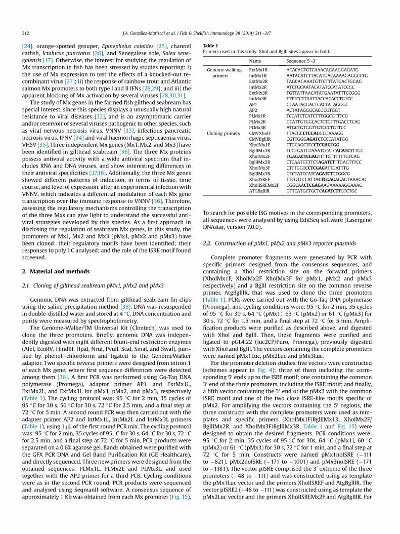
Table 1Primers used in this study. XhoI and BglII sites appear in bold.
Name Sequence 50-30
Genome walkingprimers
ExtMx1R ACACAGTGTCAAACAGAAGGAGATGIntMx1R AATACATCTTACATGACAAAAGAGGCCTGExtMx2R TAGCAGAAATGTTCTTTATGACTGGAGIntMx2R ATCTGCAATACATATCCATATCCGCExtMx3R TGTTATTAACATATGAATATTTCCGGGIntMx3R TTTTCCTTAATTACCACACCTGTCCAP1 GTAATACGACTCACTATAGGGCAP2 ACTATAGGGCACGCGTGGTPLMx1R TCCATCTCATCTTTGGCGTTTCGPLMx2R GTATTGTGGCACTCTGTTTGACCTCAGPLMx3R ATGCTGTGGTTGTCCCTGTTCC
Cloning primers CMVXhoIF TTACGCCTCGAGGCGAAAGGCMVBglIIR CGTTGGGAGATCTCCCATATGGXhoIMx1F CTGCAGCTCCCTCGAGTGGBglIIMx1R TCGTGATGTAAATCCATCAGATCTTTGGXhoIMx2F TGAGACTCGAGTTTTGTTTTTTGTCAGBglIIMx2R CTCAATGTTTCTAGATCTTTGAGTTTCCXhoIMx3F CTTTGGTCCTCGAGATTGATTTGBglIIMx3R GTCTATCCATCAGATCTGTGGCGXhoIISREF TTCGTCCCATTACTCGAGAGAGTAAAGACXhoIISREMx2F CGGCAACTCGAGAAGAAAAAGGAAAGATGBglIIR GTTCATGCTGCTCAGATCTTGTCTGC
J.A. González-Mariscal et al. / Fish & Shellfish Immunology 38 (2014) 311e317312
[24], orange-spotted grouper, Epinephelus coioides [25], channelcatfish, Ictalurus punctatus [26], and Senegalese sole, Solea sene-galensis [17]. Otherwise, the interest for studying the regulation ofMx transcription in fish has been stressed by studies reporting: i)the use of Mx expression to test the effects of a knocked-out re-combinant virus [27]; ii) the response of rainbow trout and Atlanticsalmon Mx promoters to both type I and II IFNs [28,29]; and iii) theapparent blocking of Mx activation by several viruses [28,30,31].
The study of Mx genes in the farmed fish gilthead seabream hasspecial interest, since this species displays a unusually high naturalresistance to viral diseases [32], and is an asymptomatic carrierand/or reservoir of several viruses pathogenic to other species, suchas viral nervous necrosis virus, VNNV [33], infectious pancreaticnecrosis virus, IPNV [34] and viral haemorrhagic septicaemia virus,VHSV [35]. Three independent Mx genes (Mx1, Mx2, andMx3) havebeen identified in gilthead seabream [36]. The three Mx proteinspossess antiviral activity with a wide antiviral spectrum that in-cludes RNA and DNA viruses, and show interesting differences intheir antiviral specificities [37,16]. Additionally, the three Mx genesshowed different patterns of induction, in terms of tissue, timecourse, and level of expression, after an experimental infectionwithVNNV, which indicates a differential modulation of each Mx genetranscription over the immune response to VNNV [36]. Therefore,assessing the regulatory mechanisms controlling the transcriptionof the three Mxs can give light to understand the successful anti-viral strategies developed by this species. As a first approach indisclosing the regulation of seabream Mx genes, in this study, thepromoters of Mx1, Mx2 and Mx3 (pMx1, pMx2 and pMx3) havebeen cloned; their regulatory motifs have been identified; theirresponses to poly I:C analysed; and the role of the ISRE motif foundscreened.
2. Material and methods
2.1. Cloning of gilthead seabream pMx1, pMx2 and pMx3
Genomic DNA was extracted from gilthead seabream fin clipsusing the saline precipitation method [38]. DNA was resuspendedin double-distilled water and stored at 4 �C. DNA concentration andpurity were measured by spectrophotometry.
The Genome-WalkerTM Universal Kit (Clontech) was used toclone the three promoters. Briefly, genomic DNA was indepen-dently digested with eight different blunt-end restriction enzymes(AfeI, EcoRV, HindIII, HpaI, NruI, PvuII, ScaI, SmaI, and SwaI), puri-fied by phenolechloroform and ligated to the GenomeWalkeradaptor. Two specific reverse primers were designed from intron 1of each Mx gene, where first sequence differences were detectedamong them [36]. A first PCR was performed using Go-Taq DNApolymerase (Promega), adaptor primer AP1, and ExtMx1L,ExtMx2L, and ExtMx3L for pMx1, pMx2, and pMx3, respectively(Table 1). The cycling protocol was: 95 �C for 2 min, 35 cycles of95 �C for 30 s, 56 �C for 30 s, 72 �C for 2.5 min, and a final step at72 �C for 5 min. A second round PCR was then carried out with theadapter primer AP2 and IntMx1L, IntMx2L and IntMx3L primers(Table 1), using 1 mL of the first round PCRmix. The cycling protocolwas: 95 �C for 2 min, 35 cycles of 95 �C for 30 s, 64 �C for 30 s, 72 �Cfor 2.5 min, and a final step at 72 �C for 5 min. PCR products wereseparated on a 0.6% agarose gel. Bands obtained were purified withthe GFX PCR DNA and Gel Band Purification Kit (GE Healthcare),and directly sequenced. Three new primers were designed from theobtained sequences: PLMx1L, PLMx2L and PLMx3L, and usedtogether with the AP2 primer for a third PCR. Cycling conditionswere as in the second PCR round. PCR products were sequencedand analysed using SeqmanII software. A consensus sequence ofapproximately 1 Kb was obtained from each Mx promoter (Fig. 1S).
To search for possible ISG motives in the corresponding promoters,all sequences were analysed by using EditSeq software (LasergeneDNAstar, version 7.0.0).
2.2. Construction of pMx1, pMx2 and pMx3 reporter plasmids
Complete promoter fragments were generated by PCR withspecific primers designed from the consensus sequences, andcontaining a XhoI restriction site on the forward primers(XhoIMx1F, XhoIMx2F XhoIMx3F for pMx1, pMx2 and pMx3respectively) and a BglII restriction site on the common reverseprimer, AtgBglIIR, that was used to clone the three promoters(Table 1). PCRs were carried out with the Go-Taq DNA polymerase(Promega), and cycling conditions were: 95 �C for 2 min, 35 cyclesof 95 �C for 30 s, 64 �C (pMx1), 63 �C (pMx2) or 61 �C (pMx3) for30 s, 72 �C for 1.5 min, and a final step at 72 �C for 5 min. Ampli-fication products were purified as described above, and digestedwith XhoI and BglII. Then, these fragments were purified andligated to pGL4.22 (luc2CP/Puro, Promega), previously digestedwith XhoI and BglII. The vectors containing the complete promoterswere named pMx1Luc, pMx2Luc and pMx3Luc.
For the promoter deletion studies, five vectors were constructed(schemes appear in Fig. 4): three of them including the corre-sponding 50 ends up to the ISRE motif; one containing the common30 end of the three promoters, including the ISRE motif; and finally,a fifth vector containing the 30 end of the pMx2 with the commonISRE motif and one of the two close ISRE-like motifs specific ofpMx2. For amplifying the vectors containing the 50 regions, thethree constructs with the complete promoters were used as tem-plates and specific primers (XhoIMx1F/BglIIMx1R, XhoIMx2F/BglIIMx2R, and XhoIMx3F/BglIIMx3R, Table 1 and Fig. 1S) weredesigned to obtain the desired fragments. PCR conditions were:95 �C for 2 min, 35 cycles of 95 �C for 30s, 64 �C (pMx1), 60 �C(pMx2) or 61 �C (pMx3) for 30 s, 72 �C for 1 min, and a final step at72 �C for 5 min. Constructs were named pMx1noISRE (�111to �821), pMx2noISRE (�171 to �1001) and pMx3noISRE (�171to �1181). The vector pISRE comprised the 30 extreme of the threepromoters (�48 to �111) and was constructed using as templatethe pMx1Luc vector and the primers XhoIISREF and AtgBglIIR. Thevector pISRE2 (�48 to�111) was constructed using as template thepMx2Luc vector and the primers XhoIISREMx2F and AtgBglIIR. For
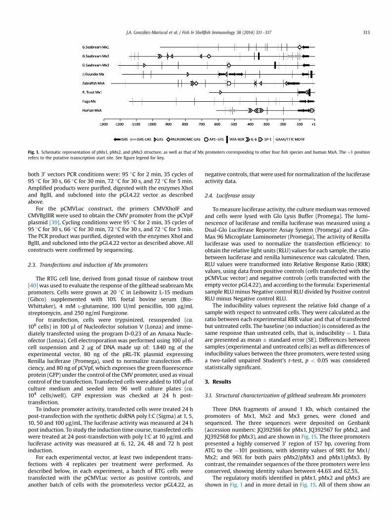
Fig. 1. Schematic representation of pMx1, pMx2, and pMx3 structure, as well as that of Mx promoters corresponding to other four fish species and human MxA. The þ1 positionrefers to the putative transcription start site. See figure legend for key.
J.A. González-Mariscal et al. / Fish & Shellfish Immunology 38 (2014) 311e317 313
both 30 vectors PCR conditions were: 95 �C for 2 min, 35 cycles of95 �C for 30 s, 66 �C for 30 min, 72 �C for 30 s, and 72 �C for 5 min.Amplified products were purified, digested with the enzymes XhoIand BglII, and subcloned into the pGL4.22 vector as describedabove.
For the pCMVLuc construct, the primers CMVXhoIF andCMVBglIIR were used to obtain the CMV promoter from the pCVpFplasmid [39]. Cycling conditions were 95 �C for 2 min, 35 cycles of95 �C for 30 s, 66 �C for 30 min, 72 �C for 30 s, and 72 �C for 5 min.The PCR product was purified, digested with the enzymes XhoI andBglII, and subcloned into the pGL4.22 vector as described above. Allconstructs were confirmed by sequencing.
2.3. Transfections and induction of Mx promoters
The RTG cell line, derived from gonad tissue of rainbow trout[40] was used to evaluate the response of the gilthead seabreamMxpromoters. Cells were grown at 20 �C in Leibowitz L-15 medium(Gibco) supplemented with 10% foetal bovine serum (Bio-Whittaker), 4 mM L-glutamine, 100 U/ml penicillin, 100 mg/mLstreptomycin, and 250 ng/ml Fungizone.
For transfection, cells were trypsinized, resuspended (ca.106 cells) in 100 ml of Nucleofector solution V (Lonza) and imme-diately transfected using the program D-0.23 of an Amaxa Nucle-ofector (Lonza). Cell electroporation was performed using 100 ml ofcell suspension and 2 mg of DNA made up of: 1.840 ng of theexperimental vector, 80 ng of the pRL-TK plasmid expressingRenilla luciferase (Promega), used to normalize transfection effi-ciency, and 80 ng of pCVpf, which expresses the green fluorescenceprotein (GFP) under the control of the CMV promoter, used as visualcontrol of the transfection. Transfected cells were added to 100 ml ofculture medium and seeded into 96 well culture plates (ca.104 cells/well). GFP expression was checked at 24 h post-transfection.
To induce promoter activity, transfected cells were treated 24 hpost-transfection with the synthetic dsRNA poly I:C (Sigma) at 1, 5,10, 50 and 100 mg/mL. The luciferase activity was measured at 24 hpost induction. To study the induction time course, transfected cellswere treated at 24 post-transfection with poly I:C at 10 mg/mL andluciferase activity was measured at 6, 12, 24, 48 and 72 h postinduction.
For each experimental vector, at least two independent trans-fections with 4 replicates per treatment were performed. Asdescribed below, in each experiment, a batch of RTG cells weretransfected with the pCMVLuc vector as positive controls, andanother batch of cells with the promoterless vector pGL4.22, as
negative controls, that were used for normalization of the luciferaseactivity data.
2.4. Luciferase assay
Tomeasure luciferase activity, the culturemediumwas removedand cells were lysed with Glo Lysis Buffer (Promega). The lumi-nescence of luciferase and renilla luciferase was measured using aDual-Glo Luciferase Reporter Assay System (Promega) and a Glo-Max 96 Microplate Luminometer (Promega). The activity of Renillaluciferase was used to normalize the transfection efficiency: toobtain the relative light units (RLU) values for each sample, the ratiobetween luciferase and renilla luminescence was calculated. Then,RLU values were transformed into Relative Response Ratio (RRR)values, using data from positive controls (cells transfected with thepCMVLuc vector) and negative controls (cells transfected with theempty vector pGL4.22), and according to the formula: Experimentalsample RLUminus Negative control RLU divided by Positive controlRLU minus Negative control RLU.
The inducibility values represent the relative fold change of asample with respect to untreated cells. They were calculated as theratio between each experimental RRR value and that of transfectedbut untreated cells. The baseline (no induction) is considered as thesame response than untreated cells, that is, inducibility ¼ 1. Dataare presented as mean � standard error (SE). Differences betweensamples (experimental and untreated cells) as well as differences ofinducibility values between the three promoters, were tested usinga two-tailed unpaired Student’s t-test, p < 0.05 was consideredstatistically significant.
3. Results
3.1. Structural characterization of gilthead seabream Mx promoters
Three DNA fragments of around 1 Kb, which contained thepromoters of Mx1, Mx2 and Mx3 genes, were cloned andsequenced. The three sequences were deposited on Genbank(accession numbers: JQ392566 for pMx1, JQ392567 for pMx2, andJQ392568 for pMx3), and are shown in Fig. 1S. The three promoterspresented a highly conserved 30 region of 157 bp, covering fromATG to the �101 positions, with identity values of 98% for Mx1/Mx2; and 96% for both pairs pMx2/pMx3 and pMx1/pMx3. Bycontrast, the remainder sequences of the three promoters were lessconserved, showing identity values between 44.6% and 62.5%.
The regulatory motifs identified in pMx1, pMx2 and pMx3 areshown in Fig. 1 and in more detail in Fig. 1S. All of them show an
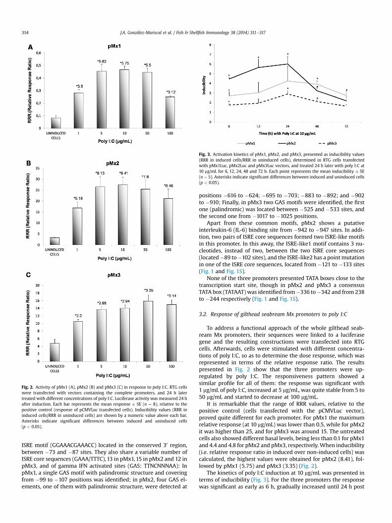
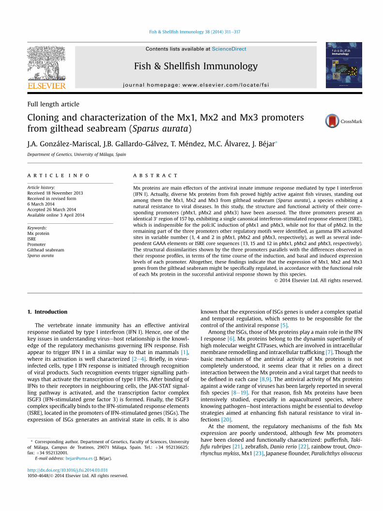
Fig. 2. Activity of pMx1 (A), pMx2 (B) and pMx3 (C) in response to poly I:C. RTG cellswere transfected with vectors containing the complete promoters, and 24 h latertreated with different concentrations of poly I:C. Luciferase activity was measured 24 hafter induction. Each bar represents the mean response � SE (n ¼ 8), relative to thepositive control (response of pCMVLuc transfected cells). Inducibility values (RRR ininduced cells/RRR in uninduced cells) are shown by a numeric value above each bar.Asterisks indicate significant differences between induced and uninduced cells(p < 0.05).
Fig. 3. Activation kinetics of pMx1, pMx2, and pMx3, presented as inducibility values(RRR in induced cells/RRR in uninduced cells), determined in RTG cells transfectedwith pMx1Luc, pMx2Luc and pMx3Luc vectors, and treated 24 h later with poly I:C at10 mg/mL for 6, 12, 24, 48 and 72 h. Each point represents the mean inducibility � SE(n ¼ 5). Asterisks indicate significant differences between induced and uninduced cells(p < 0.05).
J.A. González-Mariscal et al. / Fish & Shellfish Immunology 38 (2014) 311e317314
ISRE motif (GGAAACGAAACC) located in the conserved 30 region,between �73 and �87 sites. They also share a variable number ofISRE core sequences (GAAA/TTTC), 13 in pMx1,15 in pMx2 and 12 inpMx3, and of gamma IFN activated sites (GAS: TTNCNNNAA): InpMx1, a single GAS motif with palindromic structure and coveringfrom �99 to �107 positions was identified; in pMx2, four GAS el-ements, one of them with palindromic structure, were detected at
positions �616 to �624; �695 to �703; �883 to �892; and �902to �910; Finally, in pMx3 two GAS motifs were identified, the firstone (palindromic) was located between �525 and �533 sites, andthe second one from �1017 to �1025 positions.
Apart from these common motifs, pMx2 shows a putativeinterleukin-6 (IL-6) binding site from �942 to �947 sites. In addi-tion, two pairs of ISRE core sequences formed two ISRE-like motifsin this promoter. In this away, the ISRE-like1 motif contains 3 nu-cleotides, instead of two, between the two ISRE core sequences(located�89 to�102 sites), and the ISRE-like2 has a point mutationin one of the ISRE core sequences, located from �121 to �133 sites(Fig. 1 and Fig. 1S).
None of the three promoters presented TATA boxes close to thetranscription start site, though in pMx2 and pMx3 a consensusTATA box (TATAAT)was identified from�336 to�342 and from238to �244 respectively (Fig. 1 and Fig. 1S).
3.2. Response of gilthead seabream Mx promoters to poly I:C
To address a functional approach of the whole gilthead seab-ream Mx promoters, their sequences were linked to a luciferasegene and the resulting constructions were transfected into RTGcells. Afterwards, cells were stimulated with different concentra-tions of poly I:C, so as to determine the dose response, which wasrepresented in terms of the relative response ratio. The resultspresented in Fig. 2 show that the three promoters were up-regulated by poly I:C. The responsiveness pattern showed asimilar profile for all of them: the response was significant with1 mg/mL of poly I:C, increased at 5 mg/mL, was quite stable from 5 to50 mg/mL and started to decrease at 100 mg/mL.
It is remarkable that the range of RRR values, relative to thepositive control (cells transfected with the pCMVLuc vector),proved quite different for each promoter. For pMx1 the maximumrelative response (at 10 mg/mL) was lower than 0.5, while for pMx2it was higher than 25, and for pMx3 was around 15. The untreatedcells also showed different basal levels, being less than 0.1 for pMx1and 4.4 and 4.8 for pMx2 and pMx3, respectively.When inducibility(i.e. relative response ratio in induced over non-induced cells) wascalculated, the highest values were obtained for pMx2 (8.41), fol-lowed by pMx1 (5.75) and pMx3 (3.35) (Fig. 2).
The kinetics of poly I:C induction at 10 mg/mL was presented interms of inducibility (Fig. 3). For the three promoters the responsewas significant as early as 6 h, gradually increased until 24 h post
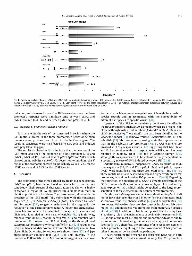
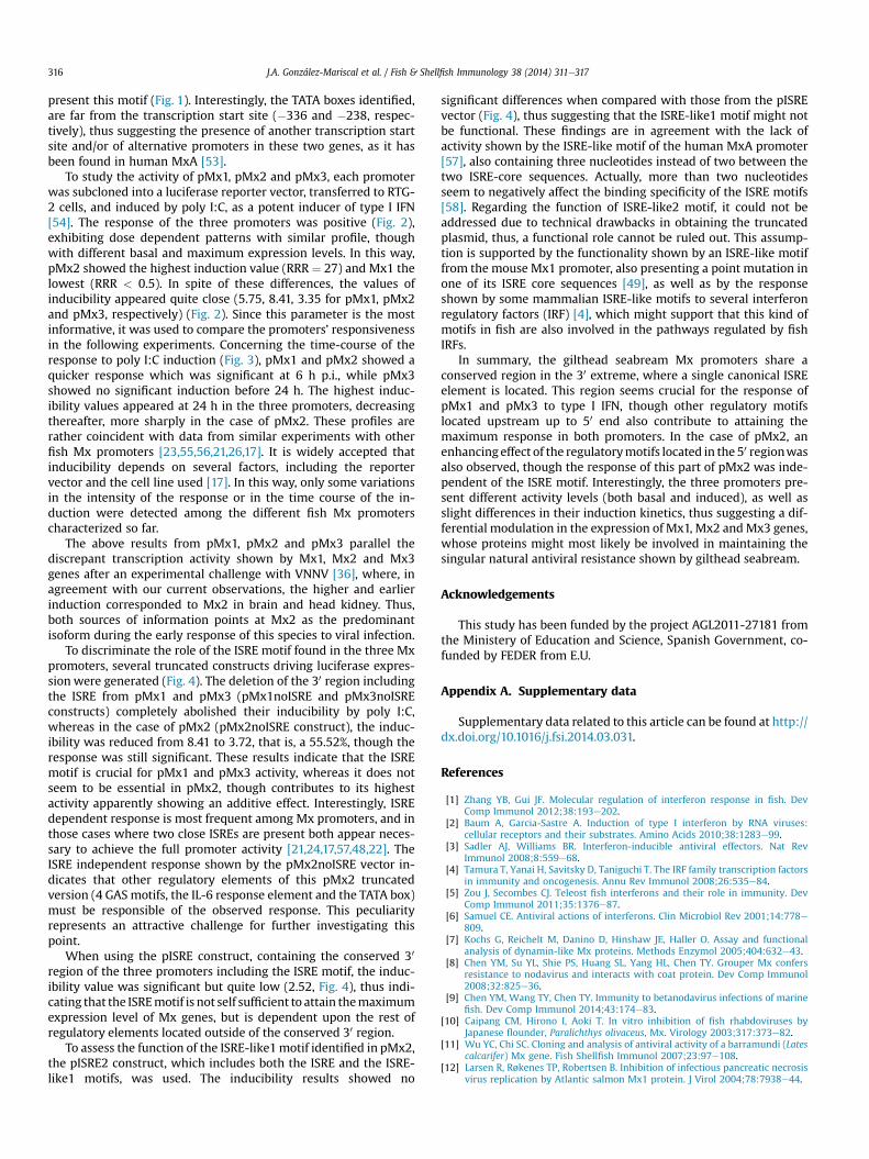
Fig. 4. Functional analysis of pMx1, pMx2 and pMx3 deletion mutants. Inducibility values (RRR in induced cells/RRR in uninduced cells) were determined in RTG transfected cells,treated 24 h later with poly I:C at 10 mg/mL for 24 h. Each point represents the mean inducibility � SE (n ¼ 8). Asterisks indicate significant differences between induced anduninduced cells (p < 0.05). Different letters denote significant differences between bars (p < 0.05).
J.A. González-Mariscal et al. / Fish & Shellfish Immunology 38 (2014) 311e317 315
induction, and decreased thereafter. Differences between the threepromoter’s response were significant only between pMx2 andpMx3 from 6 h to 48 h, and between pMx1 and pMx3 at 48 h.
3.3. Response of promoters’ deletion mutants
To characterize the role of the conserved 30 region where theISRE motif is located in the three promoters, a series of deletionmutants were produced and fused to the luciferase gene. Theresulting constructs were transfected into RTG cells and inducedwith poly I:C at 10 mg/mL.
The results displayed in Fig. 4 indicate that the deletion of theISRE motif abolished the response of pMx1 (pMx1noISRE) andpMx3 (pMx3noISRE), but not that of pMx2 (pMx2noISRE), whichshowed an inducibility value of 3.72. Vectors only containing the 30
region of the promoters showed an inducibility value of 2.52 for thepISRE vector, and of 1.93 for the pISRE2 vector.
4. Discussion
The promoters of the three gilthead seabream Mx genes (pMx1,pMx2 and pMx3) have been cloned and characterized in the pre-sent study. Their structural characterization has shown a highlyconserved 30 region of 157 bp, presenting a single ISRE motif inidentical position in all of them. The conservation, along with thepresence of the ISRE motif, fully coincident with the consensussequence (A/G/T)GAAA(N)1-2GAAA(C/G)(A/C/T) described by Colletand Secombes [23], suggest a main role for this region in theregulation of the corresponding genes. Although the characteriza-tion of Mx promoters in fish is limited to few species, the number ofISREs so far identified in them is rather variable (Fig. 1). In this way,rainbow trout Mx [23], channel catfish Mx [26] and zebrafish Mxgpromoters [41] present one ISRE, similarly to the three giltheadseabream Mx promoters, whereas pufferfish Mx [21], grouper Mx[25], and Mxa and Mxb promoters from zebrafish [41], contain twoclose ISREs. Otherwise, Senegalese sole shows three [17] and Jap-anese flounder contains four ISREs [24]. This diversity in thenumber of ISRE motifs in fish Mx promoters suggests a crucial role
for them in theMx expression regulationwhichmight be somehowspecies specific and in accordance with the susceptibility ofdifferent fish species to specific viruses [41].
Upstream of the ISRE, other regulatory motifs were identified inthe three promoters, such as GAS elements, which are present in allof them, though in different numbers (1, 4 and 2 in pMx1, pMx2 andpMx3, respectively). These motifs have also been identified in theJapanese flounder [24], rainbow trout [23], Senegalese sole [17] andzebrafish [22] Mx promoters, showing a similar representationthan in the seabream Mx promoters (Fig. 1). GAS elements areinvolved in IFN-g responsiveness [42], suggesting that Mx1, Mx2andMx3 expressionmight also respond to type II IFN, as it has beenreported in rainbow trout [28] and in Atlantic salmon [29],although this response seems to be, at least partially, dependant ona secondary release of IFN I induced by type II IFN [28,29].
Additionally, numerous independent GAAA elements or ISREcore sequences (13, 15 and 12 in pMx1, pMx2 and pMx3, respec-tively) were identified in the three promoters (Fig. 1 and Fig. 1S).These motifs are also widespread in fish and higher vertebrates Mxpromoters, as well as in other ISG promoters [43e45]. Regardingtheir function, the presence of 10 GAAA elements upstream of theISREs in zebrafish Mxa promoter, seems to play a role in enhancinggene expression [22], which might be applied to the large repre-sentation of these elements in the seabream Mx promoters.
Besides, an IL-6 response element was identified in pMx2. IL-6motifs have also been described in other fish Mx promoters, suchas rainbow trout [23], channel catfish [26], and zebrafish Mxa [22]promoters. Otherwise, they are also present in chicken Mx pro-moter [46], and in several Mx promoters from mammalian species[47e49,45,50]. In addition, it has been reported that IL-6 may havea regulatory role in themaintenance of bovineMx1 expression [45].IL-6 is one of the most pleiotropic and important cytokines due toits important role mediating the innate and adaptive immune re-sponses [51,52]. Therefore, the presence of IL-6 response elementsin Mx promoters might suggest the involvement of Mx genes inother immune response signalling pathways.
Finally, regarding the presence of a consensus TATA box in bothpMx2 and pMx3, it results unusual, as only few Mx promoters
J.A. González-Mariscal et al. / Fish & Shellfish Immunology 38 (2014) 311e317316
present this motif (Fig. 1). Interestingly, the TATA boxes identified,are far from the transcription start site (�336 and �238, respec-tively), thus suggesting the presence of another transcription startsite and/or of alternative promoters in these two genes, as it hasbeen found in human MxA [53].
To study the activity of pMx1, pMx2 and pMx3, each promoterwas subcloned into a luciferase reporter vector, transferred to RTG-2 cells, and induced by poly I:C, as a potent inducer of type I IFN[54]. The response of the three promoters was positive (Fig. 2),exhibiting dose dependent patterns with similar profile, thoughwith different basal and maximum expression levels. In this way,pMx2 showed the highest induction value (RRR ¼ 27) and Mx1 thelowest (RRR < 0.5). In spite of these differences, the values ofinducibility appeared quite close (5.75, 8.41, 3.35 for pMx1, pMx2and pMx3, respectively) (Fig. 2). Since this parameter is the mostinformative, it was used to compare the promoters’ responsivenessin the following experiments. Concerning the time-course of theresponse to poly I:C induction (Fig. 3), pMx1 and pMx2 showed aquicker response which was significant at 6 h p.i., while pMx3showed no significant induction before 24 h. The highest induc-ibility values appeared at 24 h in the three promoters, decreasingthereafter, more sharply in the case of pMx2. These profiles arerather coincident with data from similar experiments with otherfish Mx promoters [23,55,56,21,26,17]. It is widely accepted thatinducibility depends on several factors, including the reportervector and the cell line used [17]. In this way, only some variationsin the intensity of the response or in the time course of the in-duction were detected among the different fish Mx promoterscharacterized so far.
The above results from pMx1, pMx2 and pMx3 parallel thediscrepant transcription activity shown by Mx1, Mx2 and Mx3genes after an experimental challenge with VNNV [36], where, inagreement with our current observations, the higher and earlierinduction corresponded to Mx2 in brain and head kidney. Thus,both sources of information points at Mx2 as the predominantisoform during the early response of this species to viral infection.
To discriminate the role of the ISRE motif found in the three Mxpromoters, several truncated constructs driving luciferase expres-sion were generated (Fig. 4). The deletion of the 30 region includingthe ISRE from pMx1 and pMx3 (pMx1noISRE and pMx3noISREconstructs) completely abolished their inducibility by poly I:C,whereas in the case of pMx2 (pMx2noISRE construct), the induc-ibility was reduced from 8.41 to 3.72, that is, a 55.52%, though theresponse was still significant. These results indicate that the ISREmotif is crucial for pMx1 and pMx3 activity, whereas it does notseem to be essential in pMx2, though contributes to its highestactivity apparently showing an additive effect. Interestingly, ISREdependent response is most frequent among Mx promoters, and inthose cases where two close ISREs are present both appear neces-sary to achieve the full promoter activity [21,24,17,57,48,22]. TheISRE independent response shown by the pMx2noISRE vector in-dicates that other regulatory elements of this pMx2 truncatedversion (4 GAS motifs, the IL-6 response element and the TATA box)must be responsible of the observed response. This peculiarityrepresents an attractive challenge for further investigating thispoint.
When using the pISRE construct, containing the conserved 30
region of the three promoters including the ISRE motif, the induc-ibility value was significant but quite low (2.52, Fig. 4), thus indi-cating that the ISREmotif is not self sufficient to attain themaximumexpression level of Mx genes, but is dependent upon the rest ofregulatory elements located outside of the conserved 30 region.
To assess the function of the ISRE-like1motif identified in pMx2,the pISRE2 construct, which includes both the ISRE and the ISRE-like1 motifs, was used. The inducibility results showed no
significant differences when compared with those from the pISREvector (Fig. 4), thus suggesting that the ISRE-like1 motif might notbe functional. These findings are in agreement with the lack ofactivity shown by the ISRE-like motif of the human MxA promoter[57], also containing three nucleotides instead of two between thetwo ISRE-core sequences. Actually, more than two nucleotidesseem to negatively affect the binding specificity of the ISRE motifs[58]. Regarding the function of ISRE-like2 motif, it could not beaddressed due to technical drawbacks in obtaining the truncatedplasmid, thus, a functional role cannot be ruled out. This assump-tion is supported by the functionality shown by an ISRE-like motiffrom the mouse Mx1 promoter, also presenting a point mutation inone of its ISRE core sequences [49], as well as by the responseshown by some mammalian ISRE-like motifs to several interferonregulatory factors (IRF) [4], which might support that this kind ofmotifs in fish are also involved in the pathways regulated by fishIRFs.
In summary, the gilthead seabream Mx promoters share aconserved region in the 30 extreme, where a single canonical ISREelement is located. This region seems crucial for the response ofpMx1 and pMx3 to type I IFN, though other regulatory motifslocated upstream up to 50 end also contribute to attaining themaximum response in both promoters. In the case of pMx2, anenhancing effect of the regulatorymotifs located in the 50 regionwasalso observed, though the response of this part of pMx2 was inde-pendent of the ISRE motif. Interestingly, the three promoters pre-sent different activity levels (both basal and induced), as well asslight differences in their induction kinetics, thus suggesting a dif-ferential modulation in the expression of Mx1, Mx2 andMx3 genes,whose proteins might most likely be involved in maintaining thesingular natural antiviral resistance shown by gilthead seabream.
Acknowledgements
This study has been funded by the project AGL2011-27181 fromthe Ministery of Education and Science, Spanish Government, co-funded by FEDER from E.U.
Appendix A. Supplementary data
Supplementary data related to this article can be found at http://dx.doi.org/10.1016/j.fsi.2014.03.031.
References
[1] Zhang YB, Gui JF. Molecular regulation of interferon response in fish. DevComp Immunol 2012;38:193e202.
[2] Baum A, Garcia-Sastre A. Induction of type I interferon by RNA viruses:cellular receptors and their substrates. Amino Acids 2010;38:1283e99.
[3] Sadler AJ, Williams BR. Interferon-inducible antiviral effectors. Nat RevImmunol 2008;8:559e68.
[4] Tamura T, Yanai H, Savitsky D, Taniguchi T. The IRF family transcription factorsin immunity and oncogenesis. Annu Rev Immunol 2008;26:535e84.
[5] Zou J, Secombes CJ. Teleost fish interferons and their role in immunity. DevComp Immunol 2011;35:1376e87.
[6] Samuel CE. Antiviral actions of interferons. Clin Microbiol Rev 2001;14:778e809.
[7] Kochs G, Reichelt M, Danino D, Hinshaw JE, Haller O. Assay and functionalanalysis of dynamin-like Mx proteins. Methods Enzymol 2005;404:632e43.
[8] Chen YM, Su YL, Shie PS, Huang SL, Yang HL, Chen TY. Grouper Mx confersresistance to nodavirus and interacts with coat protein. Dev Comp Immunol2008;32:825e36.
[9] Chen YM, Wang TY, Chen TY. Immunity to betanodavirus infections of marinefish. Dev Comp Immunol 2014;43:174e83.
[10] Caipang CM, Hirono I, Aoki T. In vitro inhibition of fish rhabdoviruses byJapanese flounder, Paralichthys olivaceus, Mx. Virology 2003;317:373e82.
[11] Wu YC, Chi SC. Cloning and analysis of antiviral activity of a barramundi (Latescalcarifer) Mx gene. Fish Shellfish Immunol 2007;23:97e108.
[12] Larsen R, Røkenes TP, Robertsen B. Inhibition of infectious pancreatic necrosisvirus replication by Atlantic salmon Mx1 protein. J Virol 2004;78:7938e44.
J.A. González-Mariscal et al. / Fish & Shellfish Immunology 38 (2014) 311e317 317
[13] Lin C, Christopher John JA, Lin C, Chang C. Inhibition of nervous necrosis viruspropagation by fish Mx proteins. Biochem Biophys Res Commun 2006;351:534e9.
[14] Kibenge MJT, Munir K, Kibenge FSB. Constitutive expression of Atlanticsalmon Mx1 protein in CHSE-214 cells confers resistance to infectious salmonanaemia virus. Virol J 2005;2:75.
[15] Fernández-Trujillo MA, García-Rosado E, Alonso MC, Borrego JJ, Álvarez MC,Béjar J. In vitro inhibition of sole aquabirnavirus by Senegalese sole Mx. FishShellfish Immunol 2008;24:187e93.
[16] Fernandez-Trujillo MA, Garcia-Rosado E, Alonso MC, Castro D, Alvarez MC,Béjar J. Mx1, Mx2 and Mx3 proteins from the gilthead seabream (Sparusaurata) show antiviral activity against RNA and DNA viruses. Mol Immunol2013;56:630e6.
[17] Alvarez-Torres D, Bejar J, Collet B, Alonso MC, Garcia-Rosado E. Structural andfunctional characterization of the Senegalese sole (Solea senegalensis) Mxpromoter. Fish Shellfish Immunol 2013;35:1642e8.
[18] Lester K, Hall M, Urquhart K, Gahlawat S, Collet B. Development of an in vitrosystem to measure the sensitivity to the antiviral Mx protein of fish viruses.J Virol Methods 2012;182:1e8.
[19] Trobridge GD, Chiou PP, Leong JA. Cloning of the rainbow trout (Oncorhynchusmykiss) Mx2 and Mx3 cDNAs and characterization of trout Mx proteínaexpresión in salmon cells. J Virol 1997;71:5304e11.
[20] Magnadottir B. Immunological control of fish diseases. Mar Biotechnol2010;12:361e79.
[21] Yap WH, Tay A, Brenner S, Venkatesh B. Molecular cloning of the pufferfish(Takifugu rubripes) Mx gene and functional characterization of its promoter.Immunogenetics 2003;54:705e13.
[22] Altmann SM, Mellon MT, Johnson MC, Paw BH, Trede NS, Zon LI, et al. Cloningand characterization of an Mx gene and its corresponding promoter from thezebrafish, Danio rerio. Dev Comp Immunol 2004;28:295e306.
[23] Collet B, Secombes CJ. The rainbow trout (Oncorhynchus mykiss) Mx1 pro-moter: structural and functional characterization. Eur J Biochem 2001;268:1577e84.
[24] Ooi EL, Hirono I, Aoki T. Functional characterisation of the Japanese flounder,Paralichthys olivaceus, Mx promoter. Fish Shellfish Immunol 2006;21:293e304.
[25] Chen Y, Su Y, Lin JH, Yang H, Chen T. Cloning of an orange-spotted grouper(Epinephelus coioides) Mx cDNA and characterisation of its expression inresponse to nodavirus. Fish Shellfish Immunol 2006;20:58e71.
[26] Plant KP, Thune RL. Genomic organisation of the channel catfish Mx1 geneand characterisation of multiple channel catfish Mx gene promoters. FishShellfish Immunol 2008;24:575e83.
[27] KimMS, Kim KH. Effects of NV gene knock-out recombinant viral hemorrhagicsepticemia virus (VHSV) on Mx gene expression in Epithelioma papulosumcyprini (EPC) cells and olive flounder (Paralichthys olivaceus). Fish ShellfishImmunol 2012;32:459e63.
[28] Jørgensen JB, Johansen A, Hegseth MN, Zou J, Robertsen B, Collet B, et al.A recombinant CHSE-214 cell line expressing an Mx1 promoter-reportersystem responds to both interferon type I and type II from salmonids andrepresents a versatile tool to study the IFN-system in teleost fish. Fish Shell-fish Immunol 2007;23:1294e303.
[29] Sun F, Zhang YB, Liu TK, Shi J, Wang B, Gui JF. Fish MITA serves as a mediatorfor distinct fish IFN gene activation dependent on IRF3 or IRF7. J Immunol2011;187:2531e9.
[30] Collet B, Munro ES, Gahlawat S, Acosta F, Garcia J, Roemelt C, et al. Infectiouspancreatic necrosis virus suppresses type I interferon signalling in rainbowtrout gonad cell line but not in Atlantic salmon macrophages. Fish ShellfishImmunol 2007;22:44e56.
[31] Skjesol A, Aamo T, Hegseth MN, Robertsen B, Jørgensen JB. The interplaybetween infectious pancreatic necrosis virus (IPNV) and the IFN system: IFNsignaling is inhibited by IPNV infection. Virus Res 2009;143:53e60.
[32] Borrego JJ, DCastro MC, Balebona ME, García-Rosado L, López-Cortés. Patolo-gías de la dorada. Junta de Andalucía, Consejería de Agricultura y pesca; 2000.
[33] Castri J, Thiéry R, Jeffroy J, de Kinkelin P, Raymond JC. Sea bream Sparus aurata,an asymptomatic contagious fish host for nodavirus. Dis Aquat Organ2001;47:33e8.
[34] Perez-Prieto S, Garcia-Rosado E, Rodriguez S, Castro D, Borrego JJ. Antigenicproperties and experimental transmission to several fish species of a marinebirnavirus isolated from sole (Solea senegalensis). VetMicrobiol 2001;82:11e25.
[35] Esteban MA, Meseguer J, Tafalla C, Cuesta A. NK-like and oxidative burst ac-tivities are the main early cellular innate immune responses activated aftervirus inoculation in reservoir fish. Fish Shellfish Immunol 2008;25:433e8.
[36] Fernández-Trujillo MA, Novel P, Manchado M, Sepulcre MP, Mulero V,Borrego JJ, et al. Three Mx genes with differential response to VNNV infectionhave been identified in gilthead seabream (Sparus aurata). Mol Immunol2011;48:1216e23.
[37] Fernández-Trujillo MA, García-Rosado E, Alonso MC, Borrego JJ, Álvarez MC,Béjar J. Differential antiviral activity of Mx1, Mx2 and Mx3 proteins fromgilthead seabream (Sparus aurata) against Infectious Pancreatic Necrosis Virus(IPNV). Mol Immunol 2011;49:107e14.
[38] Martínez G, Shaw EM, Carrillo M, Zanuy S. A protein salting-out methodapplied in genomic DNA isolation from fish whole blood. Biotechniques1988;24:238e9.
[39] Zhao H, Li M, Purwanti YI, Liu R, Chen T, Li Z, et al. Mitf is a transcriptionalactivator of medaka germ genes in culture. Biochimie 2012;94:759e67.
[40] Wolf K, Quimby MC, Carlson CP. Infectious pancreatic necrosis virus: lyoph-ilization and subsequent stability in storage at 4 �C. Appl Microbiol 1969;17:623e4.
[41] Li G, Zhang J, Sun Y, Wang H, Wang Y. The evolutionarily dynamic IFN-inducible GTPase proteins play conserved immune functions in vertebratesand cephalochordates. Mol Biol Evol 2009;26:1619e30.
[42] Castro R, Martin SA, Bird S, Lamas J, Secombes CJ. Characterisation of gamma-interferon responsive promoters in fish. Mol Immunol 2008;45:3454e62.
[43] Chang KC, Hansen E, Foroni L, Lida J, Goldspink G. Molecular and functionalanalysis of the virus- and interferon-inducible human MxA promoter. ArchVirol 1991;117:1e15.
[44] Hug H, Costas M, Staeheli P, Aebi M, Weissmann C. Organization of the murineMx gene and characterization of its interferon- and virus-inducible promoter.Mol Cell Biol 1988;8:3065e79.
[45] Gerardin JA, Baise EA, Pire GA, Leroy M, Desmecht D. Genomic structure,organisation, and promoter analysis of the bovine (Bos taurus) Mx1 gene.Gene 2004;326:67e75.
[46] Schumacher B, Bernasconi D, Schultz U, Staeheli P. The chicken Mx promotercontains an ISRE motif and confers interferon inducibility to a reporter gene inchick and monkey cells. Virology 1994;203:144e8.
[47] Horisberger MA, Haller O, Arnheiter H. Interferon-dependent genetic resis-tance to influenza virus in mice: virus replication in macrophages is inhibitedat an early step. J Gen Virol 1980;50:205e10.
[48] Assiri AM, Ott TL. Cloning and characterizing of the ovine Mx1 gene promoter/enhancer region. Dev Comp Immunol 2007;31:847e57.
[49] Asano A, Jin HK, Watanabe T. Mouse Mx2 gene: organization, mRNAexpression and the role of the interferon-response promoter in its regulation.Gene 2003;306:105e13.
[50] Yamada K, Nakatsu Y, Onogi A, Takasuga A, Sugimoto Y, Ueda J, et al. Struc-tural and functional analysis of the bovine Mx1 promoter. J Interferon Cyto-kine Res 2009;29:217e26.
[51] Akira S, Hirano T, Taga T, Kishimoto T. Biology of multifunctional cytokines: IL6 and related molecules (IL 1 and TNF). FASEB J 1990;11:2860e7.
[52] Naka T, Nishimoto N, Kishimoto T. The paradigm of IL-6: from basic science tomedicine. Arthritis Res 2002;4:S233e42.
[53] Noguchi S, Hijikata M, Hamano E, Matsushita I, Ito H, Ohashi J, et al. MxAtranscripts with distinct first exons and modulation of gene expression levelsby single-nucleotide polymorphisms in human bronchial epithelial cells.Immunogenetics 2013;65:107e14.
[54] Biron CA, Sen GC. Interferons and other cytokines. In: Knipe DM, Howley PM,editors. Fields virology. 4th ed.. Lippincott Williams & Wilkins; 2001, p. 321e351.
[55] Collet B, Boudinot P, Benmansour A, Secombes CJ. An Mx1 promoter-reportersystem to study interferon pathways in rainbow trout. Dev Comp Immunol2004;28:793e801.
[56] Johansen A, Collet B, Sandaker E, Secombes CJ, Jørgensen JB. Quantification ofatlantic salmon type-I interferon using an Mx1 promoter reporter gene assay.Fish Shellfish Immunol 2004;16:173e84.
[57] Ronni T, Matikainen S, Lehtonen A, Palvimo J, Dellis J, Van Eylen F, et al. Theproximal interferon-stimulated response elements are essential for interferonresponsiveness: a promoter analysis of the antiviral MxA gene. J InterferonCytokine Res 1998;18:773e81.
[58] Lin R, Genin P, Mamane Y, Hiscott J. Selective DNA binding and associationwith the CREB binding protein coactivator contribute to differential activationof alpha/beta interferon genes by interferon regulatory factors 3 and 7. MolCell Biol 2000;20:6342e53.