Embed Size (px)
Citation preview
M. tuberculosis sirr characterization
Vol 45 No. 3 May 2014 689
Correspondence: Kiatichai Faksri, Department of Microbiology, Faculty of Medicine; Research and Diagnostic Center for Emerging Infectious Diseases, Khon Kaen University, Khon Kaen 40002, Thailand.Tel: +66 (0) 43 363808; Fax: +66 (0) 43 363808.E-mail: [email protected]
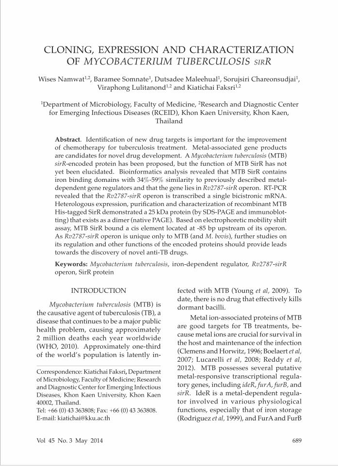
CLONING, EXPRESSION AND CHARACTERIZATION OF MYCOBACTERIUM TUBERCULOSIS sirR
Wises Namwat1,2, Baramee Somnate1, Dutsadee Maleehual1, Sorujsiri Chareonsudjai1, Viraphong Lulitanond1,2 and Kiatichai Faksri1,2
1Department of Microbiology, Faculty of Medicine, 2Research and Diagnostic Center for Emerging Infectious Diseases (RCEID), Khon Kaen University, Khon Kaen,
Thailand
Abstract. Identification of new drug targets is important for the improvement of chemotherapy for tuberculosis treatment. Metal-associated gene products are candidates for novel drug development. A Mycobacterium tuberculosis (MTB) sirR-encoded protein has been proposed, but the function of MTB SirR has not yet been elucidated. Bioinformatics analysis revealed that MTB SirR contains iron binding domains with 34%-59% similarity to previously described metal-dependent gene regulators and that the gene lies in Rv2787-sirR operon. RT-PCR revealed that the Rv2787-sirR operon is transcribed a single bicistronic mRNA. Heterologous expression, purification and characterization of recombinant MTB His-tagged SirR demonstrated a 25 kDa protein (by SDS-PAGE and immunoblot-ting) that exists as a dimer (native PAGE). Based on electrophoretic mobility shift assay, MTB SirR bound a cis element located at -85 bp upstream of its operon. As Rv2787-sirR operon is unique only to MTB (and M. bovis), further studies on its regulation and other functions of the encoded proteins should provide leads towards the discovery of novel anti-TB drugs.
Keywords: Mycobacterium tuberculosis, iron-dependent regulator, Rv2787-sirR operon, SirR protein
fected with MTB (Young et al, 2009). To date, there is no drug that effectively kills dormant bacilli.
Metal ion-associated proteins of MTB are good targets for TB treatments, be-cause metal ions are crucial for survival in the host and maintenance of the infection (Clemens and Horwitz, 1996; Boelaert et al, 2007; Lucarelli et al, 2008; Reddy et al, 2012). MTB possesses several putative metal-responsive transcriptional regula-tory genes, including ideR, furA, furB, and sirR. IdeR is a metal-dependent regula-tor involved in various physiological functions, especially that of iron storage (Rodriguez et al, 1999), and FurA and FurB
INTRODUCTION
Mycobacterium tuberculosis (MTB) is the causative agent of tuberculosis (TB), a disease that continues to be a major public health problem, causing approximately 2 million deaths each year worldwide (WHO, 2010). Approximately one-third of the world’s population is latently in-
SoutheaSt aSian J trop Med public health
690 Vol 45 No. 3 May 2014
act as negative regulators of katG (Zahrt et al, 2001), encoding a zinc-dependent reg-ulator of bacterial zinc uptake (Lucarelli et al, 2007).
MTB sirR has been suggested to en-code a 25 kDa iron-dependent dimeric reg-ulator (Saha et al, 2009). In Staphylococcus epidermidis, sirR has a 645-bp open reading frame, encoding a 25 kDa polypeptide characterized as an iron-dependent regu-lator and is located upstream of sitABC operon, which encodes a putative ABC transporter (Hill et al, 1998). This gene is a homolog of Corynebacterium diphthe-riae dtxR, a well studied iron-responsive gene in gram-positive bacteria (Kunkle and Schmitt, 2003). However, studies of sirR in MTB are rare, and there has been only one study in MTB (Saha et al, 2009). Therefore, further characterization of MTB sirR is needed.
Bioinformatics analysis suggests that Rv2787 and sirR form an operon and SirR in MTB was annotated as an iron-dependent regulatory protein (Cole et al, 2001). It is known that regulators are capable of controlling genes within their own operons (Namwat et al, 2001; Zahrt et al, 2001). Therefore, we hypothesized that MTB SirR might have similar prop-erties. The aims of this study were to characterize the Rv2787-sirR operon and determine whether SirR is able to bind this operon. Characterization of MTB SirR could lead to the discovery of a new drug target for TB therapy.
MATERIALS AND METHODS
Bioinformatics analysisThe sirR operon and binding sites
were analyzed using mycoperonDB da-tabase (Ranjan et al, 2006) and FGENESB program (http://www.softberry.com), respectively. A similarity search was per-
formed by comparing regions upstream of the Rv2787-sirR operon present in the database using BLAST (http://blast.ncbi.nlm.nih.gov/Blast.cgi). A multiple se-quence alignment between sirR and other homologous genes was performed using the ClustalW program (http://www.ebi.ac.uk/clustalw/). Conserved domains of MTB SirR were analyzed using the Pfam program (http://pfam.sanger.ac.uk/). The molecular weight and pI of SirR were calculated using the Compute pI/Mw tool from the Swiss Institute of Bioinformat-ics website (ExPASy; http://www.expasy.org/).Bacterial strains and vectors
MTB H37Rv strain was grown in Middlebrook 7H9 (Difco, Sparks, MD) liquid medium supplemented with 0.2% glycerol and 0.05% Tween 80 at 37ºC for 14 days. Escherichia coli BL21 (DE3) and E. coli XL1-Blue strains were cultured in Luria-Bertani (LB) broth (Bertani, 1951). Plasmid pET-32b (+) (Novagen, Darm-stadt, Germany) was used as a vector. The broth used to culture E. coli harboring plasmid vectors was supplemented with 50 µg/ml ampicillin.Cloning of recombinant MTB sirR
Chromosomal MTB DNA was iso-lated as previously described (van Soolin-gen et al, 1994). In brief, bacterial cells were sequentially treated with lysozyme, RNase A, SDS, and proteinase K DNA was extracted twice using phenol-chloroform and isoamyl alcohol solutions, and stored at -70ºC until used.
A 686-bp region containing MTB sirR was amplified from MTB H37Rv chro-mosomal DNA using two sets of primers bearing different restriction sites for the construction of a histidine (His)- and a thioredoxin-tagged SirR. In order to con-struct the His-tagged protein, primer pair
M. tuberculosis sirr characterization
Vol 45 No. 3 May 2014 691
(5’-AGCCATATGGTGAGGGCTGAC-GAG-3’) and (5’-ATTGCGGCCGCGCT-CACCACCCAGAT-3’), containing NdeI and NotI restriction sites (underlined), respectively, were used; and for construc-tion of the thioredoxin-tagged protein, the primer pair was (5’-ATACCATGGT-GAGGGCTGACGAGG-3’) and (5’-ATT-GCGGCCGCTCAGCTCACCACCCA-3’), bearing NcoI and NotI restriction sites (underlined), respectively. PCR thermo-cycling (using a C1000™ Thermal Cycler; Bio-Rad, Hercules, CA) was performed as follows: 25 cycles of 95ºC for 30 seconds, 62ºC for 30 seconds, and 72ºC for 1 minute.
The two 686-bp amplicons were di-gested with their respective restriction enzymes (as described above), ligated with their compatibly digested pET32b (+) plasmids and used to transform com-petent E. coli XL1-Blue cells (Hanahan, 1983). Transformants were selected by plating on LB agar supplemented with ampicillin (25 µg/ml). Heterologous expression and purification of recombinant MTB sirR proteins
Transformed E. coli BL21 (DE3) cells were cultured in LB broth containing am-picillin (25 µg/ml) at 37ºC until an OD600
nm of 0.6. Then, isopropyl-b-D-thiogalac-topyranoside (IPTG) (1 mM) was added and the cultures were incubated for an additional 4 hours. Bacterial cells were harvested by centrifugation (7,000g at 4ºC for 15 minutes), washed with 50 mM Tris-HCl (pH 7), and resuspended in lysis buffer (0.05 M Tris-HCl, pH 7 containing 0.2 M KCl, 20% v/v glycerol, 5 mM dithio-threitol and 0.5 mM p-amidinophenyl-methanesulfonyl fluoride). Following cell disruption via sonication (Soniprep 150, MSE, London, UK) for 10 seconds in an ice bath, the suspension was centrifuged (10,000g at 4ºC for 15 minutes).
Supernatant containing crude His-and thioredoxin-tagged (also His-tagged) SirR proteins were loaded onto a Ni-NTA column (HisTrap™ HP, Amersham Biosci-ences, Uppsala, Sweden). After the col-umn was washed with binding buffer [100 mM HEPES, pH 7.6, 5 mM EDTA, 50 mM (NH4)2 SO4, 5 mM dithiothreitol, 1% w/v Tween 20, 150 mM KCl], the bound pro-teins were eluted with elution buffer (0.02 M sodium phosphate, 1 M NH4Cl, pH 7.2). The eluates were analyzed by 12.5% SDS- and native PAGE. The fractions containing recombinant SirR proteins (identified by SDS-PAGE) were pooled and dialyzed against phosphate-buffered saline (PBS) overnight at 4ºC. Reverse transcription polymerase chain reaction (RT-PCR)
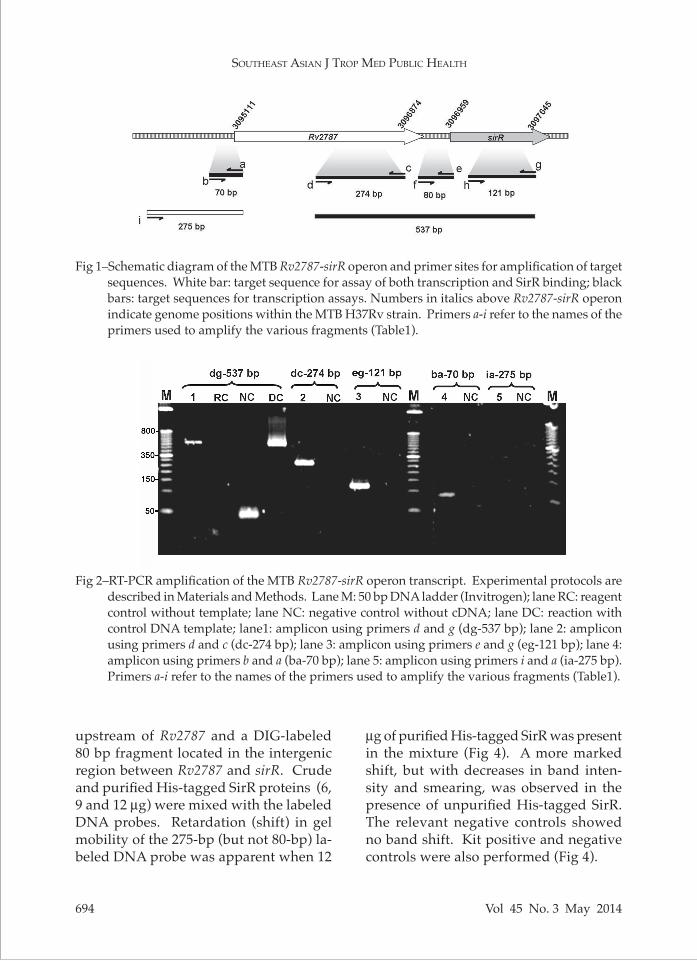
Total RNA was isolated from MTB using Trizol RNA isolation buffer (Invi-trogen, Carlsbad, CA) with sonication according to the manufacturer’s protocol. Primers used to amplify the five cDNA targets (70 bp and 275 bp fragments in the region upstream of Rv2787-sirR operon, and 274 bp, 121 bp, and 537 bp fragments in the regions within the Rv2787-sirR operon) (Fig 1) are listed in Table 1. RT-PCR was performed using SuperScript III One-Step RT-PCR System (Invitrogen, Carlsbad, CA) and C1000™ Thermal Cycler (Bio-Rad, Hercules, CA) as follows: incubation at 60ºC for 15 min-utes; 95ºC for 5 minutes; 35 cycles of 96ºC for 45 seconds, 53ºC for 45 seconds, and 72ºC for 1 minute; and a final 7 minutes at 72ºC. The amplicons were analyzed using agarose gel-electrophoresis and ethidium bromide staining. Immunoblot analysis
Purified recombinant SirR was sub-jected to 12% SDS-PAGE and transferred onto a nitrocellulose membrane. After
SoutheaSt aSian J trop Med public health
692 Vol 45 No. 3 May 2014
incubating with 3% bovine serum albu-min (BSA) in 50 mM PBS, the membrane was washed with PBS-0.05% Tween 20 and incubated for 1 hour with rabbit anti-histidine polyclonal antibodies (Gen-Script, Piscataway, NJ). After washing with PBS-Tween 20, the membrane was incubated with horseradish peroxidase-conjugated goat secondary antibodies (GenScript, Piscataway, NJ) at 37ºC for 1 hour, washed with PBS-Tween 20, and the positive signals were developed using diaminobenzidine and H2O2. Electrophoretic mobility shift assay (EMSA)
EMSA was performed as described previously (Hamoen et al, 1998; Rodriguez et al, 1999). The potential SirR binding sites (at 70 bp and 275 bp upstream of Rv2787-sirR operon and at 80 bp within the Rv2787-sirR intergenic region) were amplified using the primers listed in Table 1. The PCR conditions consisted of heating at 95ºC for 5 minutes, followed by 35 cycles of 30 seconds at 96ºC, 60 seconds at 58ºC, and 2 minutes at 72ºC, with a final 7 minute heating at 72ºC. EMSA was performed using a digoxi-genin (DIG) gel shift kit (Roche Applied Science, Basel, Switzerland). In brief, the PCR amplicons were mixed with labeling buffer (1 M potassium cacodylate, 0.125 M Tris-HCl pH 6.6, 1.25 mg/ml BSA), 5 mM CoCl2 solution, 0.05 mM DIG-11-ddUTP solution, and 1 ml of 20 U terminal trans-ferase. The mixture was incubated for 15 minutes at 37ºC and placed on ice; the mixture was then precipitated with 60 ml of ethanol at -70ºC for 30 minutes. After centrifugation at 14,000 rpm at 4ºC for 15 minutes, the pellet was washed with 500 µl of 70% ethanol, dried, and dissolved in TEN buffer (10 mM Tris-HCl pH 8.0, 1 mM EDTA, 0.1 M NaCl). The optimal amount of labeled PCR products (3.85
pmol/µl) was mixed on ice with the crude and purified His-tagged SirR proteins (at 3, 6, 9 and 12 µg) and binding buffer (100 mM HEPES pH 7.6, 5 mM EDTA, 50 mM [NH4]2SO4, 5 mM DTT, 1% (w/v) Tween 20, 150 mM KCl) (Rodriguez et al, 1999) in 20 µl of total volume. Positive and negative controls from the reagent kit, a reaction without SirR, and reactions containing competitive sequences (unlabeled PCR products for each particular sequence) were used as controls. Nonspecific bind-ing was inhibited by the addition of 1 µg of poly d(I-C) and 1 µg of poly L-lysine. After incubation for 30 minutes at room temperature, the mixtures were analyzed by 6% native PAGE. The oligonucleotide-protein complexes were blotted onto ny-lon membranes and digoxigenin-labeled probes were detected by the addition of anti-DIG Fab fragments conjugated to alkaline phosphatase and chloro-5-substituted adamantyl-1,2-dioxetane phosphate (CSPD) substrate (Roche Ap-plied Science, Basel, Switzerland). The chemiluminescent signals were detected by autoradiography.
RESULTS
Bioinformatics analysis of MTB sirRMTB sirR (NCBI accession no.
NC000962) contains 687 nucleotides and encodes a polypeptide with 228 amino acids, with a calculated molecular weight and pI of 24.95 kDa and 5.07, respectively (ExPASy web-based program). Conserved domains of SirR were analyzed using Pfam software. Several conserved do-mains in SirR included an iron-dependent repressor domain, a helix-turn-helix diph-theria toxin repressor (DTXR), a ferrous iron transport protein A (FeoA) domain, and a transcriptional repressor C-terminal domain. BLAST analysis and multiple
M. tuberculosis sirr characterization
Vol 45 No. 3 May 2014 693
sequence alignment revealed 34%-59% similarity between the amino acid se-quences of MTB SirR and other iron-de-pendent repressors from various bacterial species, highest similarity being the iron-dependent repressor of Corynebacterium glutamicum ATCC 13032 (Jakubovics et al, 2000; Ng et al, 2000; Zhang et al, 2003; Baliga et al, 2004; Monteiro-Vitorello et al, 2004; De Zoysa et al, 2005). Multiple se-quence alignment of the deduced amino acid sequences of these genes revealed moderate similarities with several highly conserved regions of MTB SirR, which suggests that these conserved residues are functionally important.Rv2787 and sirR transcription and tran-scription start sites
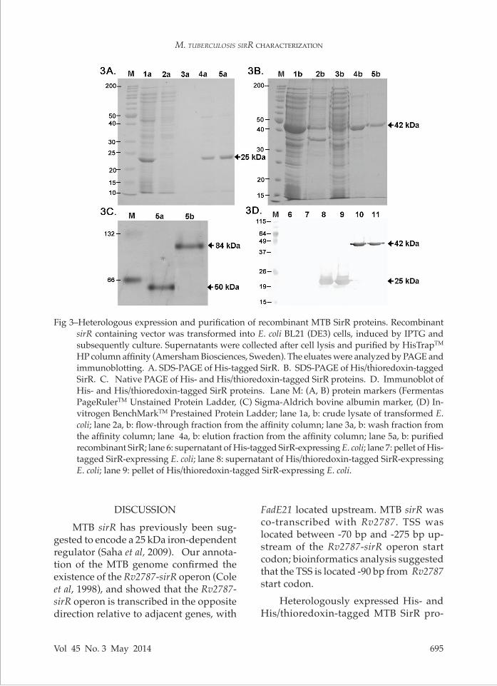
Based on information from the my-coperonDB database and FGENESB pro-gram, an operon comprising Rv2787 and sirR located downstream was predicted (Ranjan et al, 2006) and both genes are bicistronically transcribed (Fig 1). This prediction was confirmed using RT-PCR with primers that covered both Rv2787 and sirR. The size of the transcribed RNA target was as expected (537 bp), indicating the co-existence of both genes in the same mRNA (Fig 2). Bands of the expected sizes for both Rv2787 (274 bp) and sirR (121 bp) were also amplified in separate reactions.
The transcriptional start site (TSS) of Rv2787-sirR operon was demonstrated using RT-PCR with primers specific to the region located -70 bp upstream of Rv2787 start codon, but not using primers specific to the region located -275 bp upstream (Fig 2).Expression and purification of SirR pro-teins
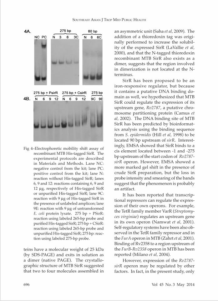
Both His- and His/thioredoxin-tagged SirR proteins were heterologously ex-pressed in E. coli BL21 (DE3) and affinity
purified. The molecular weights of the His- and His/thioredoxin-tagged SirR proteins corresponded with the calcu-lated molecular weight of 25 and 42 kDa, respectively (Fig 3A and B).
Native PAGE analysis of the purified recombinant SirR proteins revealed a putative dimer based on the apparent mo-lecular weights (50 and 84 kDa) (Fig 3C). The successful production of recombinant His-tagged SirR was confirmed immu-noblotting with anti-histidine polyclonal antibodies (Fig 3D). Sequencing of the re-combinant plasmid also confirmed correct insertion of MTB sirR (data not shown).Identification of MTB SirR putative DNA binding sites
In order to identify DNA binding sites of SirR, EMSA was performed using a 275 bp DIG-labeled DNA fragment located
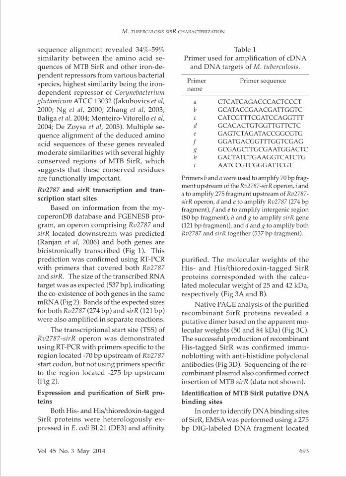
Primer Primer sequencename
a CTCATCAGACCCACTCCCT b GCATACCGAACGATTGGTC c CATCGTTTCGATCCAGGTTT d GCACACTGTGGTTGTTCTC e GAGTCTAGATACCGGCGTG f GGATGACGGTTTGGTCGAG g GCGAGCTTGCGAATGGACTC h GACTATCTGAAGGTCATCTG i AATCCGTCGGGATTCGT
Table 1Primer used for amplification of cDNA
and DNA targets of M. tuberculosis.
Primers b and a were used to amplify 70 bp frag-ment upstream of the Rv2787-sirR operon, i and a to amplify 275 fragment upstream of Rv2787-sirR operon, d and c to amplify Rv2787 (274 bp fragment), f and e to amplify intergenic region (80 bp fragment), h and g to amplify sirR gene (121 bp fragment), and d and g to amplify both Rv2787 and sirR together (537 bp fragment).
SoutheaSt aSian J trop Med public health
694 Vol 45 No. 3 May 2014
Fig 2–RT-PCR amplification of the MTB Rv2787-sirR operon transcript. Experimental protocols are described in Materials and Methods. Lane M: 50 bp DNA ladder (Invitrogen); lane RC: reagent control without template; lane NC: negative control without cDNA; lane DC: reaction with control DNA template; lane1: amplicon using primers d and g (dg-537 bp); lane 2: amplicon using primers d and c (dc-274 bp); lane 3: amplicon using primers e and g (eg-121 bp); lane 4: amplicon using primers b and a (ba-70 bp); lane 5: amplicon using primers i and a (ia-275 bp). Primers a-i refer to the names of the primers used to amplify the various fragments (Table1).
Fig 1–Schematic diagram of the MTB Rv2787-sirR operon and primer sites for amplification of target sequences. White bar: target sequence for assay of both transcription and SirR binding; black bars: target sequences for transcription assays. Numbers in italics above Rv2787-sirR operon indicate genome positions within the MTB H37Rv strain. Primers a-i refer to the names of the primers used to amplify the various fragments (Table1).
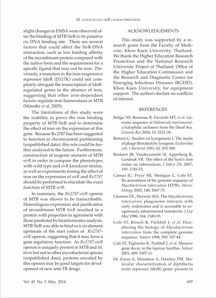
upstream of Rv2787 and a DIG-labeled 80 bp fragment located in the intergenic region between Rv2787 and sirR. Crude and purified His-tagged SirR proteins (6, 9 and 12 µg) were mixed with the labeled DNA probes. Retardation (shift) in gel mobility of the 275-bp (but not 80-bp) la-beled DNA probe was apparent when 12
µg of purified His-tagged SirR was present in the mixture (Fig 4). A more marked shift, but with decreases in band inten-sity and smearing, was observed in the presence of unpurified His-tagged SirR. The relevant negative controls showed no band shift. Kit positive and negative controls were also performed (Fig 4).
M. tuberculosis sirr characterization
Vol 45 No. 3 May 2014 695
Fig 3–Heterologous expression and purification of recombinant MTB SirR proteins. Recombinant
sirR containing vector was transformed into E. coli BL21 (DE3) cells, induced by IPTG and subsequently culture. Supernatants were collected after cell lysis and purified by HisTrapTM HP column affinity (Amersham Biosciences, Sweden). The eluates were analyzed by PAGE and immunoblotting. A. SDS-PAGE of His-tagged SirR. B. SDS-PAGE of His/thioredoxin-tagged SirR. C. Native PAGE of His- and His/thioredoxin-tagged SirR proteins. D. Immunoblot of His- and His/thioredoxin-tagged SirR proteins. Lane M: (A, B) protein markers (Fermentas PageRulerTM Unstained Protein Ladder, (C) Sigma-Aldrich bovine albumin marker, (D) In-vitrogen BenchMarkTM Prestained Protein Ladder; lane 1a, b: crude lysate of transformed E. coli; lane 2a, b: flow-through fraction from the affinity column; lane 3a, b: wash fraction from the affinity column; lane 4a, b: elution fraction from the affinity column; lane 5a, b: purified recombinant SirR; lane 6: supernatant of His-tagged SirR-expressing E. coli; lane 7: pellet of His-tagged SirR-expressing E. coli; lane 8: supernatant of His/thioredoxin-tagged SirR-expressing E. coli; lane 9: pellet of His/thioredoxin-tagged SirR-expressing E. coli.
DISCUSSION
MTB sirR has previously been sug-gested to encode a 25 kDa iron-dependent regulator (Saha et al, 2009). Our annota-tion of the MTB genome confirmed the existence of the Rv2787-sirR operon (Cole et al, 1998), and showed that the Rv2787-sirR operon is transcribed in the opposite direction relative to adjacent genes, with
FadE21 located upstream. MTB sirR was co-transcribed with Rv2787. TSS was located between -70 bp and -275 bp up-stream of the Rv2787-sirR operon start codon; bioinformatics analysis suggested that the TSS is located -90 bp from Rv2787 start codon.
Heterologously expressed His- and His/thioredoxin-tagged MTB SirR pro-
SoutheaSt aSian J trop Med public health
696 Vol 45 No. 3 May 2014
an asymmetric unit (Saha et al, 2009). The addition of a thioredoxin tag was origi-nally performed to increase the solubil-ity of the expressed SirR (LaVallie et al, 2000), and that the N-tagged thioredoxin recombinant MTB SirR also exists as a dimer, suggests that the region involved in dimerization is not located at the N-terminus.
SirR has been proposed to be an iron-responsive regulator, but because it contains a putative DNA binding do-main as well, we hypothesized that MTB SirR could regulate the expression of its upstream gene, Rv2787, a putative chro-mosome partitioning protein (Camus et al, 2002). The DNA binding site of MTB SirR has been predicted by bioinformat-ics analysis using the binding sequence from S. epidermidis (Hill et al, 1998) to be located 90 bp upstream of sirR. Interest-ingly, EMSA showed that SirR binds to a cis element located between -1 and -275 bp upstream of the start codon of Rv2787-sirR operon. However, EMSA showed a more marked gel shift in the presence of crude SirR preparation, but the loss in probe intensity and smearing of the bands suggest that the phenomenon is probably an artifact.
It has been reported that transcrip-tional repressors can regulate the expres-sion of their own operons. For example, the TetR family member VarR (Streptomy-ces virginiae) regulates an upstream gene in its own operon (Namwat et al, 2001). Self-regulatory systems have been also ob-served in the TetR family repressor and in the FurA operon in MTB (Zahrt et al, 2001). Binding of Rv2358 to a region upstream of the FurB-Rv2358 operon in MTB has been reported (Milano et al, 2004).
However, expression of the Rv2787-sirR operon may be regulated by other factors. In fact, in the present study, only
Fig 4–Electrophoretic mobility shift assay of
recombinant MTB His-tagged SirR. The experimental protocols are described in Materials and Methods. Lane NC: negative control from the kit; lane PC: positive control from the kit; lane N: reaction without His-tagged SirR; lanes 6, 9 and 12: reactions containing 6, 9 and 12 µg, respectively of His-tagged SirR or unpurified His-tagged SirR; lane 9C: reaction with 9 µg of His-tagged SirR in the presence of unlabeled amplicon; lane 9E: reaction with 9 µg of untransformed E. coli protein lysate. 275 bp + PSirR: reaction using labeled 265-bp probe and purified His-tagged SirR; 275 bp + CSirR: reaction using labeled 265-bp probe and unpurified His-tagged SirR; 275 bp: reac-tion using labeled 275-bp probe.
teins have a molecular weight of 25 kDa (by SDS-PAGE) and exits in solution as a dimer (native PAGE). The crystallo-graphic structure of MTB SirR suggested that two to four molecules assembled in
M. tuberculosis sirr characterization
Vol 45 No. 3 May 2014 697
slight changes in EMSA were observed af-ter the binding of MTB SirR to its putative cis DNA binding site. There are several factors that could affect the SirR-DNA interaction, such as low binding affinity of the recombinant protein compared with the native form and the requirement for a specific ligand that may not be iron. Pre-viously, a mutation in the iron-responsive repressor IdeR (D117K) could not com-pletely abrogate the transcription of IdeR-regulated genes in the absence of iron, suggesting that other iron-dependent factors regulate iron homeostasis in MTB (Manabe et al, 2005).
The limitations of this study were the inability to prove the iron binding property of MTB SirR and to determine the effect of iron on the expression of this gene. Because Rv2787 has been suggested to function in chromosomal partitioning (unpublished data), this role could be fur-ther analyzed in the future. Furthermore, construction of isogenic mutants of MTB sirR in order to compare the phenotypes with wild type and sirR knockout strains, as well as experiments testing the effect of iron on the expression of sirR and Rv2787 should be performed to elucidate the exact function of MTB sirR.
In summary, the Rv2787-sirR operon of MTB was shown to be transcribable. Heterologous expression and purification of recombinant MTB SirR resulted in a protein with properties in agreement with those predicted by bioinformatics analysis. MTB SirR was able to bind to a cis element upstream of the start codon of Rv2787-sirR operon, suggesting that it may have a gene regulatory function. As Rv2787-sirR operon is uniquely present in MTB and M. bovis but not in other mycobacterial species (unpublished data), proteins encoded by this operon may be good targets for devel-opment of new anti-TB drugs.
ACKNOWLEDGEMENTS
This study was supported by a re-search grant from the Faculty of Medi-cine, Khon Kaen University, Thailand. We thank the Higher Education Research Promotion and the National Research University Project of Thailand, Office of the Higher Education Commission and the Research and Diagnostic Center for Emerging Infectious Diseases (RCEID), Khon Kaen University, for equipment support. The authors declare no conflicts of interest.
REFERENCES
Baliga NS, Bonneau R, Facciotti MT, et al. Ge-nome sequence of Haloarcula marismortui: a halophilic archaeon from the Dead Sea. Genome Res 2004; 14: 2221-34.
Bertani G. Studies on lysogenesis I. The mode of phage liberation by lysogenic Escherichia coli. J Bacteriol 1951; 62: 293-300.
Boelaert JR, Vandecasteele SJ, Appelberg R, Gordeuk VR. The effect of the host’s iron status on tuberculosis. J Infect Dis 2007; 195: 1745-53.
Camus JC, Pryor MJ, Medigue C, Cole ST. Re-annotation of the genome sequence of Mycobacterium tuberculosis H37Rv. Micro-biology 2002; 148: 2967-73.
Clemens DL, Horwitz MA. The Mycobacterium tuberculosis phagosome interacts with early endosomes and is accessible to ex-ogenously administered transferrin. J Exp Med 1996; 184: 1349-55.
Cole ST, Brosch R, Parkhill J, et al. Deci-phering the biology of Mycobacterium tuberculosis from the complete genome sequence. Nature 1998; 393: 537-44.
Cole ST, Eiglmeier K, Parkhill J, et al. Massive gene decay in the leprosy bacillus. Nature 2001; 409: 1007-11.
De Zoysa A, Efstratiou A, Hawkey PM. Mo-lecular characterization of diphtheria toxin repressor (dtxR) genes present in
SoutheaSt aSian J trop Med public health
698 Vol 45 No. 3 May 2014
nontoxigenic Corynebacterium diphtheriae strains isolated in the United Kingdom. J Clin Microbiol 2005; 43: 223-8.
Hamoen LW, Van Werkhoven AF, Bijlsma JJ, Dubnau D, Venema G. The competence transcription factor of Bacillus subtilis rec-ognizes short A/T-rich sequences arranged in a unique, flexible pattern along the DNA helix. Genes Dev 1998; 12: 1539-50.
Hanahan D. Studies on transformation of Esch-erichia coli with plasmids. J Mol Biol 1983; 166: 557-80.
Hill PJ, Cockayne A, Landers P, Morrissey JA, Sims CM, Williams P. SirR, a novel iron-dependent repressor in Staphylococcus epidermidis. Infect Immun 1998; 66: 4123-9.
Jakubovics NS, Smith AW, Jenkinson HF. Expression of the virulence-related Sca (Mn2+) permease in Streptococcus gordonii is regulated by a diphtheria toxin metallo-repressor-like protein ScaR. Mol Microbiol 2000; 38: 140-53.
Kunkle CA, Schmitt MP. Analysis of the Co-rynebacterium diphtheriae DtxR regulon: identification of a putative siderophore synthesis and transport system that is similar to the Yersinia high-pathogenicity island-encoded yersiniabactin synthesis and uptake system. J Bacteriol 2003; 185: 6826-40.
LaVallie ER, Lu Z, Diblasio-Smith EA, Collins-Racie LA, McCoy JM. Thioredoxin as a fusion partner for production of soluble recombinant proteins in Escherichia coli. Meth Enzymol 2000; 326: 322-40.
Lucarelli D, Russo S, Garman E, Milano A, Mey-er-Klaucke W, Pohl E. Crystal structure and function of the zinc uptake regulator FurB from Mycobacterium tuberculosis. J Biol Chem 2007; 282: 9914-22.
Lucarelli D, Vasil ML, Meyer-Klaucke W, Pohl E. The metal-dependent regulators FurA and FurB from Mycobacterium tuberculosis. Int J Mol Sci 2008; 9: 1548-60.
Manabe YC, Hatem CL, Kesavan AK, Durack J, Murphy JR. Both Corynebacterium diphthe-
riae DtxR (E175K) and Mycobacterium tuber-culosis IdeR (D177K) are dominant positive repressors of IdeR-regulated genes in M. tuberculosis. Infect Immun 2005; 73: 5988-94.
Milano A, Branzoni M, Canneva F, Profumo A, Riccardi G. The Mycobacterium tuberculosis Rv2358-furB operon is induced by zinc. Res Microbiol 2004; 155: 192-200.
Monteiro-Vitorello CB, Camargo LE, Van Sluys MA, et al. The genome sequence of the gram-positive sugarcane pathogen Leif-sonia xyli subsp. xyli. Mol Plant Microbe Interact 2004; 17: 827-36.
Namwat W, Lee CK, Kinoshita H, Yamada Y, Nihira T. Identification of the varR gene as a transcriptional regulator of virginiamy-cin S resistance in Streptomyces virginiae. J Bacteriol 2001; 183: 2025-31.
Ng WV, Kennedy SP, Mahairas GG, et al. Ge-nome sequence of Halobacterium species NRC-1. Proc Natl Acad Sci USA 2000; 97: 12176-81.
Ranjan S, Gundu RK, Ranjan A. MycoperonDB: a database of computationally identified operons and transcriptional units in My-cobacteria. BMC Bioinformatics 2006; 7: S9.
Reddy PV, Puri RV, Khera A, Tyagi AK. Iron storage proteins are essential for the sur-vival and pathogenesis of Mycobacterium tuberculosis in THP-1 macrophages and the guinea pig model of infection. J Bacteriol 2012; 194: 567-75.
Rodriguez GM, Gold B, Gomez M, Dussurget O, Smith I. Identification and characteriza-tion of two divergently transcribed iron regulated genes in Mycobacterium tubercu-losis. Tuber Lung Dis 1999; 79: 287-98.
Saha B, Mukherjee S, Dutta D, Das AK. Ex-pression, purification, crystallization and preliminary X-ray diffraction analysis of the transcriptional repressor SirR from Mycobacterium tuberculosis H37Rv. Acta Crystallogr Sect F Struct Biol Cryst Commun 2009; 65: 154-8.
van Soolingen D, de Haas PE, Hermans PW, van Embden JD. DNA fingerprinting of Mycobacterium tuberculosis. Methods Enzy-
M. tuberculosis sirr characterization
Vol 45 No. 3 May 2014 699
mol 1994; 235: 196-205.World Health Organization (WHO). WHO
global tuberculosis control report 2010. Cent Eur J Public Health 2010; 18: 237.
Young DB, Gideon HP, Wilkinson RJ. Eliminat-ing latent tuberculosis. Trends Microbiol 2009; 17: 183-8.
Zahrt TC, Song J, Siple J, Deretic V. Mycobacte-
rial FurA is a negative regulator of cata-lase-peroxidase gene katG. Mol Microbiol 2001; 39: 1174-85.
Zhang YQ, Ren SX, Li HL, et al. Genome-based analysis of virulence genes in a non-biofilm-forming Staphylococcus epidermidis strain (ATCC 12228). Mol Microbiol 2003; 49: 1577-93.