Embed Size (px)
Citation preview
CNS ABSCESSESCNS ABSCESSES
M.MOLAVI MD
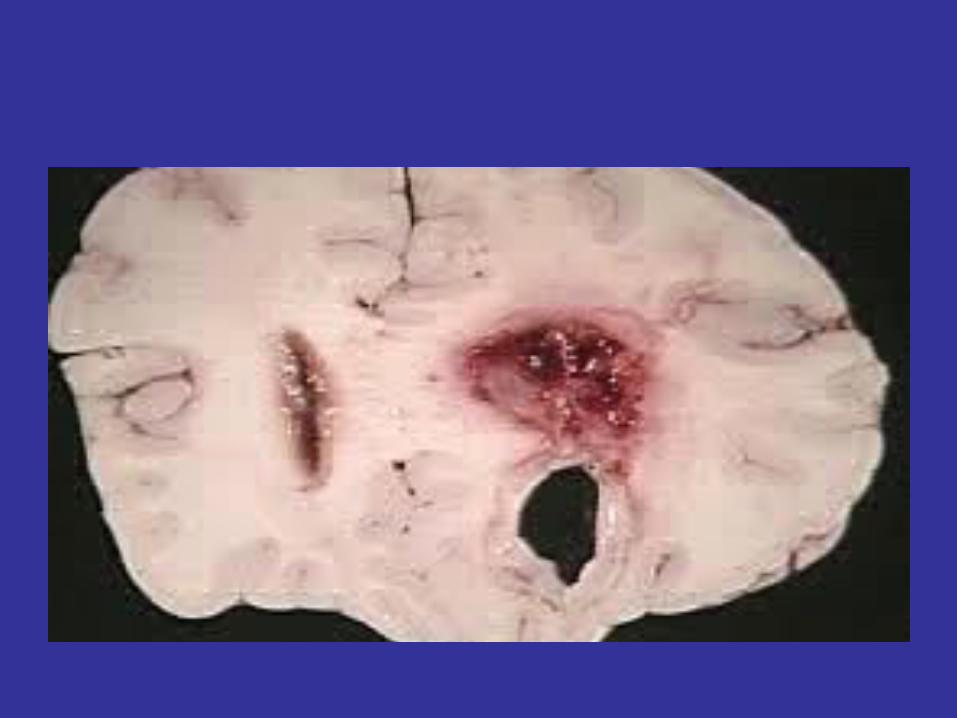
CNS ABSCESSESCNS ABSCESSES• Focal pyogenic infections of the central nervous system• Exert their effects mainly by:
– Direct involvement & destruction of the brain or spinal cord
– Compression of parenchyma– Elevation of intracranial pressure– Interfering with blood &/or CSF flow
• Include: Brain abscess, subdural empyema, intracranial epidural abscess, spinal epidural abscess, spinal cord abscess
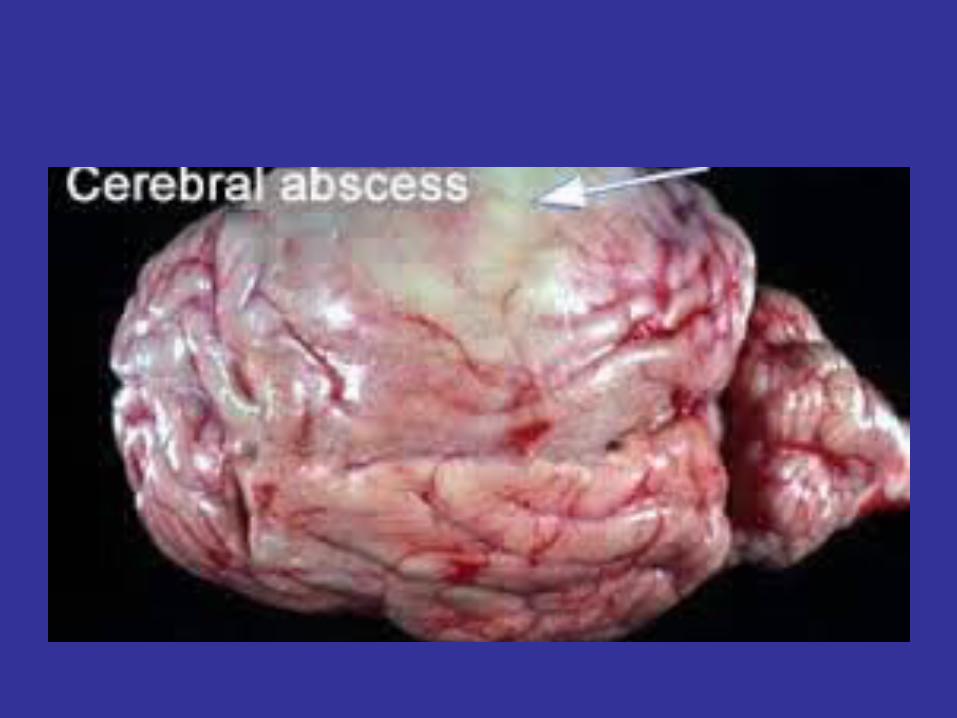
BRAIN ABSCESSBRAIN ABSCESS
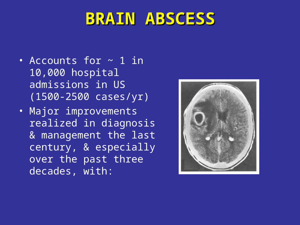
• Accounts for ~ 1 in 10,000 hospital admissions in US (1500-2500 cases/yr)
• Major improvements realized in diagnosis & management the last century, & especially over the past three decades, with:
BRAIN ABSCESSBRAIN ABSCESS
• Was uniformly fatal before the late 1800’s• Mortality down to 30-60% from WWII-1970’s
– Introduction of abx (penicillin, chloramphenicol...) – newer surgical techniques
• Mortality down to 0-24% over the past three decades, with:
– Advent of CT scanning (1974), MRI – Stereotactic brain biopsy/aspiration techniques– Further improvement in surgery – Newer abx (e.g. cephalosporins, metronidazole..)
– Better treatment of predisposing conditions
CHANGES IN EPIDEMIOLOGY CHANGES IN EPIDEMIOLOGY OF BRAIN ABSCESS OF BRAIN ABSCESS
(in the last 2-3 decades)(in the last 2-3 decades)
– Marked drop in mortality overall– Lower incidence of otogenic brain abscesses
– improved treatment of chronic ear infections
– With increase in No. of immunosuppressed patients:
• increased incidence of brain abscess seen in that population (Transplant, AIDS,…)
• More incidence of brain abscess caused by opportunistic pathogens (fungi, toxo…)
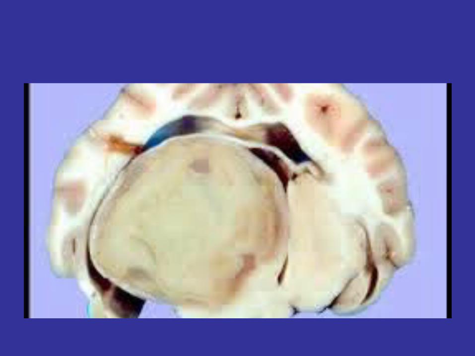
PATHOPHYSIOLOGYPATHOPHYSIOLOGY
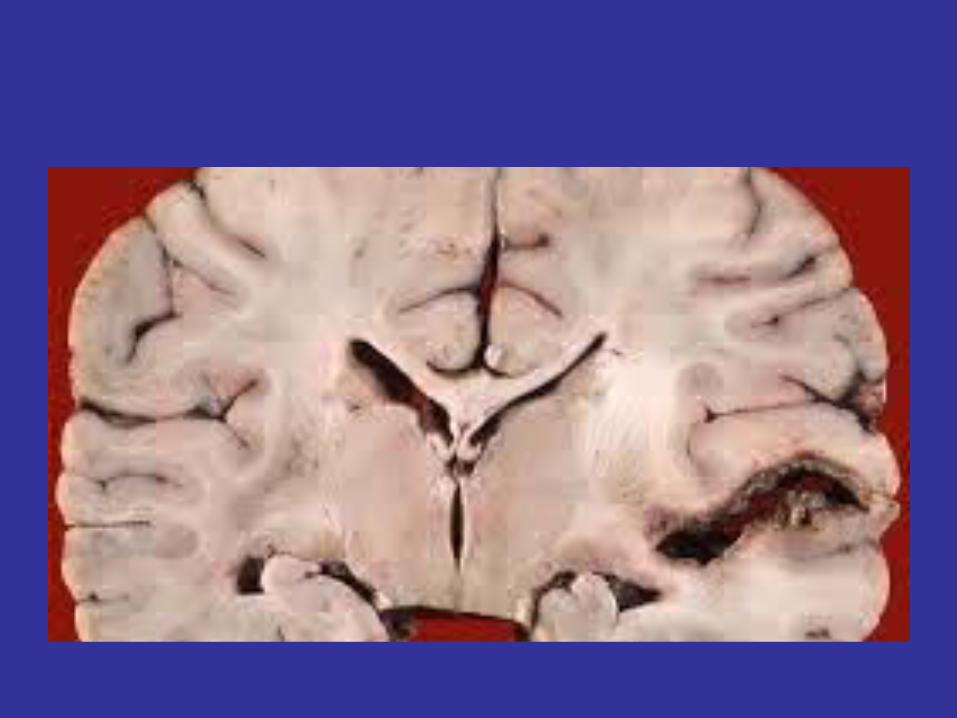
• Begins as localized cerebritis (1-2 wks)• Evolves into a collection of pus surrounded by a
well-vascularized capsule (3-4 wks)
• Lesion evolution (based on experimental animal models):– Days 1-3: “early cerebritis stage”– Days 4-9: “late cerebritis stage”– Days 10-14: “early capsule stage”– > day14: “late capsule stage”
PATHOGENESISPATHOGENESIS
• Direct spread from contiguous foci (40-50%)
• Hematogenous (25-35%)
• Penetrating trauma/surgery (10%)
• Cryptogenic (15-20%)
DIRECT SPREADDIRECT SPREAD(from contiguous foci)(from contiguous foci)
• Occurs by:– Direct extension through infected bone– Spread through emissary veins, diploic veins, local
lymphatics
• The contiguous foci include:• Otitis media/mastoiditis • Sinusitis• Dental infection (<10%), typically with molar infections • Meningitis rarely complicated by brain abscess (more
common in neonates with Citrobacter diversus meningitis, of whom 70% develop brain abscess)
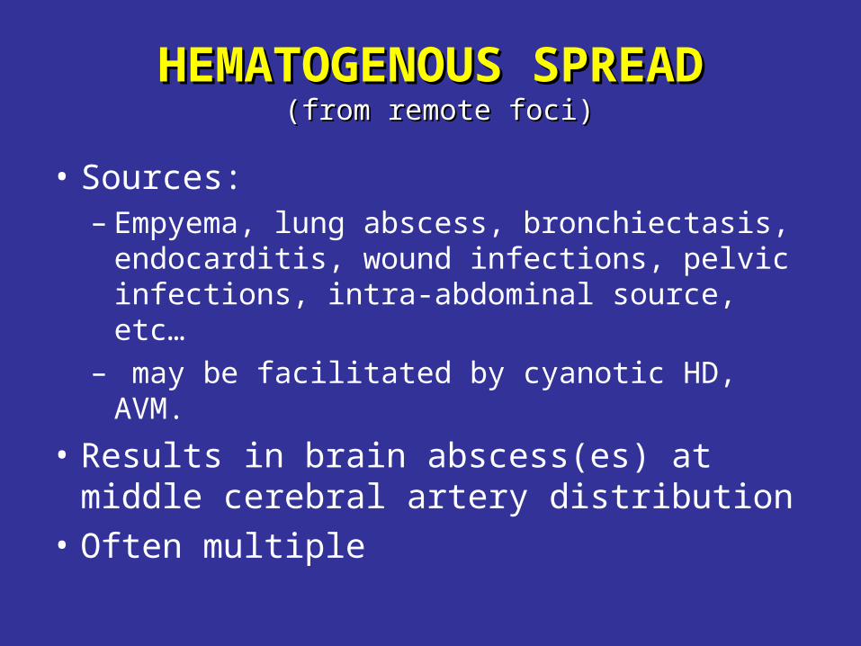
HEMATOGENOUS SPREADHEMATOGENOUS SPREAD (from remote foci)(from remote foci)
• Sources:– Empyema, lung abscess, bronchiectasis,
endocarditis, wound infections, pelvic infections, intra-abdominal source, etc…
– may be facilitated by cyanotic HD, AVM.
• Results in brain abscess(es) at middle cerebral artery distribution
• Often multiple
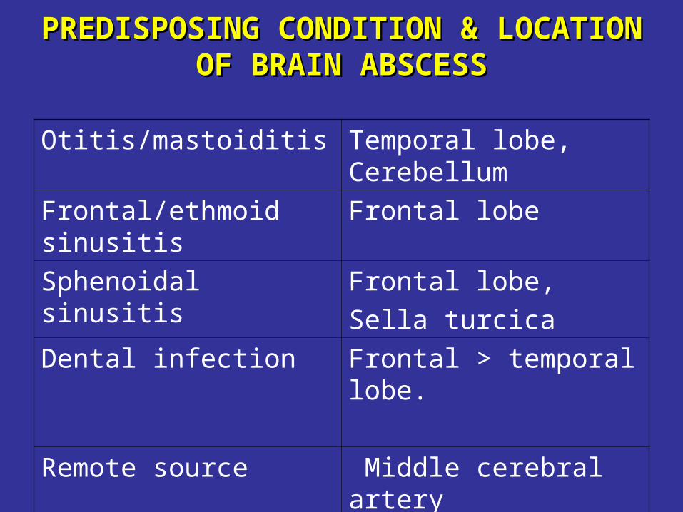
PREDISPOSING CONDITION &PREDISPOSING CONDITION & LOCATION OF BRAIN ABSCESSLOCATION OF BRAIN ABSCESS
Otitis/mastoiditis Temporal lobe, Cerebellum
Frontal/ethmoid sinusitis Frontal lobe
Sphenoidal sinusitis Frontal lobe,
Sella turcica
Dental infection Frontal > temporal lobe.
Remote source Middle cerebral artery distribution (often multiple)
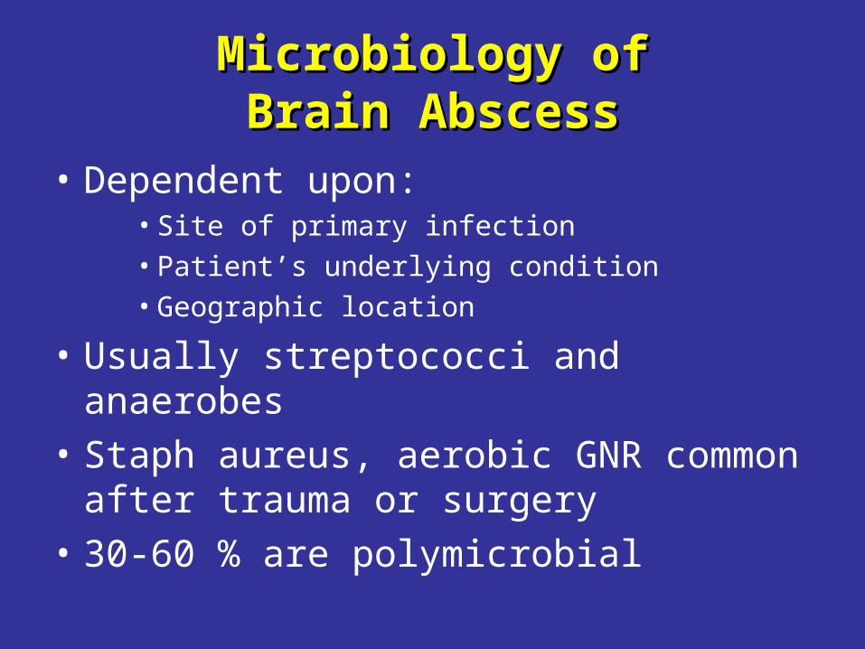
Microbiology of Microbiology of Brain AbscessBrain Abscess
• Dependent upon:• Site of primary infection• Patient’s underlying condition• Geographic location
• Usually streptococci and anaerobes
• Staph aureus, aerobic GNR common after trauma or surgery
• 30-60 % are polymicrobial
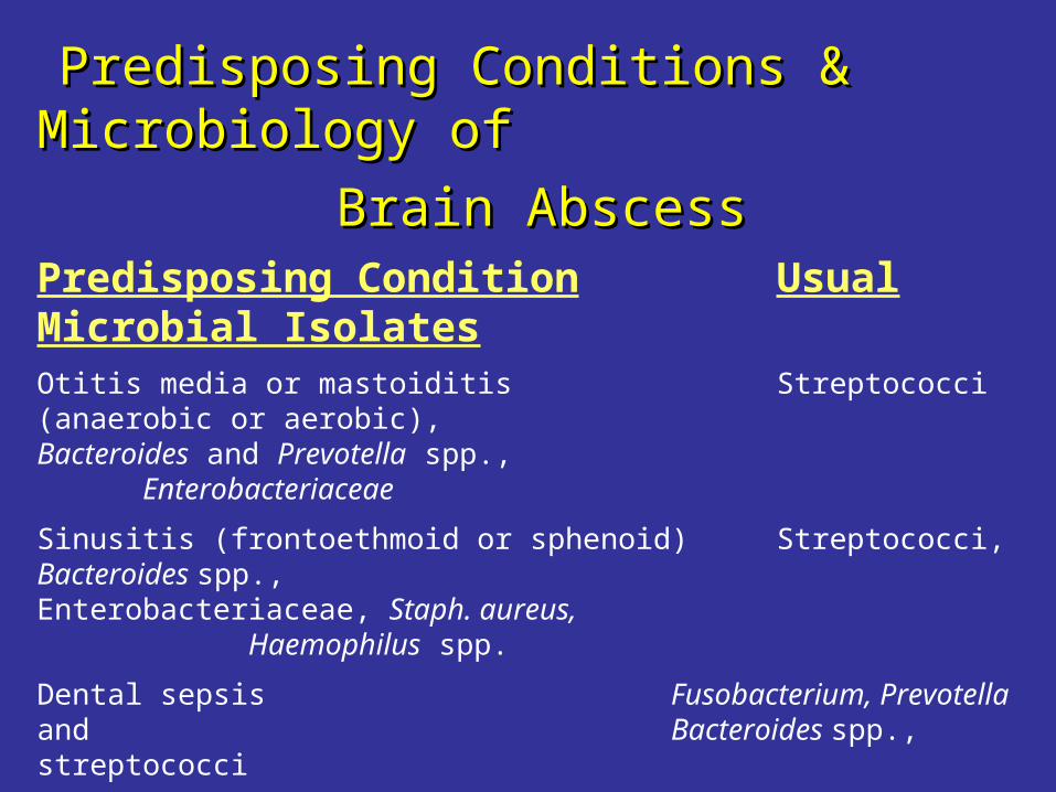
Predisposing Conditions & Microbiology of Predisposing Conditions & Microbiology of
Brain AbscessBrain AbscessPredisposing Condition Usual Microbial Isolates
Otitis media or mastoiditis Streptococci (anaerobic or aerobic), Bacteroides and Prevotella spp., Enterobacteriaceae
Sinusitis (frontoethmoid or sphenoid) Streptococci, Bacteroides spp., Enterobacteriaceae, Staph. aureus, Haemophilus spp.
Dental sepsis Fusobacterium, Prevotella and Bacteroides spp., streptococci
Penetrating trauma or postneurosurgical S. aureus, streptococci, Enterobacteriaceae, Clostridium spp.
PPID,2000
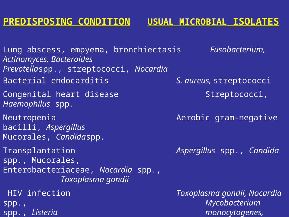
PREDISPOSING CONDITION USUAL MICROBIAL ISOLATES
Lung abscess, empyema, bronchiectasis Fusobacterium, Actinomyces, Bacteroides Prevotellaspp., streptococci, Nocardia
Bacterial endocarditis S. aureus, streptococci
Congenital heart disease Streptococci, Haemophilus spp.
Neutropenia Aerobic gram-negative bacilli, Aspergillus Mucorales, Candidaspp.
Transplantation Aspergillus spp., Candida spp., Mucorales, Enterobacteriaceae, Nocardia spp., Toxoplasma gondii
HIV infection Toxoplasma gondii, Nocardia spp., Mycobacterium spp., Listeria monocytogenes, Cryptococcus
neoformans
PPID, 2000
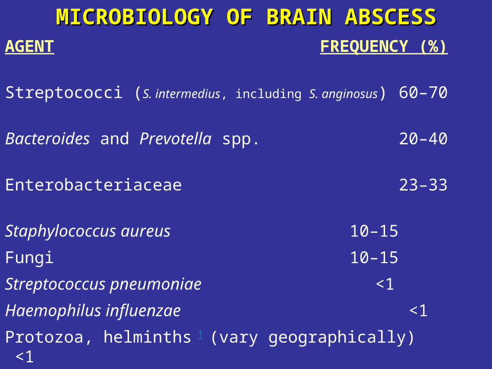
MICROBIOLOGY OFMICROBIOLOGY OF BRAIN ABSCESSBRAIN ABSCESSAGENT FREQUENCY (%)
Streptococci (S. intermedius, including S. anginosus) 60–70
Bacteroides and Prevotella spp. 20–40
Enterobacteriaceae 23–33
Staphylococcus aureus 10–15
Fungi 10–15
Streptococcus pneumoniae <1
Haemophilus influenzae <1
Protozoa, helminths † (vary geographically) <1
*Yeasts, fungi (Aspergillus Agents of mucor Candida Cryptococci Coccidiodoides Cladosporium trichoides Pseudallescheria boydii)
†Protozoa, helminths (Entamoeba histolytica, Schistosomes Paragonimus Cysticerci) CTID,2001
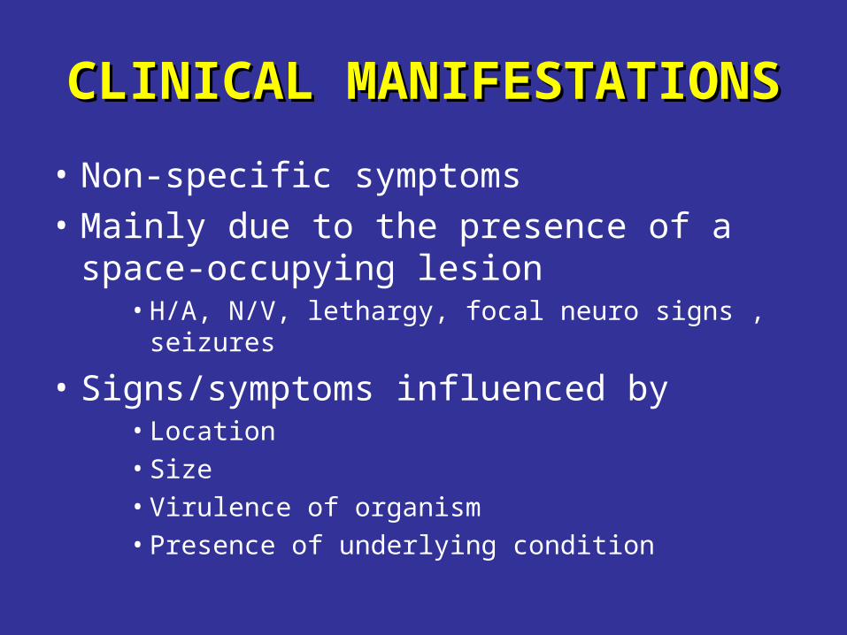
CLINICAL MANIFESTATIONSCLINICAL MANIFESTATIONS
• Non-specific symptoms
• Mainly due to the presence of a space-occupying lesion
• H/A, N/V, lethargy, focal neuro signs , seizures
• Signs/symptoms influenced by • Location • Size• Virulence of organism• Presence of underlying condition
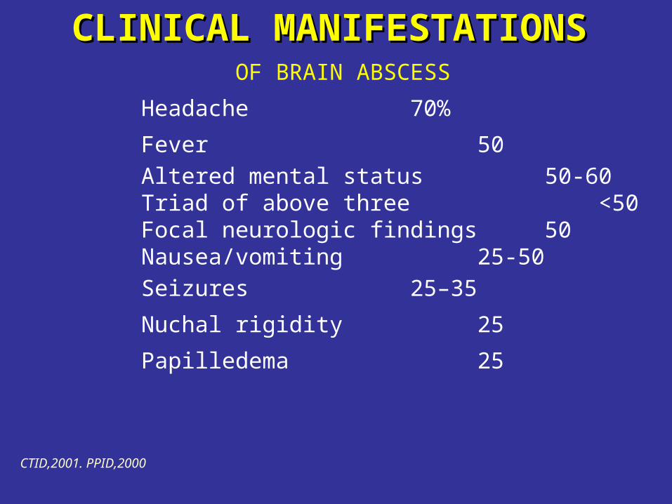
CLINICAL MANIFESTATIONSCLINICAL MANIFESTATIONS
OF BRAIN ABSCESS
Headache 70%
Fever 50
Altered mental status 50-60 Triad of above three <50Focal neurologic findings 50Nausea/vomiting 25-50
Seizures 25–35
Nuchal rigidity 25
Papilledema 25
CTID,2001. PPID,2000
CLINICAL MANIFESTATIONS CLINICAL MANIFESTATIONS
Headache • Often dull, poorly localized (hemicranial?), non-
specific– Abrupt, extremely severe H/A: think meningitis, SAH.– Sudden worsening in H/A w meningismus: think rupture
of brain abscess into ventricle (often fatal)
LOCATION & CLINICAL FEATURESLOCATION & CLINICAL FEATURES • FRONTAL LOBE: H/A, drowsiness, inattention,
hemiparesis, motor speech disorder, AMS
• TEMPORAL LOBE: Ipsilateral H/A, aphasia, visual field defect
• PARIETAL LOBE: H/A, visual field defects, endocrine disturbances
• CEREBELLUM: Nystagmus, ataxia, vomiting, dysmetria
DIFFERENTIAL DIAGNOSISDIFFERENTIAL DIAGNOSIS
• Malignancy– Abscess has hypo-dense center, with surrounding smooth, thin-
walled capsule, & areas of peripheral enhancement. – Tumor has diffuse enhancement & irregular borders.– SPECT (PET scan) may differentiate. CRP too?
• CVA• Hemorrhage• Aneurysm• Subdural empyema/ICEpidural abscess
DIAGNOSISDIAGNOSIS
• High index of suspicion
• Contrast CT or MRI
• Drainage/biopsy, if ring enhancing lesion(s) are seen
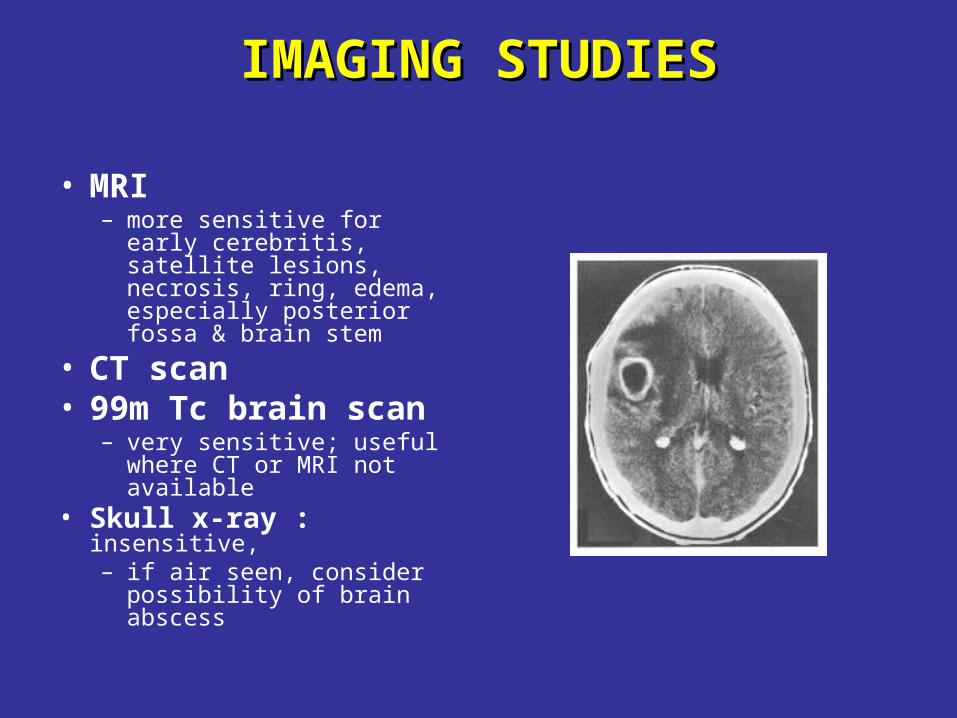
IMAGING STUDIESIMAGING STUDIES
• MRI – more sensitive for early
cerebritis, satellite lesions, necrosis, ring, edema, especially posterior fossa & brain stem
• CT scan • 99m Tc brain scan
– very sensitive; useful where CT or MRI not available
• Skull x-ray : insensitive,– if air seen, consider
possibility of brain abscess
LABORATORY TESTS LABORATORY TESTS BRAIN ABSCESS
•Aspirate: Gram/AFB/fungal stains & cultures, cytopathology (+/-PCR for TB)
•WBC Normal in 40% ( only moderate leukocytosis in ~ 50%
& only 10% have WBC >20,000)
•CRP almost invariably elevated
•ESR Usually moderately elevated
•BC Often negative BUT Should still be done
•LP Contraindicated in patients with known/suspected brainabscessRisk of herniation 15-30%
If done, may have normal CSF findings, but:Usually elevated CSF protein & cell count (lymphs)
Unremarkable glucose & CSF cultures rarely positive
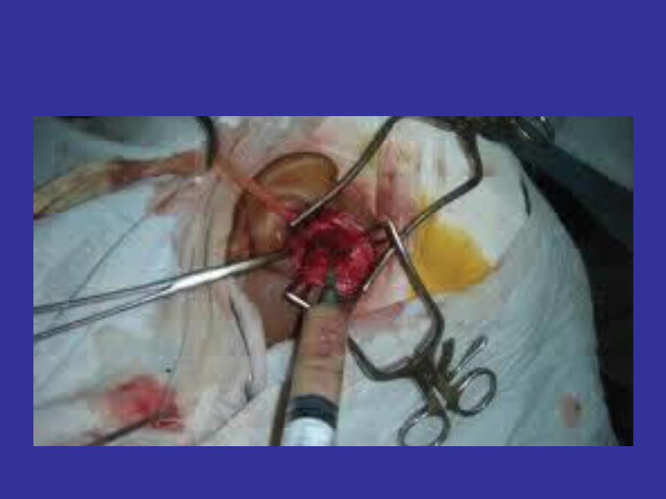
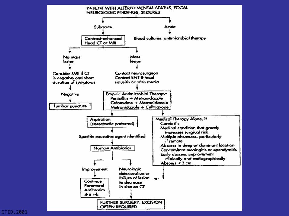
TREATMENTTREATMENT
• Combined medical & surgical• Aspiration or excision• empirical abx
• Empirical antibiotics are selected based on:• Likely pathogen (consider primary source, underlying
condition, & geography)
• Antibiotic characteristics: usual MICs, CNS penetration, activity in abscess cavity
• Modify abx based on stains• Duration: usually 6-8 wks
• after surgical excision, a shorter course may suffice
CT Scan
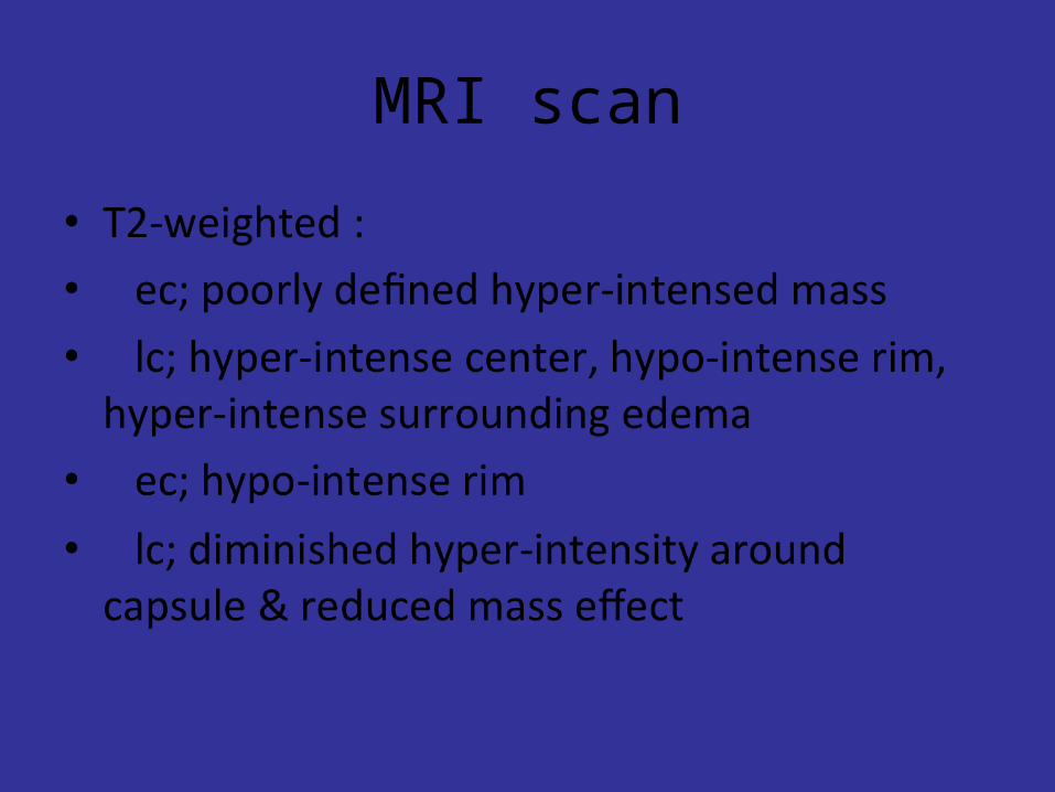
MRI scan
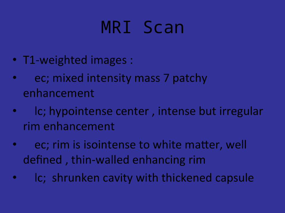
MRI Scan
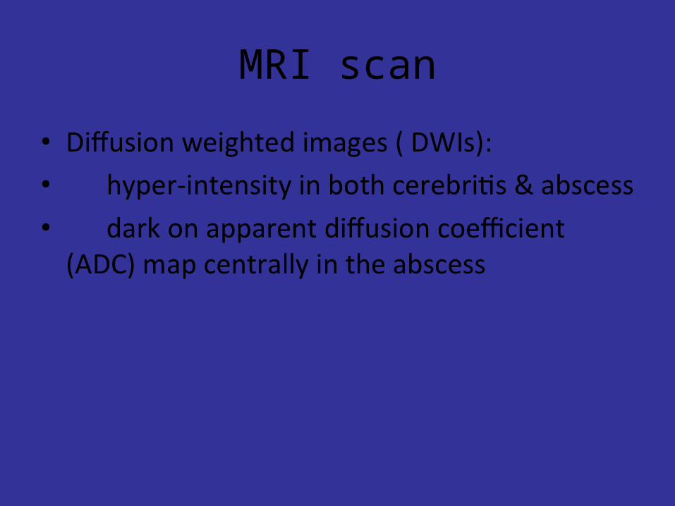
MRI scan
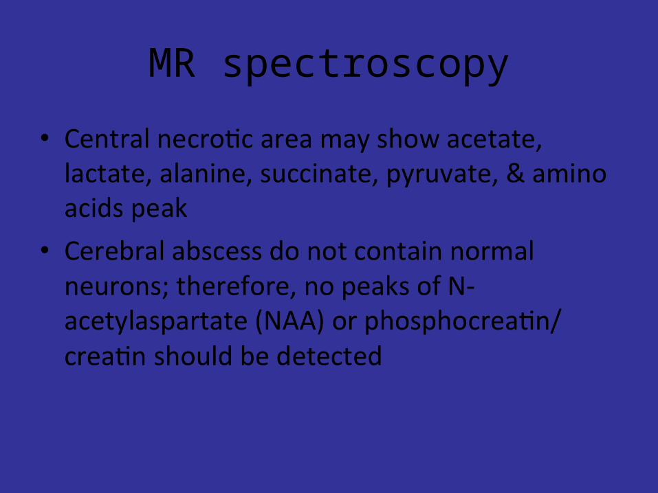
MR spectroscopy
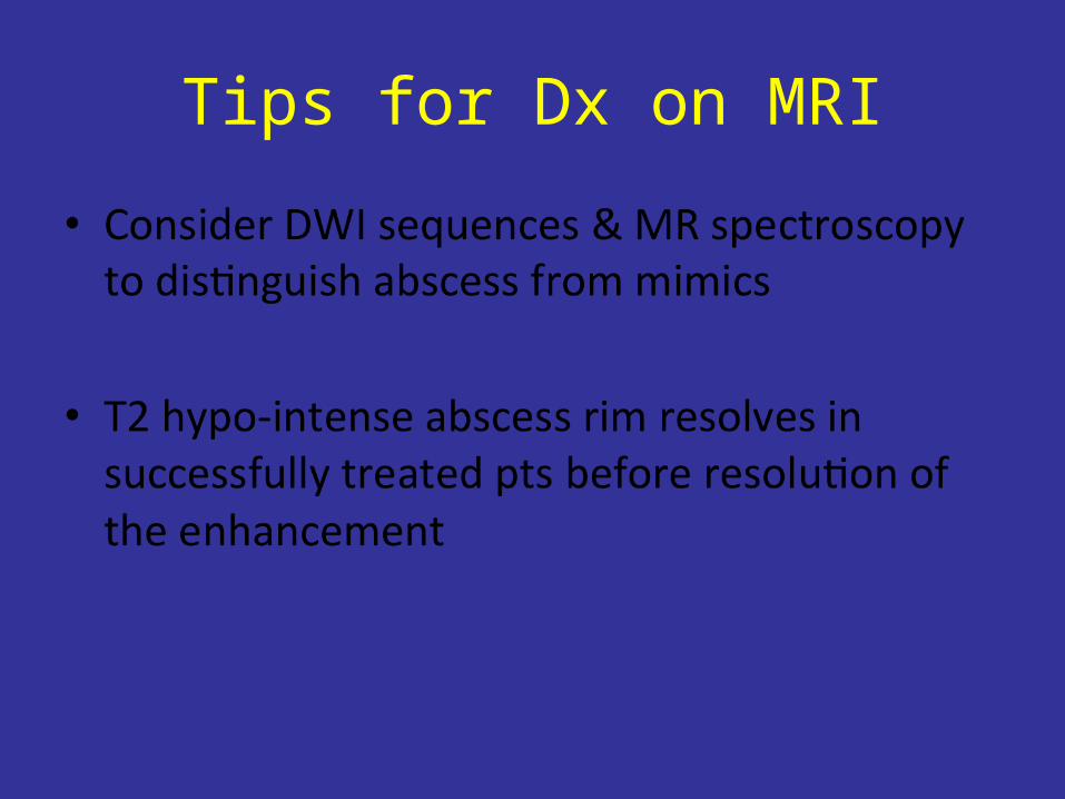
Tips for Dx on MRI
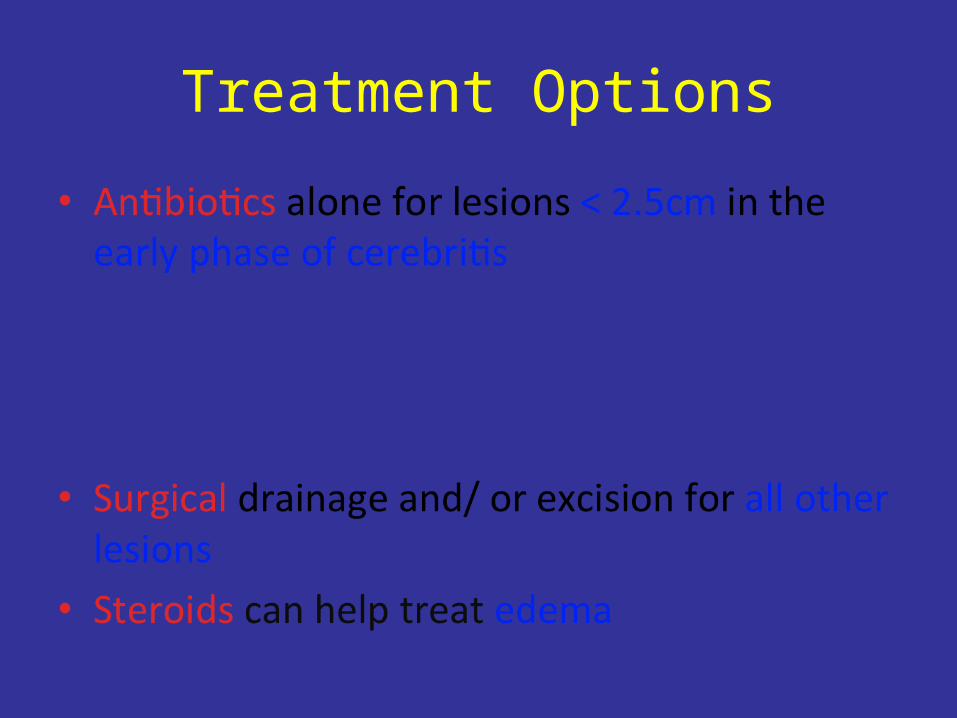
Treatment Options
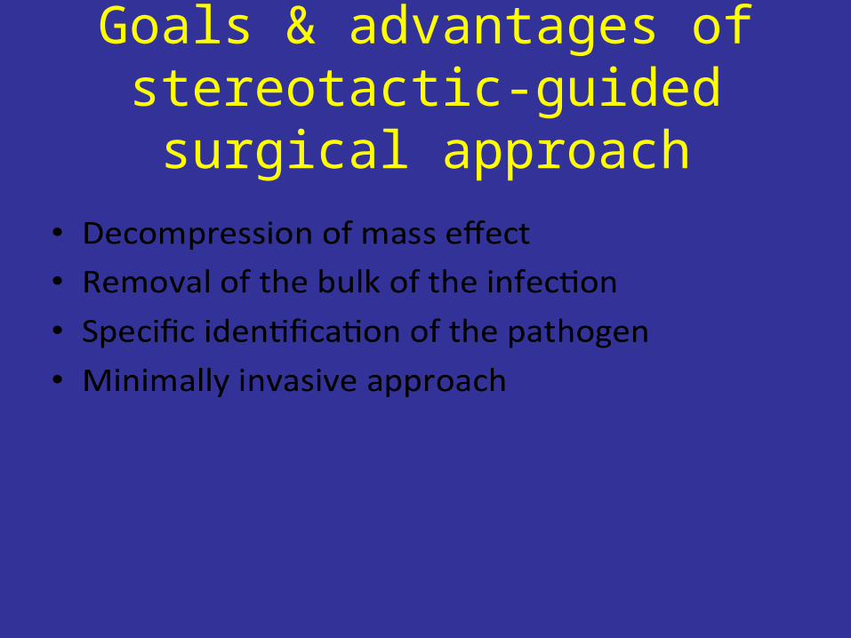
Goals & advantages of stereotactic-guided surgical
approach
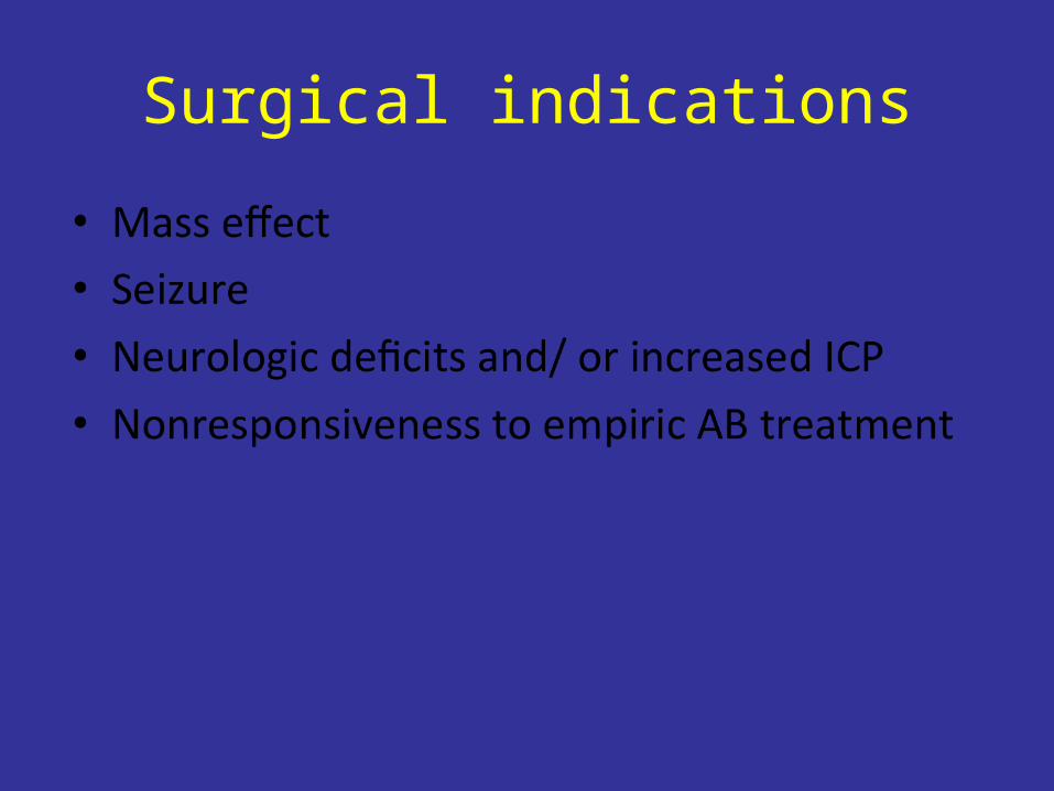
Surgical indications
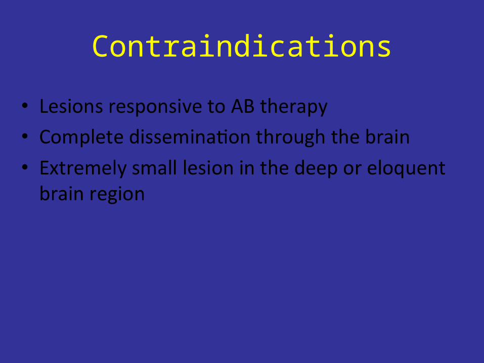
Contraindications
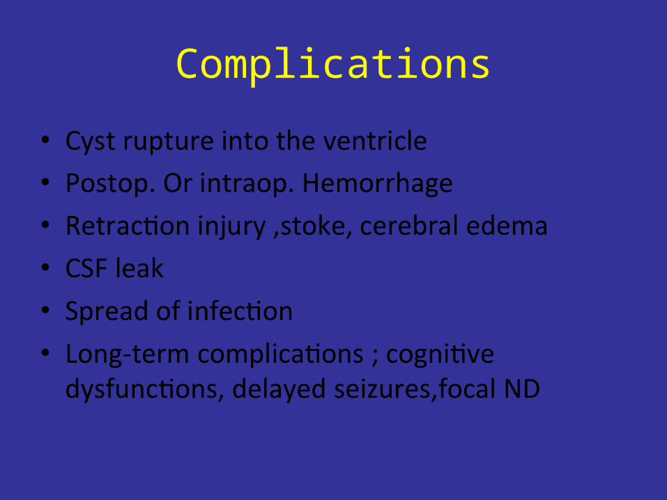
Complications
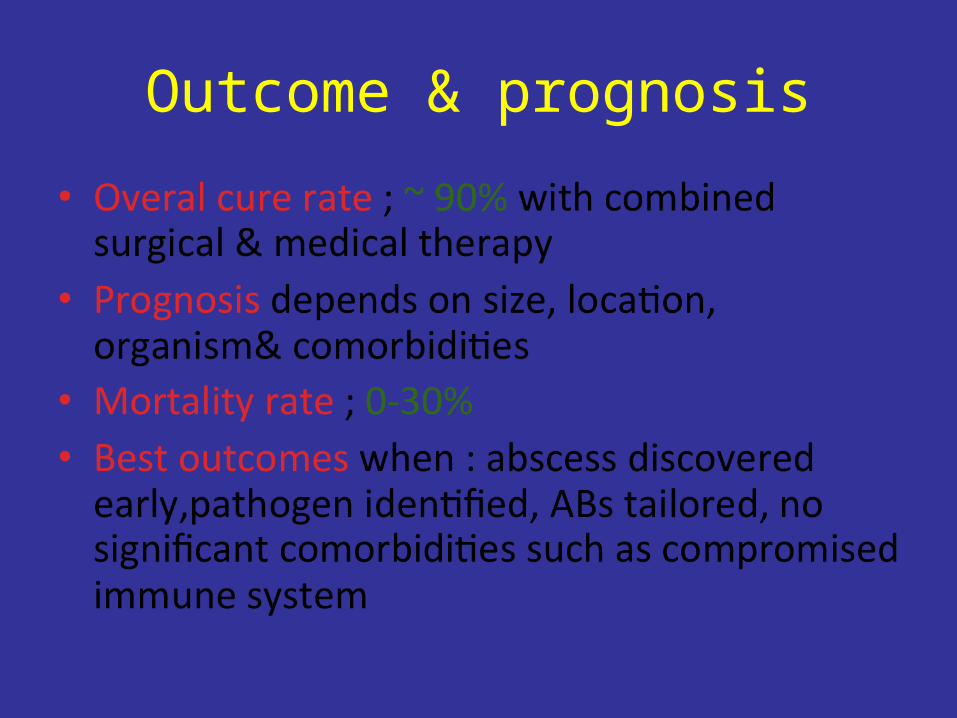
Outcome & prognosis


Armstrong ID, Mosby inc 1999
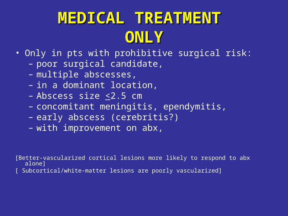
MEDICAL TREATMENTMEDICAL TREATMENT ONLYONLY
• Only in pts with prohibitive surgical risk: – poor surgical candidate,– multiple abscesses,– in a dominant location,– Abscess size <2.5 cm– concomitant meningitis, ependymitis, – early abscess (cerebritis?) – with improvement on abx,
[Better-vascularized cortical lesions more likely to respond to abx alone][ Subcortical/white-matter lesions are poorly vascularized]

CTID,2001
SERIAL IMAGING IMPORTANT TO MONITOR RESPONSE
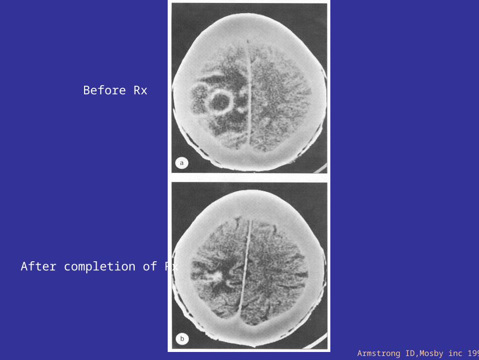
Before Rx
After completion of Rx
Armstrong ID,Mosby inc 1999
POOR PROGNOSTIC MARKERSPOOR PROGNOSTIC MARKERS•Delayed or missed diagnosis
•Inappropriate antibiotics.
•Multiple, deep, or multi-loculated abscesses
•Ventricular rupture (80%–100% mortality)
•Fungal , resistant pathogens.•Neurological compromise at presentation•Short duration w severe AMS,• Rapidly progressive neuro. Impairment•Immunosuppressed host
•Poor localization, especially in the posterior fossa (before CT)
Modified from CTID,2001

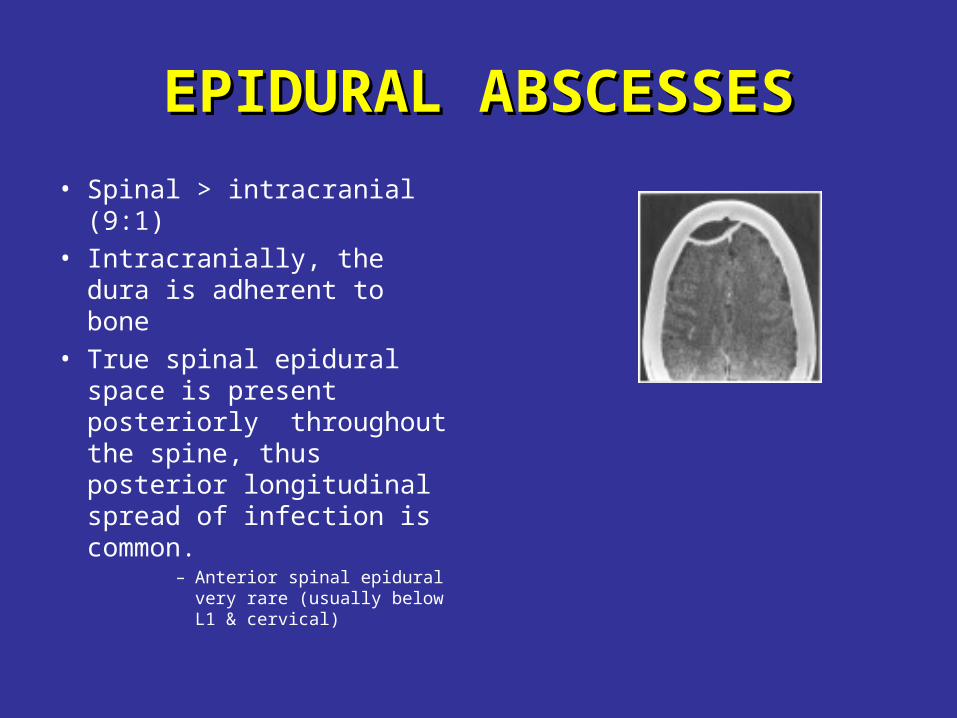
EPIDURAL ABSCESSESEPIDURAL ABSCESSES
• Spinal > intracranial (9:1)• Intracranially, the dura is
adherent to bone• True spinal epidural
space is present posteriorly throughout the spine, thus posterior longitudinal spread of infection is common.
– Anterior spinal epidural very rare (usually below L1 & cervical)
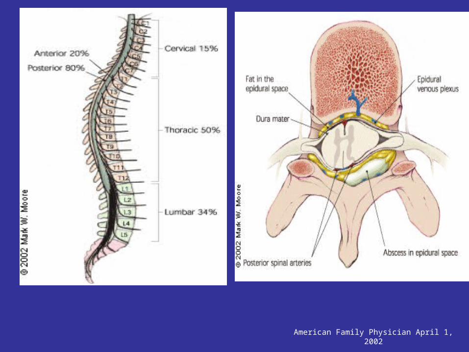
American Family Physician April 1, 2002
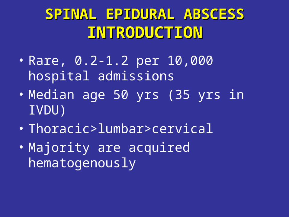
SPINAL EPIDURAL ABSCESSSPINAL EPIDURAL ABSCESSINTRODUCTIONINTRODUCTION
• Rare, 0.2-1.2 per 10,000 hospital admissions
• Median age 50 yrs (35 yrs in IVDU)
• Thoracic>lumbar>cervical
• Majority are acquired hematogenously
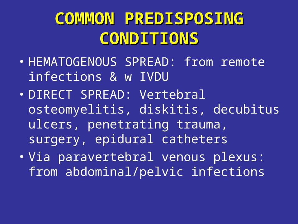
COMMON PREDISPOSING COMMON PREDISPOSING CONDITIONSCONDITIONS
• HEMATOGENOUS SPREAD: from remote infections & w IVDU
• DIRECT SPREAD: Vertebral osteomyelitis, diskitis, decubitus ulcers, penetrating trauma, surgery, epidural catheters
• Via paravertebral venous plexus: from abdominal/pelvic infections
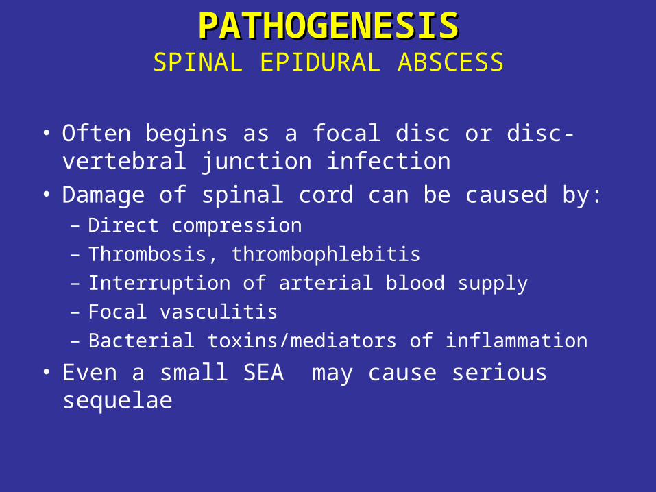
PATHOGENESISPATHOGENESISSPINAL EPIDURAL ABSCESS
• Often begins as a focal disc or disc-vertebral junction infection
• Damage of spinal cord can be caused by:– Direct compression– Thrombosis, thrombophlebitis– Interruption of arterial blood supply– Focal vasculitis– Bacterial toxins/mediators of inflammation
• Even a small SEA may cause serious sequelae
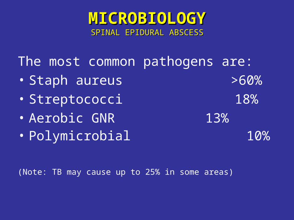
MICROBIOLOGYMICROBIOLOGYSPINAL EPIDURAL ABSCESSSPINAL EPIDURAL ABSCESS
The most common pathogens are:
• Staph aureus >60%
• Streptococci 18%
• Aerobic GNR 13%• Polymicrobial 10%
(Note: TB may cause up to 25% in some areas)
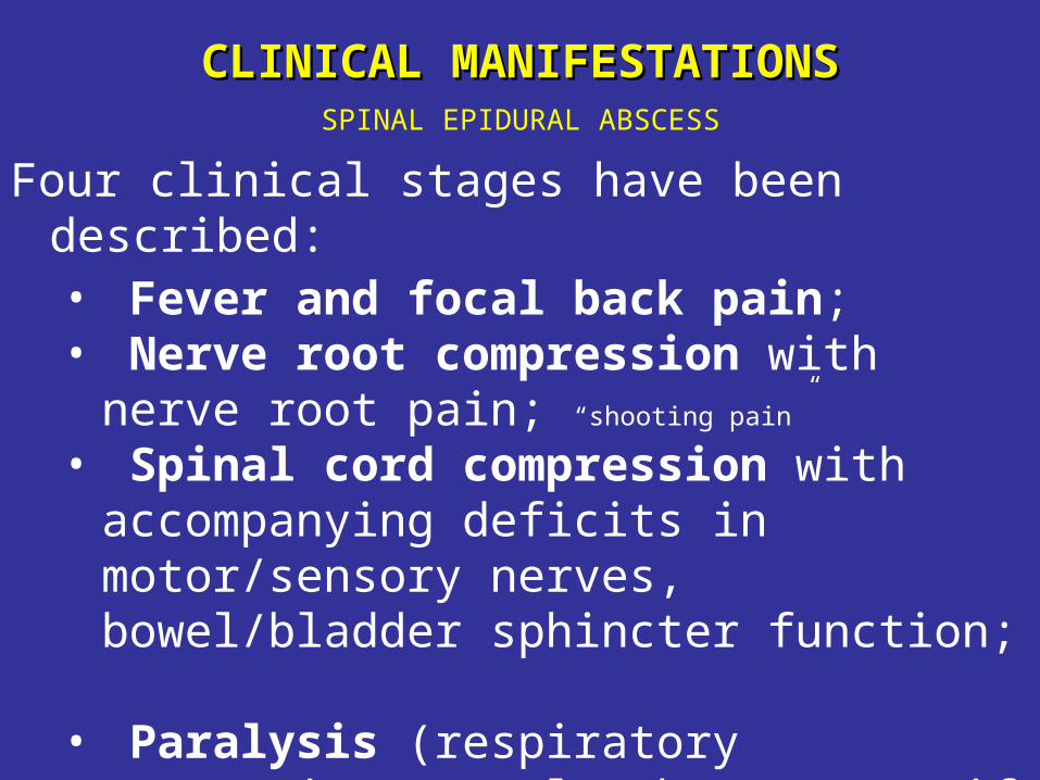
CLINICAL MANIFESTATIONSCLINICAL MANIFESTATIONSSPINAL EPIDURAL ABSCESS
Four clinical stages have been described: • Fever and focal back pain; • Nerve root compression with nerve root
pain; “shooting pain”
• Spinal cord compression with accompanying deficits in motor/sensory nerves, bowel/bladder sphincter function;
• Paralysis (respiratory compromise may also be present if the cervical cord is involved).
Armstrong, ID, Mosby inc,2000
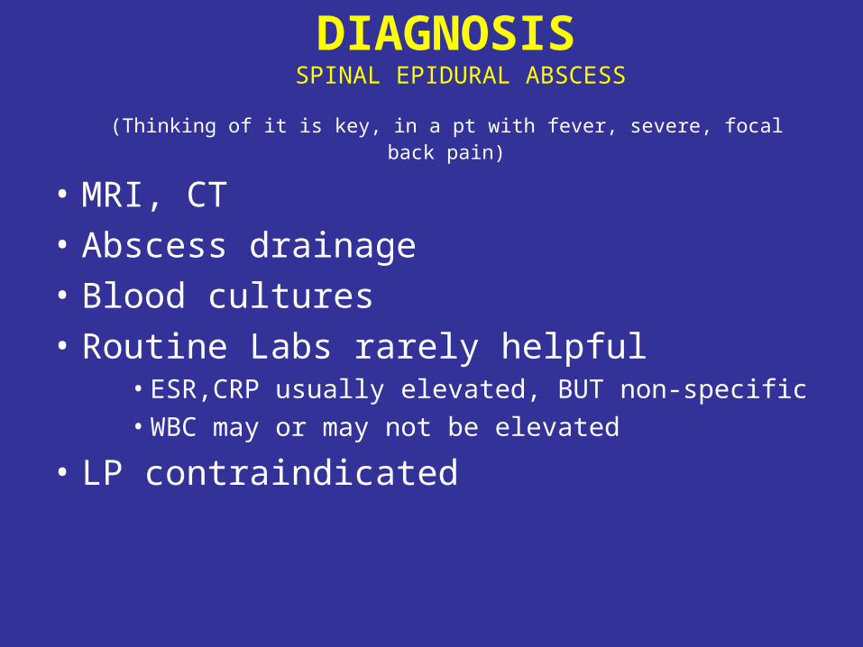
DIAGNOSIS SPINAL EPIDURAL ABSCESS
(Thinking of it is key, in a pt with fever, severe, focal back pain)
• MRI, CT
• Abscess drainage
• Blood cultures
• Routine Labs rarely helpful• ESR,CRP usually elevated, BUT non-specific• WBC may or may not be elevated
• LP contraindicated
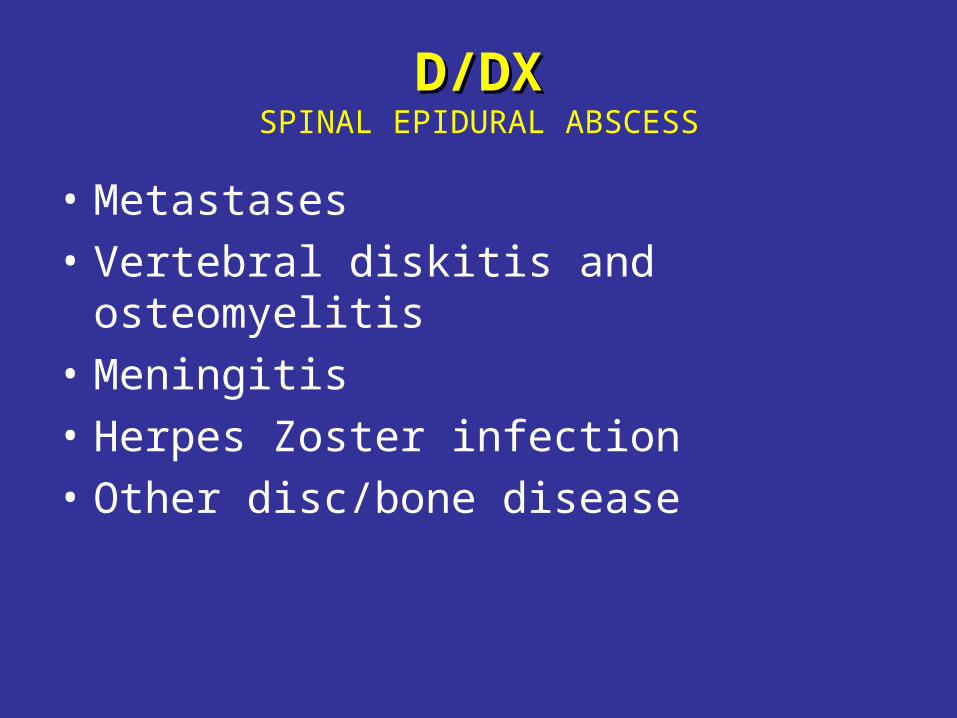
D/DXD/DXSPINAL EPIDURAL ABSCESS
• Metastases
• Vertebral diskitis and osteomyelitis
• Meningitis
• Herpes Zoster infection
• Other disc/bone disease
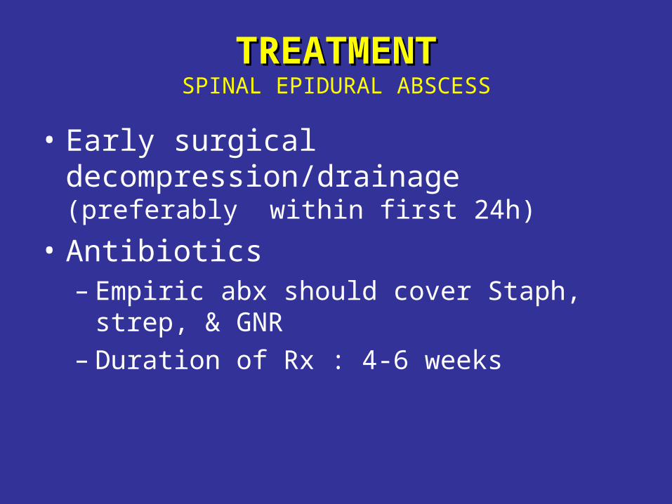
TREATMENTTREATMENTSPINAL EPIDURAL ABSCESS
• Early surgical decompression/drainage (preferably within first 24h)
• Antibiotics– Empiric abx should cover Staph, strep, &
GNR– Duration of Rx : 4-6 weeks
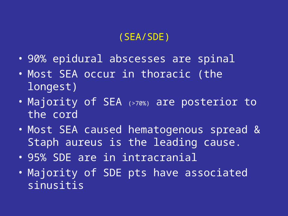
(SEA/SDE)
• 90% epidural abscesses are spinal• Most SEA occur in thoracic (the longest)• Majority of SEA (>70%) are posterior to the cord• Most SEA caused hematogenous spread &
Staph aureus is the leading cause.• 95% SDE are in intracranial• Majority of SDE pts have associated sinusitis

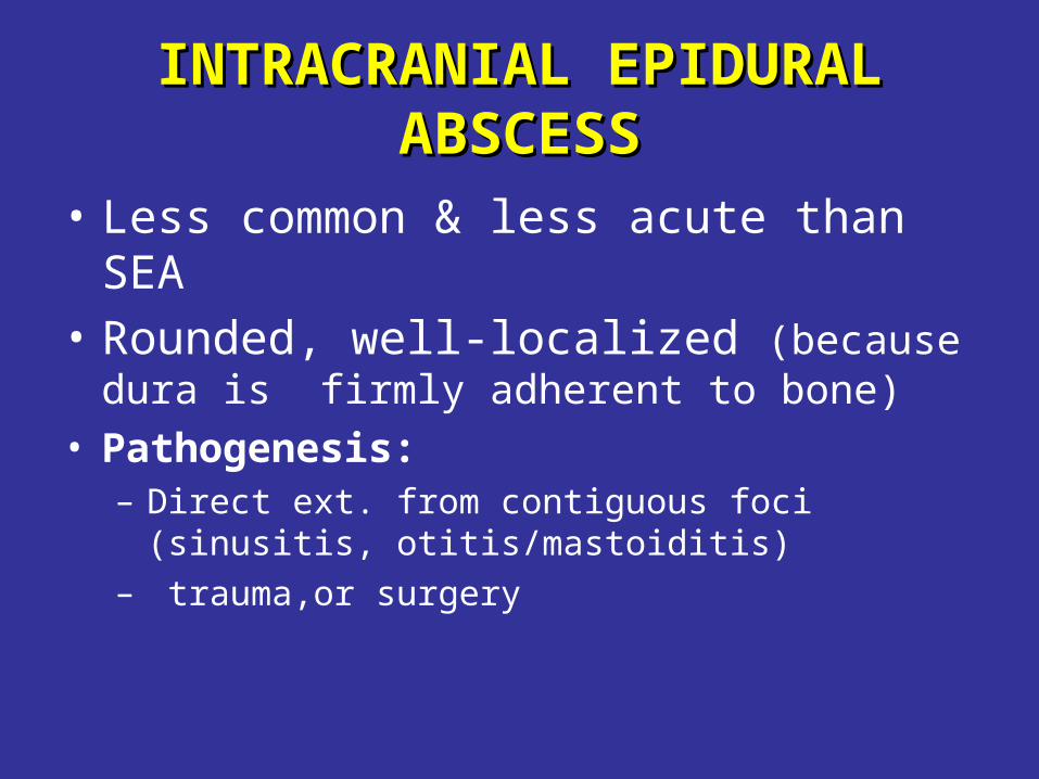
INTRACRANIAL EPIDURAL INTRACRANIAL EPIDURAL ABSCESSABSCESS
• Less common & less acute than SEA• Rounded, well-localized (because dura is
firmly adherent to bone)• Pathogenesis:
– Direct ext. from contiguous foci (sinusitis, otitis/mastoiditis)
– trauma,or surgery
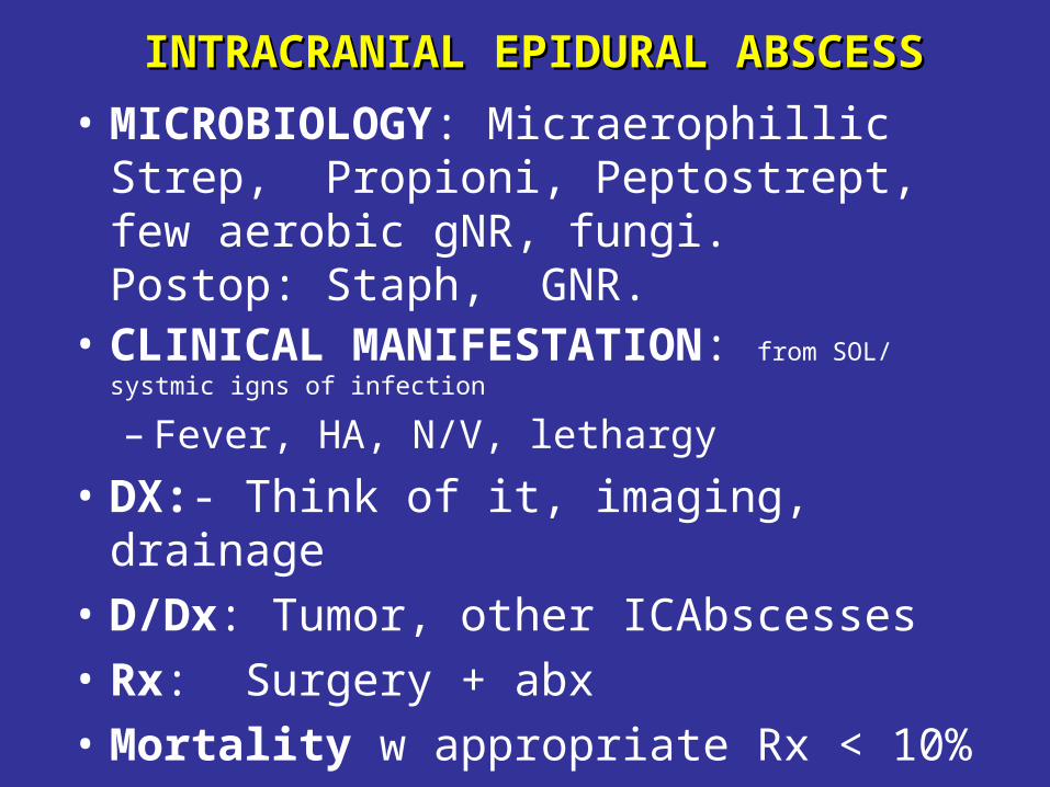
INTRACRANIAL EPIDURAL ABSCESSINTRACRANIAL EPIDURAL ABSCESS
• MICROBIOLOGY: Micraerophillic Strep, Propioni, Peptostrept, few aerobic gNR, fungi. Postop: Staph, GNR.
• CLINICAL MANIFESTATION: from SOL/ systmic igns of infection
– Fever, HA, N/V, lethargy
• DX:- Think of it, imaging, drainage
• D/Dx: Tumor, other ICAbscesses
• Rx: Surgery + abx
• Mortality w appropriate Rx < 10%
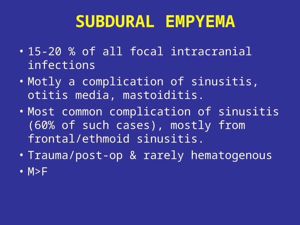
SUBDURAL EMPYEMA
• 15-20 % of all focal intracranial infections
• Motly a complication of sinusitis, otitis media, mastoiditis.
• Most common complication of sinusitis (60% of such cases), mostly from frontal/ethmoid sinusitis.
• Trauma/post-op & rarely hematogenous
• M>F
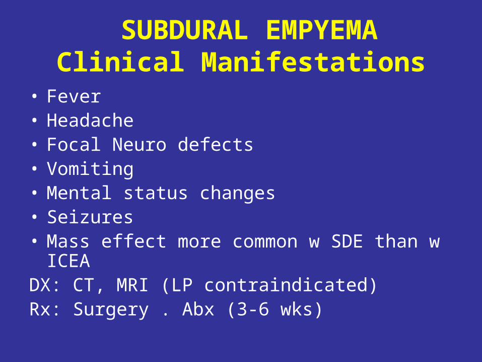
SUBDURAL EMPYEMAClinical Manifestations
• Fever• Headache• Focal Neuro defects• Vomiting• Mental status changes• Seizures• Mass effect more common w SDE than w ICEADX: CT, MRI (LP contraindicated)Rx: Surgery . Abx (3-6 wks)
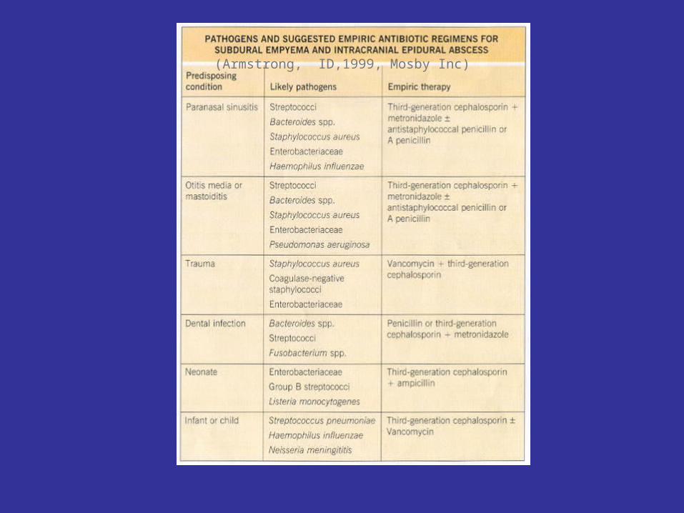
(Armstrong, ID,1999, Mosby Inc)

PARASITICPARASITIC BRAIN ABSCESS
• Toxoplasmosis
• Neurocysticercosis
• Amebic
• Echinococcal
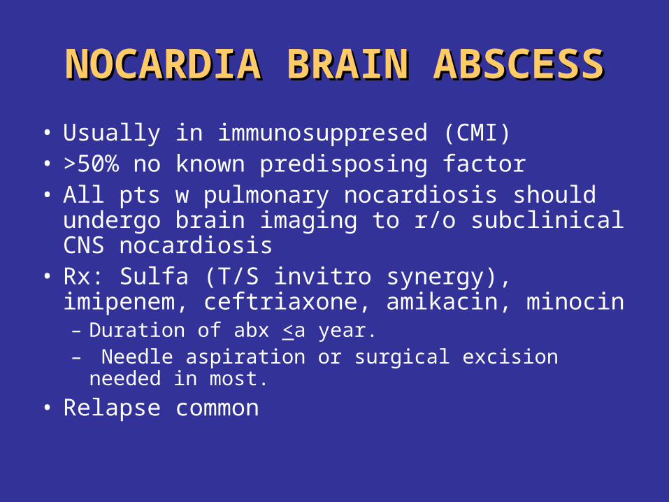
NOCARDIA BRAIN ABSCESSNOCARDIA BRAIN ABSCESS
• Usually in immunosuppresed (CMI)• >50% no known predisposing factor• All pts w pulmonary nocardiosis should undergo
brain imaging to r/o subclinical CNS nocardiosis• Rx: Sulfa (T/S invitro synergy), imipenem,
ceftriaxone, amikacin, minocin– Duration of abx <a year.– Needle aspiration or surgical excision needed in
most.
• Relapse common
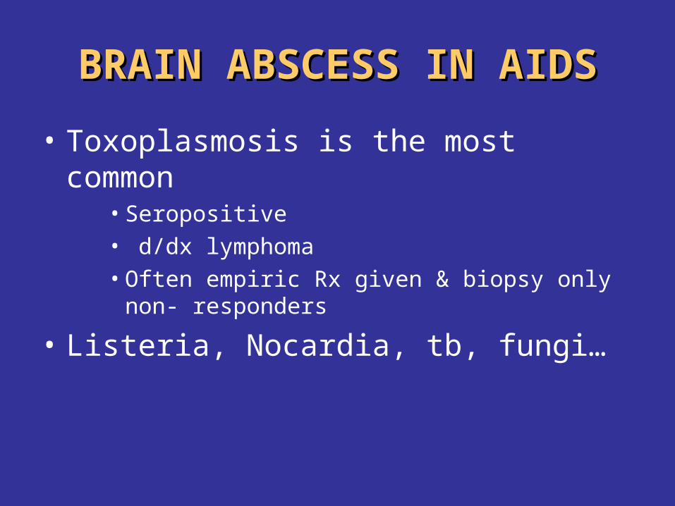
BRAIN ABSCESS IN AIDSBRAIN ABSCESS IN AIDS
• Toxoplasmosis is the most common• Seropositive• d/dx lymphoma• Often empiric Rx given & biopsy only non-
responders
• Listeria, Nocardia, tb, fungi…
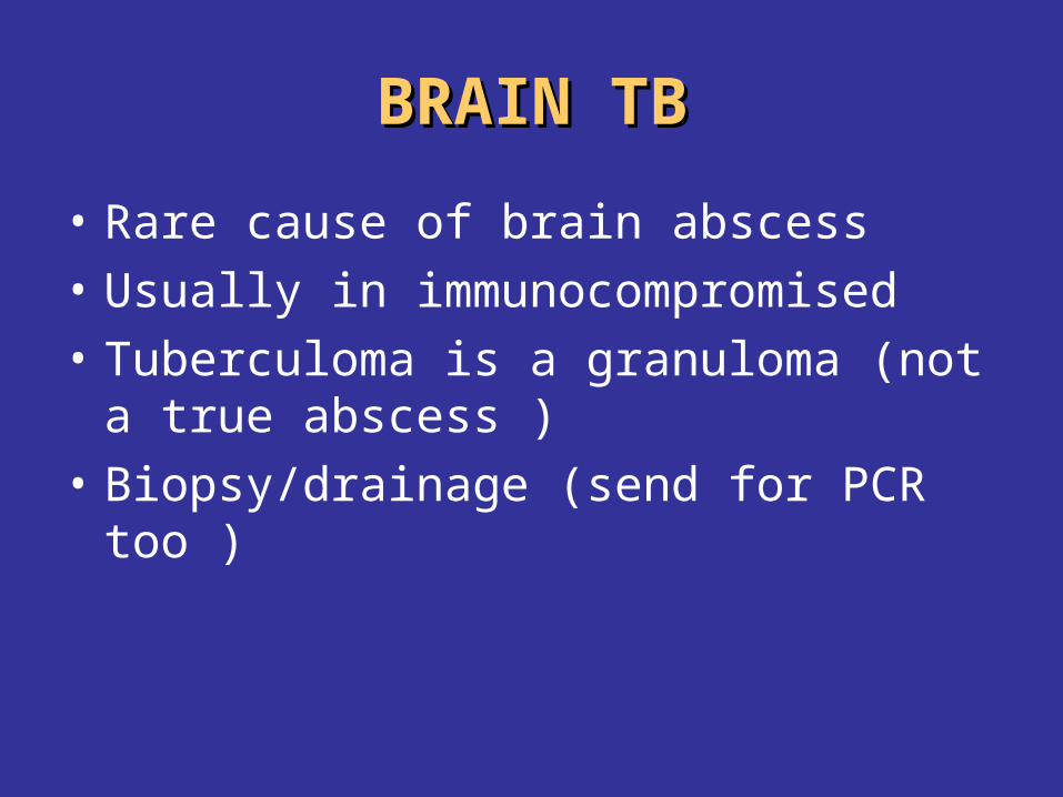
BRAIN TBBRAIN TB
• Rare cause of brain abscess
• Usually in immunocompromised
• Tuberculoma is a granuloma (not a true abscess )
• Biopsy/drainage (send for PCR too )
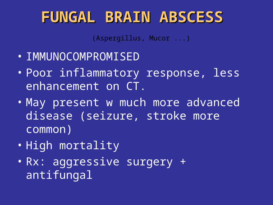
FUNGAL BRAIN ABSCESS FUNGAL BRAIN ABSCESS (Aspergillus, Mucor ...)
• IMMUNOCOMPROMISED
• Poor inflammatory response, less enhancement on CT.
• May present w much more advanced disease (seizure, stroke more common)
• High mortality
• Rx: aggressive surgery + antifungal
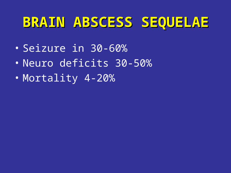
BRAIN ABSCESS SEQUELAEBRAIN ABSCESS SEQUELAE
• Seizure in 30-60%
• Neuro deficits 30-50%
• Mortality 4-20%
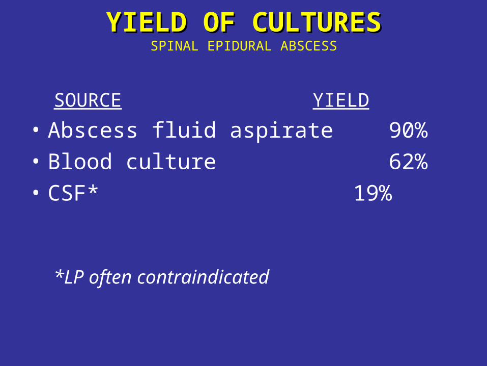
YIELD OF CULTURESYIELD OF CULTURESSPINAL EPIDURAL ABSCESS
SOURCE YIELD
• Abscess fluid aspirate 90%
• Blood culture 62%
• CSF* 19%
*LP often contraindicated


![Annals of Clinical Case Reports Case Report - anncaserep.com · pyogenic granuloma was described [5]. The Term Pyogenic granuloma is a misnomer because the The Term Pyogenic granuloma](https://img.pdfslide.net/doc/110x75/5d0a41bb88c993cf0c8b7f5f/annals-of-clinical-case-reports-case-report-pyogenic-granuloma-was-described.jpg)


![Utility of magnetic resonance imaging in the differential ... · and pyogenic spondylodiscitis Abstract ... abscesses[7] arachnoiditis, meningitis, ... Loss of cortical definition](https://img.pdfslide.net/doc/110x75/5b6ad0717f8b9af64d8c8c08/utility-of-magnetic-resonance-imaging-in-the-differential-and-pyogenic-spondylodiscitis.jpg)