Embed Size (px)
Citation preview
MOLECULAR AND CELLULAR BIOLOGY, July 2005, p. 5712–5724 Vol. 25, No. 130270-7306/05/$08.00!0 doi:10.1128/MCB.25.13.5712–5724.2005
Coactivator Recruitment Is Essential for Liganded Thyroid HormoneReceptor To Initiate Amphibian MetamorphosisBindu Diana Paul, Liezhen Fu, Daniel R. Buchholz, and Yun-Bo Shi*
Laboratory of Gene Regulation and Development, National Institute of Child Health and Human Development,National Institutes of Health, Bldg. 18T, Rm. 106, Bethesda, Maryland 20892
Received 14 September 2004/Returned for modification 19 October 2004/Accepted 25 March 2005
Thyroid hormone receptors (TRs) can repress or activate target genes depending on the absence or presenceof thyroid hormone (T3), respectively. This hormone-dependent gene regulation is mediated by recruitment ofcorepressors in the absence of T3 and coactivators in its presence. Many TR-interacting coactivators have beencharacterized in vitro. In comparison, few studies have addressed the developmental roles of these cofactors invivo. We have investigated the role of coactivators in transcriptional activation by TR during postembryonictissue remodeling by using amphibian metamorphosis as a model system. We have previously shown that ste-roid receptor coactivator 3 (SRC3) is expressed and upregulated during metamorphosis, suggesting a role ingene regulation by liganded TR. Here, we have generated transgenic tadpoles expressing a dominant negativeform of SRC3 (F-dnSRC3). The transgenic tadpoles exhibited normal growth and development throughout em-bryogenesis and premetamorphic stages. However, transgenic expression of F-dnSRC3 inhibits essentially allaspects of T3-induced metamorphosis, as well as natural metamorphosis, leading to delayed or arrested meta-morphosis or the formation of tailed frogs. Molecular analysis revealed that F-dnSRC3 functioned by blockingthe recruitment of endogenous coactivators to T3 target genes without affecting corepressor release, therebypreventing the T3-dependent gene regulation program responsible for tissue transformations during metamorpho-sis. Our studies thus demonstrate that coactivator recruitment, aside from corepressor release, is required for T3function in development and further provide the first example where a specific coactivator-dependent gene regu-lation pathway by a nuclear receptor has been shown to underlie specific developmental events.
Thyroid hormone receptors (TRs) are believed to mediatemost, if not all, of the vast, diverse biological effects of thyroidhormone (T3) (38, 56, 62, 75). TRs belong to the superfamilyof nuclear hormone receptors, which also includes steroid hor-mone receptors and 9-cis retinoic acid receptors (RXRs), andfunction in vivo most likely as heterodimers with RXRs (38, 41,66, 75). TR/RXR heterodimers bind to T3 response elements(TREs) constitutively and repress or activate gene expressionin a T3-dependent manner by recruiting corepressors or coac-tivators, respectively.
In vitro and cell culture studies have led to the isolation andcharacterization of many TR-interacting cofactor complexes(31, 34, 48, 75, 79). Among them, the best-studied corepressorcomplexes are those containing the nuclear receptor corepres-sor N-CoR (27) and the silencing mediator of retinoid andthyroid hormone receptor SMRT (7). Both N-CoR and SMRTexist in multiple histone deacetylase (HDAC)-containing com-plexes (23, 34, 40, 79). Recent studies suggest that TR mostlikely utilizes the complexes that contain HDAC3 and TBL1(for transducin beta-like protein 1) or TBLR1 (for TBL1-related protein) (23, 28, 40, 63, 64, 76, 78).
Among the coactivators that interact with TR directly, thesteroid receptor coactivator (SRC) family, which comprisesthree members (SRC1/NCoA-1, SRC2/TIF2/GRIP1, and SRC3/pCIP/ACTR/AIB-1/RAC-3/TRAM-1) has been the focus ofintense studies (6, 26, 39, 45, 61, 68). The SRC proteins bindTR and other nuclear receptors in a ligand-dependent manner
through LXXLL (L, leucine; X, any amino acid) motifs, whichare indispensable for the interaction (11, 24, 45, 67, 68). TheLXXLL motifs form short amphipathic "-helices, with theleucine residues forming a hydrophobic surface on one face ofthe helix (44, 59, 65). These motifs bind a hydrophobic cleft inthe ligand-binding domain of liganded nuclear receptors (13).Three such LXXLL motifs are localized in the central regionof these proteins and form the receptor interaction domain(RID). SRC proteins function as bridging factors to recruitchromatin-modifying enzymes, including methylases and his-tone acetyltransferases.
It remains to be determined how TR utilizes these co-activators in vivo, especially during development when TRregulates different genes in different cell types. This lack ofinformation on the in vivo function of the coactivators indevelopmental gene regulation by TR is attributed largely tothe difficulty in studying TR function in the uterus-enclosedmammalian embryos, despite the fact that T3 deficiency haslong been known to cause severe developmental defects, in-cluding cretinism (25). The effects of T3 on development takeplace mainly during perinatal period, when T3 levels in theplasma are high (4, 25, 37).
It is unclear whether and how TR mediates the developmen-tal effects of T3 because of the existence of nongenomic mech-anisms through cytosolic T3-binding proteins (10). Studies withTR knockout mice have provided some in vivo evidence tosupport a critical role of TRs in mediating T3 signal in devel-opment. Interestingly, mice lacking TR" or TR# or both havemuch less severe developmental defects than those lacking T3(15–17, 20, 22, 70). Furthermore, transgenic mice harboring adominant negative form of the corepressor N-CoR containing
* Corresponding author. Mailing address: NIH, LGRD, NICHD,Building 18T, Rm. 106, Bethesda, MD 20892. Phone: (301) 402-1004.Fax: (301) 402-1323. E-mail: [email protected].
5712
only the receptor-interacting domain in the liver exhibit up-regulated T3-inducible genes (14). Finally, many studies withtissue culture cells and frog oocytes show that the bulk of theincreases in target gene expression by T3 appears to be due tothe release of repression caused by unliganded TR. Thesestudies thus leave unresolved the possibility that corepressorrelease, not coactivator recruitment, may be the major effect ofT3 on gene regulation during development.
Here, we use amphibian metamorphosis as a model to studythe role of coactivators in T3 function during development.Amphibian metamorphosis is a T3-dependent process thatbears strong similarities to postembryonic development inmammals (56, 62). Unlike mammalian development, amphib-ian metamorphosis can be easily manipulated by blocking thesynthesis of endogenous T3 or by adding physiological concen-trations of T3 to the tadpole rearing water, even though dif-ferent organs and/or tissues undergo vastly different transfor-mations (12, 56).
Using this model system, we have shown previously that themRNAs of TR interacting cofactors SRC2, SRC3, and p300are expressed during metamorphosis, among which SRC3 isupregulated during both natural and T3-induced metamorpho-sis, supporting a role for this coactivator (47). To directlyinvestigate the role of coactivator recruitment by TR in medi-ating the developmental effects of T3 in vivo, we introduced adominant negative form of SRC3 (F-dnSRC3) into developinganimals through sperm-mediated transgenesis. We showed thatF-dnSRC3 inhibits both natural and T3-induced metamorpho-sis and the expression of TR target genes by specifically block-ing coactivator recruitment but not corepressor release. Theseresults thus identify an essential role of coactivator binding toTR beyond corepressor release in postembryonic development.
MATERIALS AND METHODS
Cloning and constructs. The dominant negative SRC3, F-dnSRC3, was gen-erated by PCR amplification of the sequences encompassing the receptor inter-action domain of Xenopus laevis SRC3 (amino acids [aa] 600 to 751) (35). Theconstruct was generated to encode a fusion protein with an N-terminal Flagepitope tag (8), followed by the simian virus 40 (SV40) nuclear localization signal(NLS) (5) using a 5$ primer containing the sequences encoding these fusionpeptides in frame with the SRC3 coding sequence. The primers used were (5$ to3$) AGATCTACCGGTGCCATGGACTACAAAGACGATGACGATAAAGGATCCCCAAAGAAGAAGAAGCGTAAGGTACTCGAGATGATATTTGAAGGGTCGGAGA (the Flag tag is underlined and the NLS is shown in boldfacetype) and CTAGTCACTAGTGAATTCTCACTTGGCCAGTGGGTCCTTCCAGTC. For expression and detection in frog oocytes, the PCR product wascloned into the T7Ts expression vector (a gift from G. J. C. Veenstra, Universityof Nijmegen), which is based on the pGEM-4Z vector (Promega) and containsthe 5$ and 3$-untranslated regions of the X. laevis #-globin gene flanking themultiple cloning sites.
For transgenesis, F-dnSRC3 was subcloned into the vector pCGCG (18) underthe control of the cytomegalovirus (CMV) promoter, replacing the original greenfluorescent protein (GFP) fragment at this location and resulting in the double-promoter construct pCF-dnSRC3CG, which also has the gene for GFP driven bythe eye lens-specific %-crystallin promoter.
Animals, transgenesis, and animal treatment. Wild-type tadpoles of the Af-rican clawed frog Xenopus laevis were obtained from Xenopus I, Inc. (Dexter,MI), and developmental stages were determined according to Nieuwkoop andFaber (43). Adult female frogs used for oocyte preparation were obtained fromNASCO (Fort Atkinson, WI).
Transgenesis was carried out using X. laevis as described previously (18) usingthe double-promoter construct pCF-dnSRC3CG. Transgenic animals were iden-tified by GFP expression in the eye lens due to the presence of the secondpromoter, the %-crystallin promoter, driving the expression of GFP.
To study the effect of transgene on T3-induced tail resorption and other
external changes, 1-week-old transgenic tadpoles were treated for 7 days with 10nM T3 diluted in 0.1& MMR (10 mM NaCl–0.2 mM KCl–0.1 mM MgCl2–0.2mM CaCl2–0.5 mM HEPES, pH 7.5). Both wild-type and transgenic animalswere treated in the same container to ensure similar conditions of treatment. Tostudy the effect of the transgene on limb development and changes in the intes-tine, stage 54 animals were treated with the indicated amount of T3 (two ani-mals per liter of deionized, dechlorinated water). These tadpoles were sacrificedby decapitation after anesthesia (cooling on ice) to isolate tissues for molecularanalyses.
Oocyte injection and immunoprecipitation. pSP64-TR, pSP64-RXR (71), andT7Ts-Flag-dnSRC3 were used to synthesize the corresponding mRNAs in vitrowith a T7 or SP6 in vitro transcription kit (mMESSAGE mMACHINE; Am-bion). The mRNA, at a concentration of 5.75 ng/oocyte, was microinjected intothe cytoplasm of 20 X. laevis stage VI oocytes. After incubation overnight at18°C, the oocytes were lysed by being pipetted in lysis buffer (20 mM HEPES, pH7.5, 5 mM KCl, 1.5 mM MgCl2, 1 mM EGTA, 10 mM glycerophosphate, 150 mMNaCl, 0.1% NP-40, 1 mM dithiothreitol, 0.2 mM phenylmethylsulfonyl fluoride,and protease inhibitor mixture [Roche Applied Science]). After centrifugation at14,000 rpm for 10 min at 4°C, the supernatant was used for immunoprecipitationwith anti-Flag-M2-agarose beads (Sigma). Each lysate was incubated with thebeads for 4 h and washed three times in the lysis buffer. The immunoprecipitateswere boiled in sodium dodecyl sulfate (SDS) loading buffer, separated on anSDS-polyacrylamide gel, and immunoblotted with indicated antibodies.
Luciferase assays. The cytoplasm of groups of stage VI oocytes was injectedwith mRNAs encoding TR, RXR, and/or F-dnSRC3 (5.75 ng per oocyte). Thereporter plasmid DNA (0.33 ng/oocyte), which contained the T3-dependentTR#A promoter driving the expression of the firefly luciferase (1) was injectedinto the oocyte nucleus, together with a control construct which contained theherpes simplex virus tk promoter driving the expression of Renilla luciferase (0.03ng/oocyte). Following incubation overnight at 18°C in the absence or presence of100 nM T3, oocytes were prepared for luciferase assay by the Dual-LuciferaseReporter Assay system (Promega), according to the manufacturer’s recommen-dations.
Protein extraction and Western blot analysis. Isolated tissues from wild-typeand transgenic tadpoles at stage 54 were homogenized on ice in lysis buffer [10mM Tris-HCl, pH 7.5, 150 mM NaCl, 5 mM EDTA, 1% Triton X-100, 200 M4-(2-aminoethyl)-benzenesulfonylfluoride HCl, 5 'g/ml aprotinin, 2 'g/ml leu-peptin, and 1 'g/ml pepstatin]. The homogenate was kept on ice for 30 minbefore centrifugation for 15 min at 4°C (11,000 & g). Equal amounts of theprotein extract were then analyzed by Western blotting with anti-Flag antibodiesto detect the expression of the transgene.
Chromatin immunoprecipitation (ChIP) assays. The ChIP assay with Xenopusoocytes was performed as described previously (63). For the ChIP assay withtadpole tissues, four tadpoles per treatment group were used. Wild-type andtransgenic tadpoles were treated with 10 nM T3 for 2 days in the same container.The ChIP assay was performed as described previously (9, 54) with minormodifications (64). The following antibodies were used in the assay: anti-XenopusTR (71), anti-acetylated histone H4 (Upstate Biotechnology, Lake Placid, N.Y.),anti-Flag M2-agarose (Sigma), anti-Xenopus SRC3 (generated in rabbits by coin-jecting the peptides SGEKRRREQESK and DHLEDGSNLDARQRYE), andanti-Xenopus SMRT antibody (generated by immunizing a rabbit with the poly-peptide KSKKQEMIKKLSTTNRSEQE, located in a 2-kb cDNA fragment cor-responding to the C-terminal part encompassing the TR-binding domain of theXenopus laevis SMRT) (64). The primers used in the oocyte ChIP assay were (5$to 3$ direction) TGCCTGTGTCTATACTGATGGGAT and CATTTTACCAACAGTACCGGAATGC, producing a 190-bp fragment containing the TRE re-gion of the construct TRE-Luc.
The DNA from the ChIP assay with tadpole tissues was analyzed by quanti-tative PCR in duplicate on an ABI 7000 (Applied Biosystems) system usingpromoter-specific primers and 6-carboxyfluorescein-labeled TaqMan probes(Applied Biosystems). To ensure the validity of the PCR for each assay, sixtwofold serial dilutions from a large batch of ChIP input DNA prepared fromintestines prepared especially to serve as standards were used for the quantifi-cation of the experimental samples. The calculated standard curves ranged inslope from (3.30 to (3.50, where theoretical amplification has a slope of (3.32.Also included was a no-template control, where double-distilled water was addedinstead of sample DNA as a control for PCR product contamination. Resultsfrom the experimental samples were within the range of the standard curve. Theprimers used for the quantitative PCR were as follows (5$ to 3$): CCCCTATCCTTGTTCGTCCTC and GCGCTGGGCTGTCCT for the TRE region of theTR#A promoter and GGACGCACTAGGGTTAAGTAAGG and TCTCCCAACCCTACAGAGTTCAA for the TRE region of the TH/bZIP promoter. The6-carboxyfluorescein-labeled probes were (5$ to 3$) CCTAGGCAGGTCATTTC
VOL. 25, 2005 PIVOTAL ROLE OF COACTIVATOR RECRUITMENT IN DEVELOPMENT 5713
and ATGAGGCTGAGCATTCA for the TR#A and the TH/bZIP promoters,respectively.
RNA isolation and reverse transcriptase PCR (RT-PCR). RNA was isolatedusing the Trizol reagent (Invitrogen) per the manufacturer’s recommendations.RT-PCRs were carried out with the Superscript One-Step RT-PCR kit (Invitro-gen). The expression of the ribosomal protein L8 (Rpl8) was used as an internalcontrol (58). The sequences of the primers used were (5$ to 3$) CGTGGTGCTCCTCTTGCCAAG and GACGACCAGTACGACGAGCAG for rpl8 (58), CCTGATGCATGCAAAACT and GTTCATCCTGGAAAGCAG for ST3 (46),CACTTAGCAACAGGGATCAGC and CTTGTCCCAGTAGCAATCATCfor TH/bZIP (19), ATAGTTAATGCGCCCGAGGGTGGA and CTTTTCTATTCTCTCCACGCTAGC for TR#) (74), CATCATGATTCCTGGTAACCGA and AAATTTCCATTTTCTGCTGTGC for BMP-4 (42), and GGGCAGTGGACATCACCAC and GTTGACCTTGGTCTGGGCC for xhh (60).The sequences of the primers used for the detection of F-dnSRC3 in trans-genic animals were (5$ to 3$) ACCGGTGCCATGGACTAC and CTAGTCACTAGTGAATTCTCACTTGGCCAGTGTCCTTCCAGTC. A total of0.5 'g of total RNA was used in a 25-'l reaction mixture, under the followingreaction conditions: 42°C for 30 min for the RT reaction, followed by 21 to 25cycles, each consisting of 94°C for 30 s, 55°C for 30 s, and 72°C for 30 s. Theresulting products were analyzed on an agarose gel stained with ethidium bro-mide.
Histological analysis of the intestine. The intestines of the tadpoles weredissected and fixed for 2 h at room temperature in 4% paraformaldehyde–60%phosphate-buffered saline, cryoprotected in 0.5 M sucrose in 60% phosphate-
buffered saline and embedded in OCT medium (TissueTek). The intestines weresectioned in a cryotome at 7.5 'm. Sections were visualized with methyl greenpyronin Y (Muto, Tokyo, Japan) (29).
RESULTS
TR recruits SRC3 during transcriptional activation in Xe-nopus laevis oocytes. Our previous expression studies haveshown that both SRC2 and SRC3 are expressed and that SRC3is upregulated during both natural and T3-induced metamor-phosis. We were thus interested in investigating the roles ofsuch TR-binding coactivators in this postembryonic process byaltering coactivator recruitment by TR through transgenesis.For this purpose, we reasoned that this might be accomplishedby introducing a dominant negative form of SRC3 to inhibitthe binding of endogenous coactivators to liganded TR. As afirst step, we generated polyclonal antibodies against peptidesderived from Xenopus laevis SRC3. The specificity of theantibody was verified by Western blotting using in vitro-translated Xenopus SRC3 and SRC2 (Fig. 1A), the two SRCproteins cloned so far from Xenopus laevis. In addition, the
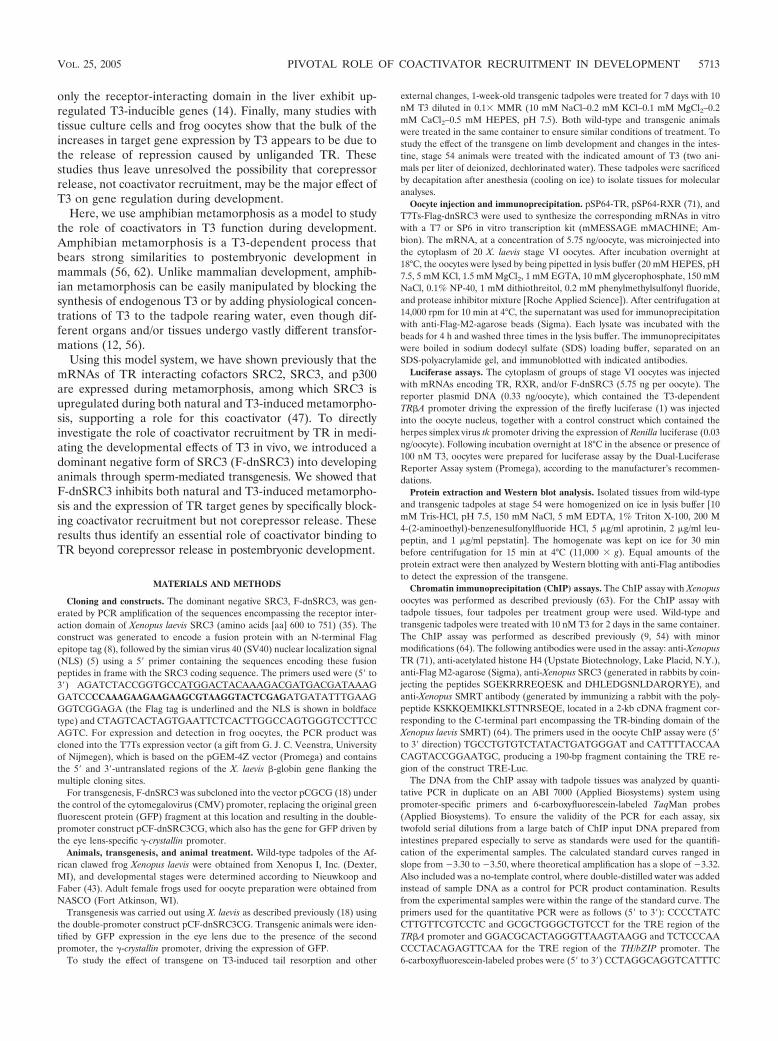
FIG. 1. The Xenopus laevis SRC3 protein is recruited by TR to target genes in the frog oocyte. (A) The anti-SRC3 antibody specificallyrecognizes the Xenopus laevis SRC3 protein. The two SRC family proteins cloned from Xenopus laevis, SRC3 and SRC2 (lanes 2 and 3), weretranslated in vitro in the presence of 35S-Met and analyzed on an 8 to 16% Tris glycine SDS gel. The proteins were transferred to a membraneand blotted with polyclonal anti-SRC3 antibodies. Top, 35S-Met signal; bottom, Western blot signal. Lane 1, control (unprogrammed rabbit reti-culocyte lysate). (B) The anti-SRC3 antibody recognizes endogenous SRC3 protein in Xenopus oocyte nuclei. Lanes 1 and 2, unprogrammed rabbitreticulocyte lysate and in vitro-translated SRC3, respectively; lane 3, protein extract from isolated nuclei (germinal vesicles) of Xenopus oocytes.(C) Transcription from the T3-inducible promoter of the Xenopus TR#A gene is regulated by TR/RXR in a ligand-dependent manner. The cyto-plasm of oocytes was injected with mRNAs for TR (5.75 ng/oocyte) and RXR (5.75 ng/oocyte). The firefly luciferase reporter construct TRE-Luc(0.33 ng/oocyte) was then injected into the nucleus together with the control Renilla luciferase plasmid (0.03 ng/oocyte), followed by incubationovernight at 18°C with or without 100 nM T3. The oocytes were harvested and lysed, and the ratio of firefly luciferase from TRE-Luc to that fromthe control Renilla luciferase plasmid was determined for each oocyte. The average and standard deviations from four measurements are plotted.(D) SRC3 is recruited to the TRE in a ligand-dependent manner. Groups of oocytes were injected with mRNAs for TR and RXR and treatedas described above. ChIP assays were carried out using antibodies against TR and SRC3 to detect the occupancy of TR and SRC3 on the promoter.The presence of TRE in the immunoprecipitated samples was detected by PCR using primers flanking the TRE. The products were analyzed ona 2% agarose gel and visualized by ethidium bromide staining. The DNA prior to immunoprecipitation was amplified by PCR as an input control.
5714 PAUL ET AL. MOL. CELL. BIOL.
antibody was raised against peptides from SRC3 that did notexhibit sequence similarity to any region of SRC1 cloned so farfrom other species (not shown), making it unlikely to recognizeendogenous Xenopus laevis SRC1.
To study the involvement of SRC3 in T3-mediated transcrip-tional activation, we utilized the frog oocyte system, whichallows the analysis of gene activation by TR in the context ofchromatin (71). First, we showed that endogenous SRC3 waspresent in the nuclei of Xenopus oocytes (Fig. 1B, lane 3). Next,a reporter construct, TRE-Luc, which harbors the T3-depen-dent Xenopus TR#A promoter fused to the firefly luciferasegene (1), was microinjected into the nuclei of Xenopus oocytestogether with a plasmid harboring the Renilla luciferase geneas an internal control. The mRNAs encoding TR and RXRwere microinjected subsequently into the cytoplasm of theoocytes. Following an overnight incubation in the presence orabsence of 100 nM T3, oocytes were harvested and assayed forluciferase activity. As expected, in the absence of T3, basalactivity from TRE-Luc was repressed by TR and RXR (71),while in the presence of T3, the promoter was activated (Fig.1C). Finally, to determine whether SRC3 was involved in this
gene activation by TR, we carried out ChIP assays using anti-bodies against TR and SRC3. The results revealed that whileTR binds to the TRE region constitutively, endogenous SRC3was recruited to the TR#A promoter only in the presence of T3(Fig. 1D), suggesting that liganded TR recruits SRC3 to acti-vate the promoter in vivo.
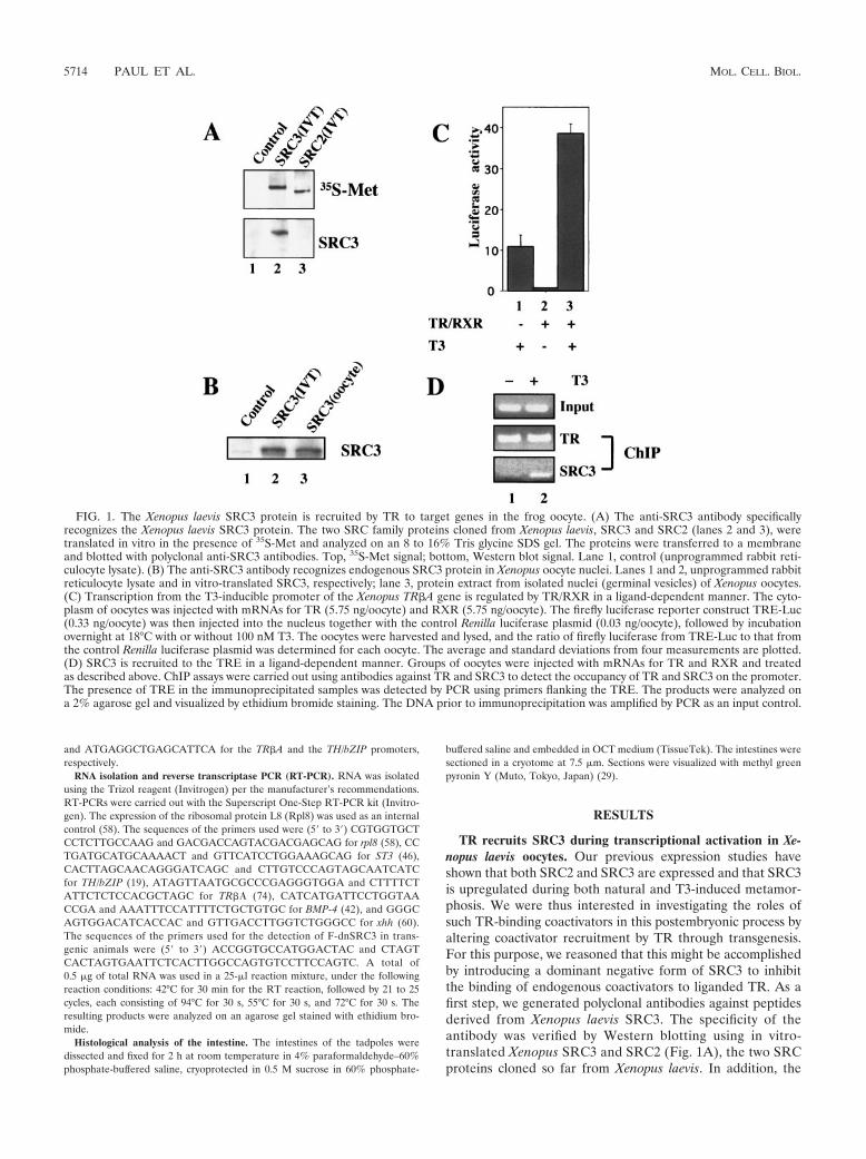
A dominant negative SRC3 inhibits gene activation by TR inthe frog oocyte. To generate a dominant negative form ofSRC3, we reasoned that a truncated protein comprising onlythe receptor interaction domain of SRC3 might be able tofunction as a dominant negative inhibitor of coactivator bind-ing to TR, thus inhibiting gene activation by liganded TR.Therefore, we generated a truncated form of SRC3 (Fig. 2A),F-dnSRC3, consisting of an N-terminal Flag tag, followed bythe SV40 NLS and the receptor interaction domain of SRC3(aa 600 to 751) (35). To verify the expression and localizationof F-dnSRC3, its mRNA was synthesized in vitro and micro-injected into the oocyte cytoplasm. As a control, the mRNAencoding Flag-tagged Xenopus TR (F-TR), which localizes tothe nucleus, was coinjected. After overnight incubation, thenuclear and cytoplasmic fractions of the injected oocytes were
FIG. 2. Generation and characterization of a dominant negative SRC3 (F-dnSRC3). (A) Schematic representation of the full-length SRC3illustrating the organization of various domains: bHLH/PAS (basic helix-loop-helix and PAS dimerization domains) RID, and CID (CBP/p300interaction domain). The LXXLL motifs present in the protein are numbered from i to vi. A glutamine-rich (Q-rich) region is present towards theC-terminal end of the protein. The dominant negative form, dnSRC3 (aa 600 to 751), which comprises the LXXLL motifs i to iii, forming the RID,and fused to an N-terminal peptide containing the Flag tag and nuclear localization sequences, is shown below. (B) The F-dnSRC3 protein islocalized in the nucleus. Oocytes were injected with mRNAs (5.75 ng/oocyte) for F-TR, RXR, and F-dnSRC3 and incubated overnight. The oocyteswere then harvested, and nuclei were dissected manually under a light microscope. The nuclei and cytoplasmic components were resuspended in1& SDS lysis buffer, boiled for 5 min, resolved on an 8 to 16% Tris glycine-SDS gel, and visualized by Western blotting using anti-Flag antibody.Lane 1, C, the cytoplasmic extracts of Xenopus oocytes overexpressing F-dnSRC3 and F-TR; lane 2, N, the corresponding nuclear extracts; lane3, IVT, in vitro-translated F-dnSRC3. The asterisks indicate cross-reacting bands recognized by the antibody. (C) F-dnSRC3 interacts withTR/RXR in a T3-dependent manner. Groups of oocytes were injected with 5.75 ng of TR, RXR, and F-dnSRC3 mRNAs and incubated overnightin the presence or absence of 100 nM T3. Twenty Xenopus oocytes were used for each immunoprecipitation with anti-Flag M2 antibody conjugatedto Sepharose beads, followed by Western blot analysis using anti-TR antibody. Lanes 1 to 3, 10% input samples; lanes 4 to 6, immunoprecipitationwith anti-Flag antibody. (D) Western blotting of input samples shown in panel C with anti-Flag antibody shows similar expression of F-dnSRC3in the injected oocytes.
VOL. 25, 2005 PIVOTAL ROLE OF COACTIVATOR RECRUITMENT IN DEVELOPMENT 5715
isolated and subjected to Western blotting using the anti-Flagantibody. The results showed that both F-TR and F-dnSRC3were efficiently expressed and localized in the nucleus (Fig.2B).
To investigate the ability of F-dnSRC3 to interact with theTR/RXR heterodimer, we then carried out coimmunoprecipi-tation assays. The mRNAs encoding Xenopus TR, RXR, andF-dnSRC3 were injected into the cytoplasm of oocytes. Afterincubation overnight in the absence or presence of 100 nM T3,extracts prepared from groups of oocytes were incubatedwith the anti-Flag antibody. Immunoprecipitated samples werethen probed with anti-TR antibodies, which revealed that F-dnSRC3 protein interacted with TR in a ligand-dependentmanner (Fig. 2C, lanes 5 and 6). Control Western blotting ofthe extracts before immunoprecipitation with anti-Flag anti-body showed that similar amounts of F-dnSRC3 were presentin oocytes incubated in the absence or presence of T3 (Fig. 2D,lanes 2 and 3).
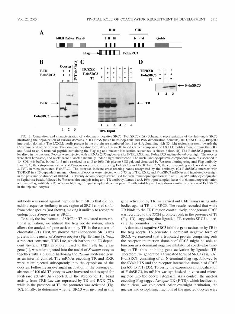
We next tested the ability of F-dnSRC3 to inhibit transcrip-tional activation by liganded TR. Again, we microinjected thereporter vector harboring the T3-dependent Xenopus TR#Apromoter into the nuclei of oocytes together with the internalcontrol plasmid harboring the Renilla luciferase gene. Thecytoplasm of groups of oocytes was injected with mRNAsencoding TR, RXR, and/or F-dnSRC3. The oocytes were in-cubated overnight in the absence or presence of T3, followedby luciferase assays. As shown in Fig. 3A, F-dnSRC3 had no
effect on basal transcription in the absence of TR/RXR (Fig.3A, lanes 1 and 2). However, the activation by TR/RXR in thepresence of T3 was inhibited by F-dnSRC3 (Fig. 3A, comparelanes 5 and 4). To investigate whether this dominant negativeeffect of F-dnSRC3 was due to its T3-dependent recruitmentto the promoter, ChIP assays were carried out using antibod-ies against TR and F-dnSRC3. The results revealed that F-dnSRC3 was recruited to the promoter in a T3-dependentfashion, whereas TR bound constitutively to the TR#A pro-moter in the oocyte (Fig. 3B). Moreover, in the presence of F-dnSRC3, the T3-dependent recruitment of endogenous SRC3to the promoter was impaired (Fig. 3B). These results suggestthat F-dnSRC3 functions as an effective dominant negative toblock endogenous coactivators in gene regulation by TR andthat the recruitment of SRC3 or other TR-binding coacti-vators by liganded TR is essential for gene activation in vivo.
Transgenic overexpression of F-dnSRC3 inhibits T3-in-duced metamorphosis. To investigate whether F-dnSRC3 mayaffect gene regulation by TR and metamorphosis in vivo, weintroduced it into developing animals through transgenesis us-ing the restriction enzyme-mediated integration method (36).The expression of F-dnSRC3 in transgenic animals was drivenby the constitutively active CMV promoter. We used a double-promoter construct that also harbored GFP under the controlof the %-crystallin promoter (Fig. 4A) (18), which is functionalonly in the eye lens. This allowed us to rear both wild-type andtransgenic animals together to avoid variations in growth and
FIG. 3. (A) F-dnSRC3 inhibits T3-induced transcription from the TR#A promoter. The mRNAs for TR, RXR, and F-dnSRC3 (5.75 ng/oocyte)were injected into the cytoplasm of oocytes. The firefly luciferase reporter vector (0.33 ng/oocyte) (TRE-Luc) was then injected into the nucleustogether with the control Renilla luciferase plasmid (0.03 ng/oocyte). After overnight incubation at 18°C with or without 100 nM T3, oocytes wereharvested and assayed for transcription from TRE-Luc. The ratio of firefly luciferase activity from TRE-Luc to that from the control Renillaluciferase plasmid was determined for each oocyte, and the average from six oocytes was plotted together with the standard deviation. Note thatas expected, unliganded TR/RXR repressed basal activity of the target promoter; the addition of T3 released the repression and further activatedthe promoter. F-dnSRC3 inhibited T3 activation but not basal activity. (B) F-dnSRC3 competes against endogenous SRC3 for recruitment to theTR#A promoter in a ligand-dependent manner. A total of 5.75 ng of TR, RXR, or F-dnSRC3 mRNAs was injected into the cytoplasm of eachXenopus oocyte. The construct TRE-Luc was injected into the nucleus. The oocytes were treated with or without 100 nM T3 as indicated. Afterovernight incubation, ChIP analysis was carried out using anti-TR, anti-SRC3, or anti-FLAG antibodies.
5716 PAUL ET AL. MOL. CELL. BIOL.
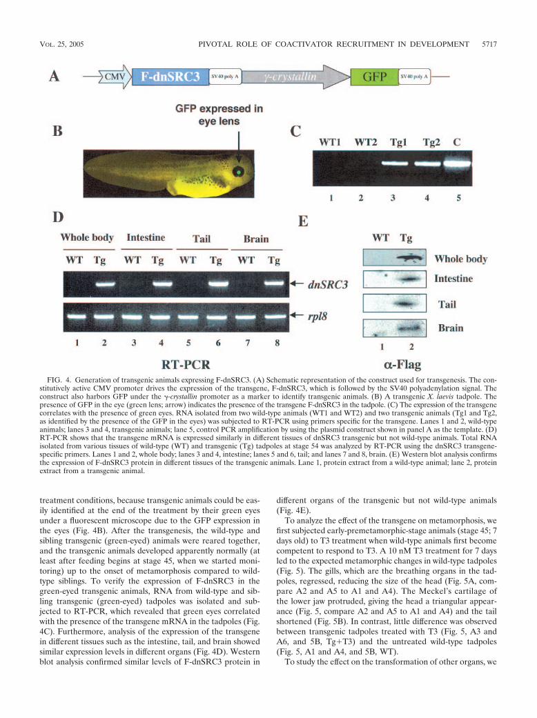
treatment conditions, because transgenic animals could be eas-ily identified at the end of the treatment by their green eyesunder a fluorescent microscope due to the GFP expression inthe eyes (Fig. 4B). After the transgenesis, the wild-type andsibling transgenic (green-eyed) animals were reared together,and the transgenic animals developed apparently normally (atleast after feeding begins at stage 45, when we started moni-toring) up to the onset of metamorphosis compared to wild-type siblings. To verify the expression of F-dnSRC3 in thegreen-eyed transgenic animals, RNA from wild-type and sib-ling transgenic (green-eyed) tadpoles was isolated and sub-jected to RT-PCR, which revealed that green eyes correlatedwith the presence of the transgene mRNA in the tadpoles (Fig.4C). Furthermore, analysis of the expression of the transgenein different tissues such as the intestine, tail, and brain showedsimilar expression levels in different organs (Fig. 4D). Westernblot analysis confirmed similar levels of F-dnSRC3 protein in
different organs of the transgenic but not wild-type animals(Fig. 4E).
To analyze the effect of the transgene on metamorphosis, wefirst subjected early-premetamorphic-stage animals (stage 45; 7days old) to T3 treatment when wild-type animals first becomecompetent to respond to T3. A 10 nM T3 treatment for 7 daysled to the expected metamorphic changes in wild-type tadpoles(Fig. 5). The gills, which are the breathing organs in the tad-poles, regressed, reducing the size of the head (Fig. 5A, com-pare A2 and A5 to A1 and A4). The Meckel’s cartilage ofthe lower jaw protruded, giving the head a triangular appear-ance (Fig. 5, compare A2 and A5 to A1 and A4) and the tailshortened (Fig. 5B). In contrast, little difference was observedbetween transgenic tadpoles treated with T3 (Fig. 5, A3 andA6, and 5B, Tg!T3) and the untreated wild-type tadpoles(Fig. 5, A1 and A4, and 5B, WT).
To study the effect on the transformation of other organs, we
FIG. 4. Generation of transgenic animals expressing F-dnSRC3. (A) Schematic representation of the construct used for transgenesis. The con-stitutively active CMV promoter drives the expression of the transgene, F-dnSRC3, which is followed by the SV40 polyadenylation signal. Theconstruct also harbors GFP under the %-crystallin promoter as a marker to identify transgenic animals. (B) A transgenic X. laevis tadpole. Thepresence of GFP in the eye (green lens; arrow) indicates the presence of the transgene F-dnSRC3 in the tadpole. (C) The expression of the transgenecorrelates with the presence of green eyes. RNA isolated from two wild-type animals (WT1 and WT2) and two transgenic animals (Tg1 and Tg2,as identified by the presence of the GFP in the eyes) was subjected to RT-PCR using primers specific for the transgene. Lanes 1 and 2, wild-typeanimals; lanes 3 and 4, transgenic animals; lane 5, control PCR amplification by using the plasmid construct shown in panel A as the template. (D)RT-PCR shows that the transgene mRNA is expressed similarly in different tissues of dnSRC3 transgenic but not wild-type animals. Total RNAisolated from various tissues of wild-type (WT) and transgenic (Tg) tadpoles at stage 54 was analyzed by RT-PCR using the dnSRC3 transgene-specific primers. Lanes 1 and 2, whole body; lanes 3 and 4, intestine; lanes 5 and 6, tail; and lanes 7 and 8, brain. (E) Western blot analysis confirmsthe expression of F-dnSRC3 protein in different tissues of the transgenic animals. Lane 1, protein extract from a wild-type animal; lane 2, proteinextract from a transgenic animal.
VOL. 25, 2005 PIVOTAL ROLE OF COACTIVATOR RECRUITMENT IN DEVELOPMENT 5717
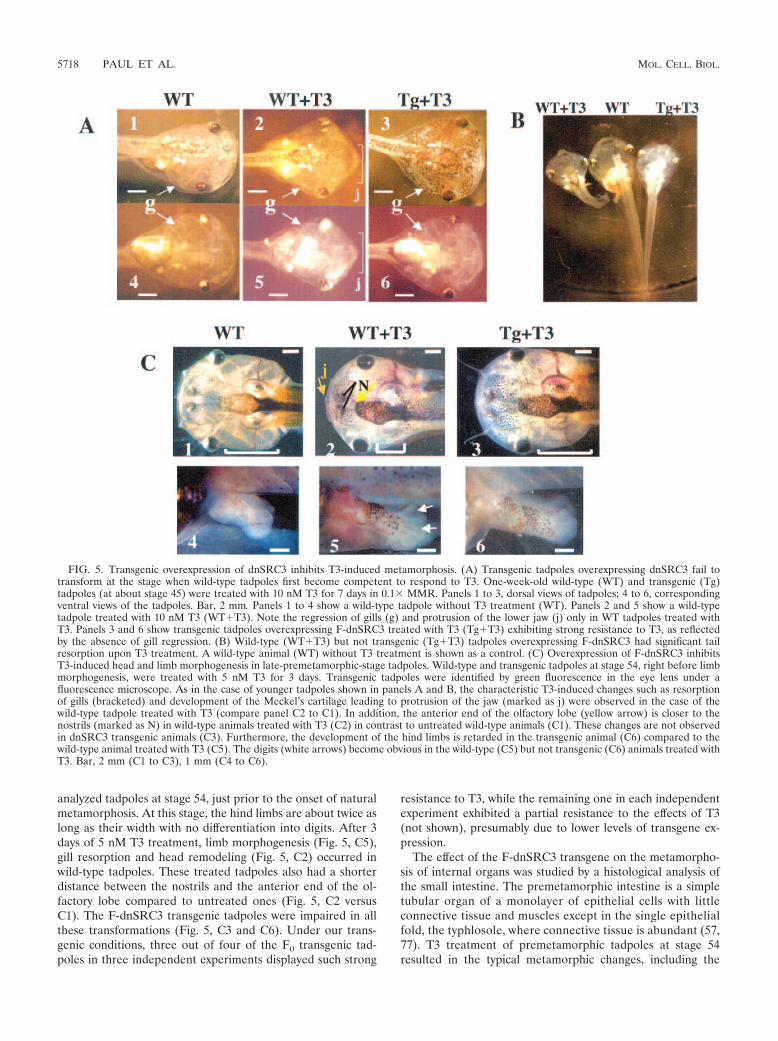
analyzed tadpoles at stage 54, just prior to the onset of naturalmetamorphosis. At this stage, the hind limbs are about twice aslong as their width with no differentiation into digits. After 3days of 5 nM T3 treatment, limb morphogenesis (Fig. 5, C5),gill resorption and head remodeling (Fig. 5, C2) occurred inwild-type tadpoles. These treated tadpoles also had a shorterdistance between the nostrils and the anterior end of the ol-factory lobe compared to untreated ones (Fig. 5, C2 versusC1). The F-dnSRC3 transgenic tadpoles were impaired in allthese transformations (Fig. 5, C3 and C6). Under our trans-genic conditions, three out of four of the F0 transgenic tad-poles in three independent experiments displayed such strong
resistance to T3, while the remaining one in each independentexperiment exhibited a partial resistance to the effects of T3(not shown), presumably due to lower levels of transgene ex-pression.
The effect of the F-dnSRC3 transgene on the metamorpho-sis of internal organs was studied by a histological analysis ofthe small intestine. The premetamorphic intestine is a simpletubular organ of a monolayer of epithelial cells with littleconnective tissue and muscles except in the single epithelialfold, the typhlosole, where connective tissue is abundant (57,77). T3 treatment of premetamorphic tadpoles at stage 54resulted in the typical metamorphic changes, including the
FIG. 5. Transgenic overexpression of dnSRC3 inhibits T3-induced metamorphosis. (A) Transgenic tadpoles overexpressing dnSRC3 fail totransform at the stage when wild-type tadpoles first become competent to respond to T3. One-week-old wild-type (WT) and transgenic (Tg)tadpoles (at about stage 45) were treated with 10 nM T3 for 7 days in 0.1& MMR. Panels 1 to 3, dorsal views of tadpoles; 4 to 6, correspondingventral views of the tadpoles. Bar, 2 mm. Panels 1 to 4 show a wild-type tadpole without T3 treatment (WT). Panels 2 and 5 show a wild-typetadpole treated with 10 nM T3 (WT!T3). Note the regression of gills (g) and protrusion of the lower jaw (j) only in WT tadpoles treated withT3. Panels 3 and 6 show transgenic tadpoles overexpressing F-dnSRC3 treated with T3 (Tg!T3) exhibiting strong resistance to T3, as reflectedby the absence of gill regression. (B) Wild-type (WT!T3) but not transgenic (Tg!T3) tadpoles overexpressing F-dnSRC3 had significant tailresorption upon T3 treatment. A wild-type animal (WT) without T3 treatment is shown as a control. (C) Overexpression of F-dnSRC3 inhibitsT3-induced head and limb morphogenesis in late-premetamorphic-stage tadpoles. Wild-type and transgenic tadpoles at stage 54, right before limbmorphogenesis, were treated with 5 nM T3 for 3 days. Transgenic tadpoles were identified by green fluorescence in the eye lens under afluorescence microscope. As in the case of younger tadpoles shown in panels A and B, the characteristic T3-induced changes such as resorptionof gills (bracketed) and development of the Meckel’s cartilage leading to protrusion of the jaw (marked as j) were observed in the case of thewild-type tadpole treated with T3 (compare panel C2 to C1). In addition, the anterior end of the olfactory lobe (yellow arrow) is closer to thenostrils (marked as N) in wild-type animals treated with T3 (C2) in contrast to untreated wild-type animals (C1). These changes are not observedin dnSRC3 transgenic animals (C3). Furthermore, the development of the hind limbs is retarded in the transgenic animal (C6) compared to thewild-type animal treated with T3 (C5). The digits (white arrows) become obvious in the wild-type (C5) but not transgenic (C6) animals treated withT3. Bar, 2 mm (C1 to C3), 1 mm (C4 to C6).
5718 PAUL ET AL. MOL. CELL. BIOL.
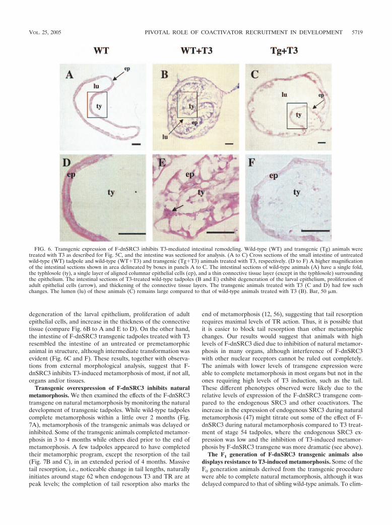
degeneration of the larval epithelium, proliferation of adultepithelial cells, and increase in the thickness of the connectivetissue (compare Fig. 6B to A and E to D). On the other hand,the intestine of F-dnSRC3 transgenic tadpoles treated with T3resembled the intestine of an untreated or premetamorphicanimal in structure, although intermediate transformation wasevident (Fig. 6C and F). These results, together with observa-tions from external morphological analysis, suggest that F-dnSRC3 inhibits T3-induced metamorphosis of most, if not all,organs and/or tissues.
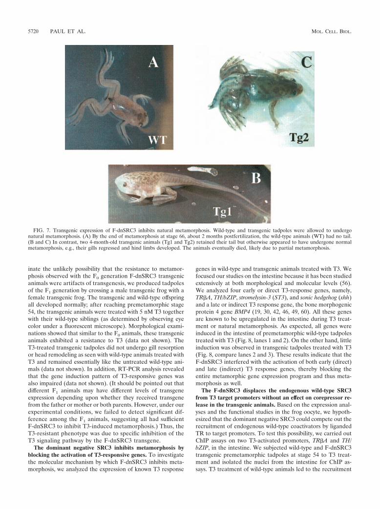
Transgenic overexpression of F-dnSRC3 inhibits naturalmetamorphosis. We then examined the effects of the F-dnSRC3transgene on natural metamorphosis by monitoring the naturaldevelopment of transgenic tadpoles. While wild-type tadpolescomplete metamorphosis within a little over 2 months (Fig.7A), metamorphosis of the transgenic animals was delayed orinhibited. Some of the transgenic animals completed metamor-phosis in 3 to 4 months while others died prior to the end ofmetamorphosis. A few tadpoles appeared to have completedtheir metamorphic program, except the resorption of the tail(Fig. 7B and C), in an extended period of 4 months. Massivetail resorption, i.e., noticeable change in tail lengths, naturallyinitiates around stage 62 when endogenous T3 and TR are atpeak levels; the completion of tail resorption also marks the
end of metamorphosis (12, 56), suggesting that tail resorptionrequires maximal levels of TR action. Thus, it is possible thatit is easier to block tail resorption than other metamorphicchanges. Our results would suggest that animals with highlevels of F-dnSRC3 died due to inhibition of natural metamor-phosis in many organs, although interference of F-dnSRC3with other nuclear receptors cannot be ruled out completely.The animals with lower levels of transgene expression wereable to complete metamorphosis in most organs but not in theones requiring high levels of T3 induction, such as the tail.These different phenotypes observed were likely due to therelative levels of expression of the F-dnSRC3 transgene com-pared to the endogenous SRC3 and other coactivators. Theincrease in the expression of endogenous SRC3 during naturalmetamorphosis (47) might titrate out some of the effect of F-dnSRC3 during natural metamorphosis compared to T3 treat-ment of stage 54 tadpoles, where the endogenous SRC3 ex-pression was low and the inhibition of T3-induced metamor-phosis by F-dnSRC3 transgene was more dramatic (see above).
The F1 generation of F-dnSRC3 transgenic animals alsodisplays resistance to T3-induced metamorphosis. Some of theF0 generation animals derived from the transgenic procedurewere able to complete natural metamorphosis, although it wasdelayed compared to that of sibling wild-type animals. To elim-
FIG. 6. Transgenic expression of F-dnSRC3 inhibits T3-mediated intestinal remodeling. Wild-type (WT) and transgenic (Tg) animals weretreated with T3 as described for Fig. 5C, and the intestine was sectioned for analysis. (A to C) Cross sections of the small intestine of untreatedwild-type (WT) tadpole and wild-type (WT!T3) and transgenic (Tg!T3) animals treated with T3, respectively. (D to F) A higher magnificationof the intestinal sections shown in area delineated by boxes in panels A to C. The intestinal sections of wild-type animals (A) have a single fold,the typhlosole (ty), a single layer of aligned columnar epithelial cells (ep), and a thin connective tissue layer (except in the typhlosole) surroundingthe epithelium. The intestinal sections of T3-treated wild-type tadpoles (B and E) exhibit degeneration of the larval epithelium, proliferation ofadult epithelial cells (arrow), and thickening of the connective tissue layers. The transgenic animals treated with T3 (C and D) had few suchchanges. The lumen (lu) of these animals (C) remains large compared to that of wild-type animals treated with T3 (B). Bar, 50 'm.
VOL. 25, 2005 PIVOTAL ROLE OF COACTIVATOR RECRUITMENT IN DEVELOPMENT 5719
inate the unlikely possibility that the resistance to metamor-phosis observed with the F0 generation F-dnSRC3 transgenicanimals were artifacts of transgenesis, we produced tadpolesof the F1 generation by crossing a male transgenic frog with afemale transgenic frog. The transgenic and wild-type offspringall developed normally; after reaching premetamorphic stage54, the transgenic animals were treated with 5 nM T3 togetherwith their wild-type siblings (as determined by observing eyecolor under a fluorescent microscope). Morphological exami-nations showed that similar to the F0 animals, these transgenicanimals exhibited a resistance to T3 (data not shown). TheT3-treated transgenic tadpoles did not undergo gill resorptionor head remodeling as seen with wild-type animals treated withT3 and remained essentially like the untreated wild-type ani-mals (data not shown). In addition, RT-PCR analysis revealedthat the gene induction pattern of T3-responsive genes wasalso impaired (data not shown). (It should be pointed out thatdifferent F1 animals may have different levels of transgeneexpression depending upon whether they received transgenefrom the father or mother or both parents. However, under ourexperimental conditions, we failed to detect significant dif-ference among the F1 animals, suggesting all had sufficientF-dnSRC3 to inhibit T3-induced metamorphosis.) Thus, theT3-resistant phenotype was due to specific inhibition of theT3 signaling pathway by the F-dnSRC3 transgene.
The dominant negative SRC3 inhibits metamorphosis byblocking the activation of T3-responsive genes. To investigatethe molecular mechanism by which F-dnSRC3 inhibits meta-morphosis, we analyzed the expression of known T3 response
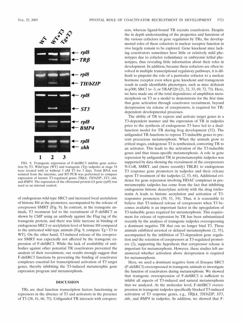
genes in wild-type and transgenic animals treated with T3. Wefocused our studies on the intestine because it has been studiedextensively at both morphological and molecular levels (56).We analyzed four early or direct T3-response genes, namely,TR#A, TH/bZIP, stromelysin-3 (ST3), and sonic hedgehog (xhh)and a late or indirect T3 response gene, the bone morphogenicprotein 4 gene BMP4 (19, 30, 42, 46, 49, 60). All these genesare known to be upregulated in the intestine during T3 treat-ment or natural metamorphosis. As expected, all genes wereinduced in the intestine of premetamorphic wild-type tadpolestreated with T3 (Fig. 8, lanes 1 and 2). On the other hand, littleinduction was observed in transgenic tadpoles treated with T3(Fig. 8, compare lanes 2 and 3). These results indicate that theF-dnSRC3 interfered with the activation of both early (direct)and late (indirect) T3 response genes, thereby blocking theentire metamorphic gene expression program and thus meta-morphosis as well.
The F-dnSRC3 displaces the endogenous wild-type SRC3from T3 target promoters without an effect on corepressor re-lease in the transgenic animals. Based on the expression anal-yses and the functional studies in the frog oocyte, we hypoth-esized that the dominant negative SRC3 could compete out therecruitment of endogenous wild-type coactivators by ligandedTR to target promoters. To test this possibility, we carried outChIP assays on two T3-activated promoters, TR#A and TH/bZIP, in the intestine. We subjected wild-type and F-dnSRC3transgenic premetamorphic tadpoles at stage 54 to T3 treat-ment and isolated the nuclei from the intestine for ChIP as-says. T3 treatment of wild-type animals led to the recruitment
FIG. 7. Transgenic expression of F-dnSRC3 inhibits natural metamorphosis. Wild-type and transgenic tadpoles were allowed to undergonatural metamorphosis. (A) By the end of metamorphosis at stage 66, about 2 months postfertilization, the wild-type animals (WT) had no tail.(B and C) In contrast, two 4-month-old transgenic animals (Tg1 and Tg2) retained their tail but otherwise appeared to have undergone normalmetamorphosis, e.g., their gills regressed and hind limbs developed. The animals eventually died, likely due to partial metamorphosis.
5720 PAUL ET AL. MOL. CELL. BIOL.
of endogenous wild-type SRC3 and increased local acetylationof histone H4 at the promoters, accompanied by the release ofcorepressor SMRT (Fig. 9). In contrast, in the transgenic an-imals, T3 treatment led to the recruitment of F-dnSRC3 asshown by ChIP using an antibody against the Flag tag of thetransgenic protein, and there was little increase in binding ofendogenous SRC3 or acetylation level of histone H4 comparedto the untreated wild-type animals (Fig. 9, compare Tg!T3 toWT). On the other hand, T3-induced release of the corepres-sor SMRT was expectedly not affected by the transgenic ex-pression of F-dnSRC3. While the lack of availability of anti-bodies against other potential TR coactivators prevented theanalysis of their recruitment, our results strongly suggest thatF-dnSRC3 functions by preventing the binding of coactivatorcomplexes essential for transcriptional activation of T3 targetgenes, thereby inhibiting the T3-induced metamorphic geneexpression program and metamorphosis.
DISCUSSION
TRs are dual function transcription factors functioning asrepressors in the absence of T3 and activators in the presenceof T3 (38, 41, 66, 75). Unliganded TR interacts with corepres-
sors, whereas ligand-bound TR recruits coactivators. Despitethe in depth understanding of the properties and functions ofthe various cofactors in gene regulation by TRs, the develop-mental roles of these cofactors in nuclear receptor function invivo largely remain to be explored. Gene knockout mice lack-ing coactivators sometimes have little or relatively mild phe-notypes due to cofactor redundancy or embryonic lethal phe-notypes, thus revealing little information about their roles indevelopment. In addition, because these cofactors are often in-volved in multiple transcriptional regulatory pathways, it is dif-ficult to pinpoint the role of a particular cofactor to a nuclearhormone receptor even when gene knockout and transgenesisresult in easily identifiable phenotypes, such as mice deficientin p300, SRC1 to -3, or TRAP220 (21, 32, 33, 69, 72, 73). Here,we have made use of the total dependence of amphibian meta-morphosis on T3 as a model to demonstrate for the first timethat gene activation through coactivator recruitment, beyondderepression via release of corepressors, is required for TR-dependent developmental processes.
The ability of TR to repress and activate target genes in aT3-dependent manner and the expression of TR in tadpolesprior to the synthesis of endogenous T3 have led to a dual-function model for TR during frog development (52). Theunliganded TR functions to repress T3-inducible genes to pre-vent precocious metamorphosis. When the animals grow tocritical stages, endogenous T3 is synthesized, converting TR toan activator. This leads to the activation of the T3-induciblegenes and thus tissue-specific metamorphosis. A role in generepression by unliganded TR in premetamorphic tadpoles wassupported by data showing the recruitment of the corepressorsN-CoR, SMRT, and (more recently) TBLR1 to endogenousT3 response gene promoters in tadpoles and their releaseupon T3 treatment of the tadpoles (2, 53, 64). Additional evi-dence for gene repression involving HDAC complexes in pre-metamorphic tadpoles has come from the fact that inhibitingendogenous histone deacetylase activity with the drug tricho-statin A leads to histone acetylation and activation of T3-responsive promoters (50, 51, 54). Thus, it is reasonable tobelieve that T3-induced release of corepressors when T3 be-comes available is an important factor in the upregulation ofT3-inducible genes required for metamorphosis. This require-ment for release of repression by TR has been substantiatedrecently by the analyses of transgenic tadpoles overexpressinga dominant negative TR that can no longer bind T3. Theseanimals exhibited arrested or delayed metamorphosis (2, 55),accompanied by the inhibition of T3-dependent gene regula-tion and the retention of corepressors at T3-regulated promot-ers (2), supporting the hypothesis that corepressor release isimportant for metamorphosis. However, these studies left un-answered whether activation above derepression is requiredfor metamorphosis.
Here, we used a dominant negative form of Xenopus SRC3(F-dnSRC3) overexpressed in transgenic animals to investigatethe function of coactivators during metamorphosis. We showedthat transgenic overexpression of F-dnSRC3 is sufficient toinhibit all aspects of T3-induced and natural metamorphosisthat we analyzed. At the molecular level, F-dnSRC3 overex-pression in transgenic tadpoles specifically blocked T3-inducedactivation of T3 response genes, e.g., TR#A, TH/bZIP, ST3,xhh, and BMP4 in tadpoles. In addition, we showed that F-
FIG. 8. Transgenic expression of F-dnSRC3 inhibits gene activa-tion by T3. Wild-type (WT) and transgenic (Tg) tadpoles at stage 54were treated with or without 5 nM T3 for 3 days. Total RNA wasisolated from the intestine, and RT-PCR was performed to compareexpression of known T3-regulated genes TR#A, TH/bZIP, ST3, xhh,and BMP4. The expression of the ribosomal protein L8 gene (rpl8) wasused as an internal control.
VOL. 25, 2005 PIVOTAL ROLE OF COACTIVATOR RECRUITMENT IN DEVELOPMENT 5721
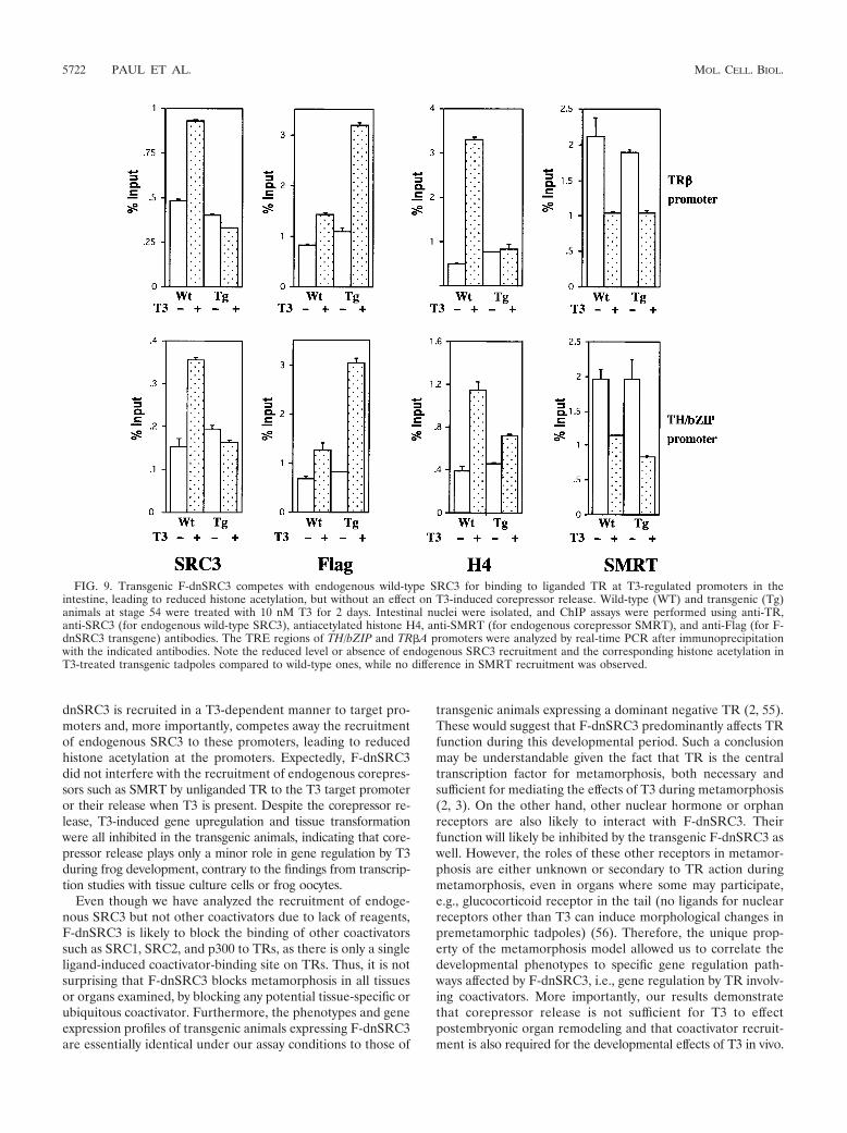
dnSRC3 is recruited in a T3-dependent manner to target pro-moters and, more importantly, competes away the recruitmentof endogenous SRC3 to these promoters, leading to reducedhistone acetylation at the promoters. Expectedly, F-dnSRC3did not interfere with the recruitment of endogenous corepres-sors such as SMRT by unliganded TR to the T3 target promoteror their release when T3 is present. Despite the corepressor re-lease, T3-induced gene upregulation and tissue transformationwere all inhibited in the transgenic animals, indicating that core-pressor release plays only a minor role in gene regulation by T3during frog development, contrary to the findings from transcrip-tion studies with tissue culture cells or frog oocytes.
Even though we have analyzed the recruitment of endoge-nous SRC3 but not other coactivators due to lack of reagents,F-dnSRC3 is likely to block the binding of other coactivatorssuch as SRC1, SRC2, and p300 to TRs, as there is only a singleligand-induced coactivator-binding site on TRs. Thus, it is notsurprising that F-dnSRC3 blocks metamorphosis in all tissuesor organs examined, by blocking any potential tissue-specific orubiquitous coactivator. Furthermore, the phenotypes and geneexpression profiles of transgenic animals expressing F-dnSRC3are essentially identical under our assay conditions to those of
transgenic animals expressing a dominant negative TR (2, 55).These would suggest that F-dnSRC3 predominantly affects TRfunction during this developmental period. Such a conclusionmay be understandable given the fact that TR is the centraltranscription factor for metamorphosis, both necessary andsufficient for mediating the effects of T3 during metamorphosis(2, 3). On the other hand, other nuclear hormone or orphanreceptors are also likely to interact with F-dnSRC3. Theirfunction will likely be inhibited by the transgenic F-dnSRC3 aswell. However, the roles of these other receptors in metamor-phosis are either unknown or secondary to TR action duringmetamorphosis, even in organs where some may participate,e.g., glucocorticoid receptor in the tail (no ligands for nuclearreceptors other than T3 can induce morphological changes inpremetamorphic tadpoles) (56). Therefore, the unique prop-erty of the metamorphosis model allowed us to correlate thedevelopmental phenotypes to specific gene regulation path-ways affected by F-dnSRC3, i.e., gene regulation by TR involv-ing coactivators. More importantly, our results demonstratethat corepressor release is not sufficient for T3 to effectpostembryonic organ remodeling and that coactivator recruit-ment is also required for the developmental effects of T3 in vivo.
FIG. 9. Transgenic F-dnSRC3 competes with endogenous wild-type SRC3 for binding to liganded TR at T3-regulated promoters in theintestine, leading to reduced histone acetylation, but without an effect on T3-induced corepressor release. Wild-type (WT) and transgenic (Tg)animals at stage 54 were treated with 10 nM T3 for 2 days. Intestinal nuclei were isolated, and ChIP assays were performed using anti-TR,anti-SRC3 (for endogenous wild-type SRC3), antiacetylated histone H4, anti-SMRT (for endogenous corepressor SMRT), and anti-Flag (for F-dnSRC3 transgene) antibodies. The TRE regions of TH/bZIP and TR#A promoters were analyzed by real-time PCR after immunoprecipitationwith the indicated antibodies. Note the reduced level or absence of endogenous SRC3 recruitment and the corresponding histone acetylation inT3-treated transgenic tadpoles compared to wild-type ones, while no difference in SMRT recruitment was observed.
5722 PAUL ET AL. MOL. CELL. BIOL.
In summary, while altering coactivator function throughknockouts can lead to developmental defects in mice, theunderlying molecular mechanisms are unknown, due to theinvolvement of the cofactors in transcriptional regulationby many diverse transcription factors and the difficulty inaccessing and manipulating postembryonic development inmammals. Our use of F-dnSRC3, comprising only the nu-clear receptor-interacting domain, restricts its effect to nu-clear receptors. More importantly, the total dependence of thetransformations of different organs and/or tissues on T3 makesTR the only transcription factor that controls initiation of thedifferent changes in various organs during this metamorphicperiod. This unique situation allowed us to illustrate the firstexample where specific developmental defects are caused by al-terations of specific gene regulation pathways involving nuclearreceptors. Our experiments connected the phenotype of organtransformation inhibition with the blockade of TR-dependentgene expression pathways via a mutant dominant negative co-activator. In addition, while derepression appears to be a majorcomponent of gene activation by T3 in tissue culture cells orfrog oocytes, it does not seem to be critical in developinganimals, as demonstrated here, further underscoring the im-portance of in vivo experiments in understanding the molecu-lar mechanisms of nuclear receptor function in development.
ACKNOWLEDGMENT
We thank other members of the group for helpful discussions.
REFERENCES
1. Amano, T., K. Leu, K. Yoshizato, and Y.-B. Shi. 2002. Thyroid hormoneregulation of a transcriptional coactivator in Xenopus laevis: implication fora role in postembryonic tissue remodeling. Dev. Dyn. 223:526–535.
2. Buchholz, D. R., V. S.-C. Hsia, L. Fu, and Y.-B. Shi. 2003. A dominantnegative thyroid hormone receptor blocks amphibian metamorphosis byretaining corepressors at target genes. Mol. Cell. Biol. 23:6750–6758.
3. Buchholz, D. R., A. Tomita, L. Fu, B. D. Paul, and Y.-B. Shi. 2004. Trans-genic analysis reveals that thyroid hormone receptor is sufficient to mediatethe thyroid hormone signal in frog metamorphosis. Mol. Cell. Biol. 24:9026–9037.
4. Burrow, G. N., D. A. Fisher, and P. R. Larsen. 1994. Maternal and fetalthyroid function. N. Engl. J. Med. 331:1072–1078.
5. Carswell, S., and J. C. Alwine. 1989. Efficiency of utilization of the simianvirus 40 late polyadenylation site: effects of upstream sequences. Mol. Cell.Biol. 9:4248–4258.
6. Chen, H., R. J. Lin, R. L. Schiltz, D. Chakravarti, A. Nash, L. Nagy, M. L.Privalsky, Y. Nakatani, and R. M. Evans. 1997. Nuclear receptor coactivatorACTR is a novel histone acetyltransferase and forms a multimeric activationcomplex with P/CAF and CBP/p300. Cell 90:569–580.
7. Chen, J. D., and R. M. Evans. 1995. A transcriptional co-repressor thatinteracts with nuclear hormone receptors. Nature 377:454–457.
8. Chubet, R. G., and B. L. Brizzard. 1996. Vectors for expression and secretionof FLAG epitope-tagged proteins in mammalian cells. BioTechniques 20:136–141.
9. Damjanovski, S., L. M. Sachs, and Y.-B. Shi. 2002. Function of thyroidhormone receptors during amphibian development, p. 153–176. In A. Bani-ahmad (ed.), Methods in molecular biology: thyroid hormone receptors, vol.202. Humana Press, Inc., Totowa, N.J.
10. Davis, P. J., and F. B. Davis. 1996. Nongenomic actions of thyroid hormone.Thyroid 6:497–504.
11. Ding, X. F., C. M. Anderson, H. Ma, H. Hong, R. M. Uht, P. J. Kushner, andM. R. Stallcup. 1998. Nuclear receptor-binding sites of coactivators glu-cocorticoid receptor interacting protein 1 (GRIP1) and steroid receptorcoactivator 1 (SRC-1): multiple motifs with different binding specificities.Mol. Endocrinol. 12:302–313.
12. Dodd, M. H. I., and J. M. Dodd. 1976. The biology of metamorphosis, p.467–599. In B. Lofts (ed.), Physiology of the amphibia. Academic Press, NewYork, N.Y.
13. Feng, W., R. C. Ribeiro, R. L. Wagner, H. Nguyen, J. W. Apriletti, R. J.Fletterick, J. D. Baxter, P. J. Kushner, and B. L. West. 1998. Hormone-dependent coactivator binding to a hydrophobic cleft on nuclear receptors.Science 280:1747–1749.
14. Feng, X., Y. Jiang, P. Meltzer, and P. M. Yen. 2001. Transgenic targeting ofa dominant negative corepressor to liver blocks basal repression by thyroidhormone receptor and increases cell proliferation. J. Biol. Chem. 276:15066–15072.
15. Flamant, F., and J. Samarut. 2003. Thyroid hormone receptors: lessons fromknockout and knock-in mutant mice. Trends Endocrinol. Metab. 14:85–90.
16. Forrest, D., E. Hanebuth, R. J. Smeyne, N. Everds, C. L. Stewart, J. M.Wehner, and T. Curran. 1996. Recessive resistance to thyroid hormone inmice lacking thyroid hormone receptor beta: evidence for tissue-specificmodulation of receptor function. EMBO J. 15:3006–3015.
17. Fraichard, A., O. Chassande, M. Plateroti, J. P. Roux, J. Trouillas, C. Dehay,C. Legrand, K. Gauthier, M. Kedinger, L. Malaval, B. Rousset, and J.Samarut. 1997. The T3R alpha gene encoding a thyroid hormone receptor isessential for post-natal development and thyroid hormone production.EMBO J. 16:4412–4420.
18. Fu, L., D. Buchholz, and Y.-B. Shi. 2002. A novel double promoter approachfor identification of transgenic animals: a tool for in vivo analysis of genefunction and development of gene-based therapies. Mol. Reprod. Dev. 62:470–476.
19. Furlow, J. D., and D. D. Brown. 1999. In vitro and in vivo analysis of theregulation of a transcription factor gene by thyroid hormone during Xenopuslaevis metamorphosis. Mol. Endocrinol. 13:2076–2089.
20. Gauthier, K., O. Chassande, M. Plateroti, J. P. Roux, C. Legrand, B. Pain,B. Rousset, R. Weiss, J. Trouillas, and J. Samarut. 1999. Different functionsfor the thyroid hormone receptors TR" and TR# in the control of thyroidhromone production and post-natal development. EMBO J. 18:623–631.
21. Gehin, M., M. Mark, C. Dennefeld, A. Dierich, H. Gronemeyer, and P.Chambon. 2002. The function of TIF2/GRIP1 in mouse reproduction isdistinct from those of SRC-1 and p/CIP. Mol. Cell. Biol. 22:5923–5927.
22. Gothe, S., Z. Wang, L. Ng, J. M. Kindblom, A. C. Barros, C. Ohlsson, B.Vennstrom, and D. Forrest. 1999. Mice devoid of all known thyroid hromonereceptors are viable but exhibit disorders of the pituitary-thyroid axis,growth, and bone maturation. EMBO J. 13:1329–1341.
23. Guenther, M. G., W. S. Lane, W. Fischle, E. Verdin, M. A. Lazar, and R.Shiekhattar. 2000. A core SMRT corepressor complex containing HDAC3and TBL1, a WD40-repeat protein linked to deafness. Genes Dev. 14:1048–1057.
24. Heery, D. M., E. Kalkhoven, S. Hoare, and M. G. Parker. 1997. A signaturemotif in transcriptional co-activators mediates binding to nuclear receptors.Nature 387:733–736.
25. Hetzel, B. S. 1989. The story of iodine deficiency: an international challengein nutrition. Oxford University Press, Oxford, United Kingdom.
26. Hong, H., K. Kohli, A. Trivedi, D. L. Johnson, and M. R. Stallcup. 1996.GRIP1, a novel mouse protein that serves as a transcriptional coactivator inyeast for the hormone binding domains of steroid receptors. Proc. Natl.Acad. Sci. USA 93:4948–4952.
27. Horlein, A. J., A. M. Naar, T. Heinzel, J. Torchia, B. Gloss, R. Kurokawa, A.Ryan, Y. Kamei, M. Soderstrom, C. K. Glass, et al. 1995. Ligand-indepen-dent repression by the thyroid hormone receptor mediated by a nuclearreceptor co-repressor. Nature 377:397–404.
28. Ishizuka, T., and M. A. Lazar. 2003. The N-CoR/histone deacetylase 3complex is required for repression by thyroid hormone receptor. Mol. Cell.Biol. 23:5122–5131.
29. Ishizuya-Oka, A., Q. Li, T. Amano, S. Damjanovski, S. Ueda, and Y.-B. Shi.2000. Requirement for matrix metalloproteinase stromelysin-3 in cell migra-tion and apoptosis during tissue remodeling in Xenopus laevis. J. Cell Biol.150:1177–1188.
30. Ishizuya-Oka, A., S. Ueda, T. Amano, K. Shimizu, K. Suzuki, N. Ueno, andK. Yoshizato. 2001. Thyroid-hormone-dependent and fibroblast-specific ex-pression of BMP-4 correlates with adult epithelial development during am-phibian intestinal remodeling. Cell Tissue Res. 303:187–195.
31. Ito, M., and R. G. Roeder. 2001. The TRAP/SMCC/Mediator complex andthyroid hormone receptor function. Trends Endocrinol. Metab. 12:127–134.
32. Ito, M., C. X. Yuan, H. J. Okano, R. B. Darnell, and R. G. Roeder. 2000.Involvement of the TRAP220 component of the TRAP/SMCC coactivatorcomplex in embryonic development and thyroid hormone action. Mol. Cell5:683–693.
33. Jepsen, K., O. Hermanson, T. M. Onami, A. S. Gleiberman, V. Lunyak, R. J.McEvilly, R. Kurokawa, V. Kumar, F. Liu, E. Seto, S. M. Hedrick, G.Mandel, C. K. Glass, D. W. Rose, and M. G. Rosenfeld. 2000. Combinatorialroles of the nuclear receptor corepressor in transcription and development.Cell 102:753–763.
34. Jones, P. L., and Y.-B. Shi. 2003. N-CoR-HDAC corepressor complexes:roles in transcriptional regulation by nuclear hormone receptors, p. 237–268.In J. L. Workman (ed.), Current topics in microbiology and immunology:protein complexes that modify chromatin, vol. 274. Springer-Verlag, Berlin,Germany.
35. Kim, H. J., S. K. Lee, S. Y. Na, H. S. Choi, and J. W. Lee. 1998. Molecularcloning of xSRC-3, a novel transcription coactivator from Xenopus, that isrelated to AIB1, p/CIP, and TIF2. Mol. Endocrinol. 12:1038–1047.
36. Kroll, K. L., and E. Amaya. 1996. Transgenic Xenopus embryos from sperm
VOL. 25, 2005 PIVOTAL ROLE OF COACTIVATOR RECRUITMENT IN DEVELOPMENT 5723
nuclear transplantations reveal FGF signaling requirements during gastru-lation. Development 122:3173–3183.
37. LaFranchi, S. 1999. Thyroid function in the preterm infant. Thyroid 9:71–78.38. Lazar, M. A. 1993. Thyroid hormone receptors: multiple forms, multiple
possibilities. Endocr. Rev. 14:184–193.39. Li, H., P. J. Gomes, and J. D. Chen. 1997. RAC3, a steroid/nuclear receptor-
associated coactivator that is related to SRC-1 and TIF2. Proc. Natl. Acad.Sci. USA 94:8479–8484.
40. Li, J., J. Wang, J. Wang, Z. Nawaz, J. M. Liu, J. Qin, and J. Wong. 2000.Both corepressor proteins SMRT and N-CoR exist in large protein com-plexes containing HDAC3. EMBO J. 19:4342–4350.
41. Mangelsdorf, D. J., C. Thummel, M. Beato, P. Herrlich, G. Schutz, K.Umesono, B. Blumberg, P. Kastner, M. Mark, and P. Chambon. 1995. Thenuclear receptor superfamily: the second decade. Cell 83:835–839.
42. Metz, A., S. Knoechel, P. Buechler, M. Koester, and W. Knoechel. 1998.Structural and functional analysis of the BMP-4 promoter in early embryosof Xenopus laevis. Mech. Dev. 74:29–39.
43. Nieuwkoop, P. D., and J. Faber. 1956. Normal table of Xenopus laevis, 1sted. North Holland Publishing, Amsterdam, The Netherlands.
44. Nolte, R. T., G. B. Wisely, S. Westin, J. E. Cobb, M. H. Lambert, R. Kuro-kawa, M. G. Rosenfeld, T. M. Willson, C. K. Glass, and M. V. Milburn. 1998.Ligand binding and co-activator assembly of the peroxisome proliferator-activated receptor-gamma. Nature 395:137–143.
45. Onate, S. A., S. Y. Tsai, M. J. Tsai, and B. W. O’Malley. 1995. Sequence andcharacterization of a coactivator for the steroid hormone receptor super-family. Science 270:1354–1357.
46. Patterton, D., W. P. Hayes, and Y. B. Shi. 1995. Transcriptional activation ofthe matrix metalloproteinase gene stromelysin-3 coincides with thyroid hor-mone-induced cell death during frog metamorphosis. Dev. Biol. 167:252–262.
47. Paul, B. D., and Y.-B. Shi. 2003. Distinct expression profiles of transcrip-tional coactivators for thyroid hormone receptors during Xenopus laevismetamorphosis. Cell Res. 13:459–464.
48. Rachez, C., and L. P. Freedman. 2001. Mediator complexes and transcrip-tion. Curr. Opin. Cell Biol. 13:274–280.
49. Ranjan, M., J. Wong, and Y. B. Shi. 1994. Transcriptional repression ofXenopus TR beta gene is mediated by a thyroid hormone response elementlocated near the start site. J. Biol. Chem. 269:24699–24705.
50. Sachs, L. M., T. Amano, N. Rouse, and Y. B. Shi. 2001. Involvement ofhistone deacetylase at two distinct steps in gene regulation during intestinaldevelopment in Xenopus laevis. Dev. Dyn. 222:280–291.
51. Sachs, L. M., T. Amano, and Y. B. Shi. 2001. An essential role of histonedeacetylases in postembryonic organ transformations in Xenopus laevis. Int.J. Mol. Med. 8:595–601.
52. Sachs, L. M., S. Damjanovski, P. L. Jones, Q. Li, T. Amano, S. Ueda, Y. B.Shi, and A. Ishizuya-Oka. 2000. Dual functions of thyroid hormone recep-tors during Xenopus development. Comp. Biochem. Physiol. Biochem. Mol.Biol. 126:199–211.
53. Sachs, L. M., P. L. Jones, E. Havis, N. Rouse, B. A. Demeneix, and Y.-B. Shi.2002. N-CoR recruitment by unliganded thyroid hormone receptor in generepression during Xenopus laevis development. Mol. Cell. Biol. 22:8527–8538.
54. Sachs, L. M., and Y.-B. Shi. 2000. Targeted chromatin binding and histoneacetylation in vivo by thyroid hormone receptor during amphibian develop-ment. Proc. Natl. Acad. Sci. USA 97:13138–13143.
55. Schreiber, A. M., B. Das, H. Huang, N. Marsh-Armstrong, and D. D. Brown.2001. Diverse developmental programs of Xenopus laevis metamorphosisare inhibited by a dominant negative thyroid hormone receptor. Proc. Natl.Acad. Sci. USA 98:10739–10744.
56. Shi, Y.-B. 1999. Amphibian metamorphosis: from morphology to molecularbiology. John Wiley & Sons, Inc., New York, N.Y.
57. Shi, Y.-B., and A. Ishizuya-Oka. 1996. Biphasic intestinal development inamphibians: embryogensis and remodeling during metamorphosis. Curr.Top. Dev. Biol. 32:205–235.
58. Shi, Y.-B., and V. C.-T. Liang. 1994. Cloning and characterization of the ri-bosomal protein L8 gene from Xenopus laevis. Biochim. Biophys. Acta 1217:227–228.
59. Shiau, A. K., D. Barstad, P. M. Loria, L. Cheng, P. J. Kushner, D. A. Agard,
and G. L. Greene. 1998. The structural basis of estrogen receptor/coactivatorrecognition and the antagonism of this interaction by tamoxifen. Cell 95:927–937.
60. Stolow, M. A., and Y. B. Shi. 1995. Xenopus sonic hedgehog as a potentialmorphogen during embryogenesis and thyroid hormone-dependent meta-morphosis. Nucleic Acids Res. 23:2555–2562.
61. Takeshita, A., G. R. Cardona, N. Koibuchi, C. S. Suen, and W. W. Chin.1997. TRAM-1, a novel 160-kDa thyroid hormone receptor activator mole-cule, exhibits distinct properties from steroid receptor coactivator-1. J. Biol.Chem. 272:27629–27634.
62. Tata, J. R. 1993. Gene expression during metamorphosis: an ideal model forpost-embryonic development. Bioessays 15:239–248.
63. Tomita, A., D. R. Buchholz, K. Obata, and Y.-B. Shi. 2003. Fusion protein ofretinoic acid receptor a with promeyelocytic leukaemia protein or promy-elocytic leukaemia zinc-finger protein recruits N-CoR-TBLR1 corepressorcomplex to repress transcription in vivo. J. Biol. Chem. 278:30788–30795.
64. Tomita, A., D. R. Buchholz, and Y.-B. Shi. 2004. Recruitment of N-CoR/SMRT-TBLR1 corepressor complex by unliganded thyroid hormone recep-tor for gene repression during frog development. Mol. Cell. Biol. 24:3337–3346.
65. Torchia, J., D. W. Rose, J. Inostroza, Y. Kamei, S. Westin, C. K. Glass, andM. G. Rosenfeld. 1997. The transcriptional co-activator p/CIP binds CBP andmediates nuclear-receptor function. Nature 387:677–684.
66. Tsai, M. J., and B. W. O’Malley. 1994. Molecular mechanisms of action ofsteroid/thyroid receptor superfamily members. Annu. Rev. Biochem. 63:451–486.
67. Voegel, J. J., M. J. Heine, M. Tini, V. Vivat, P. Chambon, and H. Gronem-eyer. 1998. The coactivator TIF2 contains three nuclear receptor-bindingmotifs and mediates transactivation through CBP binding-dependent and-independent pathways. EMBO J. 17:507–519.
68. Voegel, J. J., M. J. Heine, C. Zechel, P. Chambon, and H. Gronemeyer. 1996.TIF2, a 160 kDa transcriptional mediator for the ligand-dependent activa-tion function AF-2 of nuclear receptors. EMBO J. 15:3667–3675.
69. Wang, Z., D. W. Rose, O. Hermanson, F. Liu, T. Herman, W. Wu, D. Szeto,A. Gleiberman, A. Krones, K. Pratt, R. Rosenfeld, and C. K. Glass. 2000.Regulation of somatic growth by the p160 coactivator p/CIP. Proc. Natl.Acad. Sci. USA 97:13549–13554.
70. Wikstrom, L., C. Johansson, C. Salto, C. Barlow, A. C. Barros, F. Baas, D.Forrest, P. Thoren, and B. Veenstrom. 1998. Abnormal heart rate and bodytemperature in mice lacking thyroid hormone receptor. EMBO J. 17:455–461.
71. Wong, J., and Y.-B. Shi. 1995. Coordinated regulation of and transcriptionalactivation by Xenopus thyroid hormone and retinoid X receptors. J. Biol.Chem. 270:18479–18483.
72. Xu, J., L. Liao, G. Ning, H. Yoshida-Komiya, C. Deng, and B. W. O’Malley.2000. The steroid receptor coactivator SRC-3 (p/CIP/RAC3/AIB1/ACTR/TRAM-1) is requried for normal growth, puberty, female reproductive func-tion, and mammary gland development. Proc. Natl. Acad. Sci. USA 97:6379–6384.
73. Yao, T. P., S. P. Oh, M. Fuchs, N. D. Zhou, L. E. Ch’ng, D. Newsome, R. T.Bronson, E. Li, D. M. Livingston, and R. Eckner. 1998. Gene dosage-dependent embryonic development and proliferation defects in mice lackingthe transcriptional integrator p300. Cell 93:361–372.
74. Yaoita, Y., Y. B. Shi, and D. D. Brown. 1990. Xenopus laevis alpha and betathyroid hormone receptors. Proc. Natl. Acad. Sci. USA 87:7090–7094.
75. Yen, P. M. 2001. Physiological and molecular basis of thyroid hormoneaction. Physiol. Rev. 81:1097–1142.
76. Yoon, H.-G., D. W. Chan, Z. Q. Huang, J. Li, J. D. Fondell, J. Qin, and J.Wong. 2003. Purification and functional characterization of the human N-CoR complex: the roles of HDAC3, TBL1 and TBLR1. EMBO J. 22:1336–1346.
77. Yoshizato, K. 1989. Biochemistry and cell biology of amphibian metamor-phosis with a special emphasis on the mechanism of removal of larval organs.Int. Rev. Cytol. 119:97–149.
78. Zhang, J., M. Kalkum, B. T. Chait, and R. G. Roeder. 2002. The N-CoR-HDAC3 nuclear receptor corepressor complex inhibits the JNK pathwaythrough the integral subunit GPS2. Mol. Cell 9:611–623.
79. Zhang, J., and M. A. Lazar. 2000. The mechanism of action of thyroidhormones. Annu. Rev. Physiol. 62:439–466.
5724 PAUL ET AL. MOL. CELL. BIOL.


![On photosensitivity of liganded hemoproteins metal ...myoglobin,andperoxidase,and their metal-substituted analogues, usingthreedifferent metals ... ganded metalloporphyrins [MPor(L)]](https://img.pdfslide.net/doc/110x75/5b2719cc7f8b9a2c128b472f/on-photosensitivity-of-liganded-hemoproteins-metal-myoglobinandperoxidaseand.jpg)