Embed Size (px)
Citation preview
Coding Dermatology
Procedures
Presented by: Betty A Hovey
Director, ICD-10 Development and Training
AAPC
1
No part of this presentation may
be reproduced or transmitted in
any form or by any means
(graphically, electronically, or
mechanically, including
photocopying, recording, or taping)
without the expressed written
permission of AAPC.
2
• CPT copyright 2012 American Medical Association. All rights reserved.
• Fee schedules, relative value units, conversion factors and/or related components are not assigned by the AMA, are not part of CPT, and the AMA is not recommending their use. The AMA is not recommending their use. The AMA does not directly or indirectly practice medicine or dispense medical services. The AMA assumes no liability for data contained or not contained herein.
• CPT is a registered trademark of the American Medical Association.
• The responsibility for the content of any “National Correct Coding Policy” included in this product is with the Centers for Medicare and Medicaid Services and no endorsement by the AMA is intended or should be implied. The AMA disclaims responsibility for any consequences or liability attributable to or related to any use, nonuse or interpretation of information contained in this product.
3
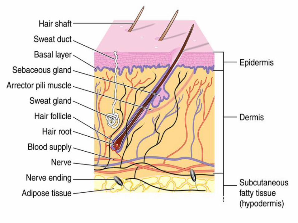
• Anatomy
• Shaving of Lesions
• Excision of Lesions
• Repairs
• Adjacent Tissue Transfer
• Destruction of Lesions
• Mohs Micrographic Surgery
AGENDA
4
5
• While skin cancers can be found on any part of
the body most (about 80%) appear on the face,
head, or neck
• The primary cause of skin cancer is ultraviolet
radiation -most often from the sun
• Also from artificial sources like sunlamps and
tanning booths
Skin Cancer
6
BCC • Basal cell carcinoma is the most common form
of skin cancer, affecting 800,000 Americans each year
• The most common of all cancers
• 1 out of every 3 new cancers is a skin cancer
• Most are basal cell carcinomas (BCC)
• These cancers arise in the basal cells, which are at the bottom of the epidermis
• More common in men, although more women are getting BCCs than in the past
Skin Cancer
7
Warning Signs of BCC
1. Open sore that bleeds, oozes, or crusts
and remains open for three or more
weeks
2. A reddish patch or irritated area,
frequently occurring on the chest,
shoulders, arms, or legs
3. Shiny bump, or nodule, that is pearly or
translucent and is often pink, red, or white
Skin Cancer
8
Warning Signs of BCC
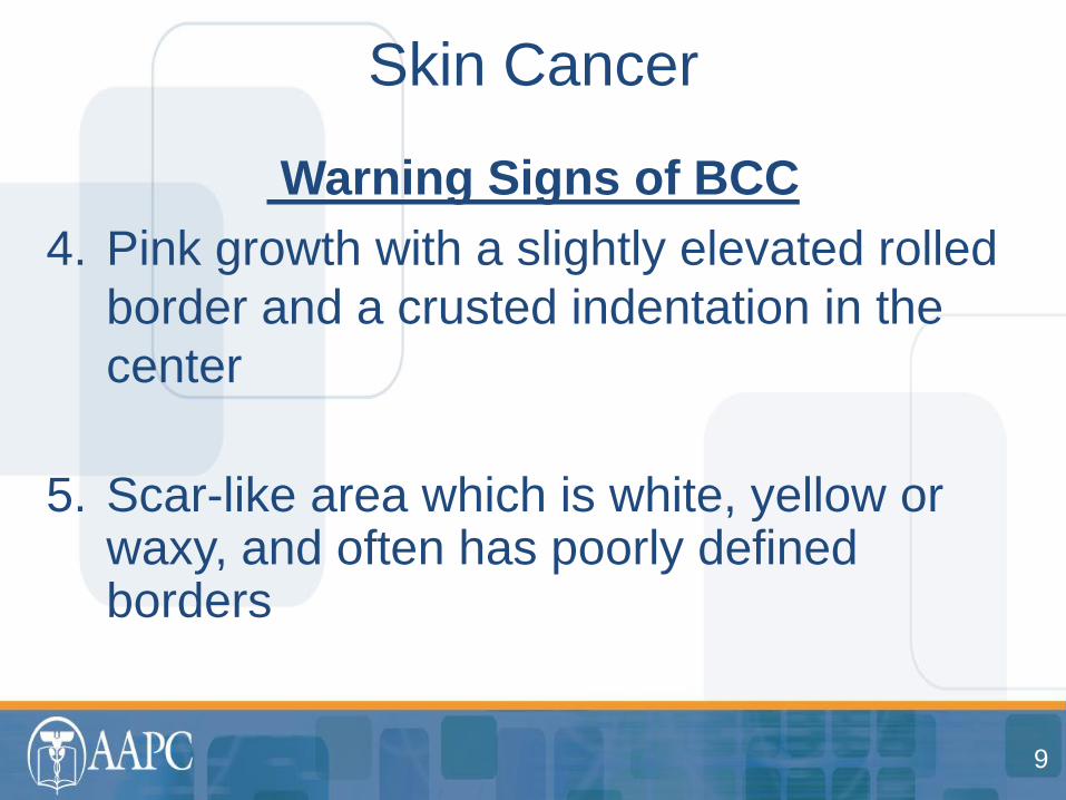
4. Pink growth with a slightly elevated rolled
border and a crusted indentation in the
center
5. Scar-like area which is white, yellow or waxy, and often has poorly defined borders
Skin Cancer
9
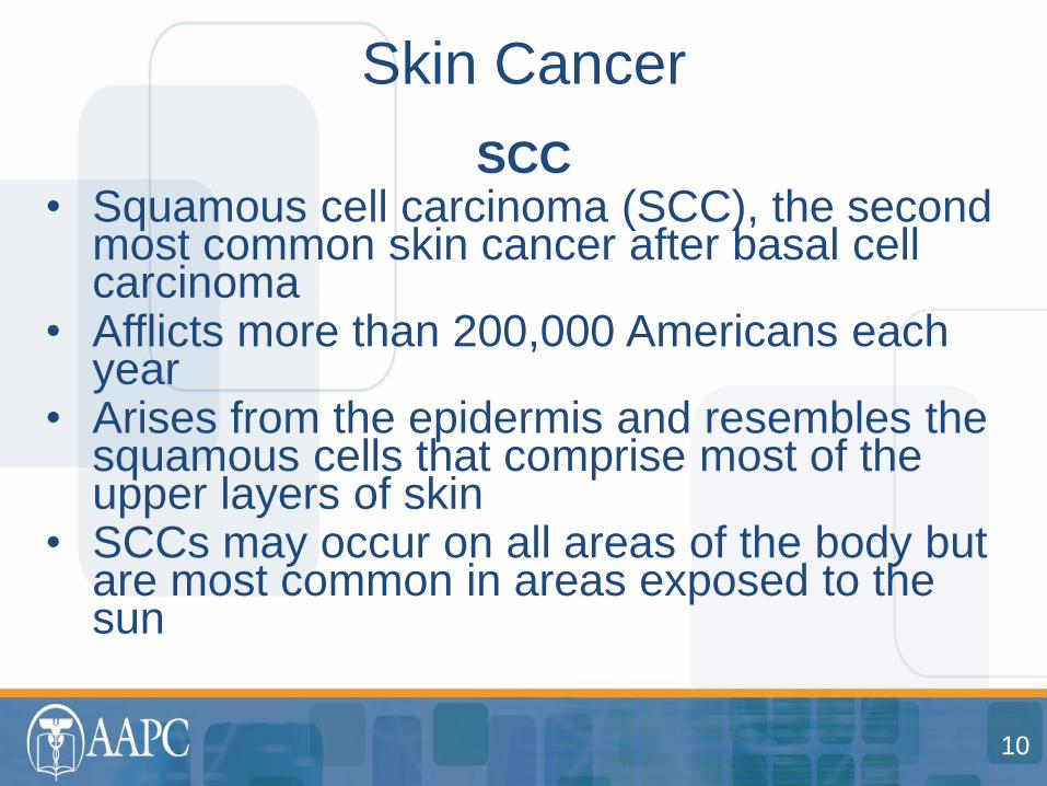
SCC • Squamous cell carcinoma (SCC), the second
most common skin cancer after basal cell carcinoma
• Afflicts more than 200,000 Americans each year
• Arises from the epidermis and resembles the squamous cells that comprise most of the upper layers of skin
• SCCs may occur on all areas of the body but are most common in areas exposed to the sun
Skin Cancer
10
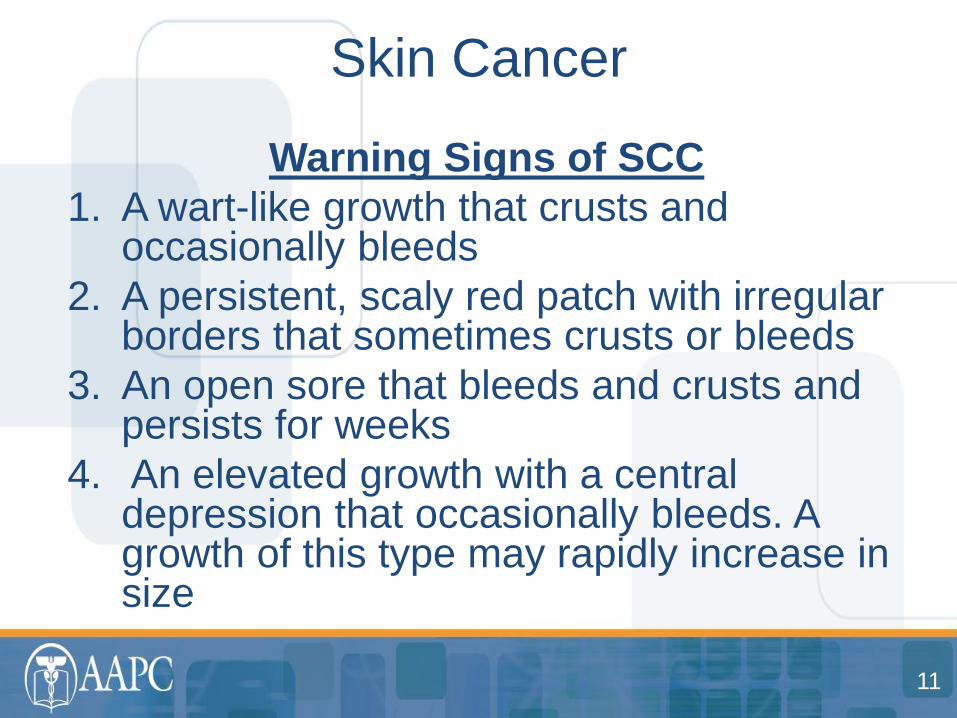
Warning Signs of SCC
1. A wart-like growth that crusts and occasionally bleeds
2. A persistent, scaly red patch with irregular borders that sometimes crusts or bleeds
3. An open sore that bleeds and crusts and persists for weeks
4. An elevated growth with a central depression that occasionally bleeds. A growth of this type may rapidly increase in size
Skin Cancer
11
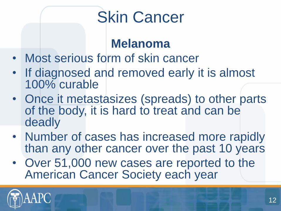
Melanoma
• Most serious form of skin cancer
• If diagnosed and removed early it is almost 100% curable
• Once it metastasizes (spreads) to other parts of the body, it is hard to treat and can be deadly
• Number of cases has increased more rapidly than any other cancer over the past 10 years
• Over 51,000 new cases are reported to the American Cancer Society each year
Skin Cancer
12
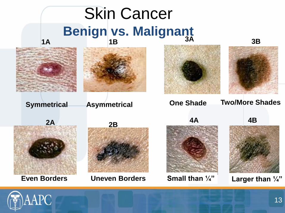
Skin Cancer Benign vs. Malignant
Symmetrical Asymmetrical
1A 1B
Even Borders Uneven Borders
2A 2B
One Shade Two/More Shades
3A 3B
Small than ¼” Larger than ¼”
4A 4B
13
ICD-9-CM Coding
• Chapter 2 of the ICD-9-CM contains the codes for most benign and all malignant neoplasms. Certain benign neoplasms, such as prostatic adenomas, may be found in the specific body system chapters. To properly code a neoplasm it is necessary to determine from the record if the neoplasm is benign, in-situ, malignant, or of uncertain histologic behavior. If malignant, any secondary (metastatic) sites should also be determined.
Skin Cancer
14
• Do not go to the Neoplasm Table first
• Reference histological term first, if given
• Melanoma a good example of when going
directly to the Table is not a good idea
Skin Cancer
15
• Primary malignancy previously excised
• When a primary malignancy has been previously excised or eradicated from its site and there is no further treatment directed to that site and there is no evidence of any existing primary malignancy, a code from category V10, Personal history of malignant neoplasm, should be used to indicate the former site of the malignancy. Any mention of extension, invasion, or metastasis to another site is coded as a secondary malignant neoplasm to that site. The secondary site may be the principal or first-listed with the V10 code used as a secondary code.
Skin Cancer
16
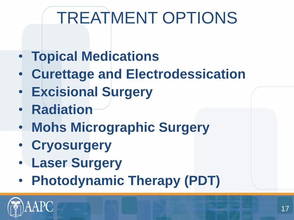
• Topical Medications
• Curettage and Electrodessication
• Excisional Surgery
• Radiation
• Mohs Micrographic Surgery
• Cryosurgery
• Laser Surgery
• Photodynamic Therapy (PDT)
TREATMENT OPTIONS
17
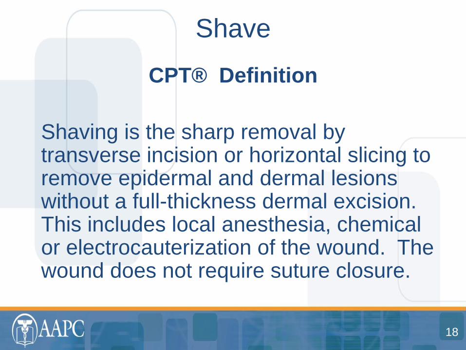
CPT® Definition
Shaving is the sharp removal by transverse incision or horizontal slicing to remove epidermal and dermal lesions without a full-thickness dermal excision. This includes local anesthesia, chemical or electrocauterization of the wound. The wound does not require suture closure.
Shave
18
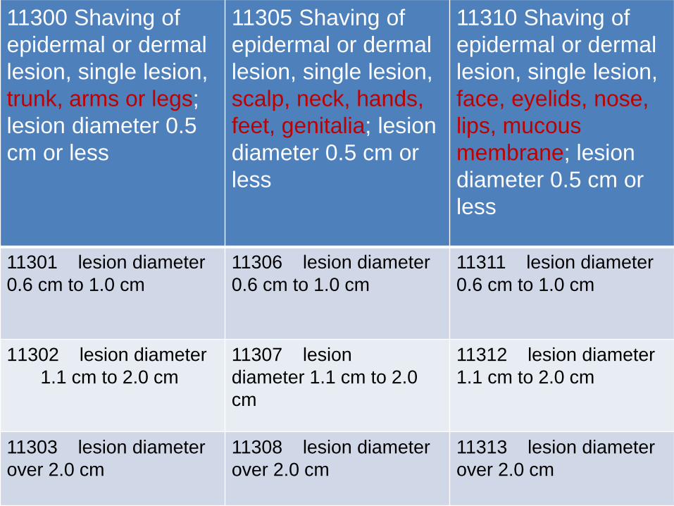
11300 Shaving of
epidermal or dermal
lesion, single lesion,
trunk, arms or legs;
lesion diameter 0.5
cm or less
11305 Shaving of
epidermal or dermal
lesion, single lesion,
scalp, neck, hands,
feet, genitalia; lesion
diameter 0.5 cm or
less
11310 Shaving of
epidermal or dermal
lesion, single lesion,
face, eyelids, nose,
lips, mucous
membrane; lesion
diameter 0.5 cm or
less
11301 lesion diameter
0.6 cm to 1.0 cm
11306 lesion diameter
0.6 cm to 1.0 cm
11311 lesion diameter
0.6 cm to 1.0 cm
11302 lesion diameter
1.1 cm to 2.0 cm
11307 lesion
diameter 1.1 cm to 2.0
cm
11312 lesion diameter
1.1 cm to 2.0 cm
11303 lesion diameter
over 2.0 cm
11308 lesion diameter
over 2.0 cm
11313 lesion diameter
over 2.0 cm
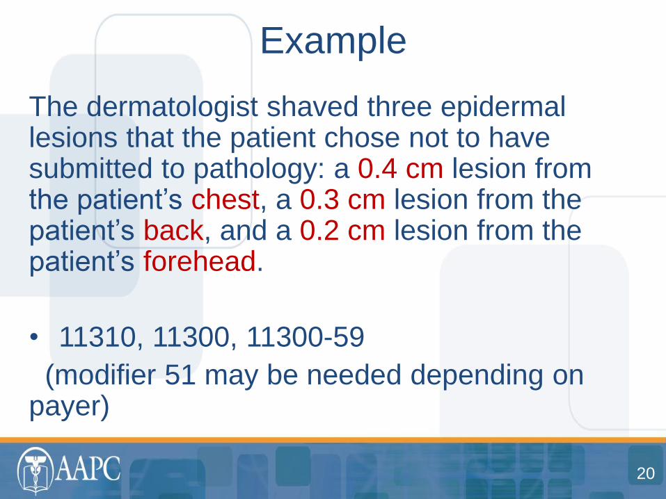
The dermatologist shaved three epidermal lesions that the patient chose not to have submitted to pathology: a 0.4 cm lesion from the patient’s chest, a 0.3 cm lesion from the patient’s back, and a 0.2 cm lesion from the patient’s forehead.
• 11310, 11300, 11300-59
(modifier 51 may be needed depending on payer)
Example
20
CPT® Definition
Excision is defined as full-thickness (through the dermis) removal of lesion, including margins, and includes simple (non-layered) closure when performed
• Deeper than a shave (partial thickness)
Excision
21
Code selection is determined by measuring the greatest clinical diameter of the apparent lesion plus that margin required for complete excision (lesion diameter plus the most narrow margins required equals the excised diameter).
The margins refer to the most narrow margin
required to adequately excise the lesion, based on individual judgment.
The measurement of the lesion plus margin
is made prior to excision.
Excision
22
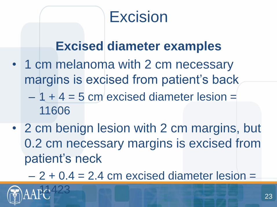
Excised diameter examples
• 1 cm melanoma with 2 cm necessary
margins is excised from patient’s back
– 1 + 4 = 5 cm excised diameter lesion =
11606
• 2 cm benign lesion with 2 cm margins, but
0.2 cm necessary margins is excised from
patient’s neck
– 2 + 0.4 = 2.4 cm excised diameter lesion =
11423
Excision
23
Coding Lesion Excisions
• Benign v Malignant
• Anatomic Site
• Size (excised diameter)
• Type of Repair
Excision
24
• The type of repair is important with excision of lesions as simple repairs are bundled into the excision codes per CPT® guidelines.
• Layered and complex repairs are separately reportable.
• When an excision and repair are separately reported, modifier 51 may be necessary when reporting (payer issue).
Excision
25
A physician refers a patient to the dermatologist for excision of a “mole” on the patient’s left cheek. The dermatologist suspects that the mole is a small basal cell carcinoma (later confirmed pathologically). She performs an excision to remove the 0.9 cm excised diameter lesion in the office. She then closes the wound via simple repair.
• 11641 (repair not separately reported)
• 173.31
Example
26
A patient is seen for excision of a biopsy-
proven squamous cell carcinoma on his
back. The 4.2 cm excised diameter lesion
requires a 6.3 cm intermediate repair.
• 11606, 12032 (possible modifier 51)
• 173.52
Example
27
VIDEO DEMONSTRATING
LESION EXCISION WITH
INTERMEDIATE REPAIR
28
Repair Coding
• Type of Repair
• Site of Repair
• Size of Repair
• When to Add Repairs
Repair
29
• CPT® defines a wound closure as a closure
“utilizing sutures, staples, or tissue adhesives
(eg, 2-cyanoacrylate), either singly or in
combination with each other, or in combination
with adhesive strips.
• If adhesive strips (i.e., butterfly) alone are used,
then it is bundled in to the E/M service.
Repair
30
Types of Repair
• Simple repair
• Intermediate repair
– Single-layer closure of heavily contaminated wounds that have required extensive cleaning or removal of particulate matter also constitutes intermediate repair.
• Complex repair
Repair
31
• According to the CPT® manual we add
together repairs when they are the same
classification (simple, intermediate, complex)
and the same anatomic grouping (scalp, arms,
etc.).
• For example, you would add together a 4.0 cm
simple repair of the abdomen, a 5.6 cm simple
repair of the back, and a 2.2 cm simple repair of
the chest as one 11.8 cm simple repair to the
trunk (12004).
Repair
32
• But, when more than one classification of wound
is repaired, they are reported separately. The
most complicated repair is listed as the primary
procedure and the less complicated is listed as
the secondary procedure, with the modifier 51
attached (depending on the payer).
Repair
33
A patient has 2 benign lesions excised. The
first one is a 2.1 cm excised diameter lesion
on the forehead, the second is a 2.5 cm on
the cheek. They both require intermediate
repair -2.6 cm on the forehead and 3.0 cm
on the cheek.
• 12053, 11443, 11443-59
• 216.3
Example
34
• Codes 14000-14302 are used for excision
(including lesion) and/or repair by adjacent
tissue transfer or rearrangement
• Z-plasty, W-plasty, V-Y-plasty
• Rotation flap
• Random island flap
• Advancement flap
Adjacent Tissue Transfer
35
• What’s not an ATT?
• Secondary defect closure
• Size for code selection
Adjacent Tissue Transfer
36
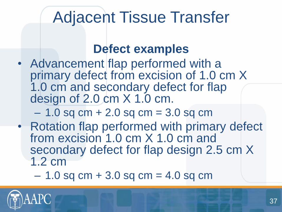
Defect examples
• Advancement flap performed with a primary defect from excision of 1.0 cm X 1.0 cm and secondary defect for flap design of 2.0 cm X 1.0 cm. – 1.0 sq cm + 2.0 sq cm = 3.0 sq cm
• Rotation flap performed with primary defect from excision 1.0 cm X 1.0 cm and secondary defect for flap design 2.5 cm X 1.2 cm – 1.0 sq cm + 3.0 sq cm = 4.0 sq cm
Adjacent Tissue Transfer
37
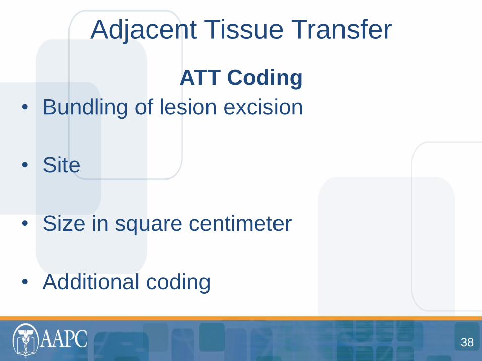
ATT Coding
• Bundling of lesion excision
• Site
• Size in square centimeter
• Additional coding
Adjacent Tissue Transfer
38
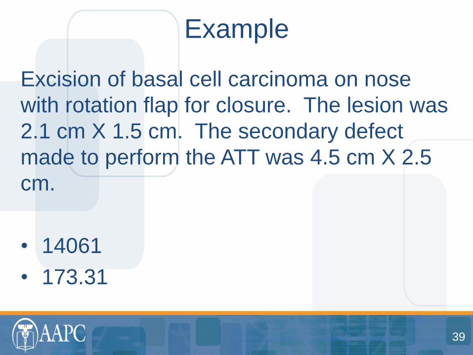
Excision of basal cell carcinoma on nose
with rotation flap for closure. The lesion was
2.1 cm X 1.5 cm. The secondary defect
made to perform the ATT was 4.5 cm X 2.5
cm.
• 14061
• 173.31
Example
39
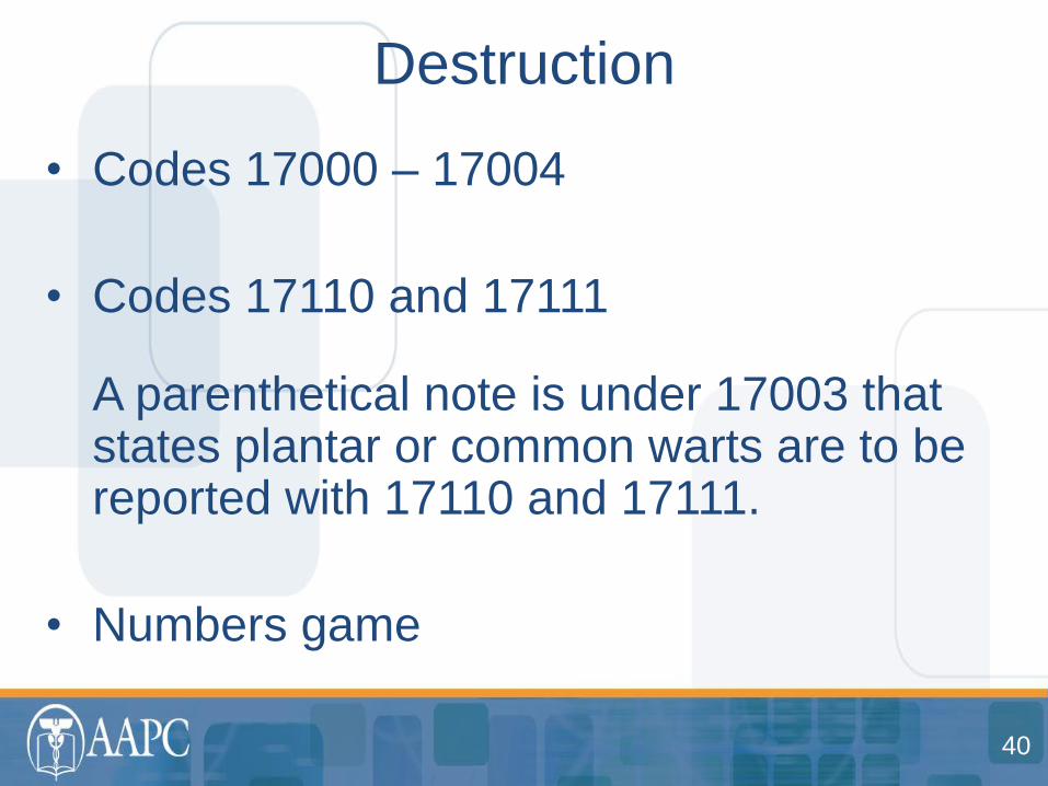
• Codes 17000 – 17004
• Codes 17110 and 17111
A parenthetical note is under 17003 that states plantar or common warts are to be reported with 17110 and 17111.
• Numbers game
Destruction
40
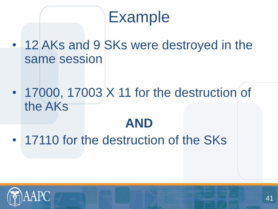
• 12 AKs and 9 SKs were destroyed in the same session
• 17000, 17003 X 11 for the destruction of the AKs
AND
• 17110 for the destruction of the SKs
Example
41
• Mohs is a highly specialized procedure for treatment of skin cancers.
• Mohs allows for complete removal of skin
cancer at one session.
• It has the highest cure rates for squamous and basal cell carcinomas.
• The physician acts as surgeon and pathologist.
Mohs Micrographic Surgery
42
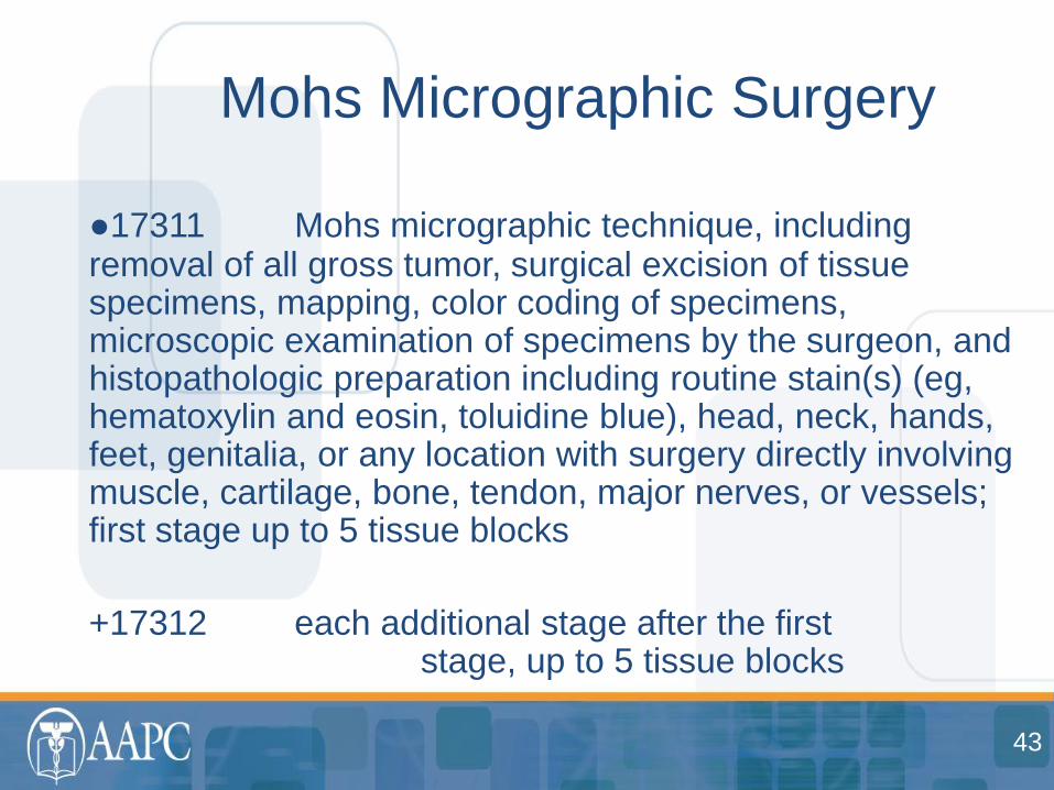
●17311 Mohs micrographic technique, including removal of all gross tumor, surgical excision of tissue specimens, mapping, color coding of specimens, microscopic examination of specimens by the surgeon, and histopathologic preparation including routine stain(s) (eg, hematoxylin and eosin, toluidine blue), head, neck, hands, feet, genitalia, or any location with surgery directly involving muscle, cartilage, bone, tendon, major nerves, or vessels; first stage up to 5 tissue blocks
+17312 each additional stage after the first stage, up to 5 tissue blocks
Mohs Micrographic Surgery
43
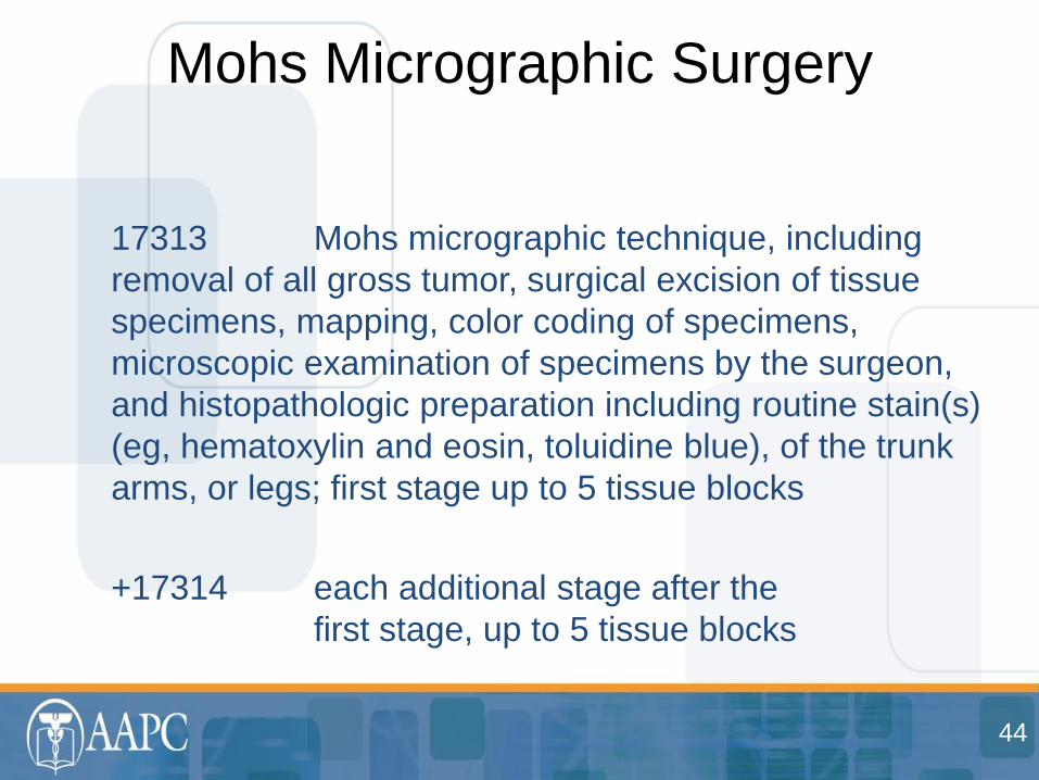
17313 Mohs micrographic technique, including
removal of all gross tumor, surgical excision of tissue
specimens, mapping, color coding of specimens,
microscopic examination of specimens by the surgeon,
and histopathologic preparation including routine stain(s)
(eg, hematoxylin and eosin, toluidine blue), of the trunk
arms, or legs; first stage up to 5 tissue blocks
+17314 each additional stage after the
first stage, up to 5 tissue blocks
Mohs Micrographic Surgery
44
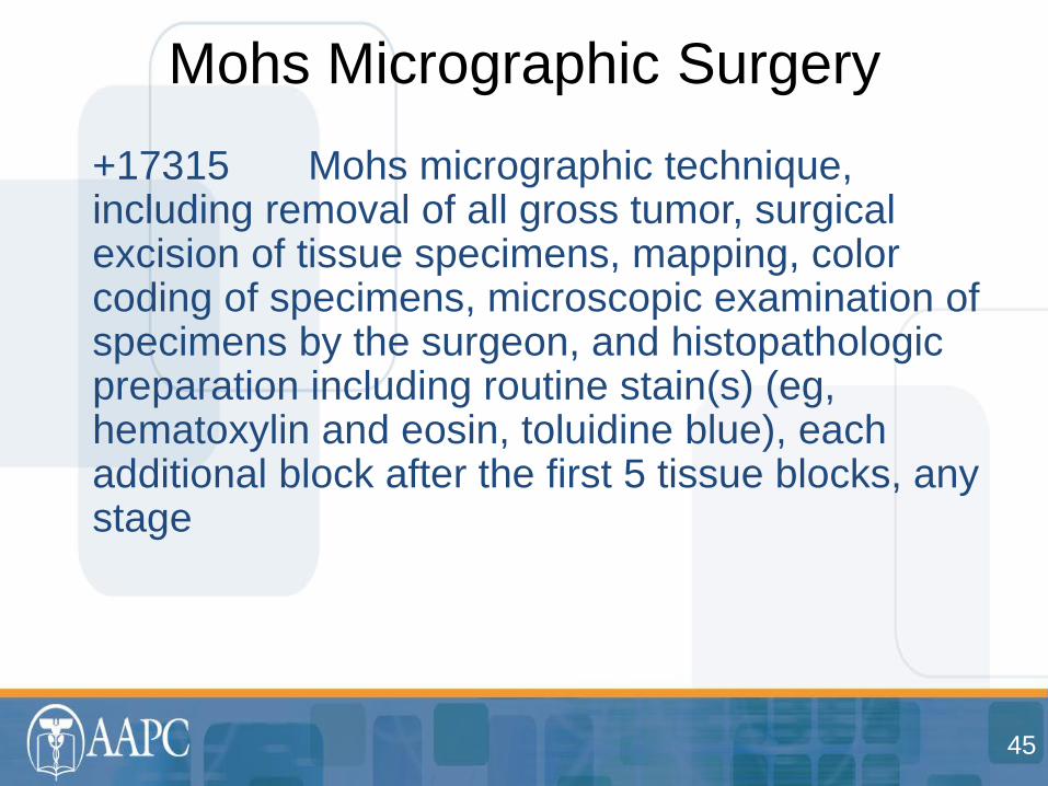
+17315 Mohs micrographic technique, including removal of all gross tumor, surgical excision of tissue specimens, mapping, color coding of specimens, microscopic examination of specimens by the surgeon, and histopathologic preparation including routine stain(s) (eg, hematoxylin and eosin, toluidine blue), each additional block after the first 5 tissue blocks, any stage
Mohs Micrographic Surgery
45
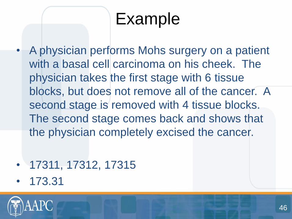
• A physician performs Mohs surgery on a patient
with a basal cell carcinoma on his cheek. The
physician takes the first stage with 6 tissue
blocks, but does not remove all of the cancer. A
second stage is removed with 4 tissue blocks.
The second stage comes back and shows that
the physician completely excised the cancer.
• 17311, 17312, 17315
• 173.31
Example
46
LIVE SURGICAL
PICTURES AND
VIDEO OF MOHS
SURGERY
Mohs
47

ENJOY THE
REST OF
CONFERENCE!
Thank You
48
• Mohs video allowed by permission of Richard
DeAngelis, M.D. from Skin Cancer Centre in
Anderson, S.C. www.skincancercentre.com
• Lesion excision video allowed by permission
of Adrian Richards, M.D. from Aurora Plastic
Surgery and Cosmetic Centres, United
Kingdom www.aurora-clinics.co.uk
• CPT® 2013 Professional Edition
• ICD-9-CM 2013
Sources
49