Embed Size (px)
Citation preview
COFACTOR REQUIREMENTS OF MICROSOMAL 3~-HYDRO~-STEROID OXIDOREDU~ASE,
5-ENE-~SOMERASE FROM HUMAN PLACENTA
JOSEPH WEIDENFELD, RUTH BEN-UZILIO and YEHUDA NE’EMAN
Department of Endocrinology, The Hebrew University-Hadassah Medical School, Jerusalem, Israel
(Received II June 1974)
SUMMARY
Cofactor requirements of 3/3-hydroxysteroid oxidoreductase, Sene-isomerase were studied in lyophilized microsomes and in supernatants obtained by washings with buffer solution. Following 1 h incubation with lyophilized microsomes the activity with NAD or NADP was almost equal. However. rate measurements using 1 or 5 min incubations showed that NAD was the most active cofactor. In 120,OOQ g supernatants of three successive washings the activity was mainly NAD dependent.
The possibility that the activity found in the presence of NADP is mediated by a microsomal transhydrogenase is discussed.
Human placental microsomes contain an enzymic system which catalyses the conversion of 5-ene-3fl- hydroxysteroids to 4-ene-3-keto-steroids (Koide and Torres, 1965; Davies and Ryan, 1972). This conver- sion consists of two reactions: (1) oxidation of the hydroxy group at C-3 (3~~l~ehy~ogena~~ and (2) a shift of double bond from C-5 to C-4 (&ene- isomerase) (Samueis, 1960; Cheatum and Warren, 1966). Whereas it is generally accepted that the above conversion is NAD dependent (Do&an and Ungar, 1965; Davies and Ryan, 1972), Koide and Tort-es (1965) have shown that both NAD and NADP may be cofactors in the conversion of certain 5-ene-38- hydroxysteroids to 4ene-3-keto-steroids. In order to investigate these observations in more detail, we have attempted to determine the cofactor requirements of 3jGhydroxysteroid oxidoreductase, 5-ene-isomerase (3j?OH, 5-I) of human placental microsomes and of soluble extracts derived from these microsomes.
EXPERIMENTAL
All reagents and solvents were of analytical grade. Solvents were redistilled before use; water was double distilled. Pyridine nucleotide cofactors were obtained from Sigma Chemical Co. The steroids were obtained from Ikapharm (Ramat-Gan, Israel). Radioactivity was counted in 10 ml of a scintillation fluid consisting of 4 g PPO and Ql g POPOP (Packard) per liter of toluene. The counting efficiency for ‘H was 53% and the S.D. was 25% in a Packard scintillator spectrometer, model 3380. Scanning of radioactivity on chromatograms was performed on a Berthold, LB 2723 Dunnschicht scanner.
METHODS
Preparation of placental microsomes
All procedures were performed at 04°C. Fresh human placentas, obtained up to 1 h after delivery, were washed with ice-cold Hz0 and cut into small pieces; blood vessels and connective tissue were excluded. The tissue (3OOg) was suspended in 150 ml phosphate buffer (0.2 M pH 7.2) containing O-25 M sucrose and OG4 M nicotinamide and was homo- genized for 1 min in a Waring Blender. The cell debris was removed by centrifuging for 30 min at 1000 g; the resulting supernatant was then centrifuged for 60 min at 10,000 g to remove the mit~hon~ia. Micro- somes were obtained by centrifuging the second supernatant for 60 min at 105,ooO g. The precipitate, containing the microsomes, was washed twice with buffer solution and once with H,O by resuspension in 10 ml of the appropriate fluid, and centrifuged for 60 min at 105,ooO g. The final precipitate was sus- pended in 20 ml of H,O and then lyophilized. The yield of lyophilized microsomes was about 500 mg per 300 g wet weight placenta. The dry preparation consisted of approximately 50% protein. (Protein was determined according to the procedure of Lowry et al., 1951).
Extraction of 3flOEI,5-1 Jkm microsixnes
The microsomes (125 mg) were suspended in 16 ml 0.2 M phosphate buffer containing 0.154 M KCl. The suspension was then homogenized by 2-3 pas- sages of a Teflon piston homogenizer and then centri- fuged for 60 min at 120,000 g. The resulting superna- tant (referred to as supernatant I) was decanted. The precipitate was resuspended twice in 16 ml of buffer and centrifuged each time at 120,000 g for 60 min. The supernatants (referred to as supernatants II and III, respectively) were decanted and kept separately.
1243
1244 J. W~ID~~F~LD, R. BEN-UZILIO and Y. NE’EMAN
Table 1. Activity of 3fi-hydroxysteroid oxidoreductose, 5- 50 pg pregnenolone and 50 ,ug progesterone or 50 ene-isomerase in lyophilized microsomes and 12O.OOOg a DHA and 50 pg androstenedione.
supernatants. following 3 successive washings
Extraction arzd separation qf’ preynenolorw and progcs- Prone
Enzymic activity was measured as the conversion of {7- “HJ-pregnenolone to [7-3H] progesterone and as the con- version of [7-3H]-DHA to r7-3H]-andros~enedione. Each beaker contained 0.2 &i/2 pg of the appropriate substrate, 1.5 pmoles of either NAD or NADP and 0.15 mg protein. Incubations were carried out in I.2 ml of 0.2 M phosphate buffer pH 7,2 containing 0.154 M KC1 at 37°C for 60 min.
*The values shown are the means of two duplicates which in no case differed from one another by more than IO”/’ of the mean value. 0
Enzymic activity was determined in the first micro-
somal suspension, in the 3 supernatants and in the microsomes remaining after the third centrifugation. In the supernatants the protein concentration gradu-
ally decreased with each successive washing of the microsomes (Table 1). Thus the first extract contained approximately 20% of the protein estimated in the original microsomes, the second extract contained loo/,, and the third contained 5%, respectively.
The incubation mixture was extracted in 10 ml ether. The ether was washed with 1 ml of H,O. trans- ferred to a small tube and evaporated to dryness un- der a stream of Nz. The extracted steroids were chro- matographed on thin layer (Ladany and Finkelstein, 1963). Pregnenolone was separated from progesterone by developing the chromatograms in chloroform- ethyl acetate (3: 1, v/v) while DHA was separated from androstenedione in the solvent system of ben- zene-methanol (9: 1, v/v). The radioactive zones were located by scanning; standards of pregnenolone and DHA were detected by their reaction with concen- trated suIphuric acid (Finkelstein, 1968). The progcs- terone and androstenedione standards were located under U.V. light (240 nm). The zones of radioactivity corresponding to the crystalline standards were scraped, eluted with ethanol and their radioactivity counted. The sum of the radioactivity in the zones accounted for about 957; of the total radioactivity taken for incubation. In the control incubation (in absence of enzyme) about 9.57{, of the radioactivity was found in the pregnenolone or DHA zones. No radioactivity could be detected in either the zone cor- responding to progesterone or androstenedione.
~etermi~tioll of enzymic activity
Incubatiorl procedure
The activity of 3,GOH,5-I was dete~ined by mea- suring the conversion of pregnenolone to progester- one and also by the conversion of dehydroisoandro- sterone (DHA) to androstenedione. An ethanolic solu- tion of 0.2 &i [7-3H] (S.A. 19.8 Ci/mmol) and 2 pg of crystalline pregnenolone or @2 ,&i of [7-3H] DHA (S.A. 20-5 Ci/mmol) and 2 pg of crystalline DHA, was transferred to 10 ml beakers. The ethanol was evapor- ated under a stream of N, and the steroid residue dissolved in O-2 ml of 0.2 M phosphate buffer (pH 7.2) containing 0.154 M KC1 and 15 pm01 of either NAD or NADP. Samples of 0.15 mg protein from the various enzymic preparations in phosphate buffer were added to a final volume of 1.2 ml. The incuba- tions were carried out in air at 37°C for 1 h in a Dubnoff metabolic shaker. Rate measurements for the conversion of pregnenolone to progesterone (Fig. 1) in presence of NAD or NADP, were carried out by incubating 0.1 PCi of [7-3H]-pregnenolone at 5 or 6 different ~n~ntrations of crystalline pregnenolone for 1 min (NAD) or 5 min (NADP) at 37°C in air. The protein content was 0025 mg (with NAD) or 0.25 mg (with NADP) in a total incubation volume of 0.7 ml. The incubations were terminated by placing the samples in an ice bath and adding @2 ml of eth- anol containing the appropriate standard carriers i.e.
The enzymic activity was determine’d by measuring the percent of conversion of [7-3H]-pregnenolone or [7-3H]-DHA of a known S.A. to [7-3H]-progesterone or [7-3H]-androstenedione respectively. Based on the percent conversion, the amount of product was calcu- lated. The enzymic activity was expressed as nmol
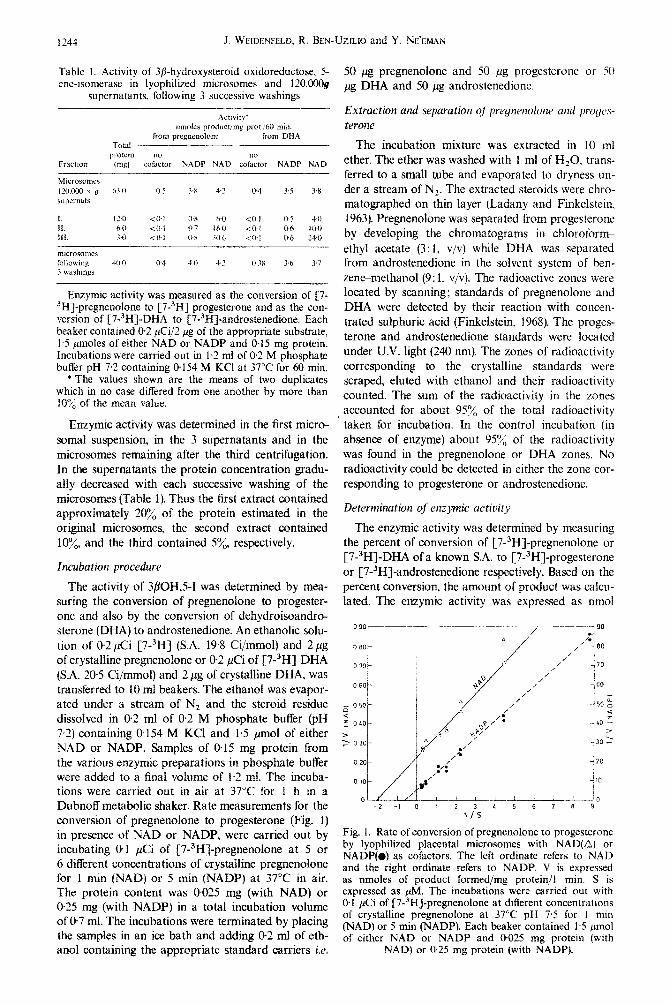
Fig. 1. Rate of conversion of pregnenolone to progesterone by lyophilized placental microsomes with NAD(Q) or NADP(# as cofactors. The left ordinate refers to NAD and the right ordinate refers to NADP. V is expressed as nmoles of product formed/mg protein/l min. S is expressed as PM. The incubations were carried out with 0.1 &i of [7-3H]-pregnenolone at different concentrations of crystalline pregnenolone at 37°C pH 7.5 for I min (NAD) or 5 min (NADP). Each beaker contained 1.5 pmol of either NAD or NADP and 0.025 mg protein (with
NAD) or 0.25 mg protein (with NADP).
Human placental 3/Ghydroxysteroid oxidoreductase, 5-ene-isomerase 1245
of progesterone or androstenedione produced per 60 min per mg of protein. The kinetic data were plotted according to the method of Lineweaver-Burk (1934). The Michaelis constant (K,) and the maximal vel- ocity (V,,,) were calculated by an unweighed linear regression and computed.
RESULTS
The activity of 3fiOH,5-I in lyophilized microsomes and in 120,000 g supernatants following 1 h incuba- tion is summarized in Table 1. In the native micro- somes the activity of 3bOH,5-I was approximately equal with either NAD or NADP as cofactors; the activities measured by the conversion of pregnenolone to progesterone were 4.2 and 3.8 nmol/mg/h, respect- ively. In the absence of cofactors only a relatively low activity was found (0.5 nmol/mg/h). Similar results (Table 1) were obtained when the conversion of DHA to androstenedione was measured.
In the presence of NAD the amount of progester- one formed in the successive supernatants were 6, 16 and 30 nmol/mg/h, respectively. When these superna- tants were examined for 3/?OH,5-I with NADP the activities were significantly lower (0.8 nmol/mg/h) and were constant. In the absence of exogenous cofactors no activity could be detected (less than 0.1 nmol/mg/ h). When the activity was measured by the conversion of DHA to androstenedione in the presence of NAD or NADP, a similar pattern was found although the values were 2&30% lower than those found for the production of progesterone (Table 1).
The activity of 3/?OH,5-I remaining in the microso- ma1 pellet after the third extraction was comparable to that found in original microsomes (Table 1): for pregnenolone in the presence of either NAD or NADP the activities were 4.2 and 40 nmol/mg/h, re- spectively; in the absence of NAD or NADP, the acti- vity of 3jIOH,5-I was 04 nmol/mg/h. Similar but slightly lower results were found for the conversion of DHA (Table 1).
Initial rate measurements of conversion of preg- nenolone to progesterone in presence of either NAD or NADP are shown in Fig. 1. The K, values were 044 $vI with NAD and 1.73 $4 with NADP. The V,,, values were 4.2 and @2 nmol/mg protein/min with NAD and NADP, respectively.
DISCUSSION
The results of the present study show that in lyo- philized human placental microsomes either NAD or NADP may act as a cofactor in the conversion of pregnenolone to progesterone or of DHA to andro- stenedione. One hour incubation with either of these cofactors increased the activity of 3flOH,5-I approxi- mately 7 fold (Table 1). However, initial rate measure- ments of the conversion of pregnenolone to progester- one (Fig. 1) showed that NAD was more efficient than NADP as a cofactor. This is indicated by the marked difference in both K, and if,‘,,, for the reaction in
presence of NAD or NADP. While the activity of 3/3OH,5-I in the microsomes in 1 h incubation was almost equal with either NAD or NADP, the activity in the 120,000g supernatant was mainly NAD depen- dent. Thus, the activity in the 120,OOOg supernatant, following the third wash, was 40-fold higher with NAD than with NADP. In addition, the activity with NAD as the cofactor increased with each successive washing whereas the activity with NADP remained consistently low. This increase in activity with NAD (as the cofactor) may be because the more readily soluble extraneous proteins are extracted in the ear- lier washings and contribute to the total protein mea- sured whereas in the last supernatant only a small amount of protein is extracted containing a relatively high proportion of 3/?OH,5-I.
A review of the literature indicates that the cofactor requirements for the conversion of 3/I-hydroxyster- oids to 4ene-3-ketosteroids appears to depend on the preparation used. Evidence for activity in the presence of either NAD or NADP has been found by Koide and Torres (1965) for human placental microsomes, by Tamaoki et al. (1969) for rat testicular microsomes and by Sulimovici and Boyd (1969) for rat ovarian microsomes. On the other hand others have found solely a NAD dependence: Cheatum and Warren (1966) in a purified microsomal preparation of bovine corpus luteum, Davenport and Mallette (1966) in rab- bit ovarian microsomes and Weidenfeld and Ben-Uzi- lio (1973) in the microsomal fraction of the human ovary.
The results of the present study suggest that 3flOH,5-I of human placental microsomes are primar- ily NAD dependent. This follows from the marked difference in K, and V,, values in the presence of the respective cofactors; the increase in specific acti- vity of 3fiOH,5-I with NAD in the sequential 120,ooO g supernatants supports this idea.
The activity of 3@OH,5-I with NADP as cofactor may be the result of a dual cofactor specificity of the enzyme, however, the great difference in activities in the presence of the respective cofactors observed with the 120,OCOg supernatants indicate that this is probably not the case. Another possibility is that the microsomes contain different forms of 3/3OH,S-I hav- ing different cofactor specificities and which also differ in the ease with which they can be solubilized. This would account for the difference in cofactor depen- dent activity found in the microsomes, supernatants. and final pellet. A third possibility is that the activity of 3fl-OH,5-I observed in the presence of NADP is due to the activity of a microsomal transhydrogenase catalyzing the reaction NADP+NADH+ NADPH + NAD. As we have shown, there is a small amount of conversion of pregnenolone to progester- one in the absence of added cofactors which suggests the presence of a small amount of endogenous cofac- tor bound to the microsomes. If this bound cofactor were NAD, then, its regeneration from NADH by a transhydrogenase would be sufficient to account for the activity seen in the presence of NADP. Similarly,
I’36 J. WEIDENFELD. R. BEN-UZILIO and Y. NE’EMAN
the low activity of the 3POH.SI in the 120,OOOg supernatant with NADP as a cofactor may be explained on the basis that only residual transhydro- gcnase activity was released to the 120,000 g superna- tant.
The presence of 3POH.5-I in the 120,000 g superna- tants raises the question whether these enzymes are truly solubilized or whether they are still bound to small microsomal particles which do not sediment at 120,000 g. The latter possibility has been suggested by Neville and Engel (1968), who worked with deoxycho- late treated bovine adrenal microsomes, and by others who used other solubilization techniques (Ewald et al.. 196&a, 1964b). It would be expected, however, that if the 12O.OOOg supernatant contained small microso- ma1 particles, other membrane bound enzymes such as the enzyme complex catalyzing the aromatization of testosterone to estradiol-17P would be detectable. We have attempted to estimate such activity in both the microsomes and in the 12O.OOOg supernatant (Weidenfeld and Ben-Uzilio. 1973) but aromatizing activity was not found in the supernatants. Moreover, the difference in cofactor specificity between the 3/jOH.5-I found in the native microsomes and that found in the 120.000 g supernatants suggests that the supernatant enzyme is no longer microsomally bound; were it bound. it would be expected that the cofactor requirements would remain the same as in the native microsomes. These arguments, however, do not exclude the possibility that the supernatant 3POH.5-I activity is still bound to small membrane or particulate components, not necessarily identical with the native microsomes, and which can utilize NADP as a cofactor.
Ack,lowledgrnlerlts-We wish to thank Prof. M. Finkelstein for his encouragement and advice in carrying out these
studies. The aid of Dr. J. Shaefer in preparing the manu- script and Dr. A. Samuni in carrying out the kinetic studies is gratefully acknowledged.
REFERENCES
I. Cheatum S. G. and Warren J. C.: Biochem. hiophys. Acta 122 (1961) 1-13.
2. Davenport G. R. and Mallette L. E.: Endocrinology 78 (1966) 672-678.
3. Davies J. and Ryan K. J.: Vitam. Horm. 30 (1972) 223 271.
4. Dorfman R. I. and Ungar F.: Metabolism of Steroid Hormones. Academic Press. New York (1965) pp. 394 405.
5. Ewald W., Werbin H. and Chaikoff I. L.: Steroids 4 (1964a) 759-776.
6. Ewald W., Werbin H. and Chaikoff ,I. L.: Biochim. bio- phys. Acta 81 (1964b) 199-201.
7. Finkelstein M.: In Methods in Hormone Research, Vol. 1 2nd ed (Edited by R. I. Dorfman). Academic Press, New Yo& (1968) pi. 451-487.
8. Koide S. S. and Torres M. T.: Biochim. bioohvs. Acta . , 105 (1965) 115-120.
9. Ladany S. and Finkelstein M.: Steroids 2 (1963) 297- 318.
10. Lineweaver H. and Burk D.: d. Am. Chem. Sot. 56 (1934) 658.
Il. Lowry 0. H., Rosebrough N. J.. Farr A. L. and Ran- dall R. J.: J. biol. Chem. 193 (19511 26s-275.
12. Neville H. M. and Engel L. L.: Endbcrinology 83 (1968) 864-872.
13. Samuels L. T.: In Metabolic Pathways (Edited by M. D. Greenberg). Academic Press, New York (1960) pp. 431-480.
14. Sulimovici S. and Boyd G. S.: Eur. J. Biochem. 7 (1969) 549-558.
15. Tamaoki B. I., Inano H. and Nakano H.: In The Gonads (Edited by K. W. McKerns). North Holland Publishing Co., Amsterdam (1969) pp. 547-613.
16. Weidenfeld J. and Ben-Uzilio R.: (Unpublished data) (1973).
![Pyrethrin Biosynthesis: The Cytochrome P450 Oxidoreductase ...Pyrethrin Biosynthesis: The Cytochrome P450 Oxidoreductase CYP82Q3 Converts Jasmolone To Pyrethrolone1[OPEN] Wei Li,a](https://img.pdfslide.net/doc/110x75/5e2d08c0200c602a86070292/pyrethrin-biosynthesis-the-cytochrome-p450-oxidoreductase-pyrethrin-biosynthesis.jpg)