Embed Size (px)
Citation preview
Ana Maria Carmona-Ribeiro
Biocolloids Lab
Universidade de São Paulo
Colloid stability, DLVO and
dynamic light scattering
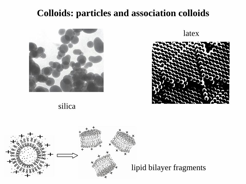
Colloids: particles and association colloids
DODAB vesicle DODAB BF DODAB BF/antigen
+ +
+
+
++
+
+
+ ++
+ +
+
+
++
+
+
+ ++
+
-
-
-
--
-
-
--
-
-
--
-
-
-
silica
latex
lipid bilayer fragments
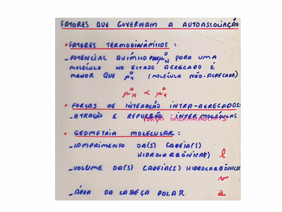
COLOIDES DE ASSOCIAÇÃO
Autoassociação de moléculas anfifílicas.
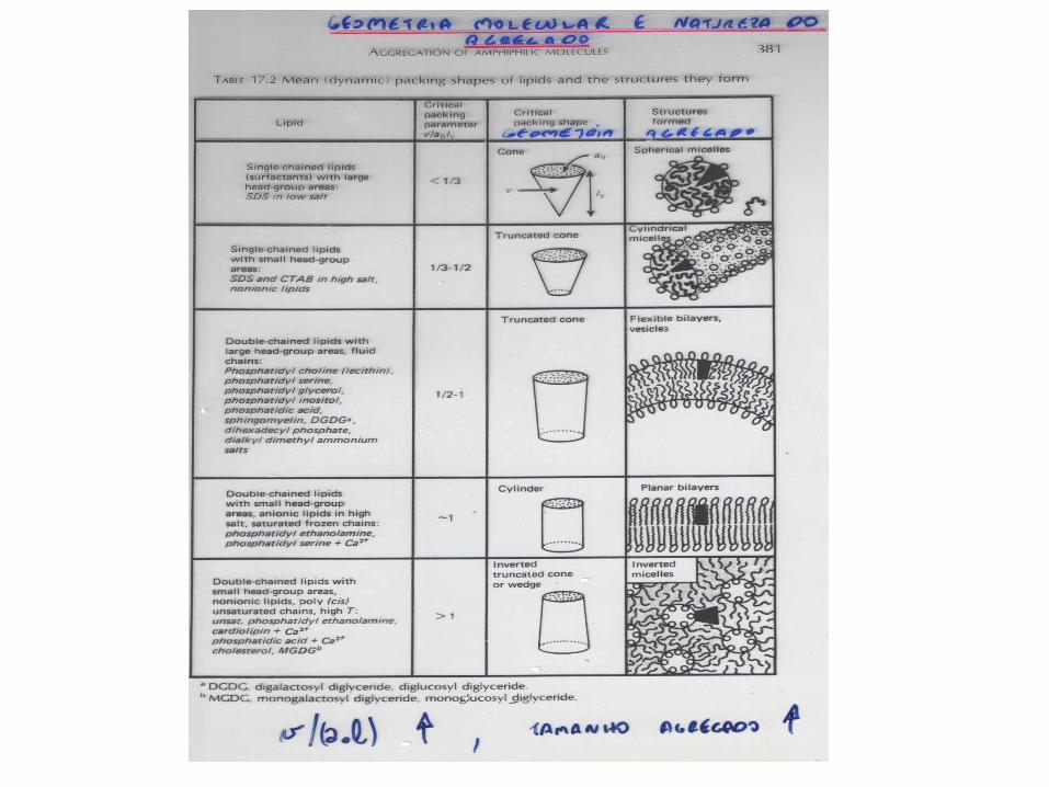
Agregados supramoleculares e geometria molecular.
Micelas, monocamadas, bicamadas, vesículas, lipossomos e
membranas celulares.
Biomembranas.
Forças de interação intra- e interagregados (eletrostática, van der
Waals, hidrofóbica, solvatação).
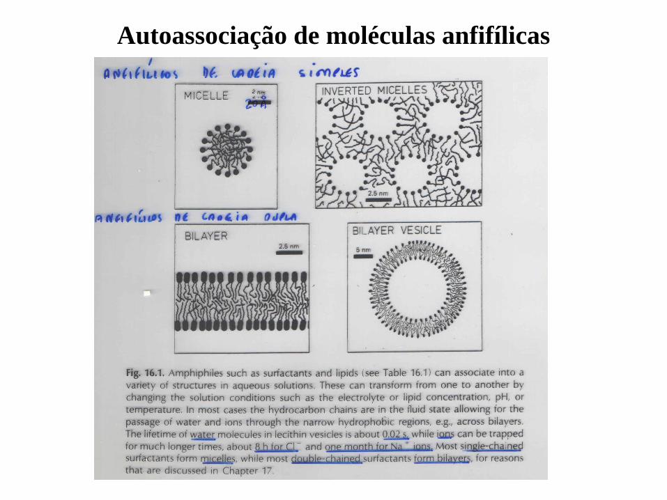
Autoassociação de moléculas anfifílicas
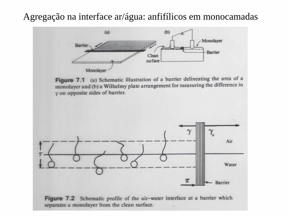
Agregação na interface ar/água: anfifílicos em monocamadas
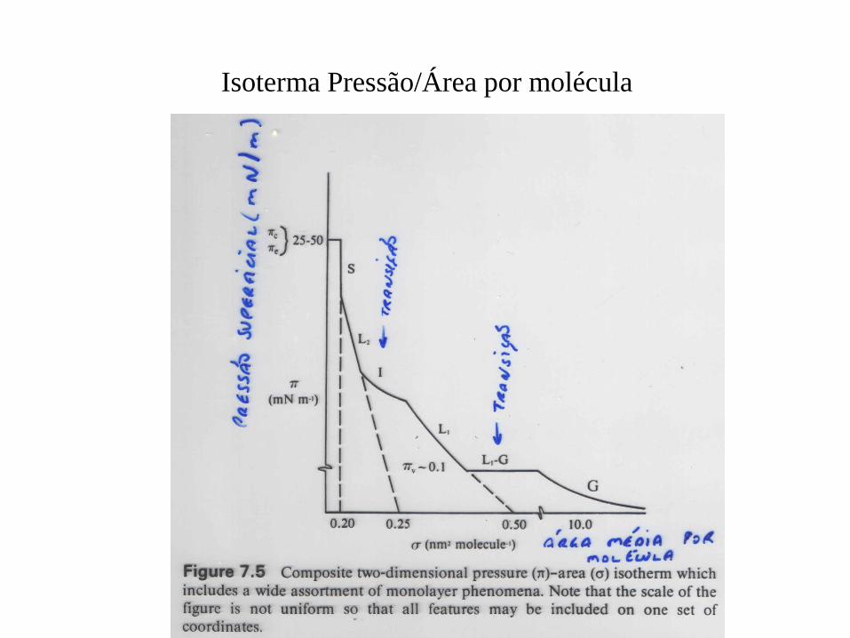
Isoterma Pressão/Área por molécula
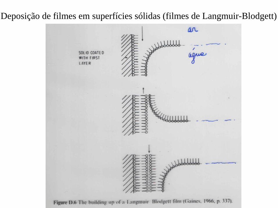
Deposição de filmes em superfícies sólidas (filmes de Langmuir-Blodgett)
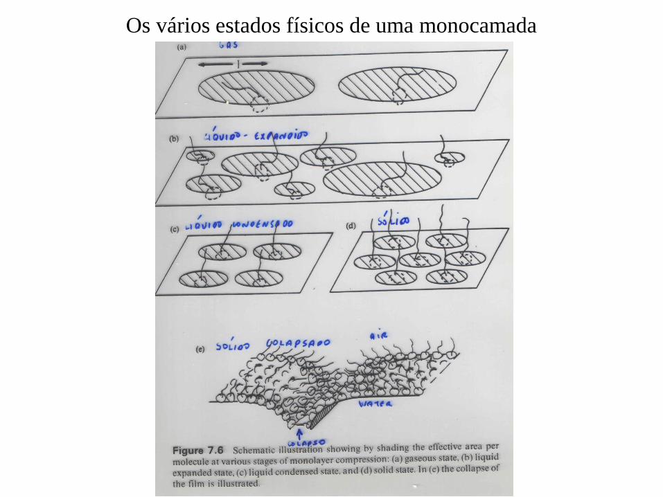
Os vários estados físicos de uma monocamada
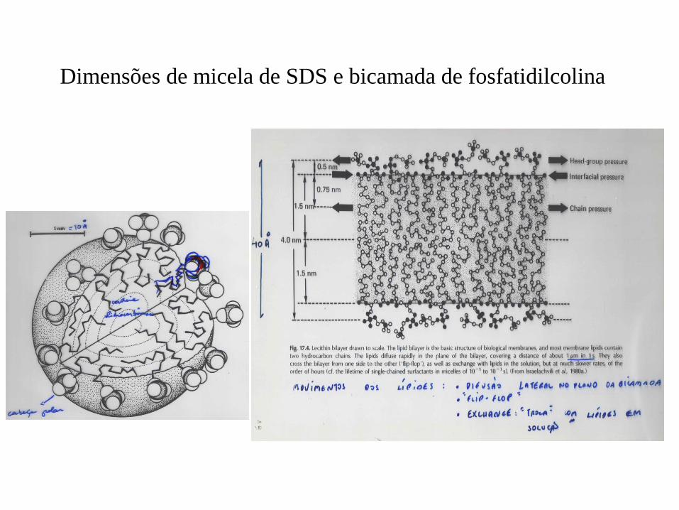
Dimensões de micela de SDS e bicamada de fosfatidilcolina
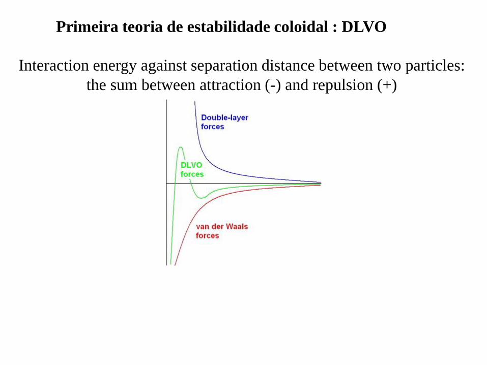
Interaction energy against separation distance between two particles:
the sum between attraction (-) and repulsion (+)
Primeira teoria de estabilidade coloidal : DLVO
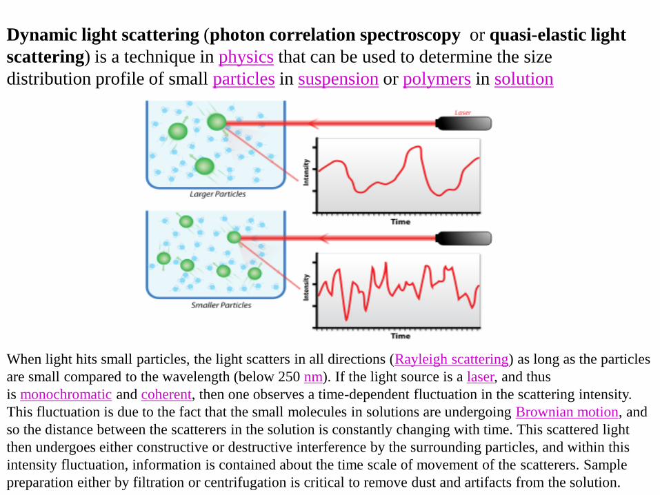
Dynamic light scattering (photon correlation spectroscopy or quasi-elastic light
scattering) is a technique in physics that can be used to determine the size
distribution profile of small particles in suspension or polymers in solution
When light hits small particles, the light scatters in all directions (Rayleigh scattering) as long as the particles
are small compared to the wavelength (below 250 nm). If the light source is a laser, and thus
is monochromatic and coherent, then one observes a time-dependent fluctuation in the scattering intensity.
This fluctuation is due to the fact that the small molecules in solutions are undergoing Brownian motion, and
so the distance between the scatterers in the solution is constantly changing with time. This scattered light
then undergoes either constructive or destructive interference by the surrounding particles, and within this
intensity fluctuation, information is contained about the time scale of movement of the scatterers. Sample
preparation either by filtration or centrifugation is critical to remove dust and artifacts from the solution.
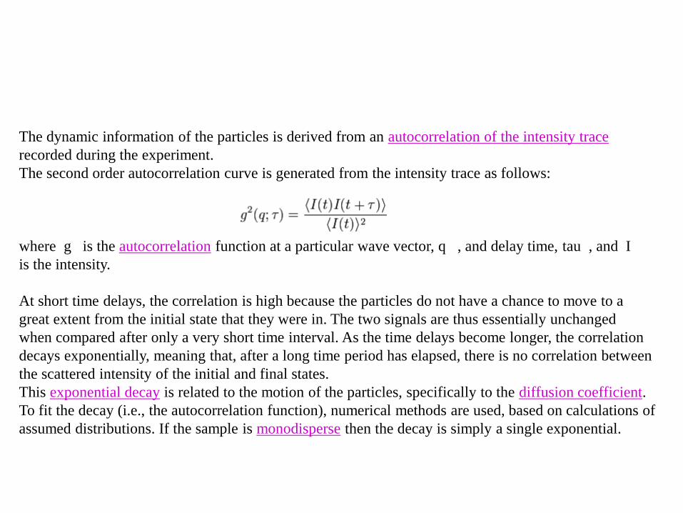
The dynamic information of the particles is derived from an autocorrelation of the intensity trace
recorded during the experiment.
The second order autocorrelation curve is generated from the intensity trace as follows:
where g is the autocorrelation function at a particular wave vector, q , and delay time, tau , and I
is the intensity.
At short time delays, the correlation is high because the particles do not have a chance to move to a
great extent from the initial state that they were in. The two signals are thus essentially unchanged
when compared after only a very short time interval. As the time delays become longer, the correlation
decays exponentially, meaning that, after a long time period has elapsed, there is no correlation between
the scattered intensity of the initial and final states.
This exponential decay is related to the motion of the particles, specifically to the diffusion coefficient.
To fit the decay (i.e., the autocorrelation function), numerical methods are used, based on calculations of
assumed distributions. If the sample is monodisperse then the decay is simply a single exponential.
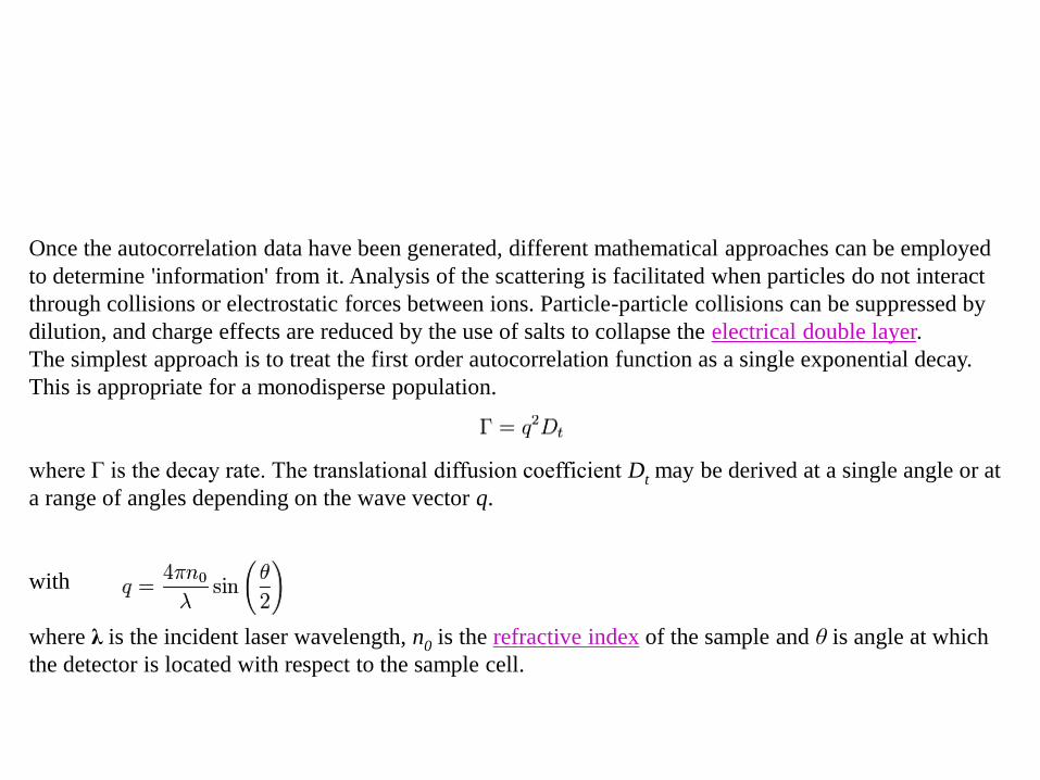
Once the autocorrelation data have been generated, different mathematical approaches can be employed
to determine 'information' from it. Analysis of the scattering is facilitated when particles do not interact
through collisions or electrostatic forces between ions. Particle-particle collisions can be suppressed by
dilution, and charge effects are reduced by the use of salts to collapse the electrical double layer.
The simplest approach is to treat the first order autocorrelation function as a single exponential decay.
This is appropriate for a monodisperse population.
where Γ is the decay rate. The translational diffusion coefficient Dt may be derived at a single angle or at
a range of angles depending on the wave vector q.
with
where λ is the incident laser wavelength, n0 is the refractive index of the sample and θ is angle at which
the detector is located with respect to the sample cell.
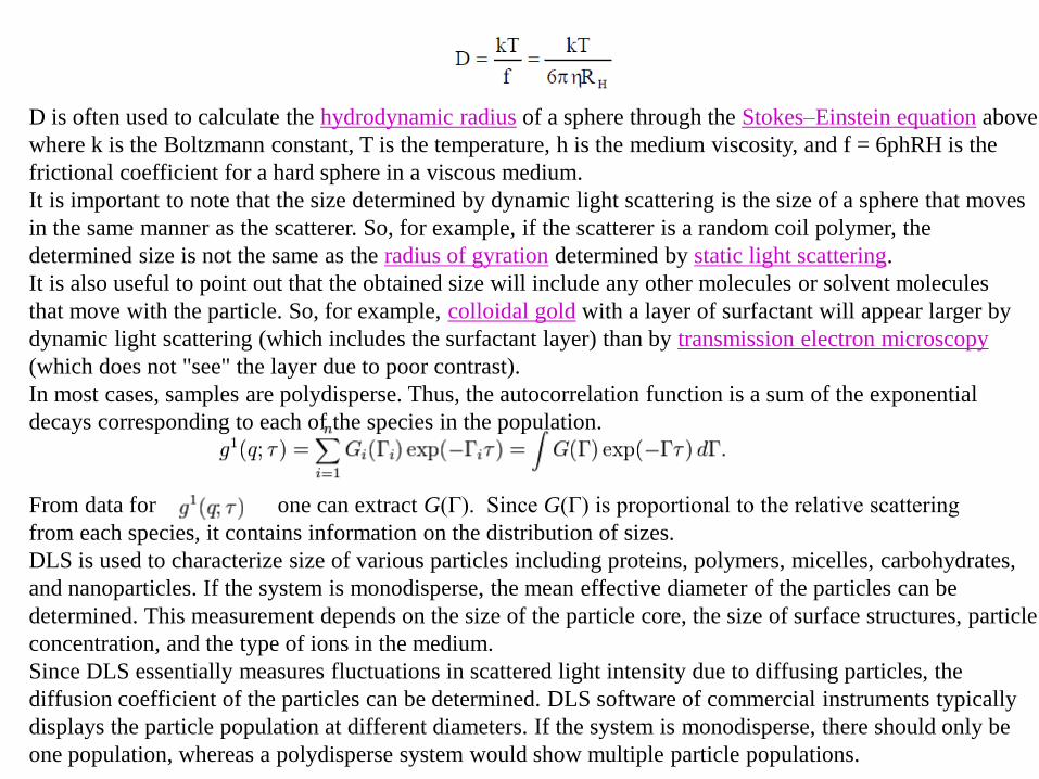
D is often used to calculate the hydrodynamic radius of a sphere through the Stokes–Einstein equation above
where k is the Boltzmann constant, T is the temperature, h is the medium viscosity, and f = 6phRH is the
frictional coefficient for a hard sphere in a viscous medium.
It is important to note that the size determined by dynamic light scattering is the size of a sphere that moves
in the same manner as the scatterer. So, for example, if the scatterer is a random coil polymer, the
determined size is not the same as the radius of gyration determined by static light scattering.
It is also useful to point out that the obtained size will include any other molecules or solvent molecules
that move with the particle. So, for example, colloidal gold with a layer of surfactant will appear larger by
dynamic light scattering (which includes the surfactant layer) than by transmission electron microscopy
(which does not "see" the layer due to poor contrast).
In most cases, samples are polydisperse. Thus, the autocorrelation function is a sum of the exponential
decays corresponding to each of the species in the population.
From data for one can extract G(Γ). Since G(Γ) is proportional to the relative scattering
from each species, it contains information on the distribution of sizes.
DLS is used to characterize size of various particles including proteins, polymers, micelles, carbohydrates,
and nanoparticles. If the system is monodisperse, the mean effective diameter of the particles can be
determined. This measurement depends on the size of the particle core, the size of surface structures, particle
concentration, and the type of ions in the medium.
Since DLS essentially measures fluctuations in scattered light intensity due to diffusing particles, the
diffusion coefficient of the particles can be determined. DLS software of commercial instruments typically
displays the particle population at different diameters. If the system is monodisperse, there should only be
one population, whereas a polydisperse system would show multiple particle populations.
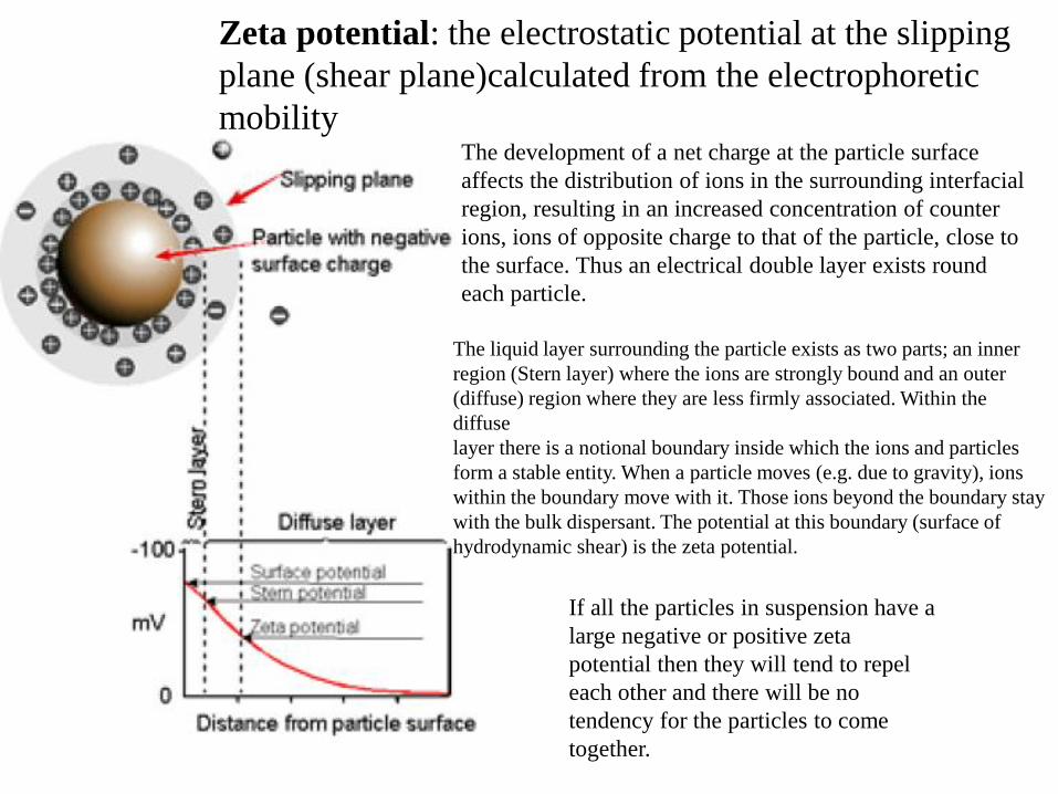
The development of a net charge at the particle surface
affects the distribution of ions in the surrounding interfacial
region, resulting in an increased concentration of counter
ions, ions of opposite charge to that of the particle, close to
the surface. Thus an electrical double layer exists round
each particle.
The liquid layer surrounding the particle exists as two parts; an inner
region (Stern layer) where the ions are strongly bound and an outer
(diffuse) region where they are less firmly associated. Within the
diffuse
layer there is a notional boundary inside which the ions and particles
form a stable entity. When a particle moves (e.g. due to gravity), ions
within the boundary move with it. Those ions beyond the boundary stay
with the bulk dispersant. The potential at this boundary (surface of
hydrodynamic shear) is the zeta potential.
If all the particles in suspension have a
large negative or positive zeta
potential then they will tend to repel
each other and there will be no
tendency for the particles to come
together.
Zeta potential: the electrostatic potential at the slipping
plane (shear plane)calculated from the electrophoretic
mobility
Alguns exemplos de estabilidade e instabilidade coloidal
Importância do DLS para avaliar estabilidade coloidal
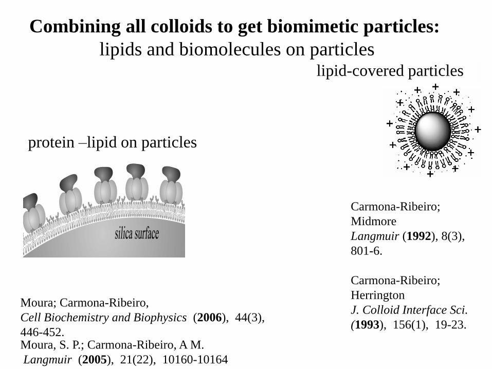
Combining all colloids to get biomimetic particles:
lipids and biomolecules on particles
Particle
DODAB vesicle DODAB BF DODAB BF/antigen
+ +
+
+
++
+
+
+ ++
+ +
+
+
++
+
+
+ ++
+
-
-
-
--
-
-
--
-
-
--
-
-
-
lipid-covered particles
protein –lipid on particles
Moura; Carmona-Ribeiro,
Cell Biochemistry and Biophysics (2006), 44(3),
446-452.
Carmona-Ribeiro;
Midmore
Langmuir (1992), 8(3),
801-6.
Carmona-Ribeiro;
Herrington
J. Colloid Interface Sci.
(1993), 156(1), 19-23.
Moura, S. P.; Carmona-Ribeiro, A M.
Langmuir (2005), 21(22), 10160-10164
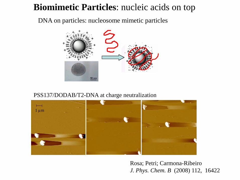
Biomimetic Particles: nucleic acids on top
1 m
(B)
A B
A B
DNA on particles: nucleosome mimetic particles
PSS137/DODAB/T2-DNA at charge neutralization
Rosa; Petri; Carmona-Ribeiro
J. Phys. Chem. B (2008) 112, 16422
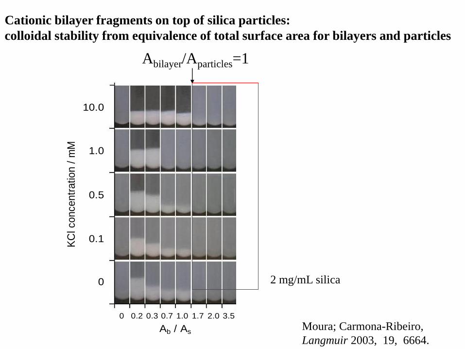
0 0.2 0.3 0.7 1.0 1.7 2.0 3.5
Ab / As
KC
l co
nce
ntr
atio
n /
mM
10.0
1.0
0.5
0.1
0 2 mg/mL silica
Cationic bilayer fragments on top of silica particles:
colloidal stability from equivalence of total surface area for bilayers and particles
Abilayer/Aparticles=1
Moura; Carmona-Ribeiro,
Langmuir 2003, 19, 6664.
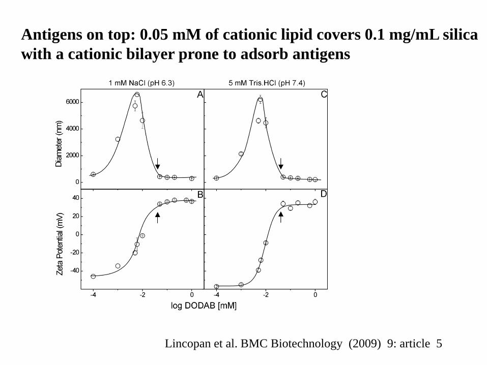
Antigens on top: 0.05 mM of cationic lipid covers 0.1 mg/mL silica
with a cationic bilayer prone to adsorb antigens
Lincopan et al. BMC Biotechnology (2009) 9: article 5
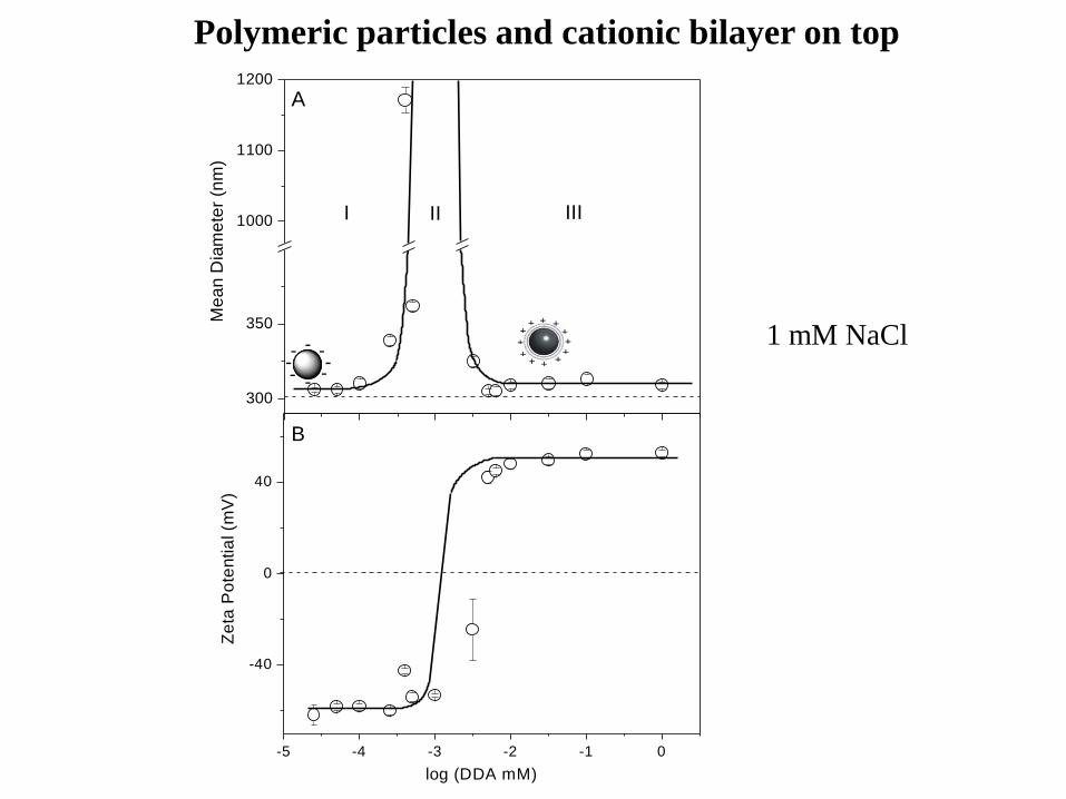
Polymeric particles and cationic bilayer on top
-5 -4 -3 -2 -1 0
-40
0
40
log (DDA mM)
B
Ze
ta P
ote
ntia
l (m
V)
300
350
1000
1100
1200
Me
an
Dia
me
ter
(nm
)
A
IIIIII
+
+
+
+
+ +
+ +
+ +
+ +
-
-
- - - -
- - 1 mM NaCl
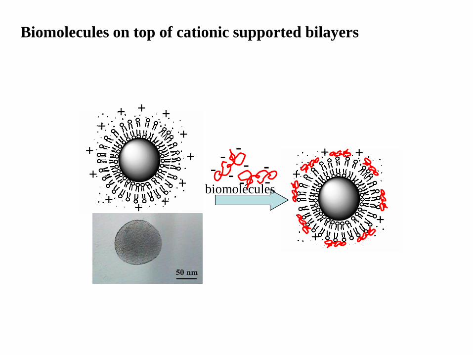
Biomolecules on top of cationic supported bilayers
-
-
--
-
-
--
-
-
--
-
-
--
+
+
+
+
+
++
+
+
+ +
+ + +
+ + +
+
+ +
+ + +
+
+ +
+ + +
+ + +
+
+
+
--
-- -
-- -biomolécules
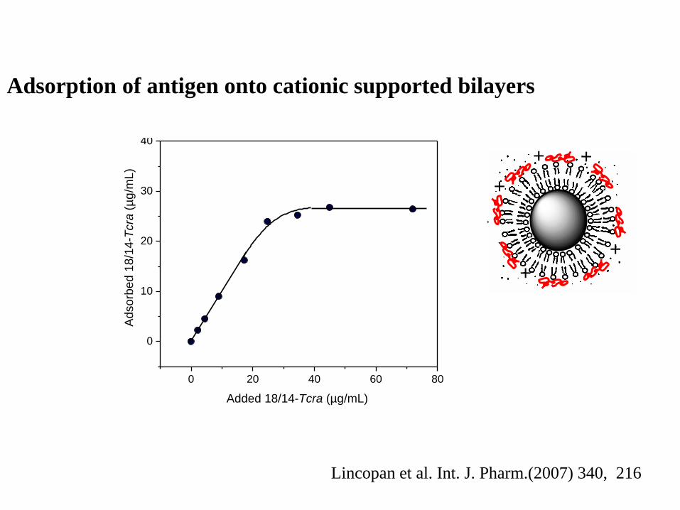
Adsorption of antigen onto cationic supported bilayers
0 20 40 60 80
0
10
20
30
40A
dso
rbe
d 1
8/1
4-T
cra
(µ
g/m
L)
Added 18/14-Tcra (µg/mL)
-
-
--
-
-
--
-
-
--
-
-
--
+
+
+
+
+
++
+
+
+ +
+ + +
+ + +
+
+ +
+ + +
+
+ +
+ + +
+ + +
+
+
+
--
-- -
-- -
Lincopan et al. Int. J. Pharm.(2007) 340, 216
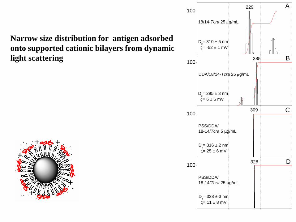
100
5 50 500 5000
DDA/18/14-Tcra 25 g/mL
18/14-Tcra 25 g/mL
Dz= 310 ± 5 nm
= -52 ± 1 mV
229
100
Dz= 295 ± 3 nm
= 6 ± 6 mV
385
100
PSS/DDA/
18-14/Tcra 5 µg/mL
1883
Dz= 316 ± 2 nm
= 25 ± 6 mV
309
100
PSS/DDA/
18-14/Tcra 50 µg/mL
PSS/DDA/
18-14/Tcra 25 µg/mL
Dz= 328 ± 3 nm
= 11 ± 8 mV
Diâmetro (nm)
Inte
nsid
ad
e
328
5
50
500
5000
0
100E
D
C
B
A
Dz= 1261 ± 22 nm
= -35 ± 4 mV
Narrow size distribution for antigen adsorbed
onto supported cationic bilayers from dynamic
light scattering
-
-
--
-
-
--
-
-
--
-
-
--
+
+
+
+
+
++
+
+
+ +
+ + +
+ + +
+
+ +
+ + +
+
+ +
+ + +
+ + +
+
+
+
--
-- -
-- -
Sample DDA
(M)
Ag
(μg/mL)
Mean diameter
(nm) Zeta-Potential (mV) Polydispersity index
PSS - - 301 ± 2 - 60 ± 1 0.064 ± 0.020
DDA 2,000 - 81 ± 1 45 ± 2 0.230 ± 0.006
PSS/DDA 7 - 309 ± 2 48 ± 2 0.040 ± 0.010
PSS/DDA/18/14-Tcra 7 25 328 ± 3 11 ± 8 0.060 ± 0.020
Biomimetic particles: artificial viruses?-
-
--
-
-
--
-
-
--
-
-
--
+
+
+
+
+
++
+
+
+ +
+ + +
+ + +
+
+ +
+ + +
+
+ +
+ + +
+ + +
+
+
+
--
-- -
-- -
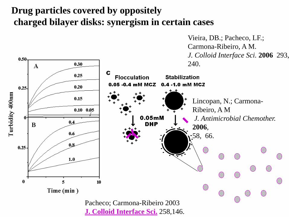
Drug particles covered by oppositely
charged bilayer disks: synergism in certain cases
Pacheco; Carmona-Ribeiro 2003
J. Colloid Interface Sci. 258,146.
Vieira, DB.; Pacheco, LF.;
Carmona-Ribeiro, A M.
J. Colloid Interface Sci. 2006 293,
240.
Lincopan, N.; Carmona-
Ribeiro, A M
J. Antimicrobial Chemother.
2006,
58, 66.
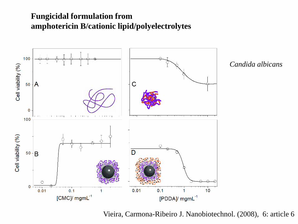
Fungicidal formulation from
amphotericin B/cationic lipid/polyelectrolytes
Vieira, Carmona-Ribeiro J. Nanobiotechnol. (2008), 6: article 6
Candida albicans
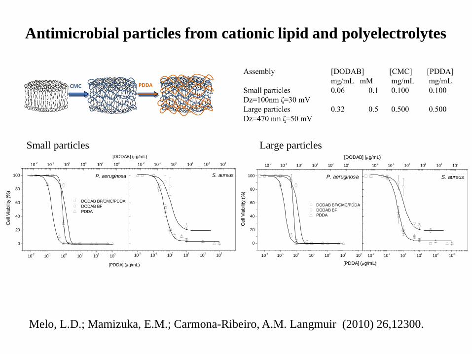
Antimicrobial particles from cationic lipid and polyelectrolytes
Assembly [DODAB]
mg/mL mM
[CMC]
mg/mL
[PDDA]
mg/mL
Small particles
Dz=100nm ζ=30 mV
0.06 0.1 0.100 0.100
Large particles
Dz=470 nm ζ=50 mV
0.32 0.5 0.500 0.500
10-2
10-1
100
101
102
103
0
20
40
60
80
100
Cell
Via
bili
ty (
%)
DODAB BF/CMC/PDDA
DODAB BF
PDDA
10-2
10-1
100
101
102
103
10-2
10-1
100
101
102
103
10-2
10-1
100
101
102
103
S. aureusP. aeruginosa
[PDDA] (g/mL)
[DODAB] (g/mL)
10-2
10-1
100
101
102
103
10-2
10-1
100
101
102
103
104
0
20
40
60
80
100
DODAB BF/CMC/PDDA
DODAB BF
PDDA
Cell
Via
bili
ty (
%)
10-2
10-1
100
101
102
103
10-2
10-1
100
101
102
103
S. aureusP. aeruginosa
[PDDA] (g/mL)
[DODAB] (g/mL)
CMC PDDA
Small particles Large particles
Melo, L.D.; Mamizuka, E.M.; Carmona-Ribeiro, A.M. Langmuir (2010) 26,12300.
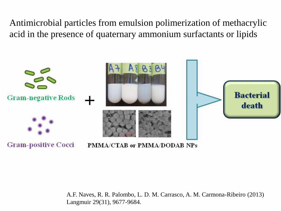
Antimicrobial particles from emulsion polimerization of methacrylic
acid in the presence of quaternary ammonium surfactants or lipids
A.F. Naves, R. R. Palombo, L. D. M. Carrasco, A. M. Carmona-Ribeiro (2013)
Langmuir 29(31), 9677-9684.
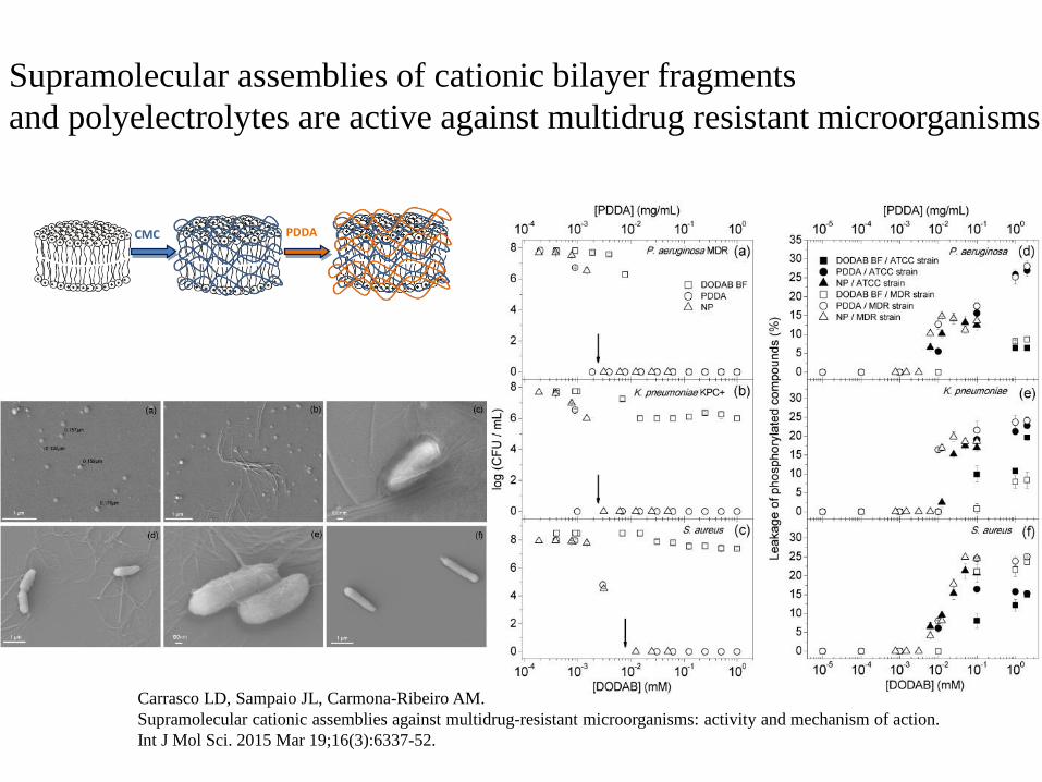
Carrasco LD, Sampaio JL, Carmona-Ribeiro AM.
Supramolecular cationic assemblies against multidrug-resistant microorganisms: activity and mechanism of action.
Int J Mol Sci. 2015 Mar 19;16(3):6337-52.
Supramolecular assemblies of cationic bilayer fragments
and polyelectrolytes are active against multidrug resistant microorganisms
CMC PDDA
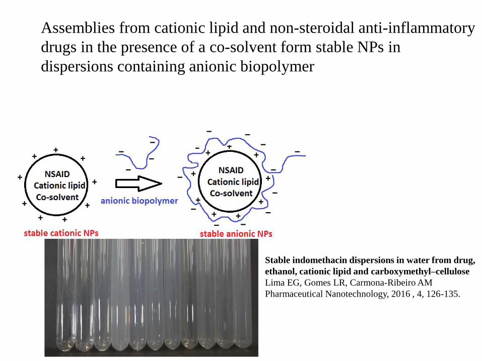
Assemblies from cationic lipid and non-steroidal anti-inflammatory
drugs in the presence of a co-solvent form stable NPs in
dispersions containing anionic biopolymer
Stable indomethacin dispersions in water from drug,
ethanol, cationic lipid and carboxymethyl–cellulose
Lima EG, Gomes LR, Carmona-Ribeiro AM
Pharmaceutical Nanotechnology, 2016 , 4, 126-135.
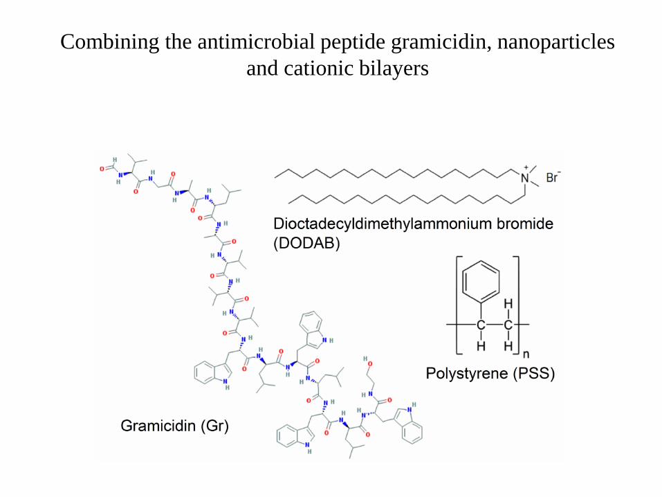
Combining the antimicrobial peptide gramicidin, nanoparticles
and cationic bilayers
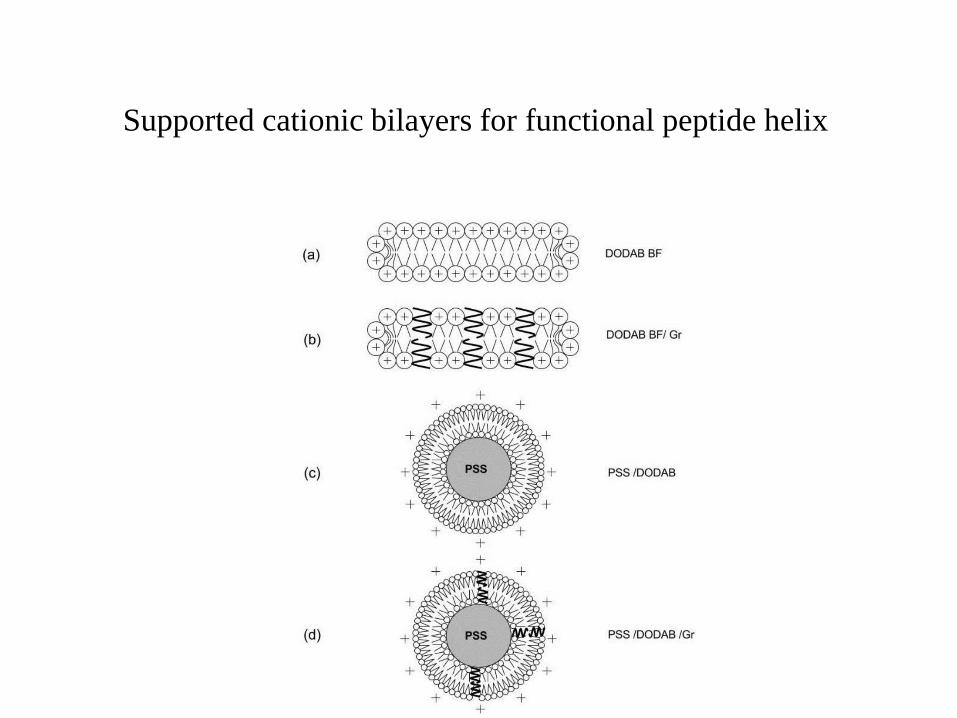
Supported cationic bilayers for functional peptide helix
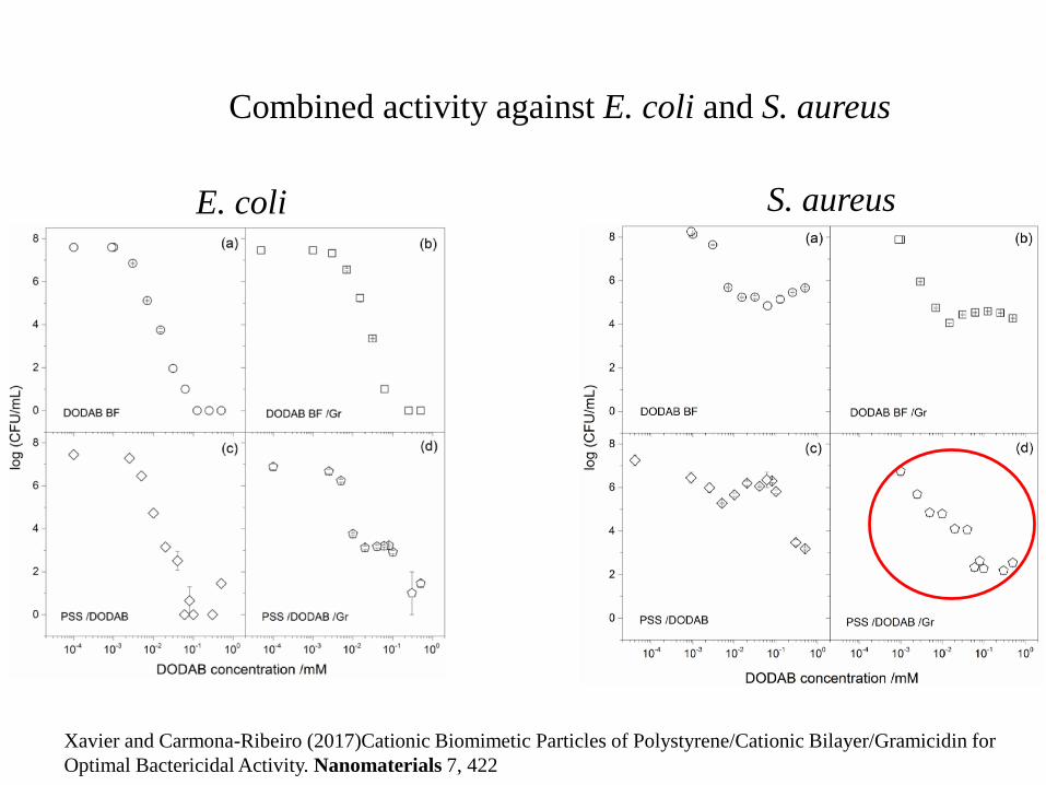
Combined activity against E. coli and S. aureus
S. aureusE. coli
Xavier and Carmona-Ribeiro (2017)Cationic Biomimetic Particles of Polystyrene/Cationic Bilayer/Gramicidin for
Optimal Bactericidal Activity. Nanomaterials 7, 422
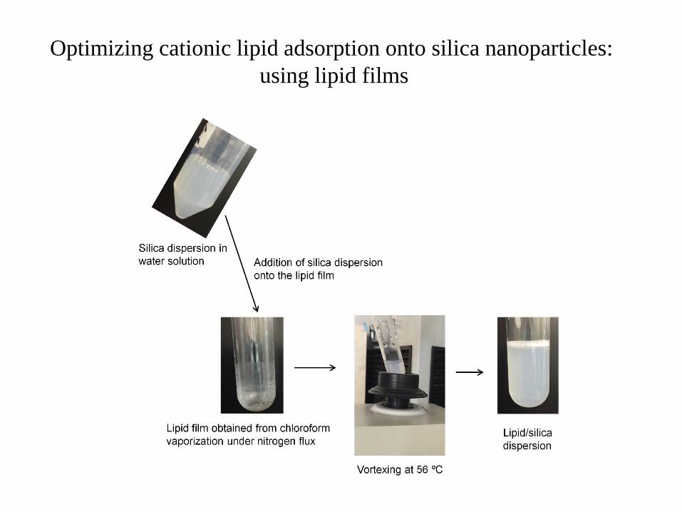
Optimizing cationic lipid adsorption onto silica nanoparticles:
using lipid films
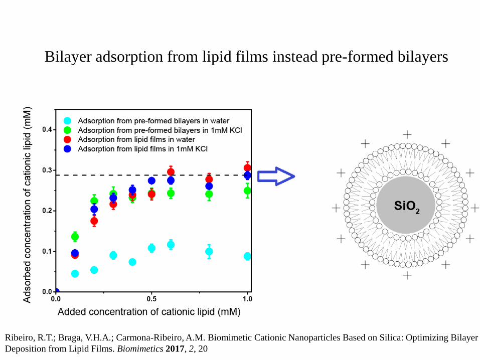
Bilayer adsorption from lipid films instead pre-formed bilayers
Ribeiro, R.T.; Braga, V.H.A.; Carmona-Ribeiro, A.M. Biomimetic Cationic Nanoparticles Based on Silica: Optimizing Bilayer
Deposition from Lipid Films. Biomimetics 2017, 2, 20
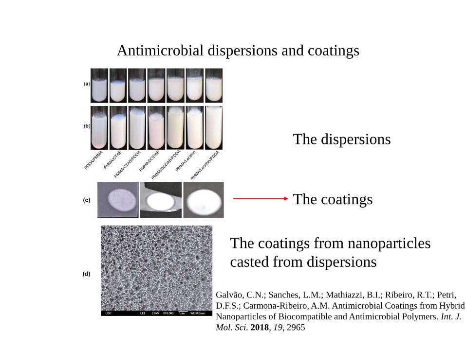
The coatings
The dispersions
Antimicrobial dispersions and coatings
The coatings from nanoparticles
casted from dispersions
Galvão, C.N.; Sanches, L.M.; Mathiazzi, B.I.; Ribeiro, R.T.; Petri,
D.F.S.; Carmona-Ribeiro, A.M. Antimicrobial Coatings from Hybrid
Nanoparticles of Biocompatible and Antimicrobial Polymers. Int. J.
Mol. Sci. 2018, 19, 2965