Embed Size (px)
Citation preview
Color Image Processing with Biomedical Applications
Rangaraj M. Rangayyan, Begoña Acha, and Carmen Serrano
University of Calgary, Calgary, Alberta, Canada University of Seville, Spain
2
SPIE Press 2011 434 pages
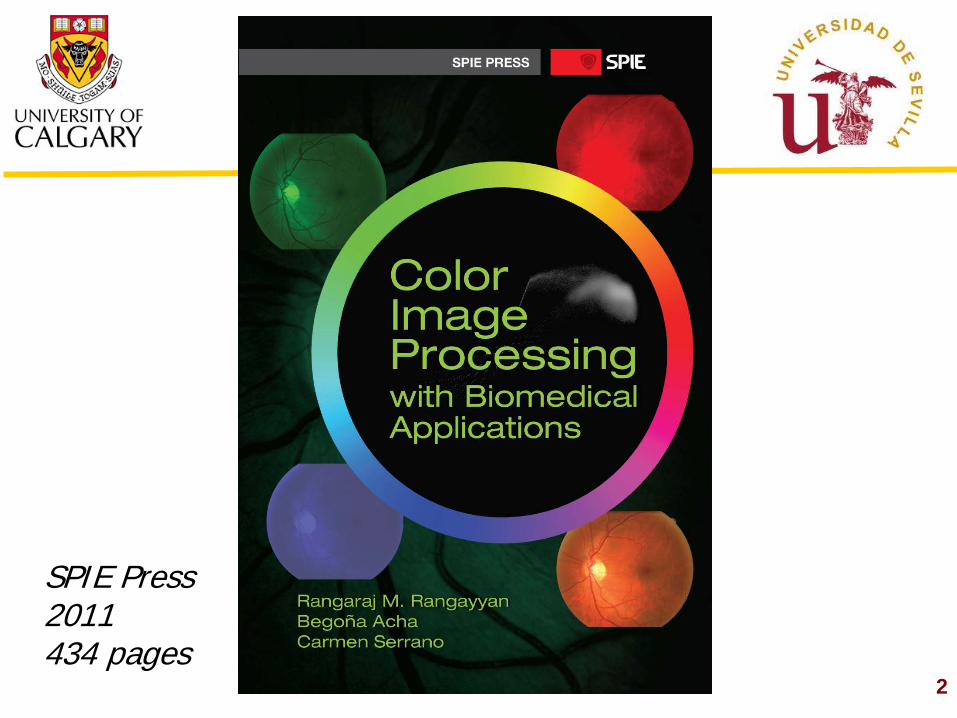
The Nature of Color Images
3
Photo courtesy of Chris Pawluk
Color Attributes
4
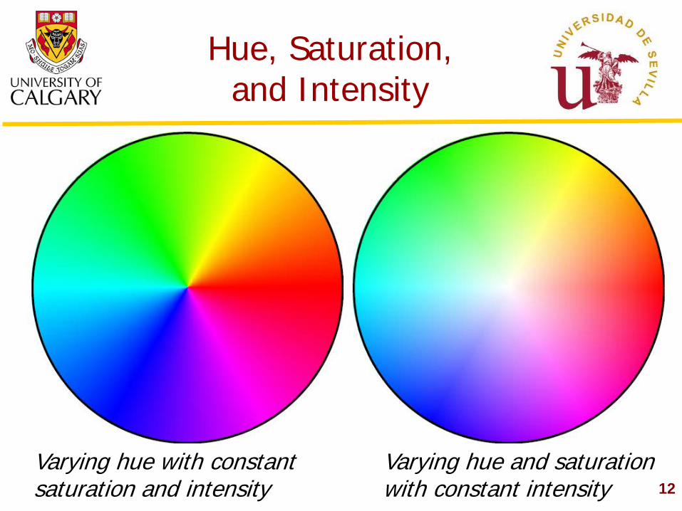
Hue: dominant wavelength or band Saturation: quality or colorfulness, not diluted with white Intensity or Brightness: primary visual sensation related to physical luminance Also used: Chroma, Lightness
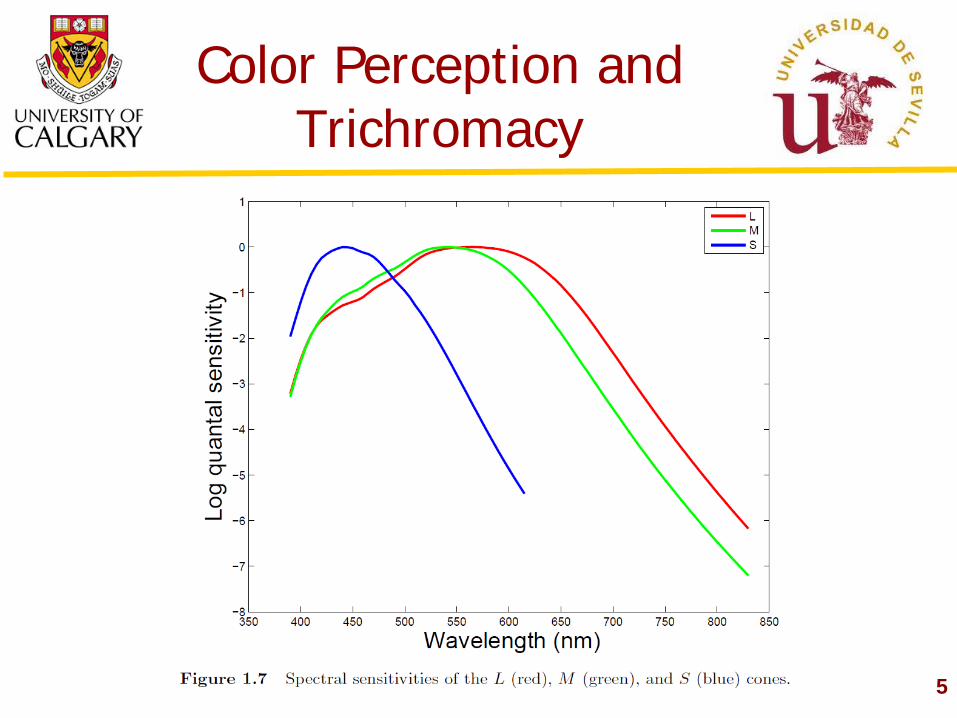
Color Perception and Trichromacy
5
6
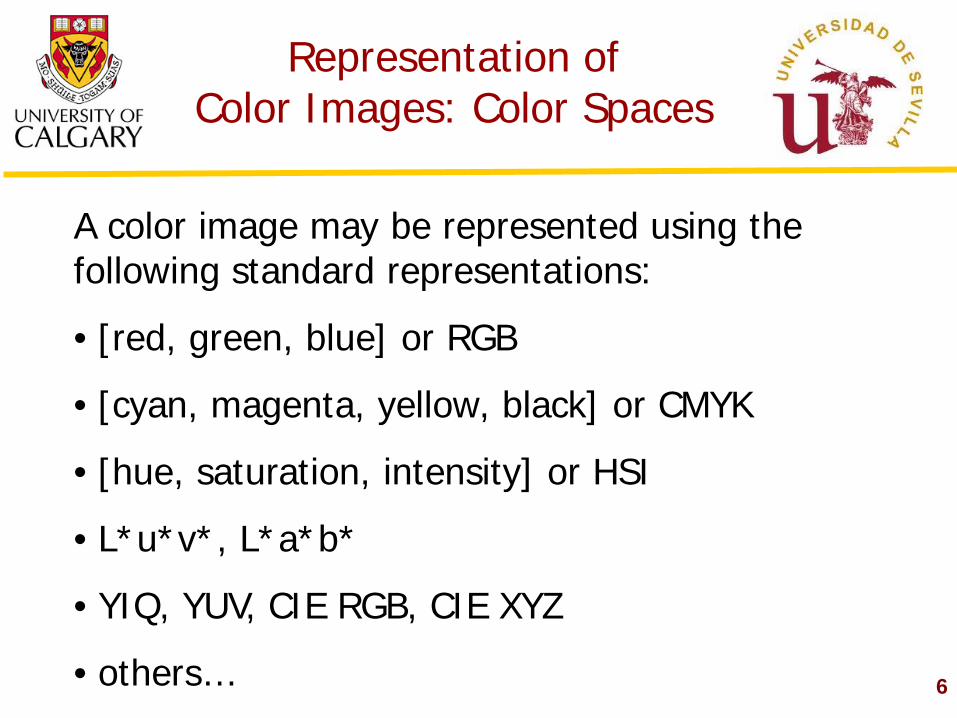
Representation of Color Images: Color Spaces
A color image may be represented using the following standard representations:
• [red, green, blue] or RGB
• [cyan, magenta, yellow, black] or CMYK
• [hue, saturation, intensity] or HSI
• L*u*v*, L*a*b*
• YIQ, YUV, CIE RGB, CIE XYZ
• others…
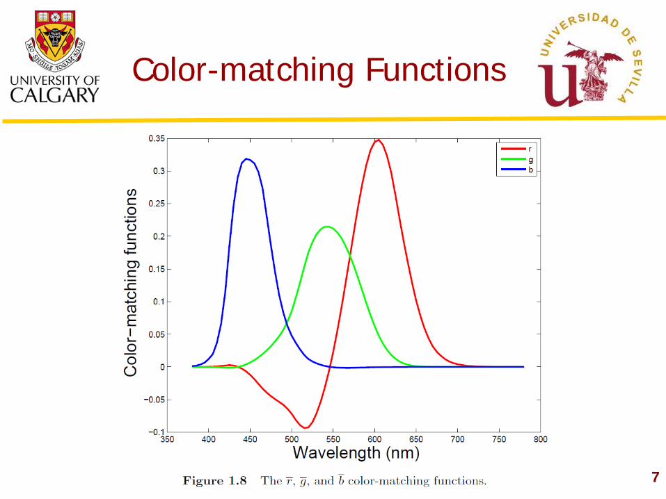
Color-matching Functions
7
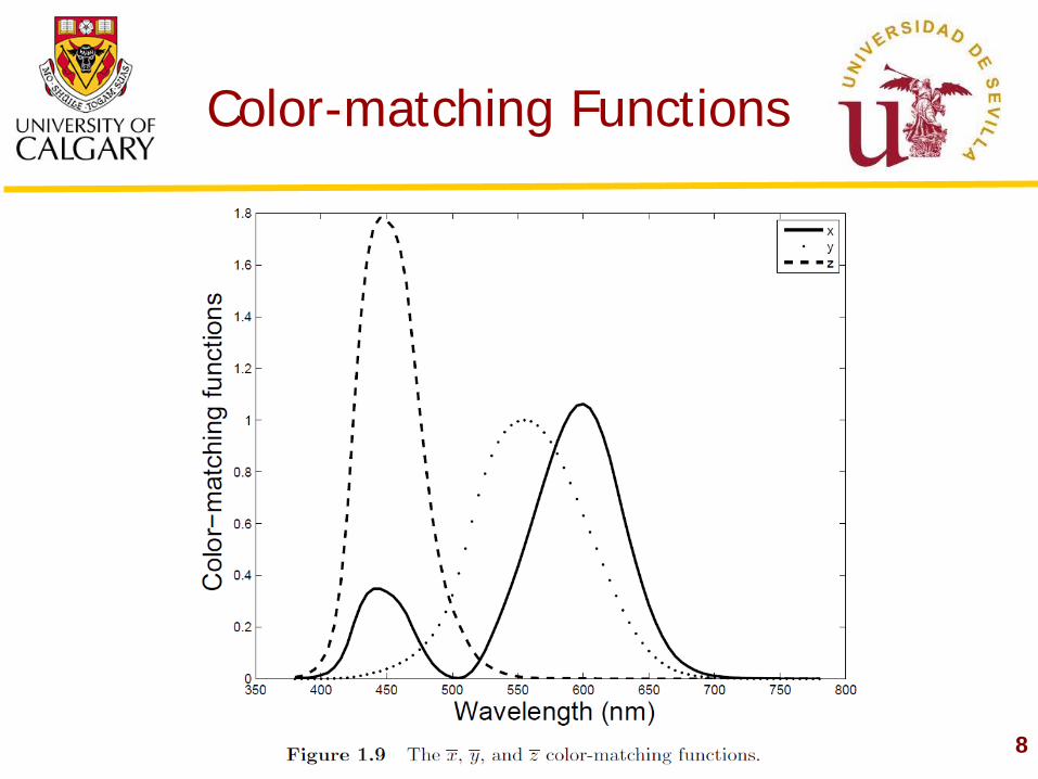
Color-matching Functions
8
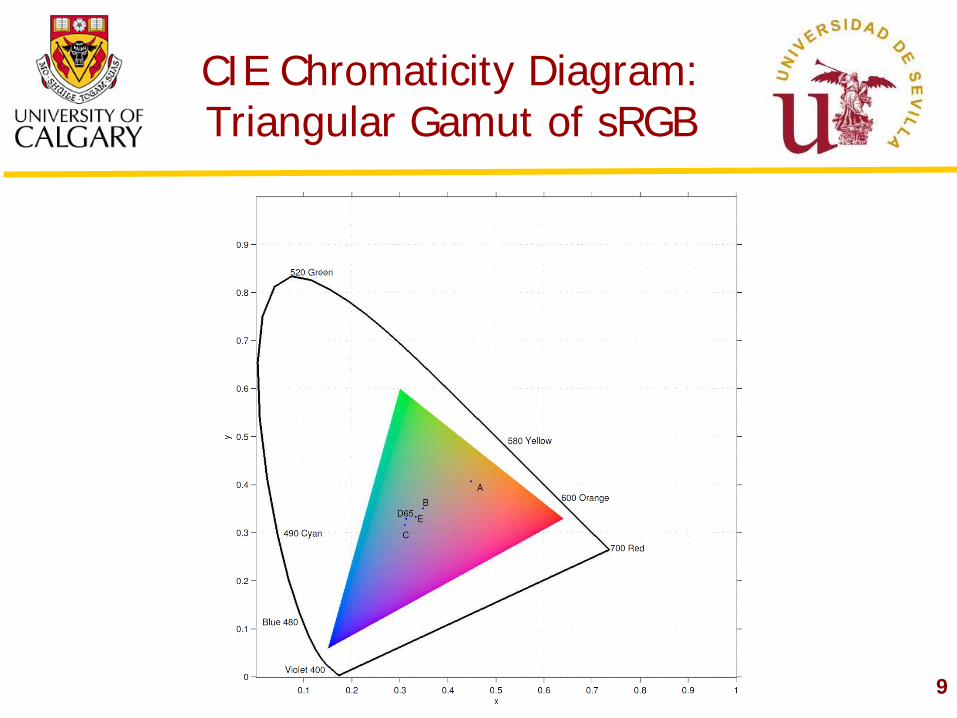
CIE Chromaticity Diagram: Triangular Gamut of sRGB
9
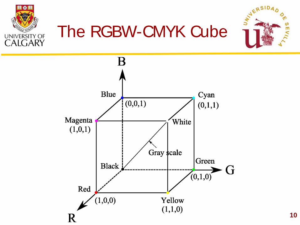
The RGBW-CMYK Cube
10
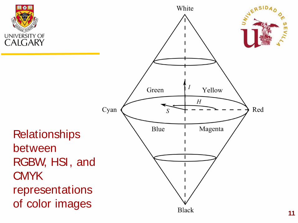
11
Relationships between RGBW, HSI, and CMYK representations of color images
12
Hue, Saturation, and Intensity
Varying hue with constant saturation and intensity
Varying hue and saturation with constant intensity
13
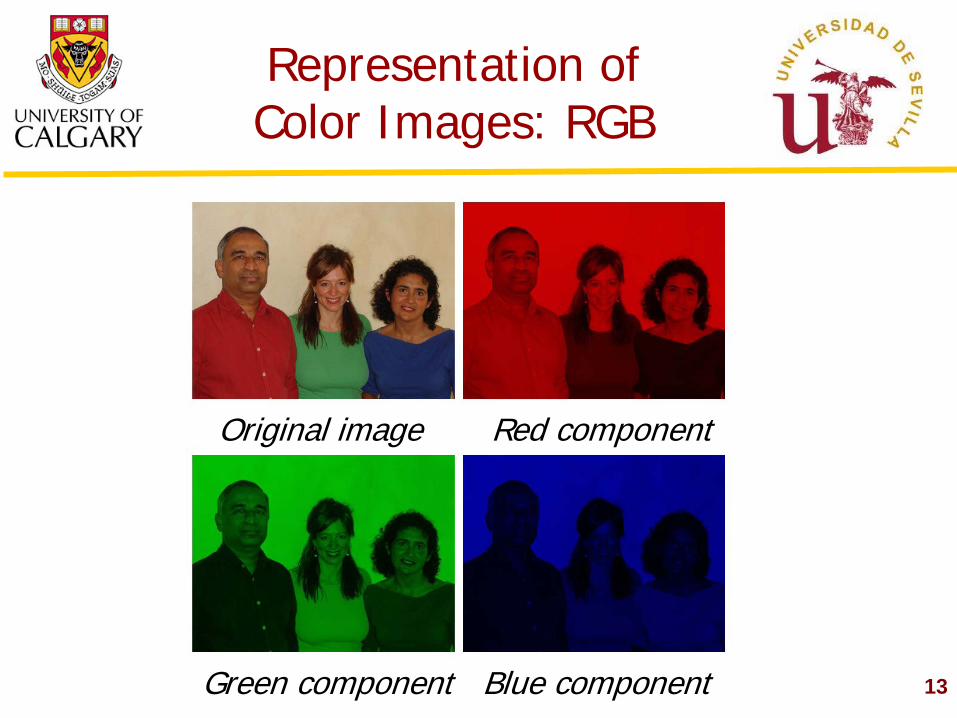
Representation of Color Images: RGB
Original image Red component
Green component Blue component
14
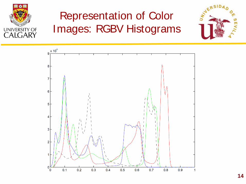
Representation of Color Images: RGBV Histograms
15
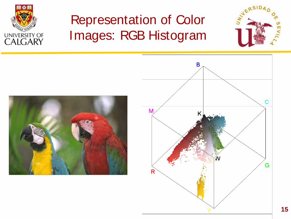
Representation of Color Images: RGB Histogram
16
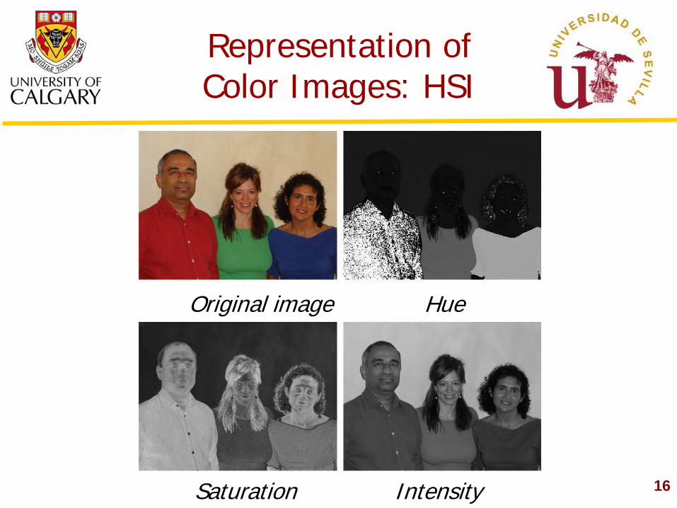
Representation of Color Images: HSI
Original image Hue
Saturation Intensity
17
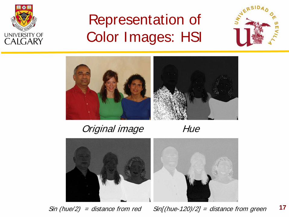
Representation of Color Images: HSI
Original image Hue
Sin (hue/2) = distance from red Sin[(hue-120)/2] = distance from green
18
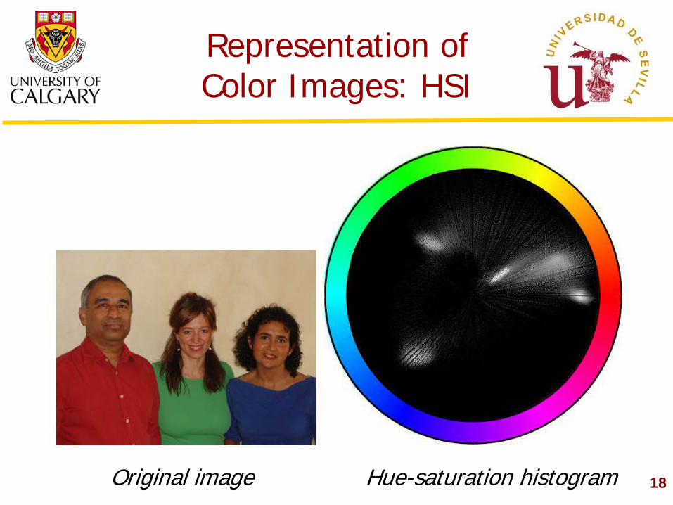
Representation of Color Images: HSI
Original image Hue-saturation histogram
19
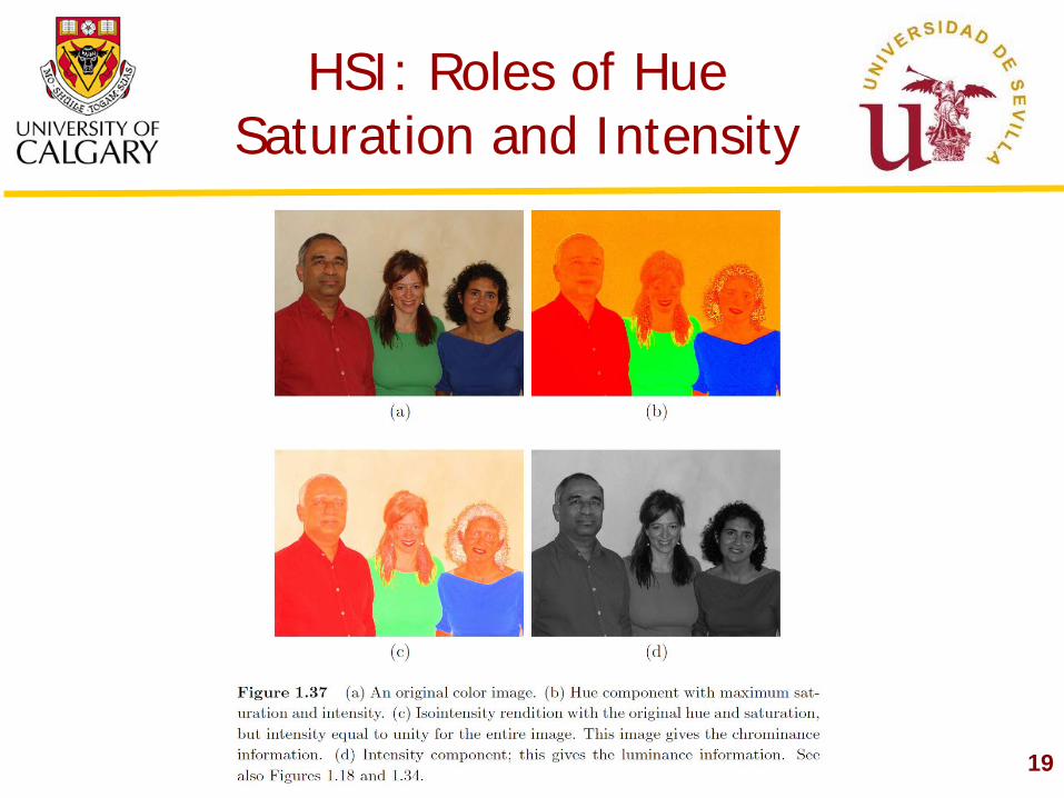
HSI: Roles of Hue Saturation and Intensity
20
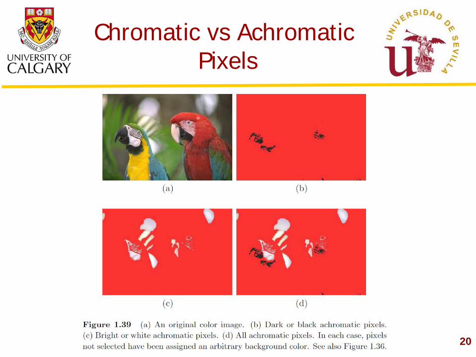
Chromatic vs Achromatic Pixels
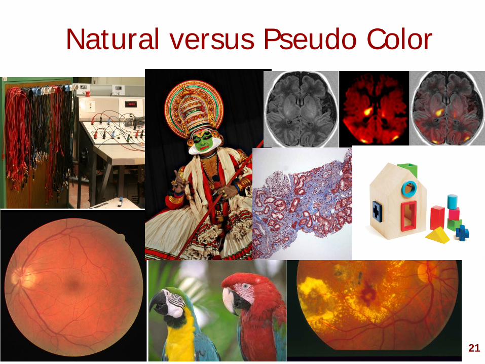
Natural versus Pseudo Color
21
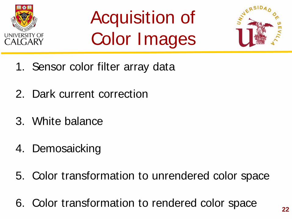
Acquisition of Color Images
22
1. Sensor color filter array data
2. Dark current correction
3. White balance
4. Demosaicking
5. Color transformation to unrendered color space
6. Color transformation to rendered color space
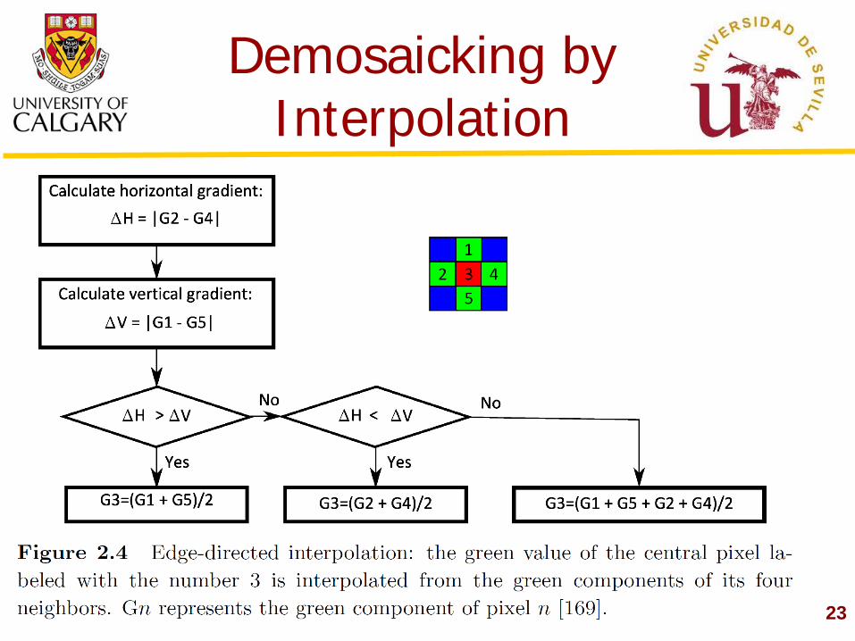
Demosaicking by Interpolation
23
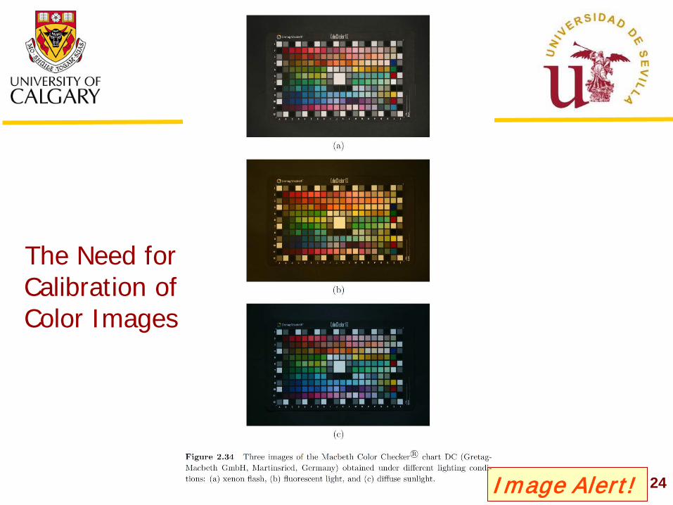
The Need for Calibration of Color Images
24 Image Alert!
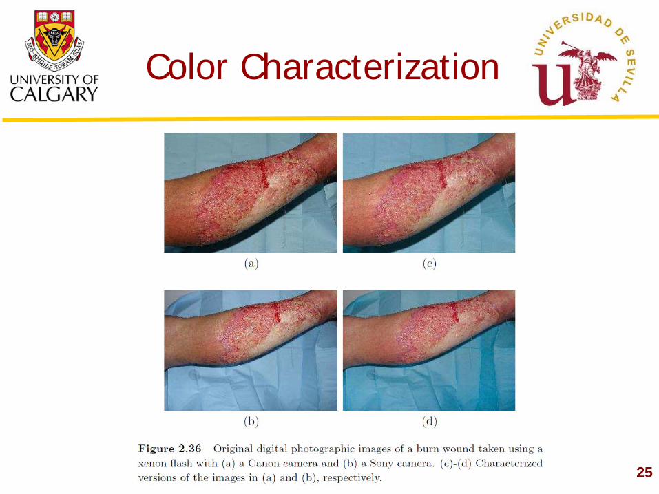
Color Characterization
25
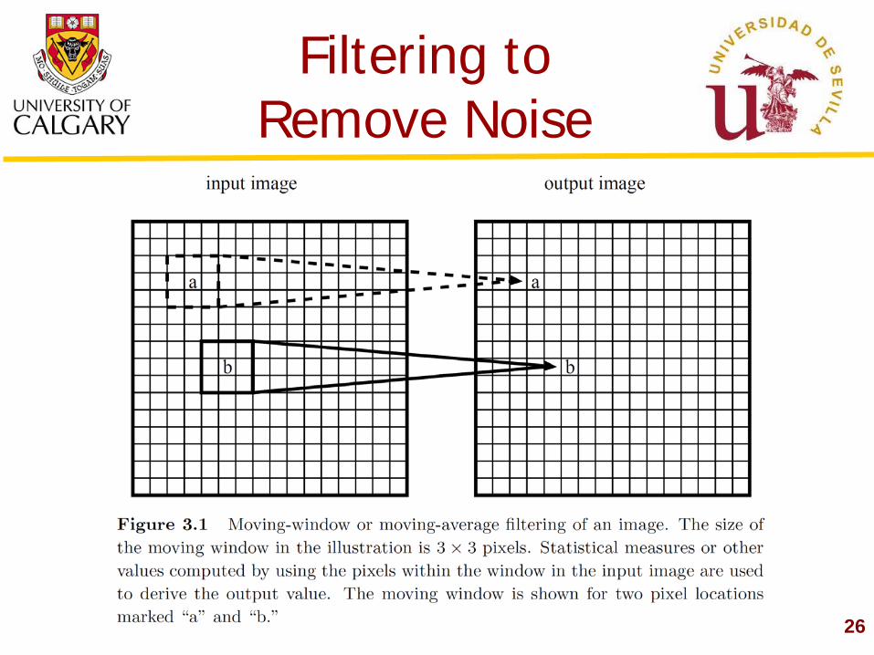
Filtering to Remove Noise
26
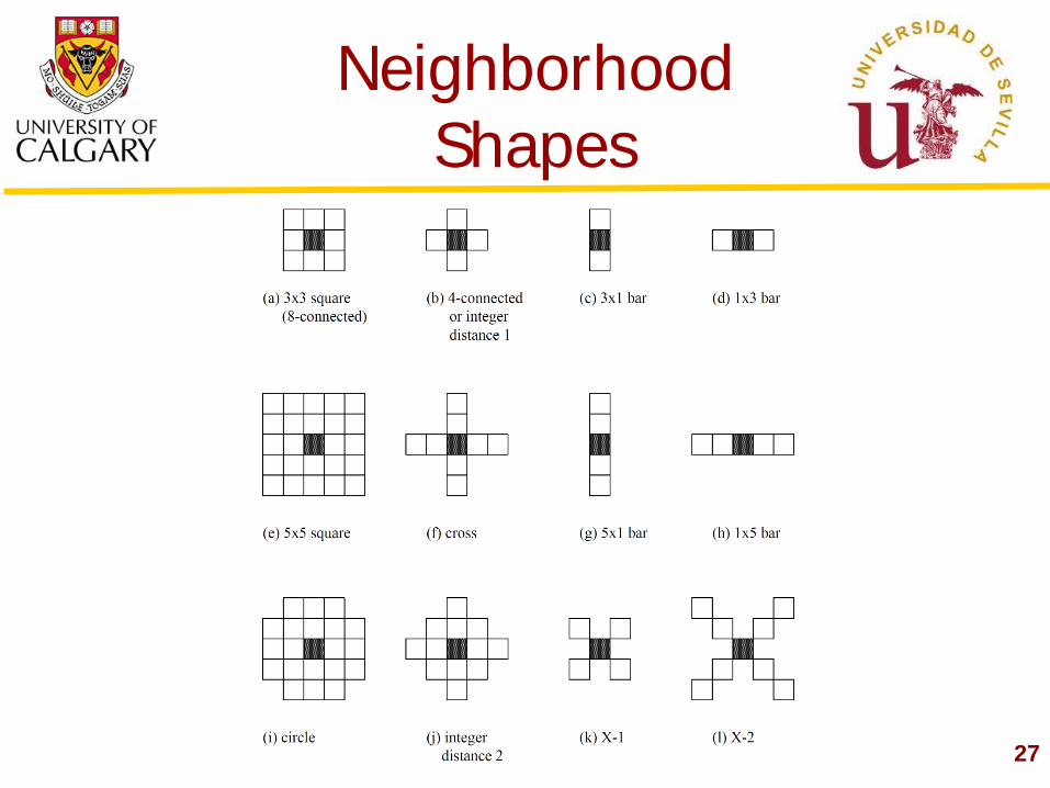
Neighborhood Shapes
27

Mean and Median Filtering
28
Mean = 90.67 Median = 87
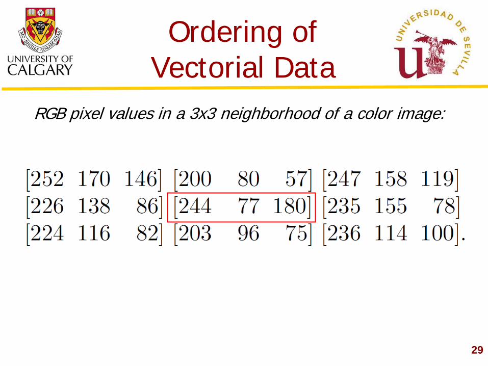
Ordering of Vectorial Data
29
RGB pixel values in a 3x3 neighborhood of a color image:
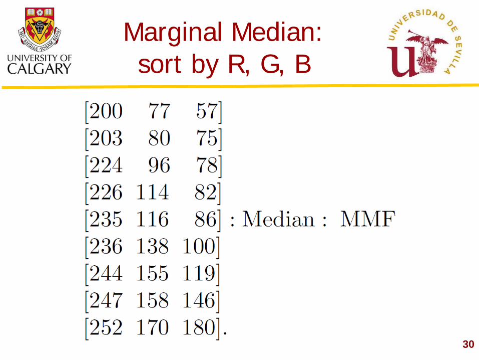
Marginal Median: sort by R, G, B
30
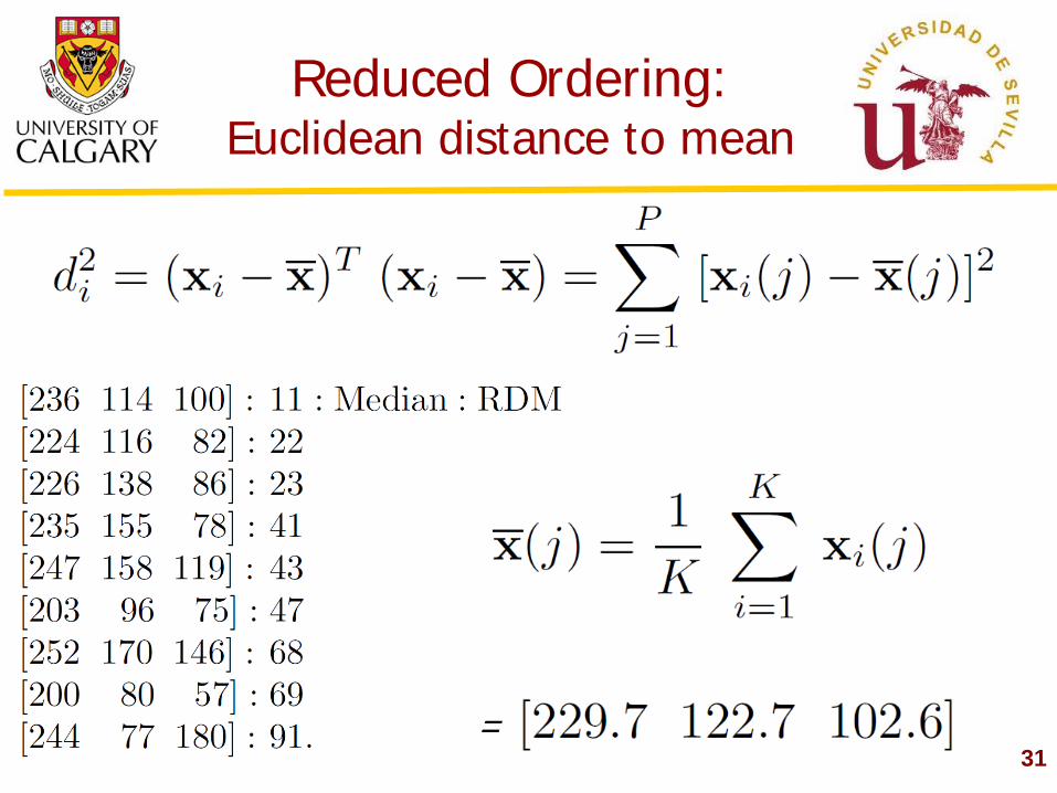
Reduced Ordering: Euclidean distance to mean
31 =
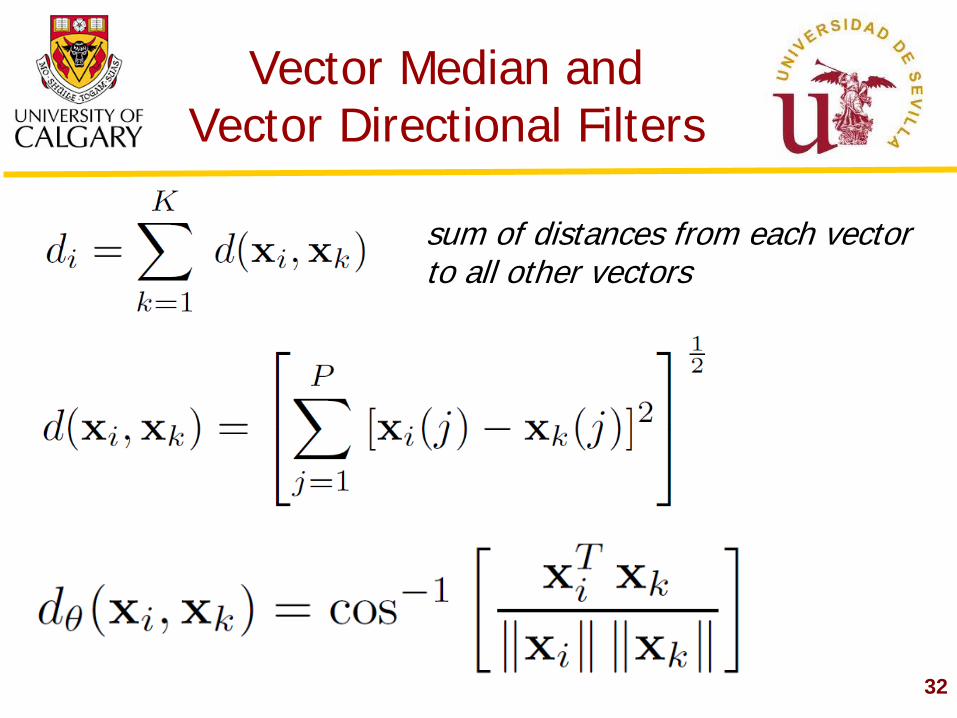
Vector Median and Vector Directional Filters
32
sum of distances from each vector to all other vectors
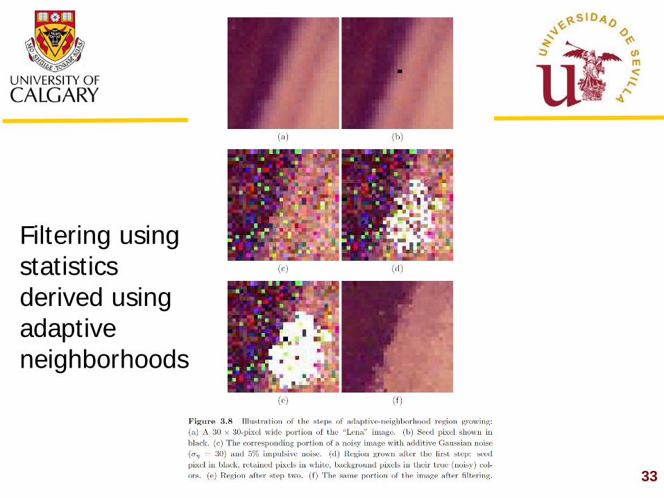
33
Filtering using statistics derived using adaptive neighborhoods
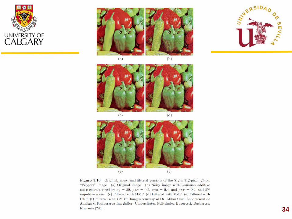
34
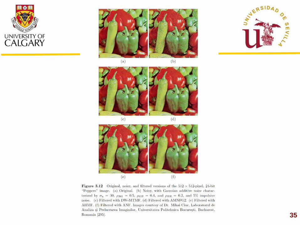
35
Enhancement of Color Images
36
Quite often, the enhancement required would be only in the intensity component: Gamma correction, Histogram equalization. Sometimes, saturation may need to be increased. Rarely would we want to alter the hue component. Processing the RGB components individually is not usually recommended.
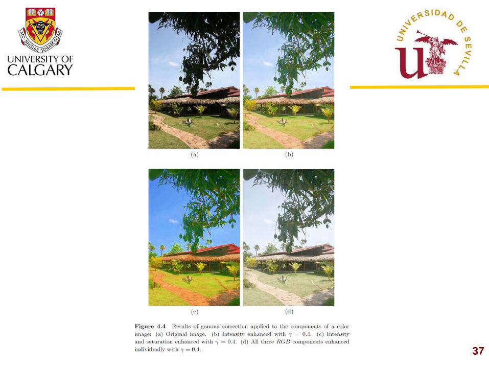
37
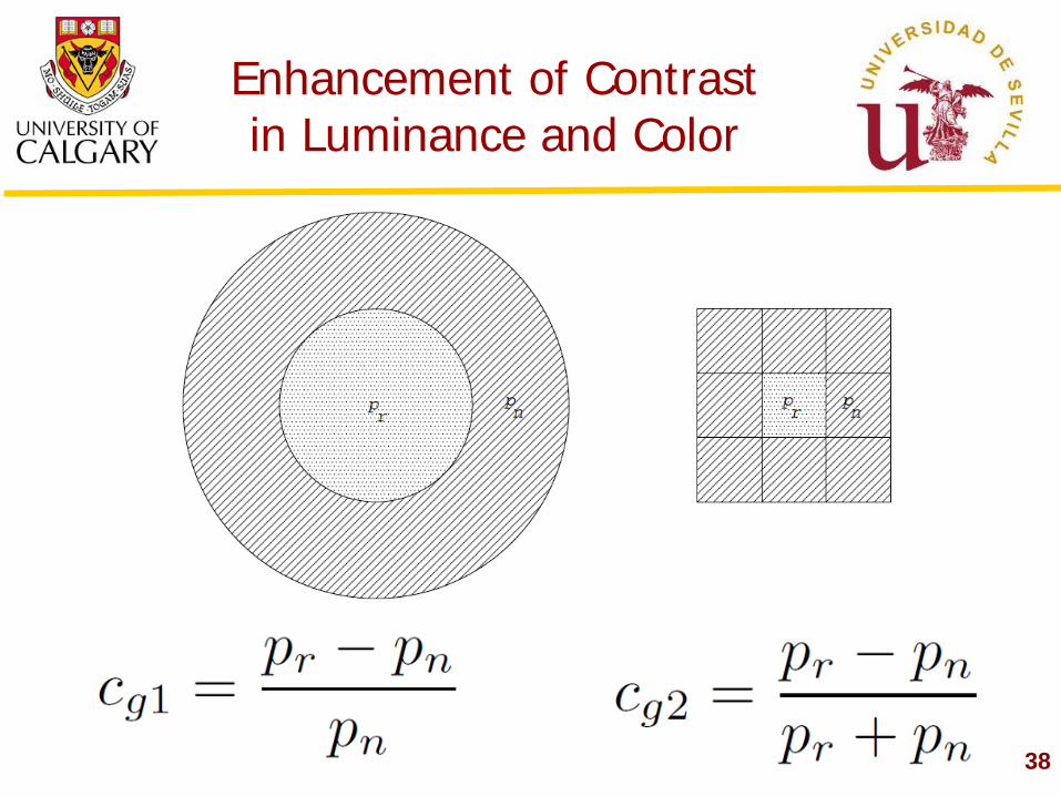
Enhancement of Contrast in Luminance and Color
38
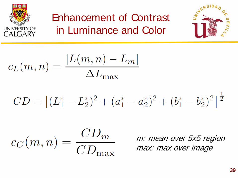
39
m: mean over 5x5 region max: max over image
Enhancement of Contrast in Luminance and Color
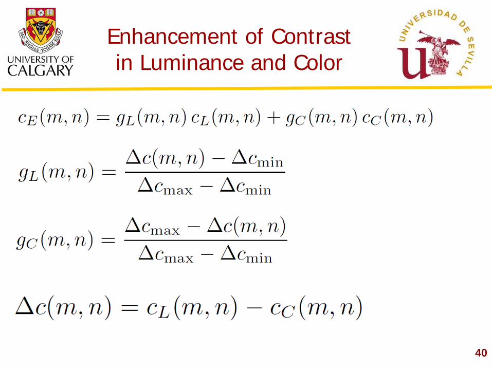
40
Enhancement of Contrast in Luminance and Color
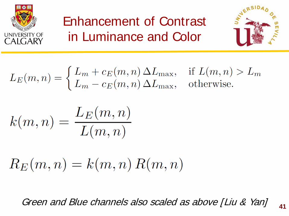
41 Green and Blue channels also scaled as above [Liu & Yan]
Enhancement of Contrast in Luminance and Color
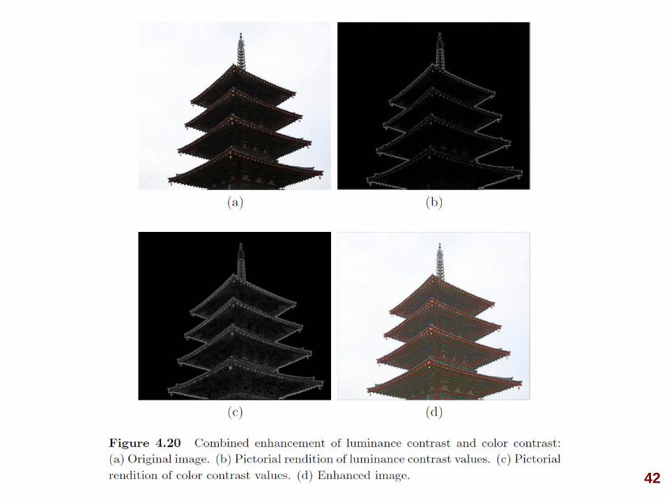
42
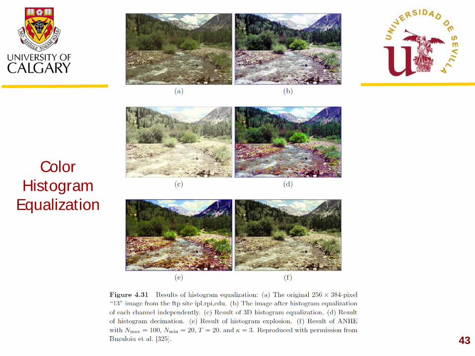
43
Color Histogram
Equalization
44
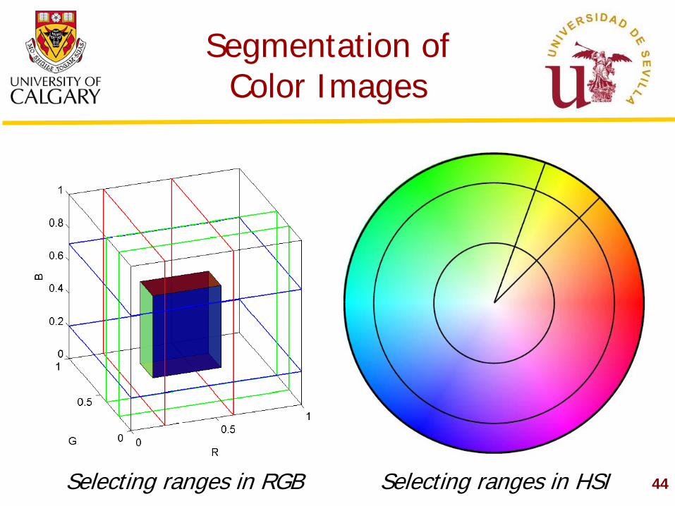
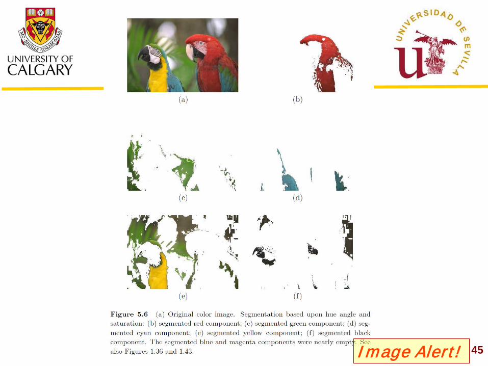
Segmentation of Color Images
Selecting ranges in RGB Selecting ranges in HSI
45 Image Alert!
46
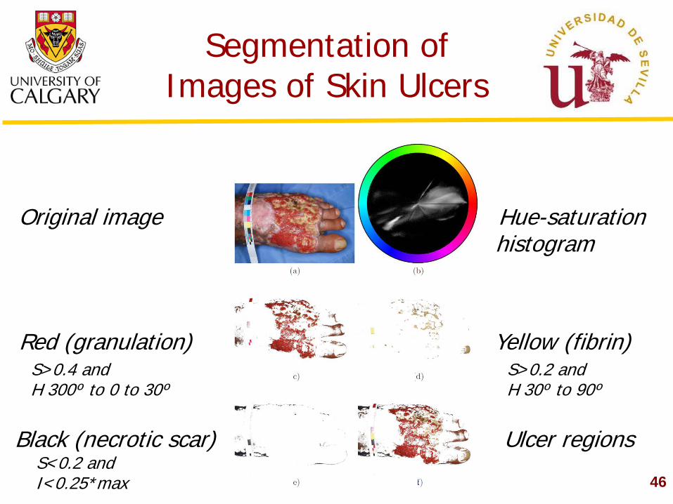
Segmentation of Images of Skin Ulcers
Black (necrotic scar) Ulcer regions
Red (granulation) Yellow (fibrin)
Original image Hue-saturation histogram
S>0.4 and H 300º to 0 to 30º
S>0.2 and H 30º to 90º
S<0.2 and I<0.25*max
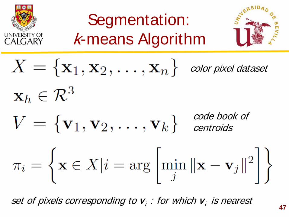
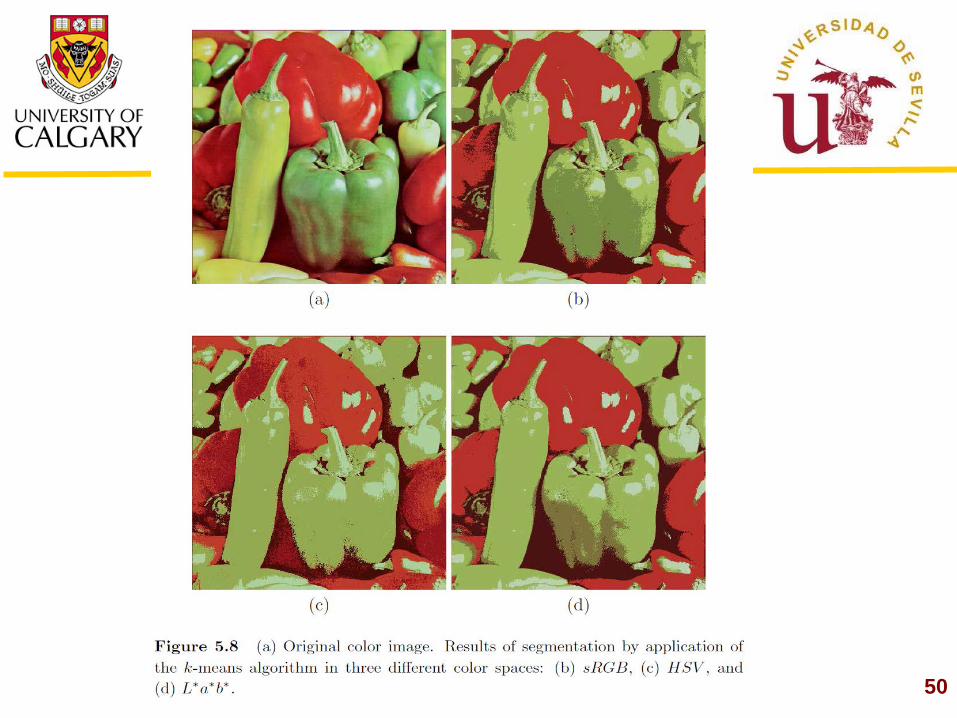
Segmentation: k-means Algorithm
47
color pixel dataset
code book of centroids
set of pixels corresponding to vi : for which vi is nearest
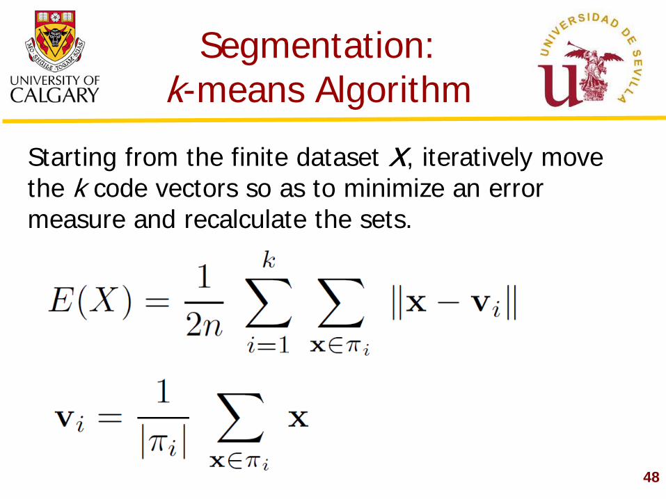
Segmentation: k-means Algorithm
48
Starting from the finite dataset X, iteratively move the k code vectors so as to minimize an error measure and recalculate the sets.
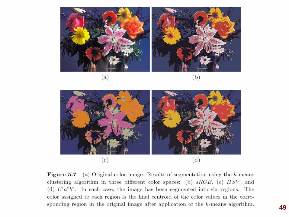
49
50
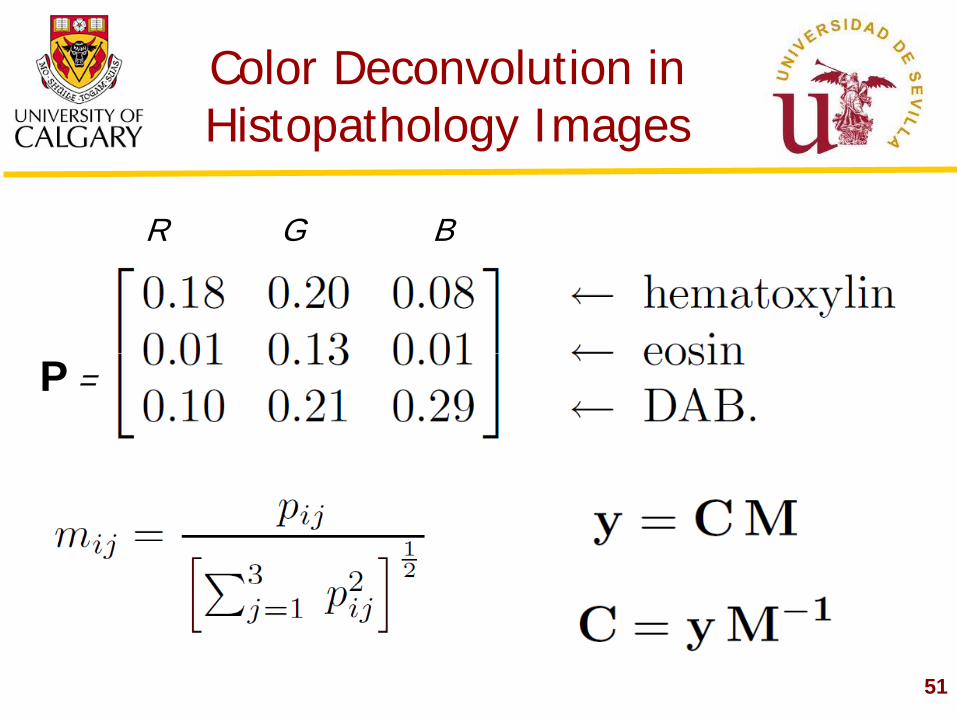
Color Deconvolution in Histopathology Images
51
R G B
P =
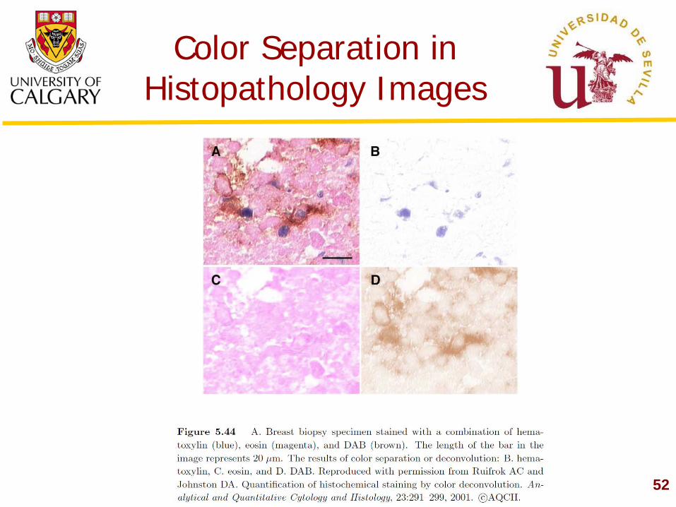
Color Separation in Histopathology Images
52
Additional Topics
53
Edge detection in color Region growing in color Morphological image processing in color Hyperspectral image processing Analysis of texture in color Coding and data compression of multispectral data Analysis of burn wounds Analysis of skin ulcers Teledermatology Telepathology Aerial photogrammetry...
Thank You!
Please see the book for details, references, and credits


![IJECT V . 7, I 1, J - M 2016 Proposed Algorithm for ... · [1] “Lesson 17: Heart Sound”, Biopac Student Lab 3.7.6. [2] Lehner RJ, Rangayyan RM.,“A three-channel microcomputer](https://img.pdfslide.net/doc/110x75/5e30b9f46b4afa240e4cbe39/iject-v-7-i-1-j-m-2016-proposed-algorithm-for-1-aoelesson-17-heart.jpg)
![IEEE TRANSACTIONS ON PATTERN ANALYSIS AND …ds/Papers/BoCS08.pdfpattern used to probe the input image. ... was proposed by Gordon and Rangayyan [20] ... topological properties of](https://img.pdfslide.net/doc/110x75/5aa8cfe07f8b9a8b188c0588/ieee-transactions-on-pattern-analysis-and-dspapers-used-to-probe-the-input.jpg)