Embed Size (px)
Citation preview
Common problems in Gastroenterology for Internal medicine
Julajak Limsrivilai
Siriraj Hospital
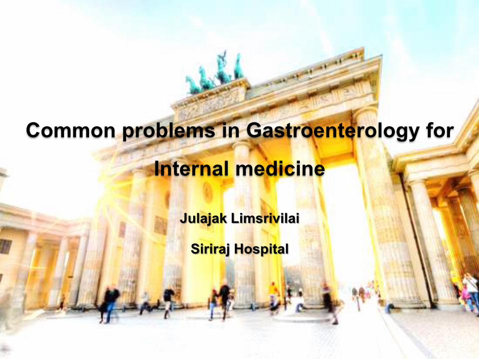
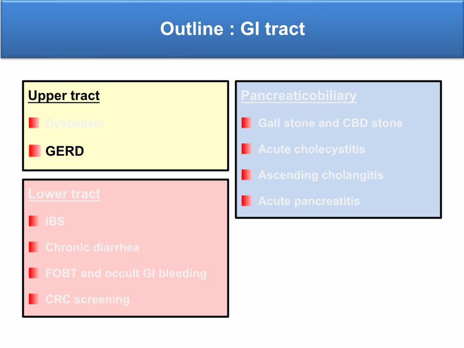
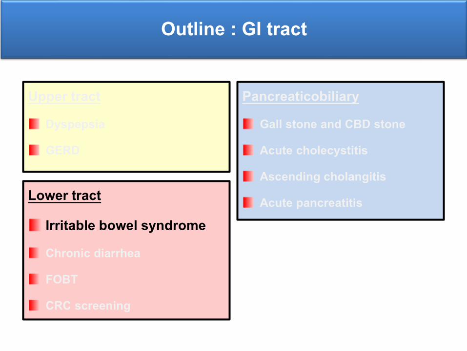
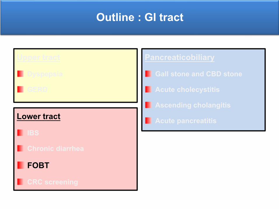
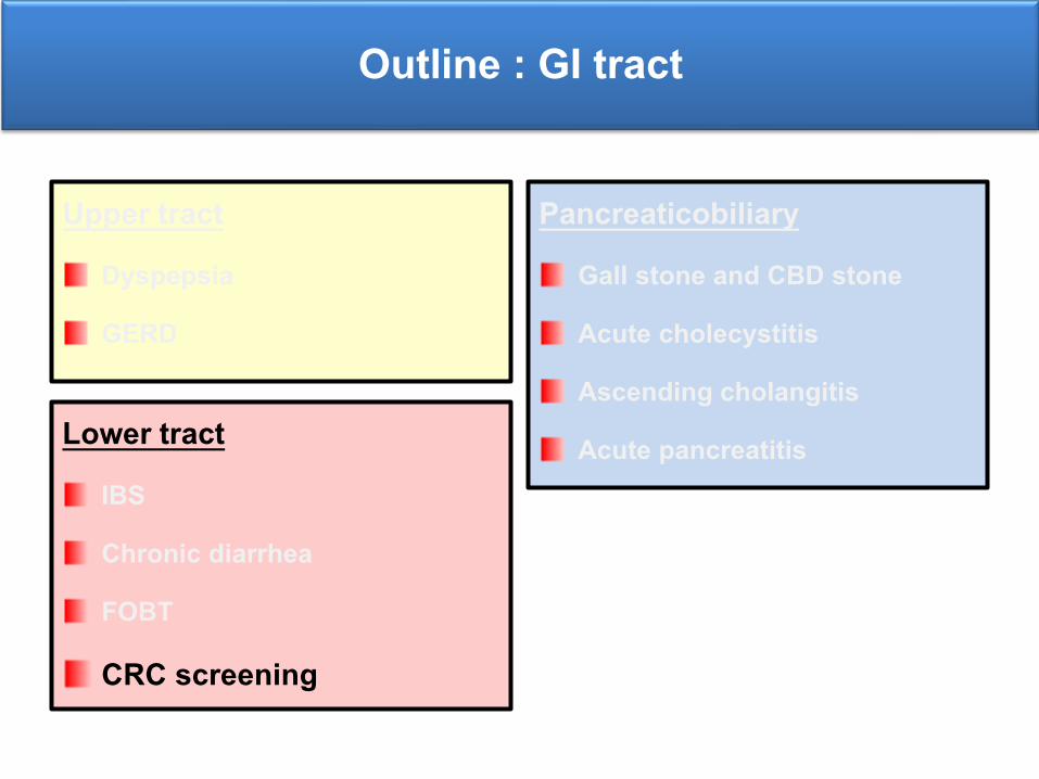
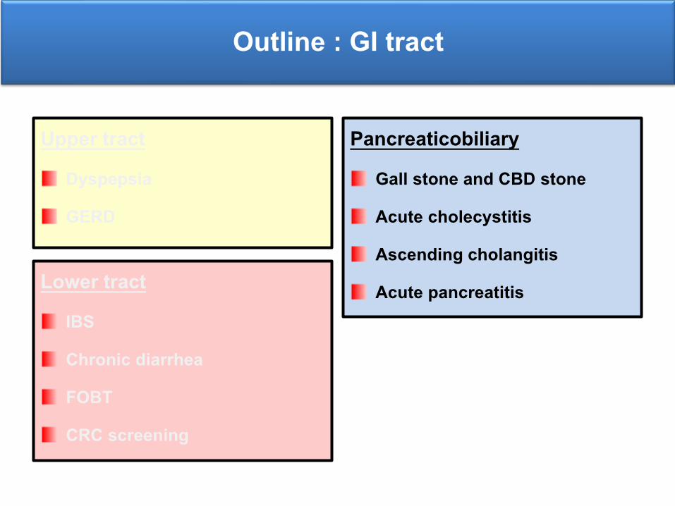
Outline : GI tract
Upper tract Dyspepsia
GERD
PancreaticobiliaryGall stone and CBD stone
Acute cholecystitis
Ascending cholangitis
Acute pancreatitis Lower tract IBS
Chronic diarrhea
FOBT
CRC screening
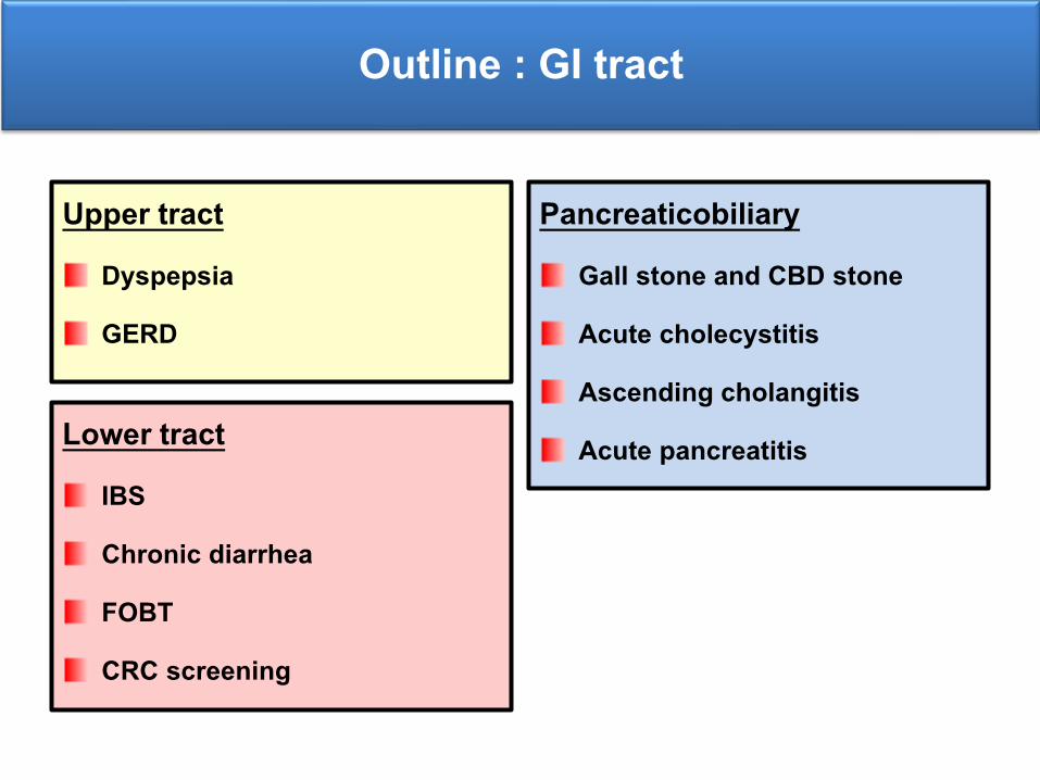
Outline : Liver disease
Chronic liver disease Viral hepatitis
Alcoholic hepatitis
NAFLD
Autoimmune liver disease
Metabolic liver disease
Cirrhosis Portal hypertension
Variceal bleeding
Ascites and complication
Hepatorenal syndrome
Hepatocellular carcinoma
Question 1
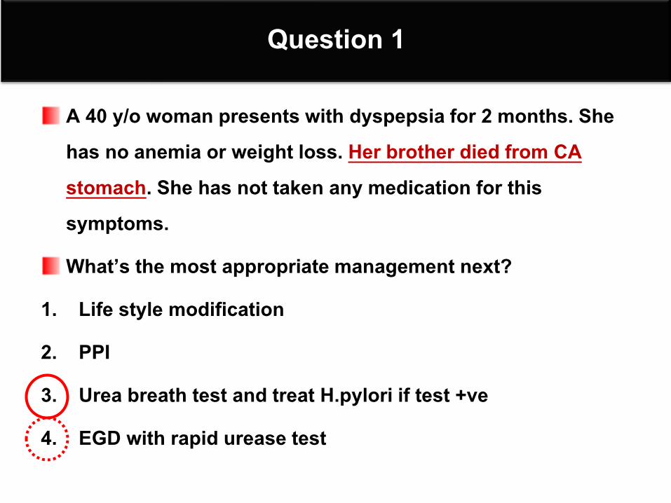
A 40 y/o woman presents with dyspepsia for 2 months. She has no anemia or weight loss. Her brother died from CA stomach. She has not taken any medications for this symptoms.
What’s the most appropriate management next?
1. Life style modification
2. PPI
3. Urea breath test and treat H.pylori if test +ve
4. EGD with rapid urease test
Dyspepsia
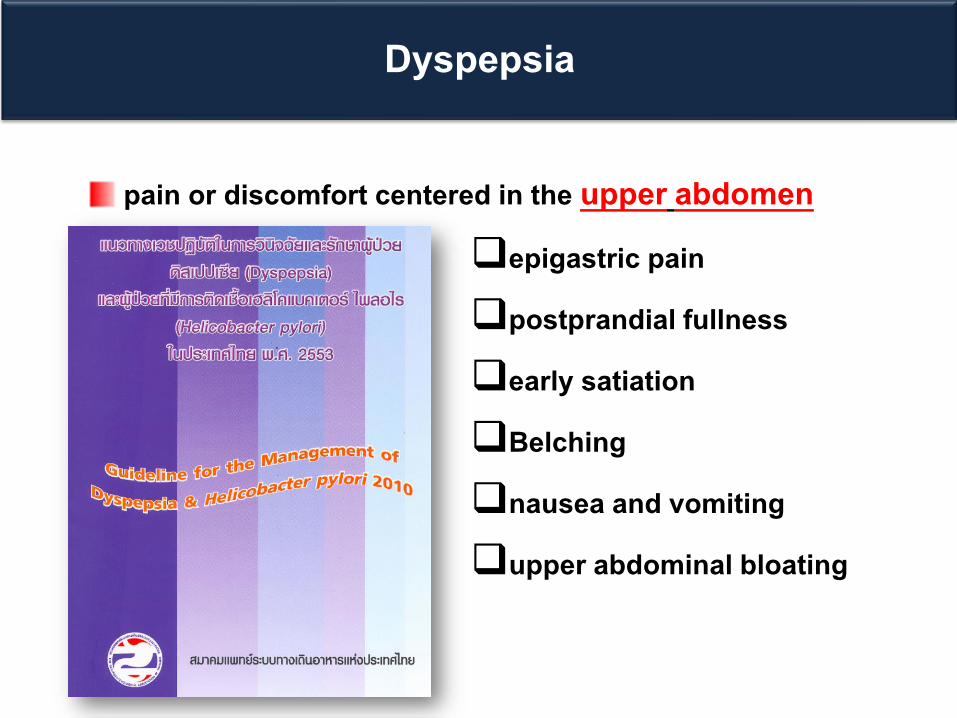
pain or discomfort centered in the upper abdomenepigastric pain
postprandial fullness
early satiation
Belching
nausea and vomiting
upper abdominal bloating
Dyspepsia
Etiology
Guideline
Etiology
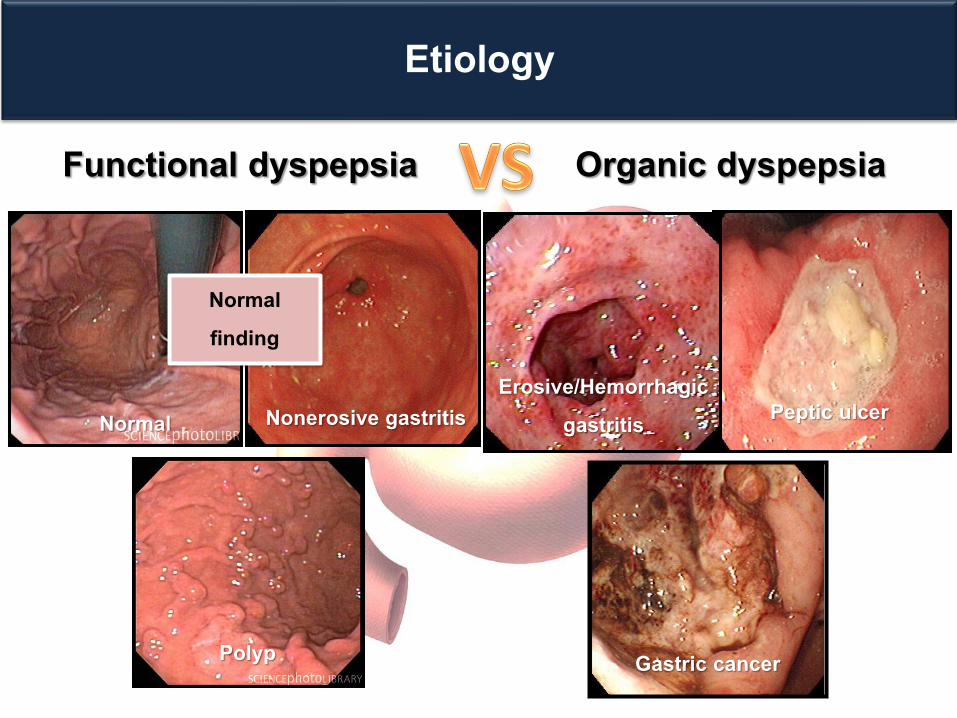
Functional dyspepsia Organic dyspepsia
Normal Nonerosive gastritis
Polyp
Erosive/Hemorrhagic gastritis Peptic ulcer
Gastric cancer
Normal finding
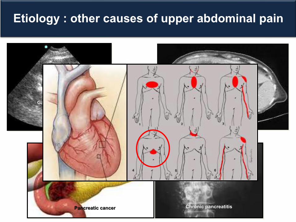
Etiology : other causes of upper abdominal pain
Chronic pancreatitisPancreatic cancer
Gall stone
Liver tumor
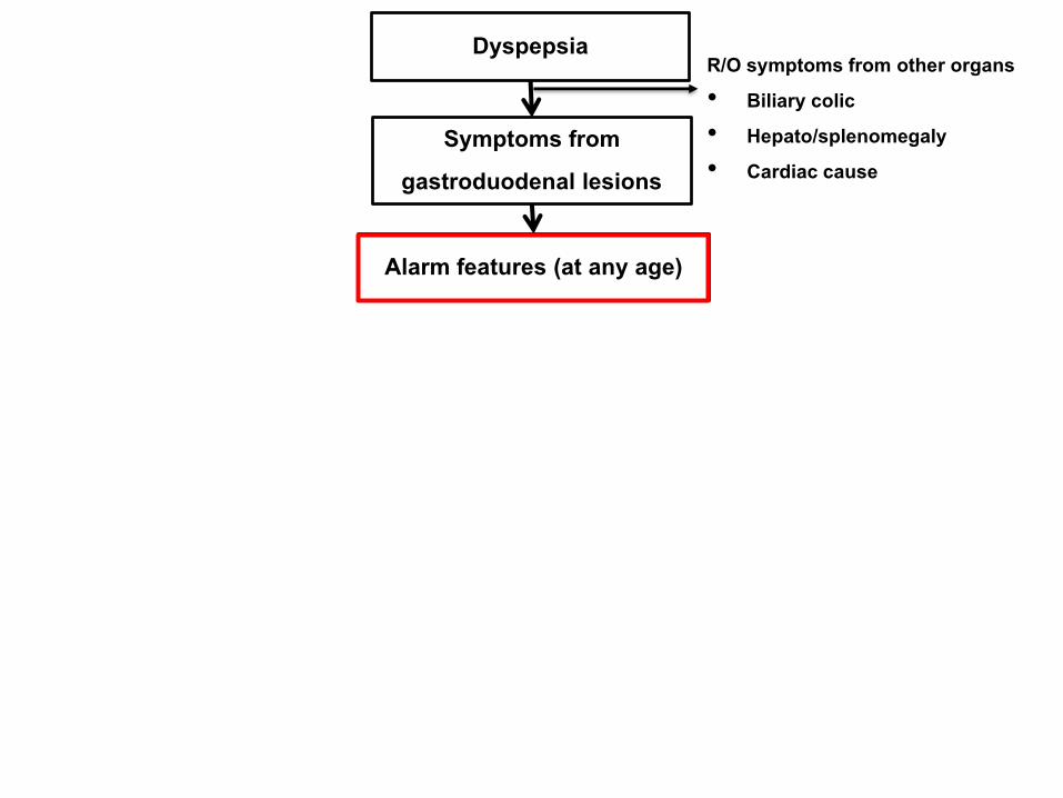
Dyspepsia
Alarm features (at any age)
R/O symptoms from other organs• Biliary colic • Hepato/splenomegaly • Cardiac cause
Symptoms from gastroduodenal lesions
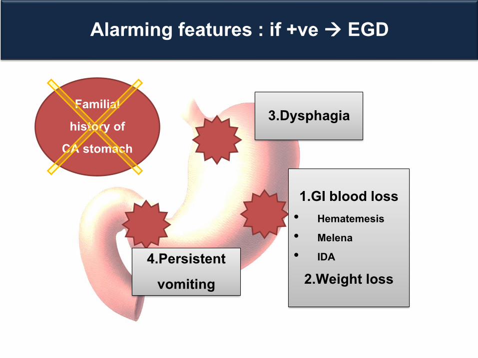
Alarming features : if +ve EGD
3.Dysphagia
1.GI blood loss• Hematemesis • Melena • IDA
2.Weight loss 4.Persistent
vomiting
Familial history of
CA stomach
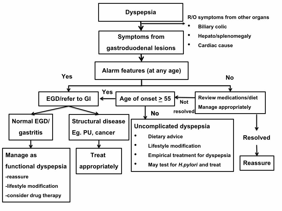
Dyspepsia
Alarm features (at any age)
R/O symptoms from other organs• Biliary colic • Hepato/splenomegaly • Cardiac cause
EGD/refer to GI
Structural diseaseEg. PU, cancer
Treat appropriately
Normal EGD/gastritis
Manage as functional dyspepsia-reassure -lifestyle modification -consider drug therapy
Symptoms from gastroduodenal lesions
Yes No
Review medications/diet Manage appropriately
Reassure
Resolved
Age of onset > 55 Not resolved
Yes
Uncomplicated dyspepsia• Dietary advice• Lifestyle modification• Empirical treatment for dyspepsia• May test for H.pylori and treat
No
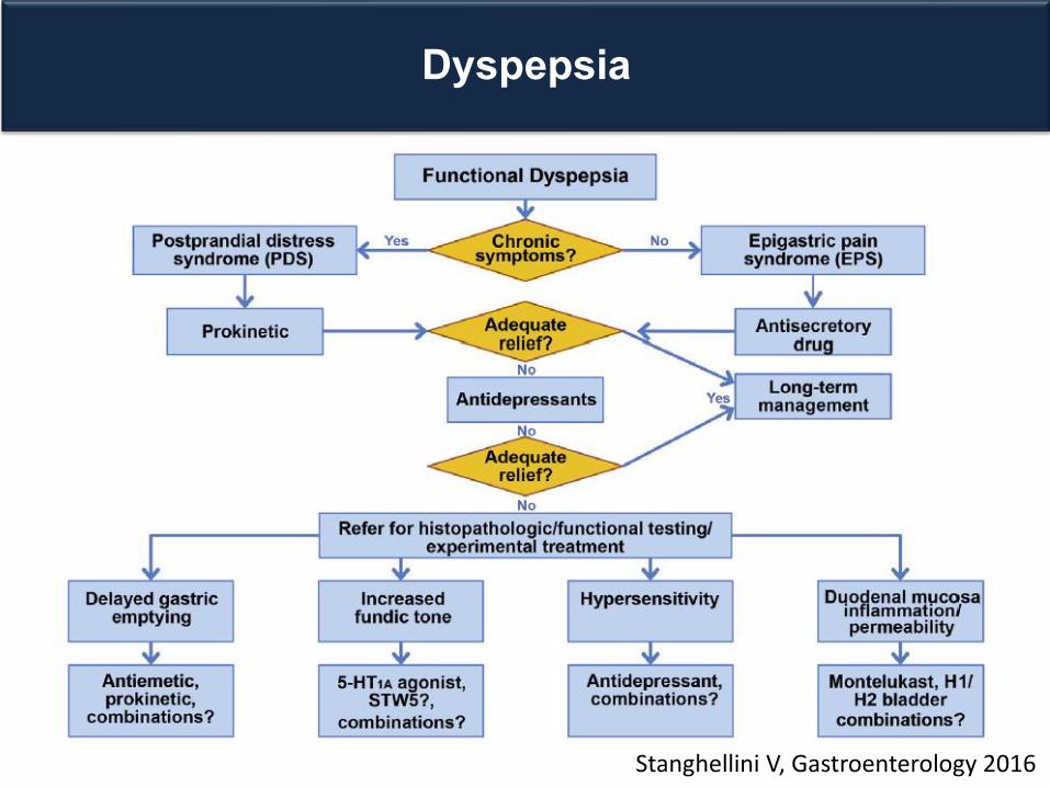
Stanghellini V, Gastroenterology 2016
Dyspepsia
Who should be treated for H. pylori?
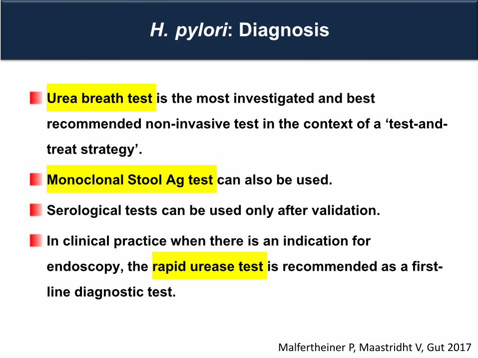
H. pylori: Diagnosis
Urea breath test is the most investigated and best recommended non-invasive test in the context of a ‘test-and-treat strategy’.
Monoclonal Stool Ag test can also be used.
Serological tests can be used only after validation.
In clinical practice when there is an indication for endoscopy, the rapid urease test is recommended as a first-line diagnostic test.
Malfertheiner P, Maastridht V, Gut 2017
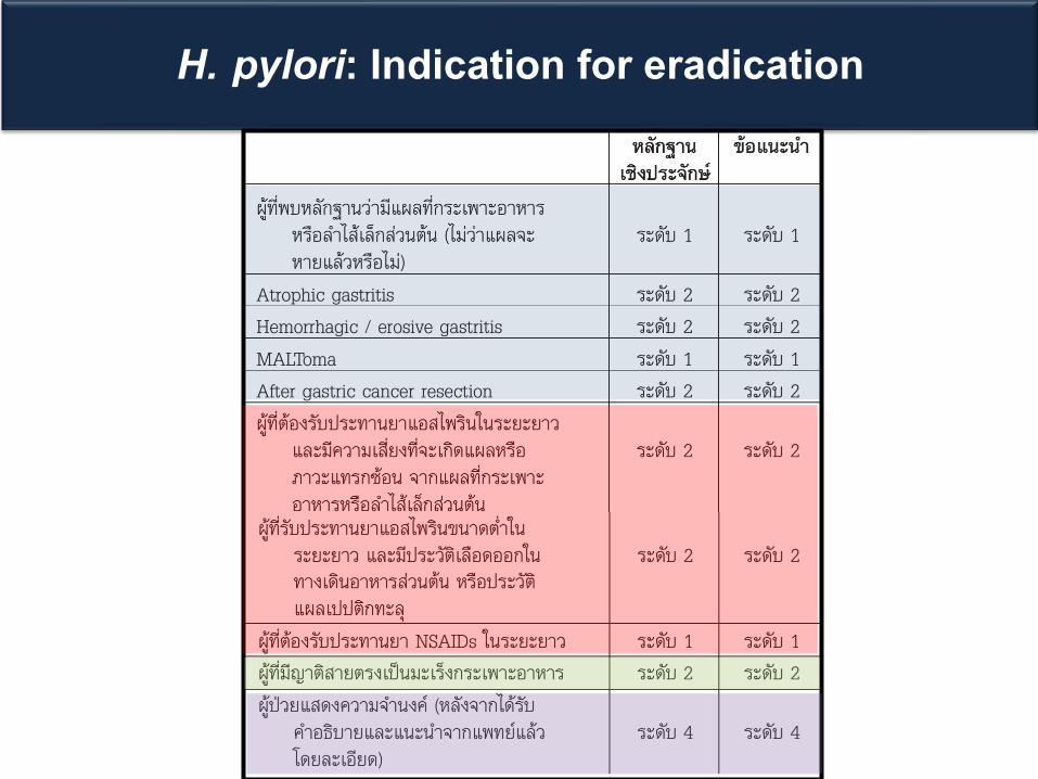
H. pylori: Indication for eradication
Question 1
A 40 y/o woman presents with dyspepsia for 2 months. She has no anemia or weight loss. Her brother died from CA stomach. She has not taken any medication for this symptoms.
What’s the most appropriate management next?
1. Life style modification
2. PPI
3. Urea breath test and treat H.pylori if test +ve
4. EGD with rapid urease test
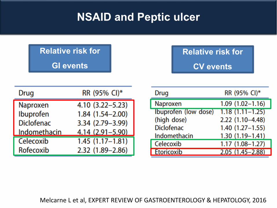
NSAID and Peptic ulcer
Melcarne L et al, EXPERT REVIEW OF GASTROENTEROLOGY & HEPATOLOGY, 2016
Relative risk for GI events
Relative risk for CV events
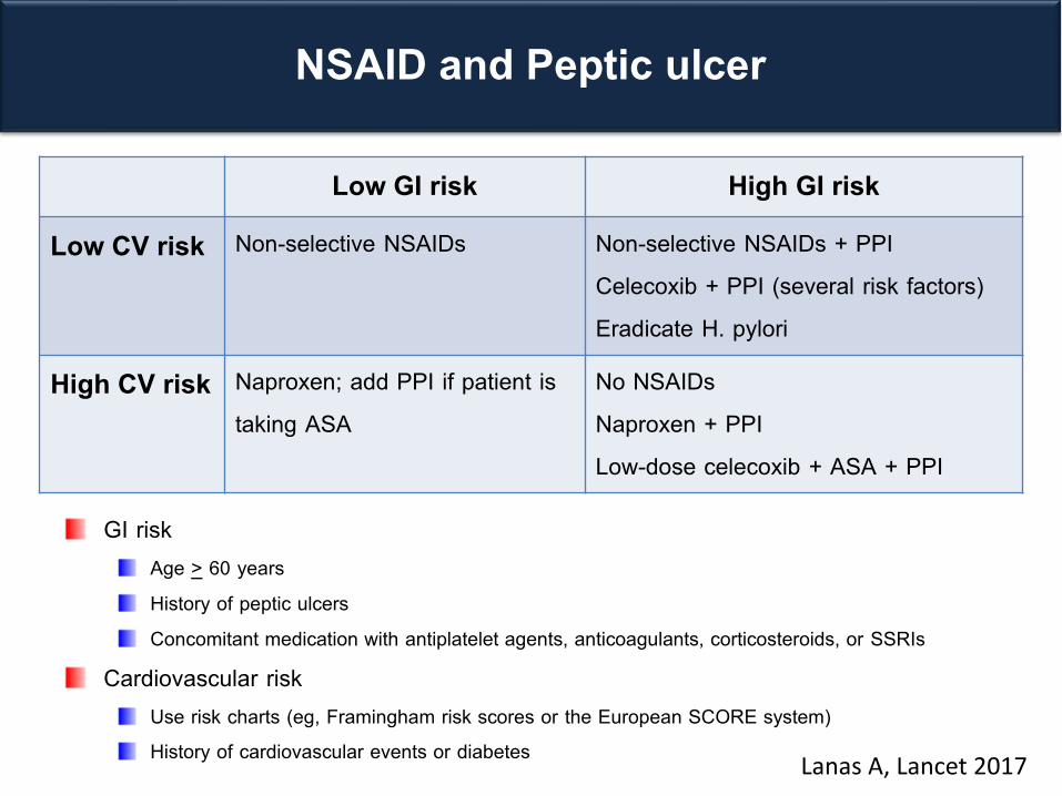
NSAID and Peptic ulcer
Low GI risk High GI risk
Low CV risk Non-selective NSAIDs Non-selective NSAIDs + PPICelecoxib + PPI (several risk factors) Eradicate H. pylori
High CV risk Naproxen; add PPI if patient is taking ASA
No NSAIDsNaproxen + PPI Low-dose celecoxib + ASA + PPI
Lanas A, Lancet 2017
GI risk Age > 60 years History of peptic ulcers Concomitant medication with antiplatelet agents, anticoagulants, corticosteroids, or SSRIs
Cardiovascular riskUse risk charts (eg, Framingham risk scores or the European SCORE system) History of cardiovascular events or diabetes
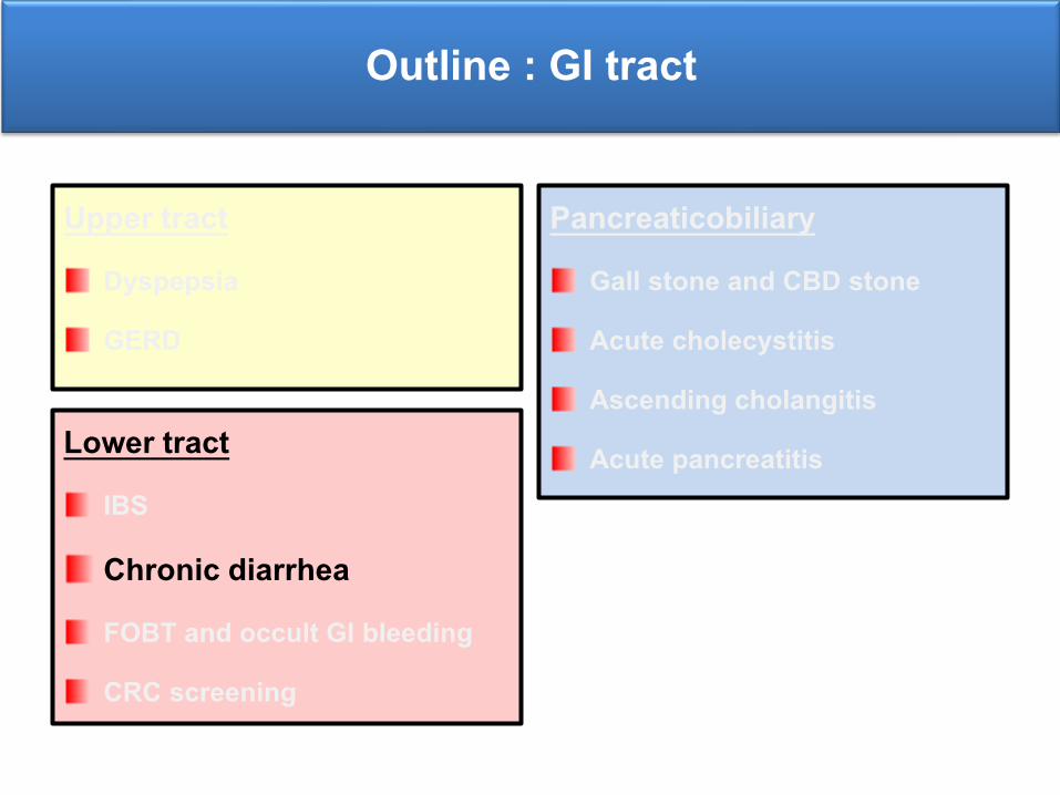
Outline : GI tract
Upper tract Dyspepsia
GERD
PancreaticobiliaryGall stone and CBD stone
Acute cholecystitis
Ascending cholangitis
Acute pancreatitis Lower tract IBS
Chronic diarrhea
FOBT and occult GI bleeding
CRC screening
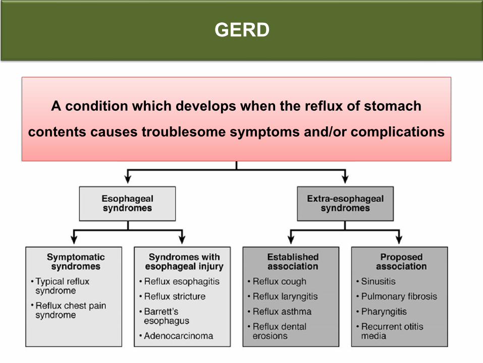
GERD
A condition which develops when the reflux of stomach contents causes troublesome symptoms and/or complications
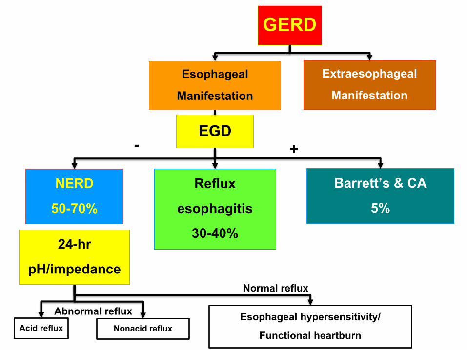
GERD
EsophagealManifestation
EGD
Reflux esophagitis
30-40%
NERD50-70%
Acid reflux
ExtraesophagealManifestation
Barrett’s & CA5%
Nonacid reflux
- +
Abnormal reflux
24-hr pH/impedance
Normal reflux
Esophageal hypersensitivity/ Functional heartburn
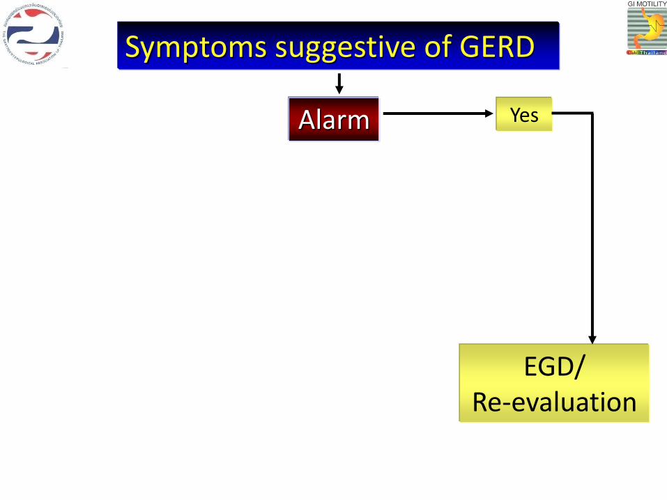
Symptoms suggestive of GERD
Alarm Yes
EGD/ Re-evaluation
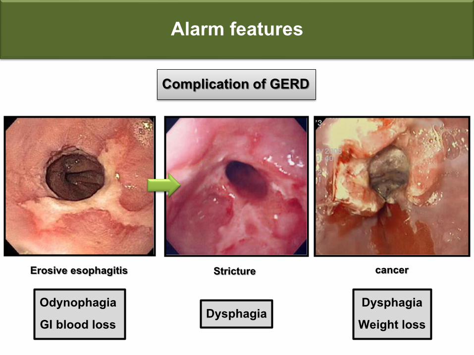
Alarm features
Complication of GERD
Erosive esophagitis Stricture cancer
Odynophagia GI blood loss
DysphagiaDysphagiaWeight loss
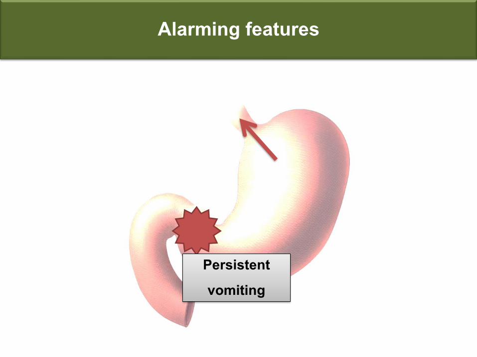
Alarming features
Persistent vomiting
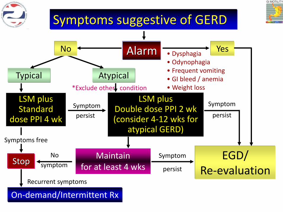
Symptoms suggestive of GERD
Alarm Yes
Typical Atypical
LSM plusStandard
dose PPI 4 wk
LSM plusDouble dose PPI 2 wk(consider 4-12 wks for
atypical GERD)
StopMaintain
for at least 4 wks
On-demand/Intermittent Rx
EGD/ Re-evaluation
No
symptom
persist
Symptom
persist
Symptom
persist
Symptom
No
Symptoms free
Recurrent symptoms
• Dysphagia• Odynophagia• Frequent vomiting• GI bleed / anemia• Weight loss*Exclude other condition
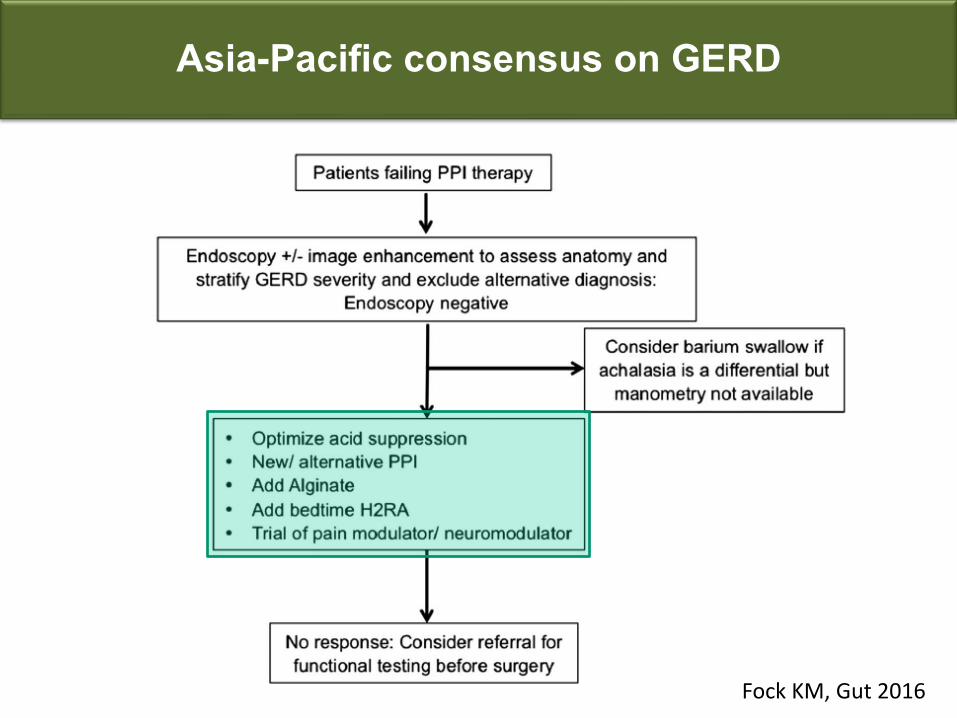
Asia-Pacific consensus on GERD
Fock KM, Gut 2016
Outline : GI tract
Upper tract Dyspepsia
GERD
PancreaticobiliaryGall stone and CBD stone
Acute cholecystitis
Ascending cholangitis
Acute pancreatitis Lower tract
Irritable bowel syndrome Chronic diarrhea
FOBT
CRC screening
Irritable bowel syndrome (IBS)
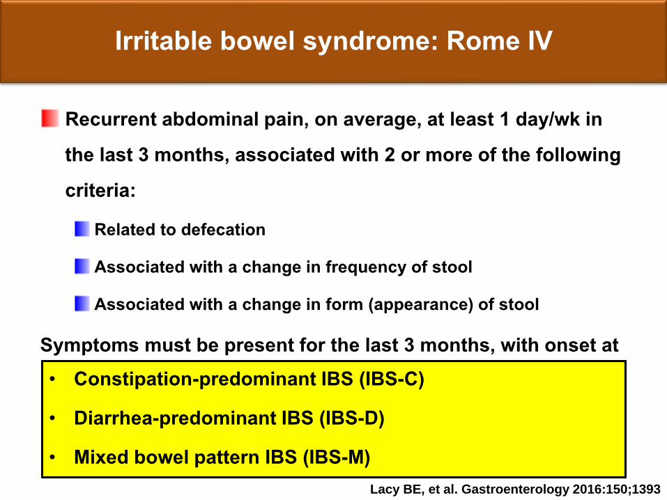
Irritable bowel syndrome: Rome IV
Recurrent abdominal pain, on average, at least 1 day/wk in the last 3 months, associated with 2 or more of the following criteria:
Related to defecation
Associated with a change in frequency of stool
Associated with a change in form (appearance) of stool
Symptoms must be present for the last 3 months, with onset at least 6 months before diagnosis • Constipation-predominant IBS (IBS-C)• Diarrhea-predominant IBS (IBS-D)• Mixed bowel pattern IBS (IBS-M)
Lacy BE, et al. Gastroenterology 2016:150;1393
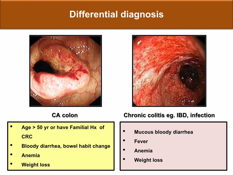
Differential diagnosis
CA colon Chronic colitis eg. IBD, infection• Age > 50 yr or have Familial Hx of
CRC • Bloody diarrhea, bowel habit change • Anemia • Weight loss
• Mucous bloody diarrhea • Fever • Anemia • Weight loss
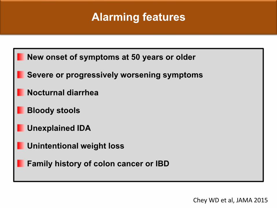
Alarming features
New onset of symptoms at 50 years or older
Severe or progressively worsening symptoms
Nocturnal diarrhea
Bloody stools
Unexplained IDA
Unintentional weight loss
Family history of colon cancer or IBD
Chey WD et al, JAMA 2015
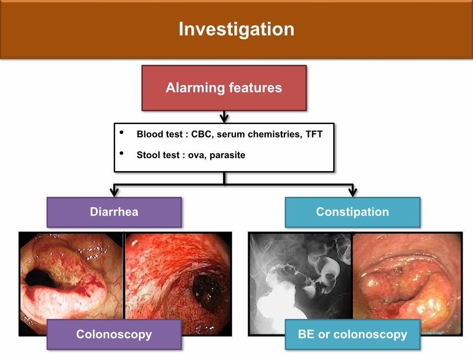
Investigation
Alarming features
• Blood test : CBC, serum chemistries, TFT
• Stool test : ova, parasite
Diarrhea Constipation
Colonoscopy BE or colonoscopy
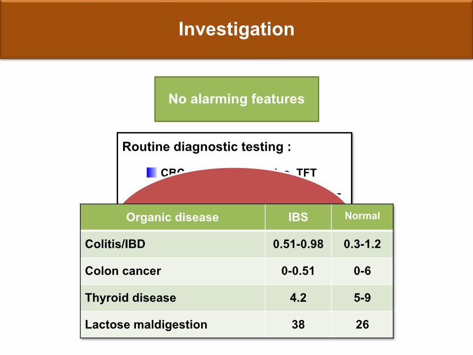
Investigation
Routine diagnostic testing : CBC, serum chemistries, TFT
Stool for ova and parasites, FOBT
Abdominal imaging
No alarming features
May not be required Organic disease IBS Normal
Colitis/IBD 0.51-0.98 0.3-1.2
Colon cancer 0-0.51 0-6
Thyroid disease 4.2 5-9
Lactose maldigestion 38 26
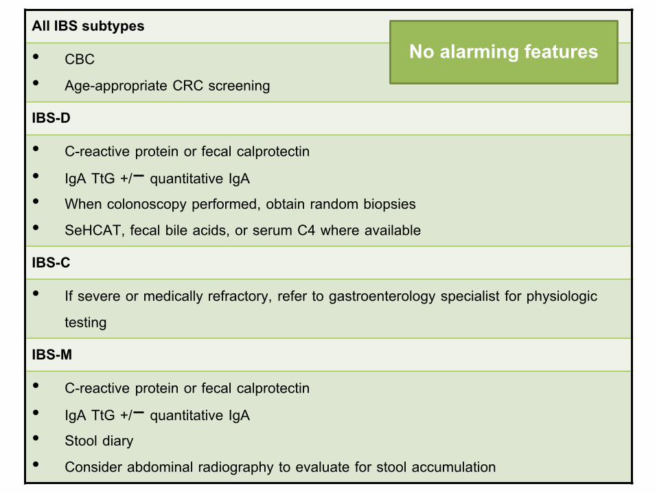
All IBS subtypes
• CBC • Age-appropriate CRC screening
IBS-D
• C-reactive protein or fecal calprotectin• IgA TtG +/− quantitative IgA• When colonoscopy performed, obtain random biopsies• SeHCAT, fecal bile acids, or serum C4 where available
IBS-C
• If severe or medically refractory, refer to gastroenterology specialist for physiologic testing
IBS-M
• C-reactive protein or fecal calprotectin• IgA TtG +/− quantitative IgA• Stool diary• Consider abdominal radiography to evaluate for stool accumulation
No alarming features
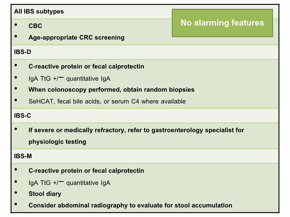
All IBS subtypes
• CBC• Age-appropriate CRC screening
IBS-D
• C-reactive protein or fecal calprotectin• IgA TtG +/− quantitative IgA• When colonoscopy performed, obtain random biopsies• SeHCAT, fecal bile acids, or serum C4 where available
IBS-C
• If severe or medically refractory, refer to gastroenterology specialist for physiologic testing
IBS-M
• C-reactive protein or fecal calprotectin• IgA TtG +/− quantitative IgA• Stool diary• Consider abdominal radiography to evaluate for stool accumulation
No alarming features
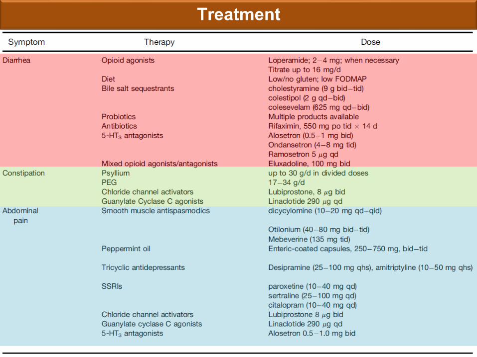
Treatment
Outline : GI tract
Upper tract Dyspepsia
GERD
PancreaticobiliaryGall stone and CBD stone
Acute cholecystitis
Ascending cholangitis
Acute pancreatitis Lower tract IBS
Chronic diarrhea FOBT and occult GI bleeding
CRC screening
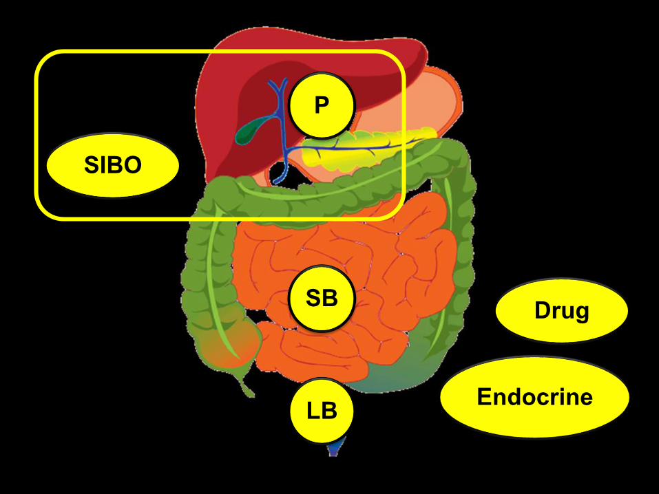
Chronic diarrhea
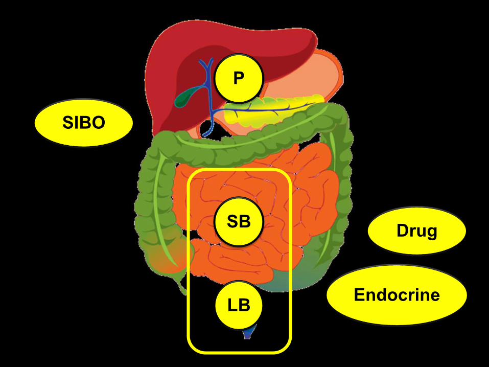
P
LB
SB
SIBO
Endocrine
Drug
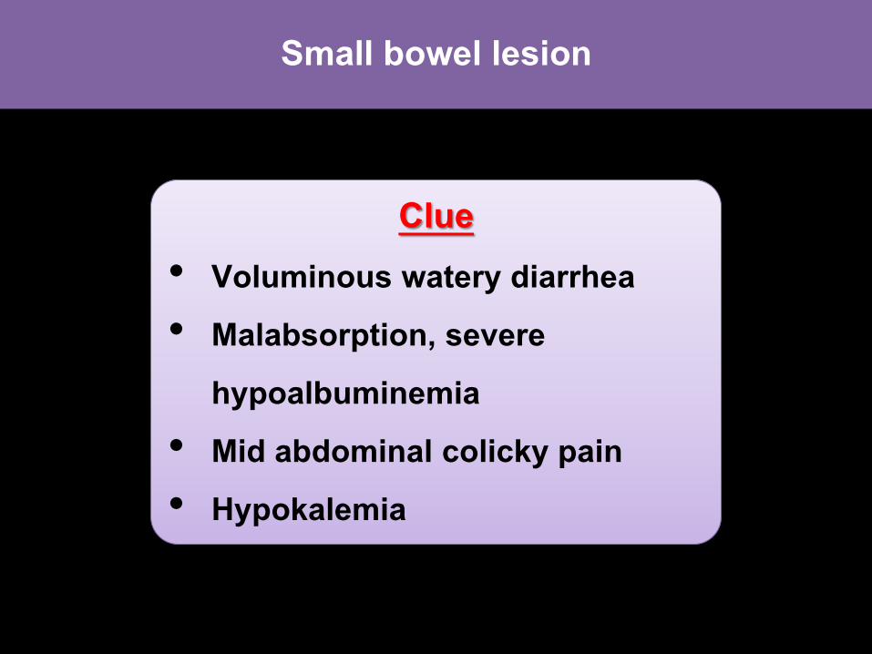
Small bowel lesion
Clue• Voluminous watery diarrhea • Malabsorption, severe
hypoalbuminemia• Mid abdominal colicky pain • Hypokalemia
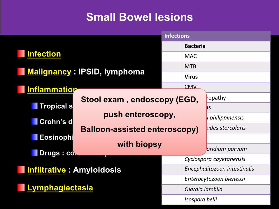
Small Bowel lesions
Infection
Malignancy : IPSID, lymphoma
InflammationTropical sprue
Crohn’s disease
Eosinophilic enteritis
Drugs : colchicine, ponstand
Infiltrative : Amyloidosis
Lymphagiectasia
Infections
Bacteria
MAC
MTB
Virus
CMV
HIV enteropathy
Helminths
Capillaria philippinensis
Strongyloides stercolaris
Protozoa
Cryptosporidium parvum
Cyclospora cayetanensis
Encephalitozoon intestinalis
Enterocytozoon bieneusi
Giardia lamblia
Isospora belli
Stool exam , endoscopy (EGD, push enteroscopy,
Balloon-assisted enteroscopy) with biopsy
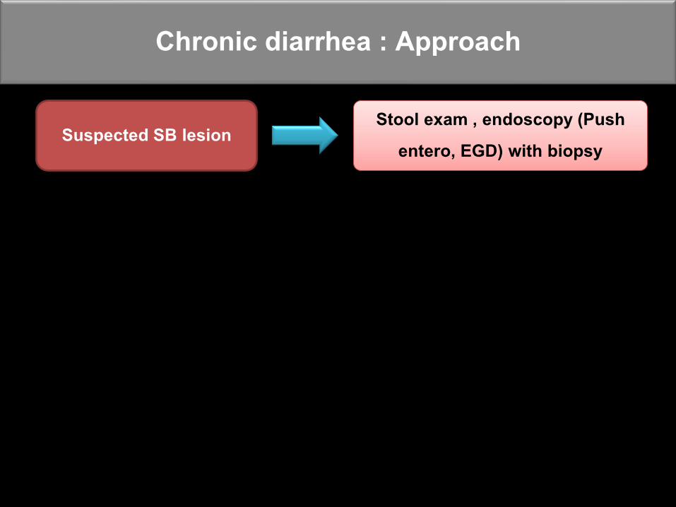
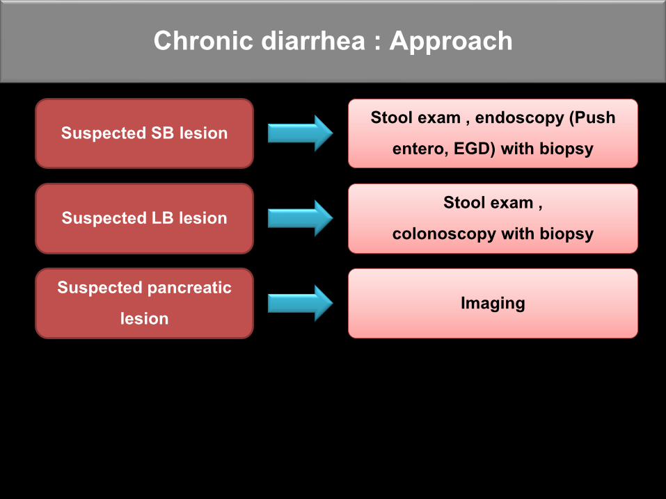
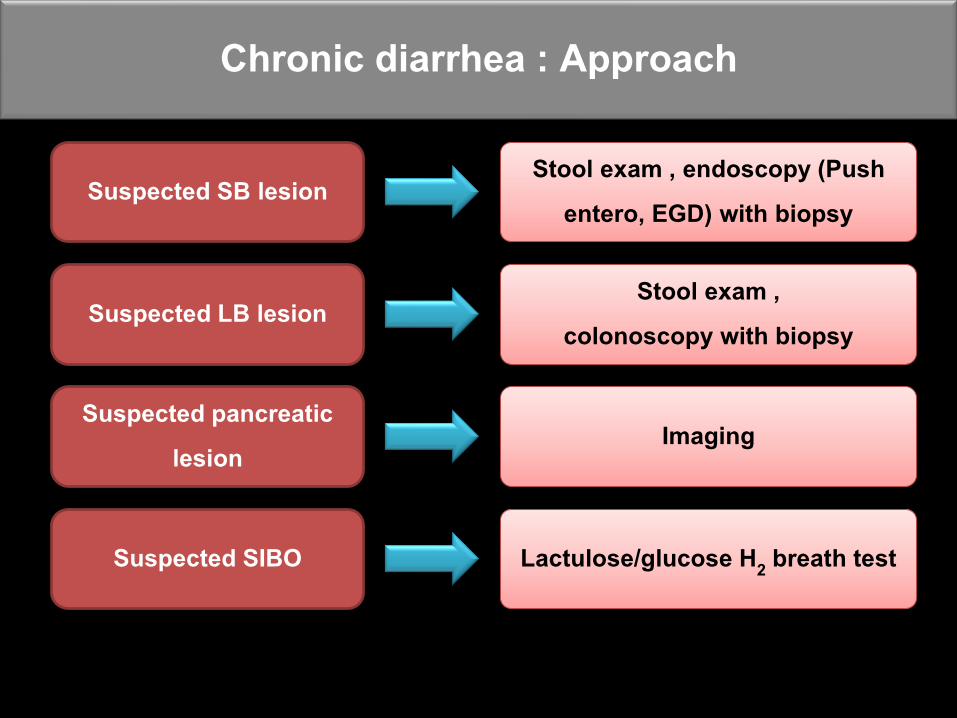
Suspected SB lesion Stool exam , endoscopy (Push
entero, EGD) with biopsy
Chronic diarrhea : Approach
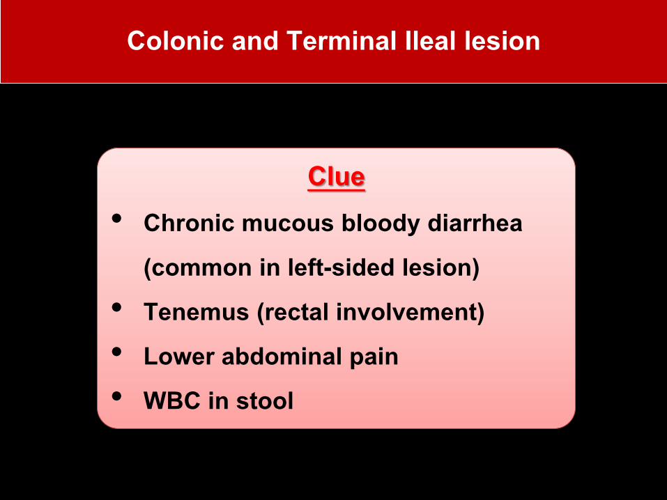
Colonic and Terminal Ileal lesion
Clue• Chronic mucous bloody diarrhea
(common in left-sided lesion) • Tenemus (rectal involvement)• Lower abdominal pain• WBC in stool
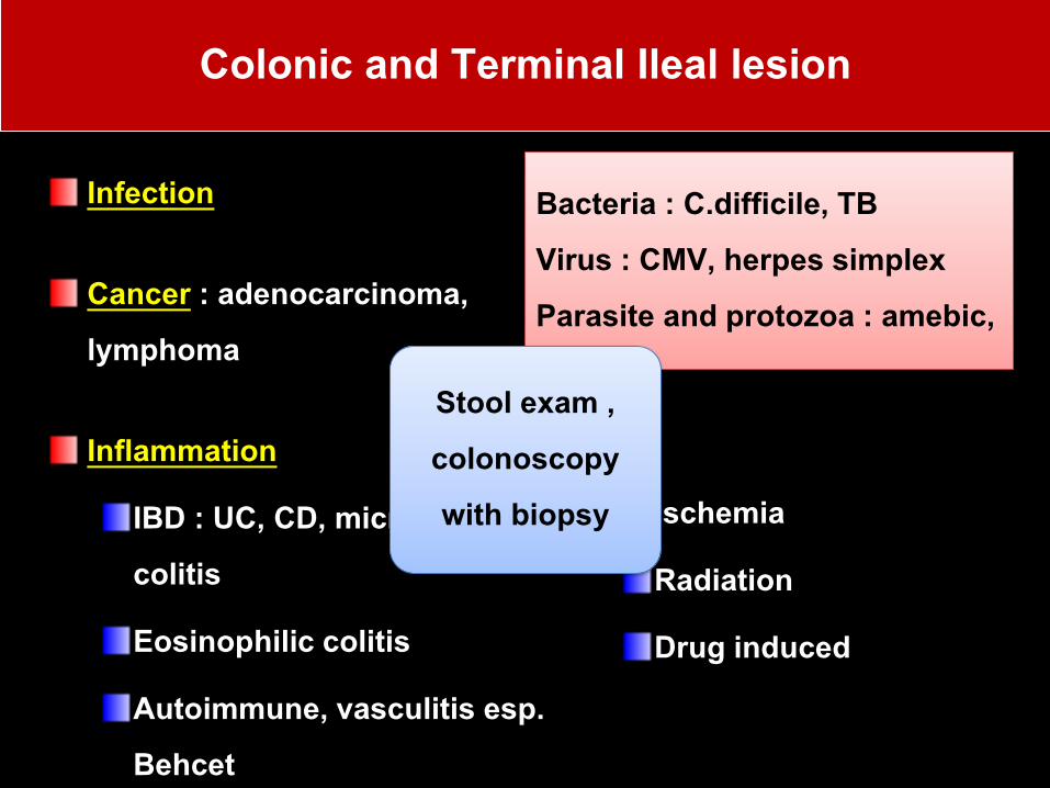
Colonic and Terminal Ileal lesion
Infection
Cancer : adenocarcinoma, lymphoma
Inflammation
IBD : UC, CD, microscopic colitis
Eosinophilic colitis
Autoimmune, vasculitis esp. Behcet
Bacteria : C.difficile, TB Virus : CMV, herpes simplex Parasite and protozoa : amebic,
Ischemia
Radiation
Drug induced
Stool exam , colonoscopy with biopsy
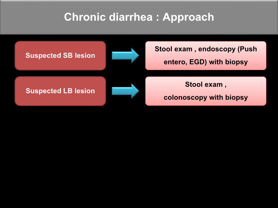
Suspected SB lesion Stool exam , endoscopy (Push
entero, EGD) with biopsy
Chronic diarrhea : Approach
Suspected LB lesion Stool exam ,
colonoscopy with biopsy
P
LB
SB
SIBO
Endocrine
Drug
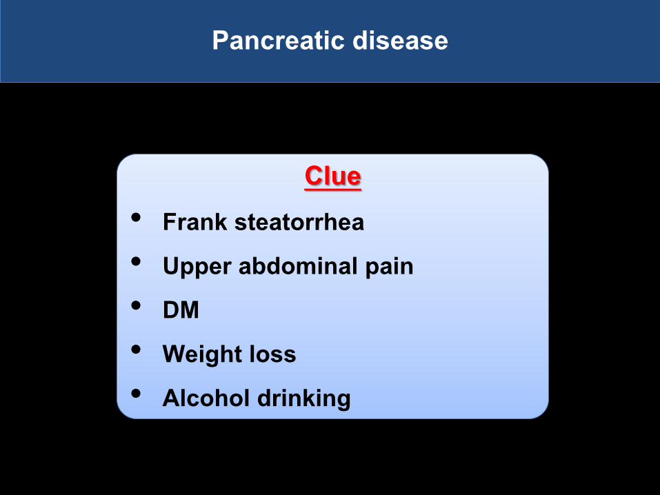
Pancreatic disease
Clue• Frank steatorrhea• Upper abdominal pain • DM• Weight loss• Alcohol drinking
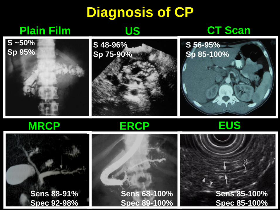
Plain FilmS ~50%
Sp 95%
US
S 48-96%
Sp 75-90%
CT Scan
S 56-95%
Sp 85-100%
ERCP
Sens 68-100%
Spec 89-100%
EUS
Sens 85-100%
Spec 85-100%
Diagnosis of CP
MRCP
Sens 88-91%
Spec 92-98%
Suspected SB lesion Stool exam , endoscopy (Push
entero, EGD) with biopsy
Chronic diarrhea : Approach
Suspected LB lesion Stool exam ,
colonoscopy with biopsy
Suspected pancreatic lesion
Imaging
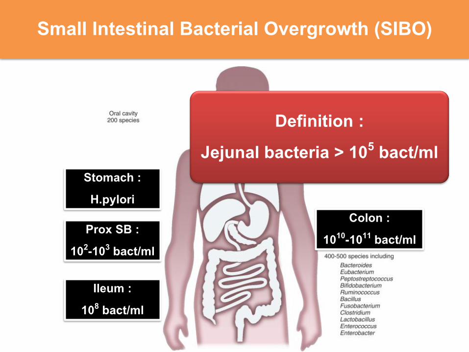
Small Intestinal Bacterial Overgrowth (SIBO)
Stomach : H.pylori
Prox SB : 102-103 bact/ml
Ileum : 108 bact/ml
Colon : 1010-1011 bact/ml
Definition : Jejunal bacteria > 105 bact/ml
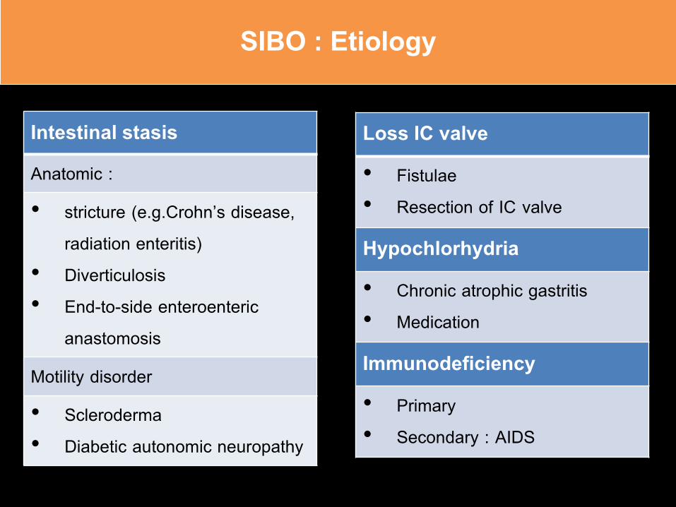
SIBO : Etiology
Intestinal stasisAnatomic :
• stricture (e.g.Crohn’s disease, radiation enteritis)
• Diverticulosis• End-to-side enteroenteric
anastomosis
Motility disorder
• Scleroderma• Diabetic autonomic neuropathy
Loss IC valve • Fistulae • Resection of IC valve
Hypochlorhydria• Chronic atrophic gastritis • Medication
Immunodeficiency• Primary • Secondary : AIDS
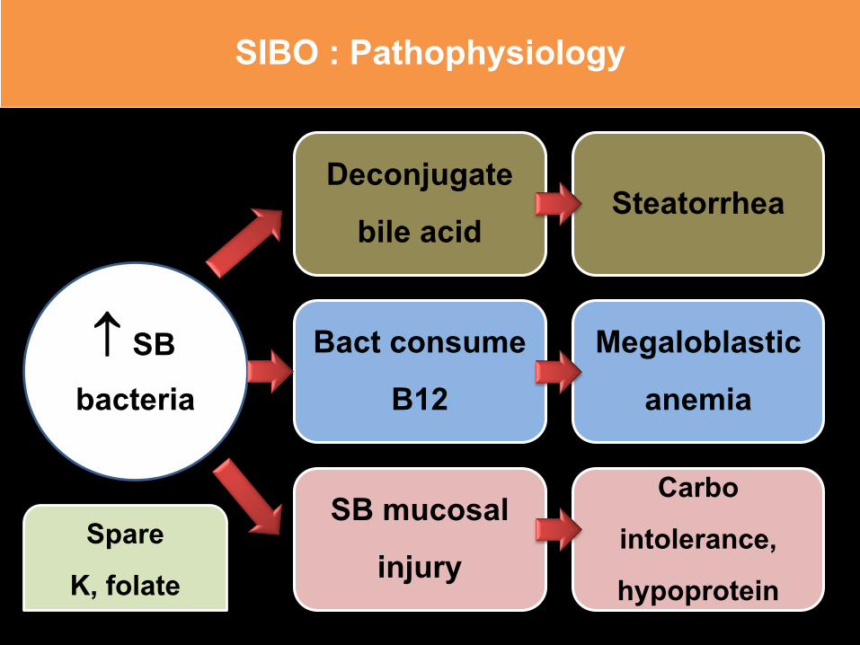
SIBO : Pathophysiology
SB bacteria
Deconjugatebile acid
Steatorrhea
Bact consume B12
Megaloblasticanemia
SB mucosal injury
Carbointolerance, hypoprotein
Spare K, folate
Suspected SB lesion Stool exam , endoscopy (Push
entero, EGD) with biopsy
Chronic diarrhea : Approach
Suspected LB lesion Stool exam ,
colonoscopy with biopsy
Suspected pancreatic lesion
Imaging
Suspected SIBO Lactulose/glucose H2 breath test
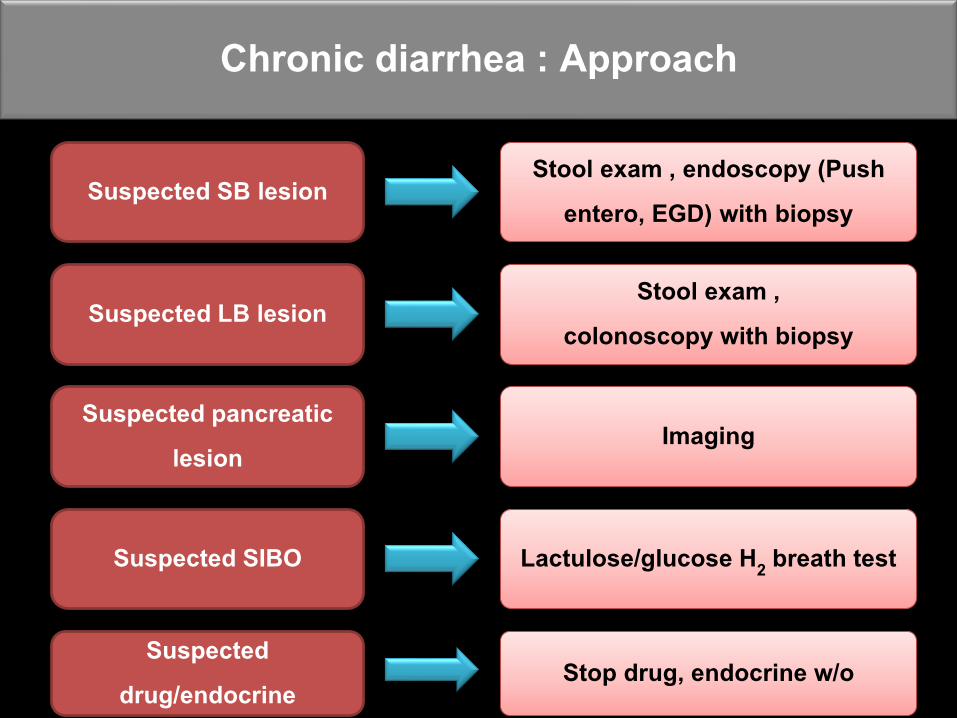
Suspected SB lesion Stool exam , endoscopy (Push
entero, EGD) with biopsy
Chronic diarrhea : Approach
Suspected LB lesion Stool exam ,
colonoscopy with biopsy
Suspected pancreatic lesion
Imaging
Suspected SIBO Lactulose/glucose H2 breath test
Suspected drug/endocrine
Stop drug, endocrine w/o
Outline : GI tract
Upper tract Dyspepsia
GERD
PancreaticobiliaryGall stone and CBD stone
Acute cholecystitis
Ascending cholangitis
Acute pancreatitis Lower tract IBS
Chronic diarrhea
FOBTCRC screening
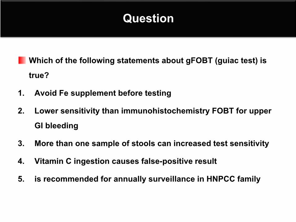
Question
Which of the following statements about gFOBT (guiac test) is true?
1. Avoid Fe supplement before testing
2. Lower sensitivity than immunohistochemistry FOBT for upper GI bleeding
3. More than one sample of stools can increased test sensitivity
4. Vitamin C ingestion causes false-positive result
5. is recommended for annually surveillance in HNPCC family
FOBT
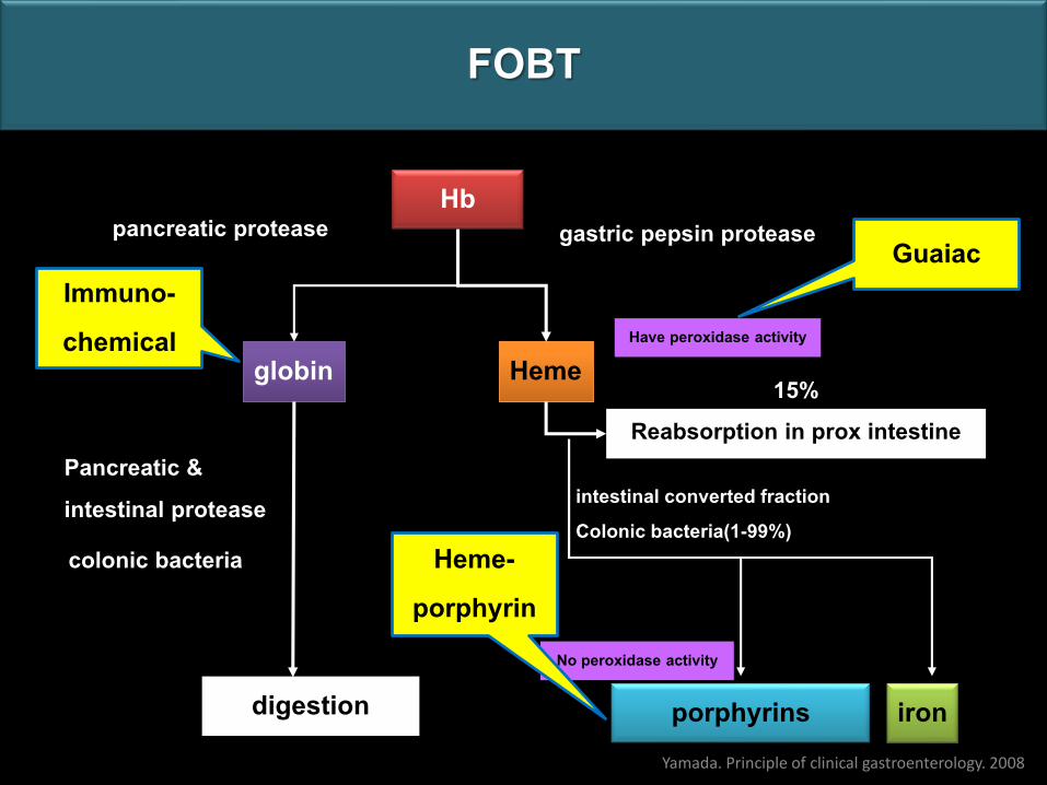
FOBT
Hb
globin Heme
Reabsorption in prox intestine
porphyrins iron
pancreatic protease gastric pepsin protease
Pancreatic & intestinal protease
digestion
colonic bacteria
intestinal converted fractionColonic bacteria(1-99%)
15%
Have peroxidase activity
No peroxidase activity
Yamada. Principle of clinical gastroenterology. 2008
GuaiacImmuno-chemical
Heme-porphyrin
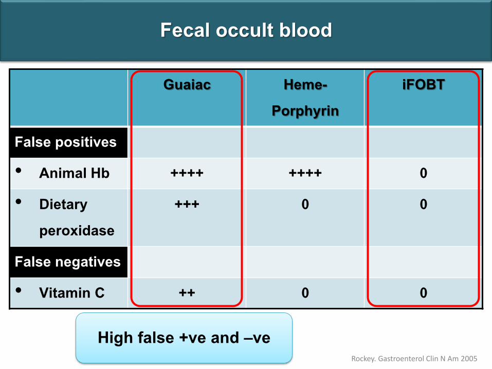
Fecal occult blood
Rockey. Gastroenterol Clin N Am 2005
Guaiac Heme-Porphyrin
iFOBT
False positives
• Animal Hb ++++ ++++ 0
• Dietary peroxidase
+++ 0 0
False negatives
• Vitamin C ++ 0 0
High false +ve and –ve
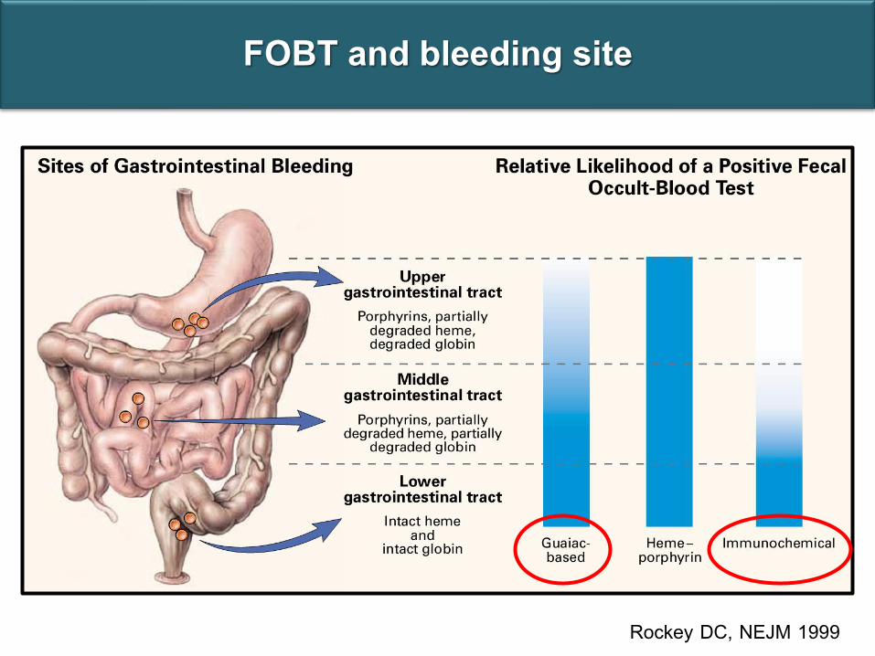
FOBT and bleeding site
Rockey DC, NEJM 1999
Fecal occult blood test : indication
Colorectal cancer screening :Decreased mortality from CRC
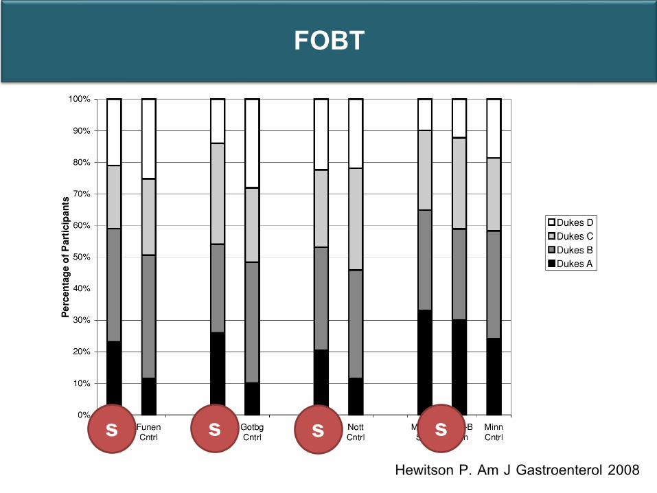
FOBT
Hewitson P. Am J Gastroenterol 2008
s s s s
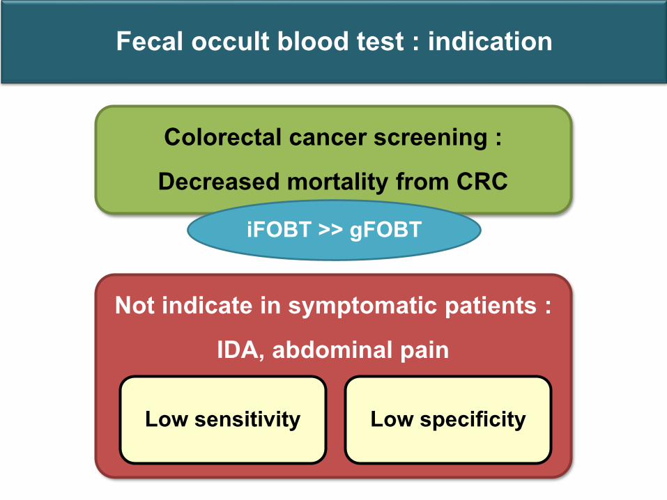
Fecal occult blood test : indication
Colorectal cancer screening :Decreased mortality from CRC
Not indicate in symptomatic patients : IDA, abdominal pain
Low sensitivity Low specificity
iFOBT >> gFOBT
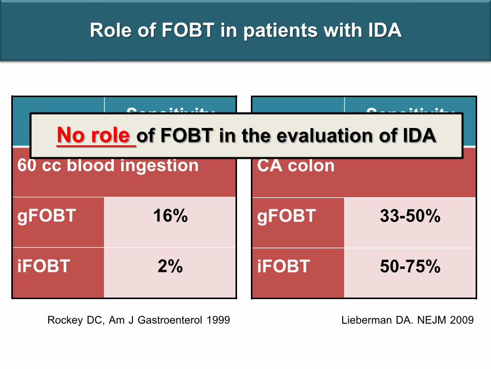
Role of FOBT in patients with IDA
Sensitivity
CA colon
gFOBT 33-50%
iFOBT 50-75%
Sensitivity
60 cc blood ingestion
gFOBT 16%
iFOBT 2%
Lieberman DA. NEJM 2009Rockey DC, Am J Gastroenterol 1999
No role of FOBT in the evaluation of IDA
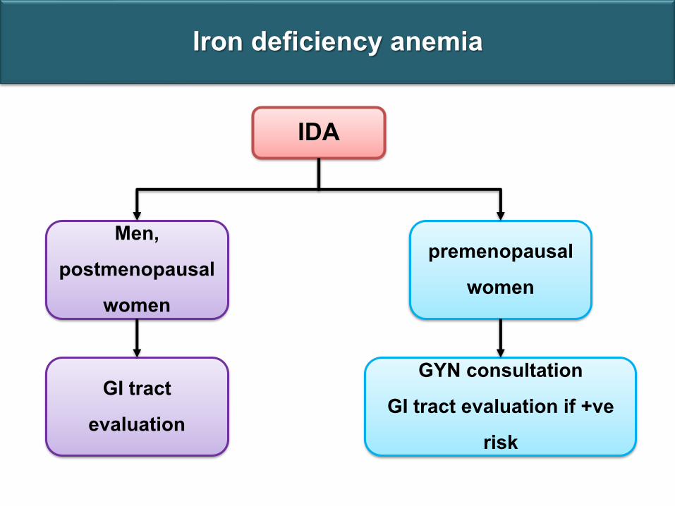
Iron deficiency anemia
IDA
Men, postmenopausal
women
GI tract evaluation
premenopausal women
GYN consultationGI tract evaluation if +ve
risk
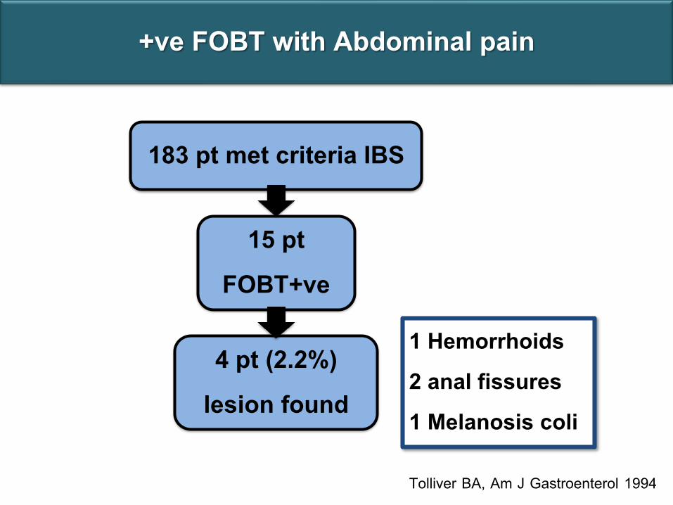
183 pt met criteria IBS
15 ptFOBT+ve
4 pt (2.2%)lesion found
1 Hemorrhoids 2 anal fissures 1 Melanosis coli
Tolliver BA, Am J Gastroenterol 1994
+ve FOBT with Abdominal pain
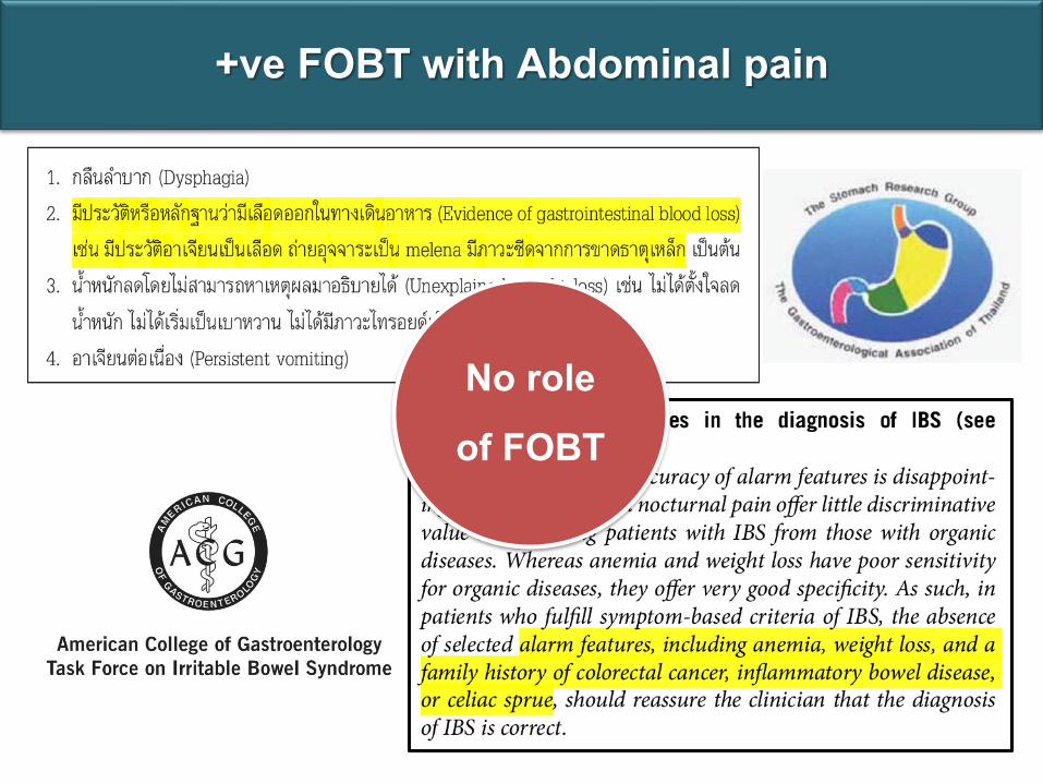
No role of FOBT
+ve FOBT with Abdominal pain
Question
Which of the following statement about gFOBT (guiac test) is true?
1. Avoid Fe supplement before testing
2. Lower sensitivity than immunohistochemistry FOBT for upper GI bleeding
3. More than one sample of stools can increased test sensitivity
4. Vitamin C ingestion causes false-positive result
5. is recommended for annually surveillance in HNPCC family
Outline : GI tract
Upper tract Dyspepsia
GERD
PancreaticobiliaryGall stone and CBD stone
Acute cholecystitis
Ascending cholangitis
Acute pancreatitis Lower tract IBS
Chronic diarrhea
FOBT
CRC screening
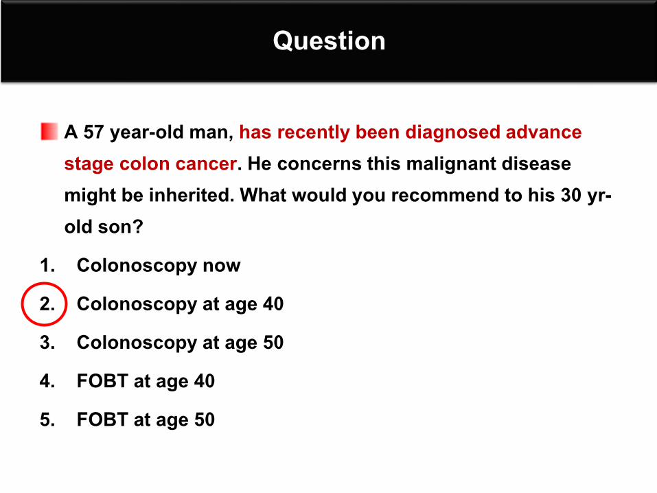
Question
A 57 year-old man, has recently been diagnosed advance stage colon cancer. He concerns that this malignant disease might be inherited. What would you recommend to his 30 yr-old son?
1. Colonoscopy now 2. Colonoscopy at age 40 3. Colonoscopy at age 504. FOBT at age 40 5. FOBT at age 50
Colon cancer screening
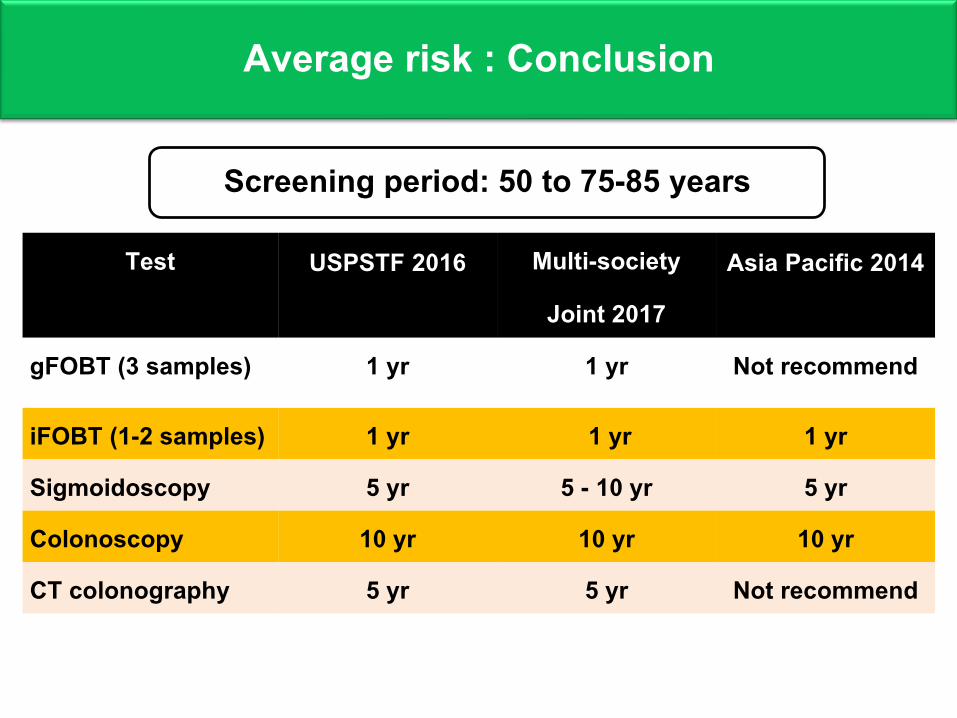
Test USPSTF 2016 Multi-society
Joint 2017Asia Pacific 2014
gFOBT (3 samples) 1 yr 1 yr Not recommend
iFOBT (1-2 samples) 1 yr 1 yr 1 yrSigmoidoscopy 5 yr 5 - 10 yr 5 yr Colonoscopy 10 yr 10 yr 10 yr CT colonography 5 yr 5 yr Not recommend
Average risk : Conclusion
Screening period: 50 to 75-85 years
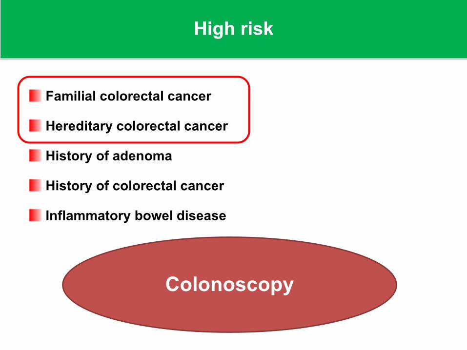
High risk
Familial colorectal cancer
Hereditary colorectal cancer
History of adenoma
History of colorectal cancer
Inflammatory bowel disease
Colonoscopy
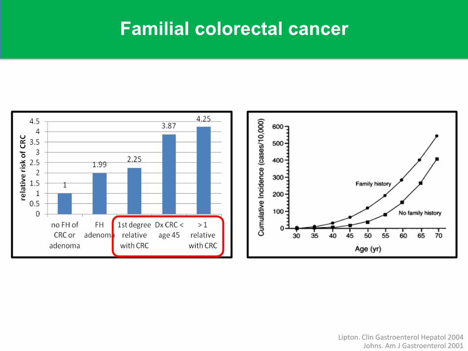
Familial colorectal cancer
Lipton. Clin Gastroenterol Hepatol 2004Johns. Am J Gastroenterol 2001
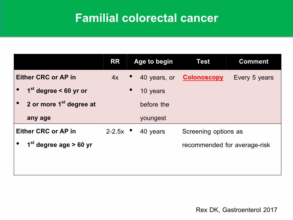
Familial colorectal cancer
RR Age to begin Test Comment
Either CRC or AP in • 1st degree < 60 yr or • 2 or more 1st degree at
any age
4x • 40 years, or • 10 years
before the youngest
Colonoscopy Every 5 years
Either CRC or AP in • 1st degree age > 60 yr
2-2.5x • 40 years Screening options as recommended for average-risk
Rex DK, Gastroenterol 2017
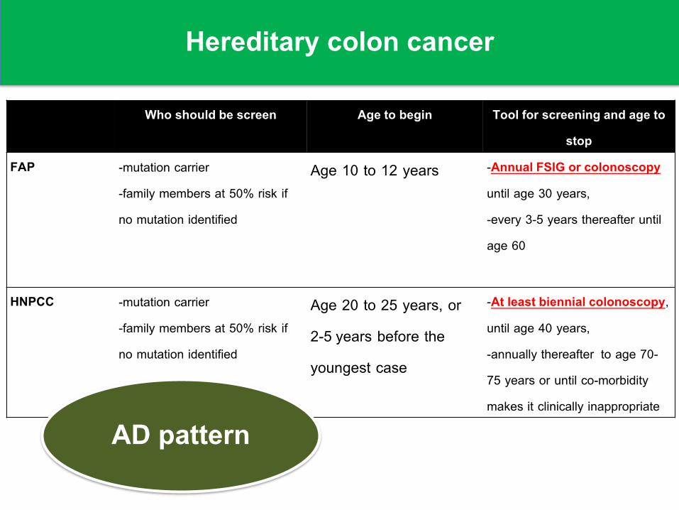
Hereditary colon cancer
Who should be screen Age to begin Tool for screening and age to stop
FAP -mutation carrier-family members at 50% risk if no mutation identified
Age 10 to 12 years -Annual FSIG or colonoscopy until age 30 years, -every 3-5 years thereafter until age 60
HNPCC -mutation carrier-family members at 50% risk if no mutation identified
Age 20 to 25 years, or 2-5 years before the youngest case
-At least biennial colonoscopy, until age 40 years, -annually thereafter to age 70-75 years or until co-morbidity makes it clinically inappropriate
AD pattern
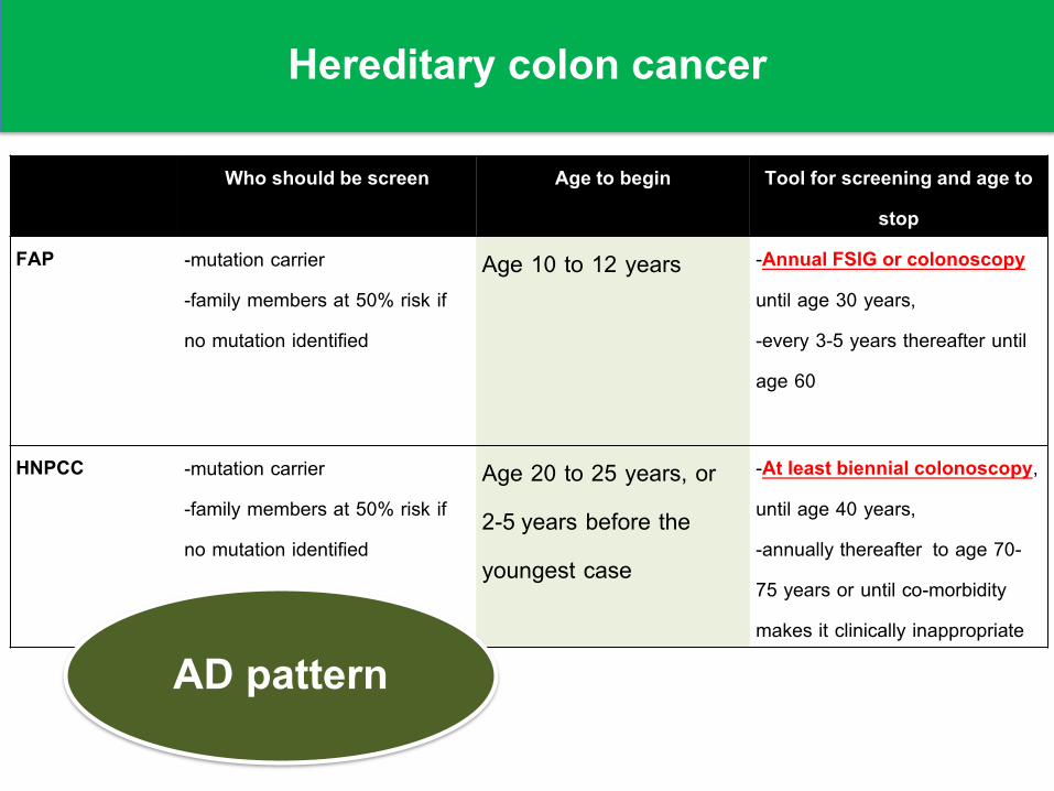
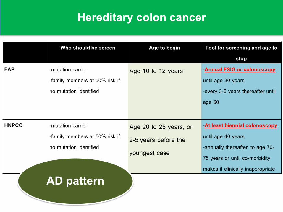
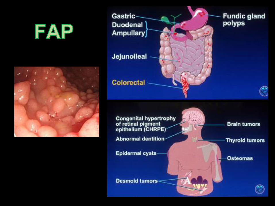
HNPCC
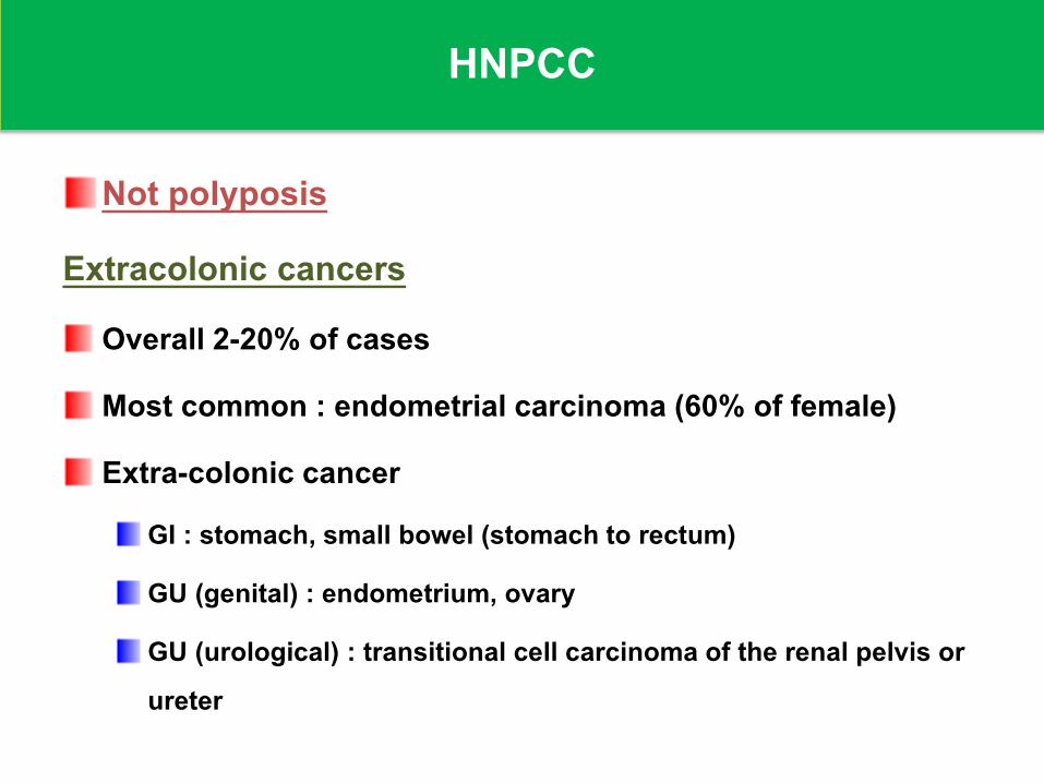
Not polyposis
Extracolonic cancers Overall 2-20% of cases
Most common : endometrial carcinoma (60% of female)
Extra-colonic cancer GI : stomach, small bowel (stomach to rectum)
GU (genital) : endometrium, ovary
GU (urological) : transitional cell carcinoma of the renal pelvis or ureter
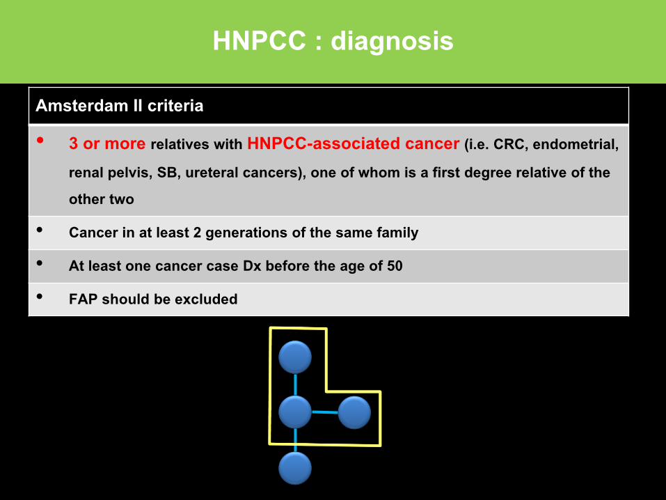
HNPCC : diagnosis
Amsterdam II criteria
• 3 or more relatives with HNPCC-associated cancer (i.e. CRC, endometrial, renal pelvis, SB, ureteral cancers), one of whom is a first degree relative of the other two
• Cancer in at least 2 generations of the same family
• At least one cancer case Dx before the age of 50
• FAP should be excluded
Question
A 57 year-old man, has recently been diagnosed advance stage colon cancer. He concerns this malignant disease might be inherited. What would you recommend to his 30 yr-old son?
1. Colonoscopy now 2. Colonoscopy at age 40 3. Colonoscopy at age 504. FOBT at age 40 5. FOBT at age 50
Outline : GI tract
Upper tract Dyspepsia
GERD
PancreaticobiliaryGall stone and CBD stone
Acute cholecystitis
Ascending cholangitis
Acute pancreatitis Lower tract IBS
Chronic diarrhea
FOBT
CRC screening
Question
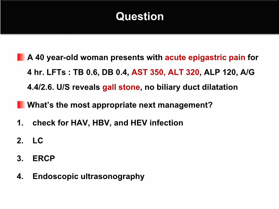
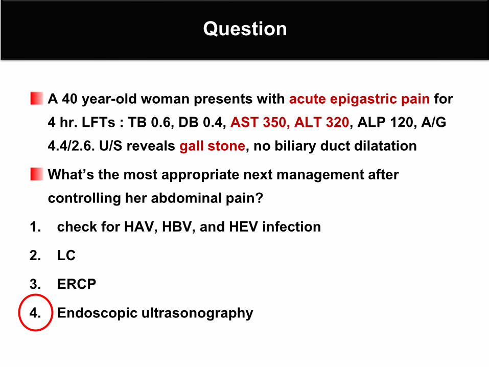
A 40 year-old woman presents with acute epigastric pain for 4 hr. LFTs : TB 0.6, DB 0.4, AST 350, ALT 320, ALP 120, A/G 4.4/2.6. U/S reveals gall stone, no biliary duct dilatation
What’s the most appropriate next management?
1. check for HAV, HBV, and HEV infection
2. LC
3. ERCP
4. Endoscopic ultrasonography
Gall stone and complications

Asymptomatic GS60-80%
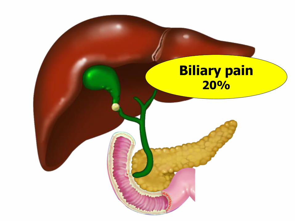
Biliary pain20%
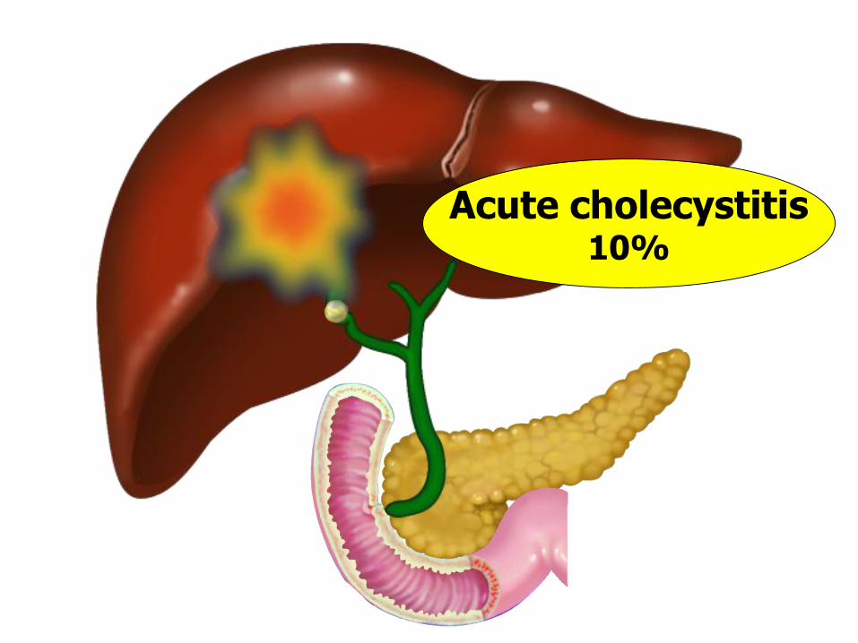
Acute cholecystitis10%
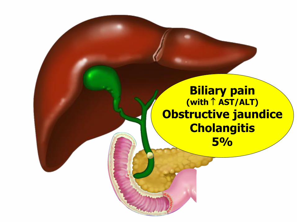
Biliary pain(with AST/ALT)
Obstructive jaundiceCholangitis
5%
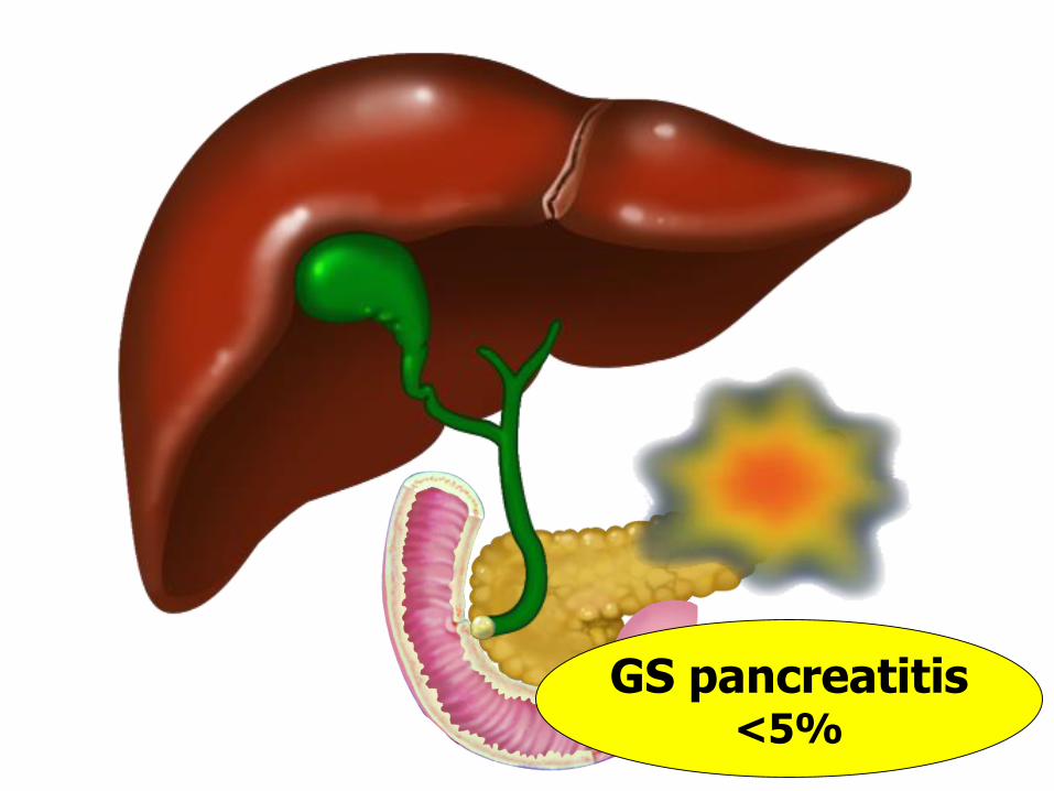
GS pancreatitis<5%
Gall stone and CBD stone
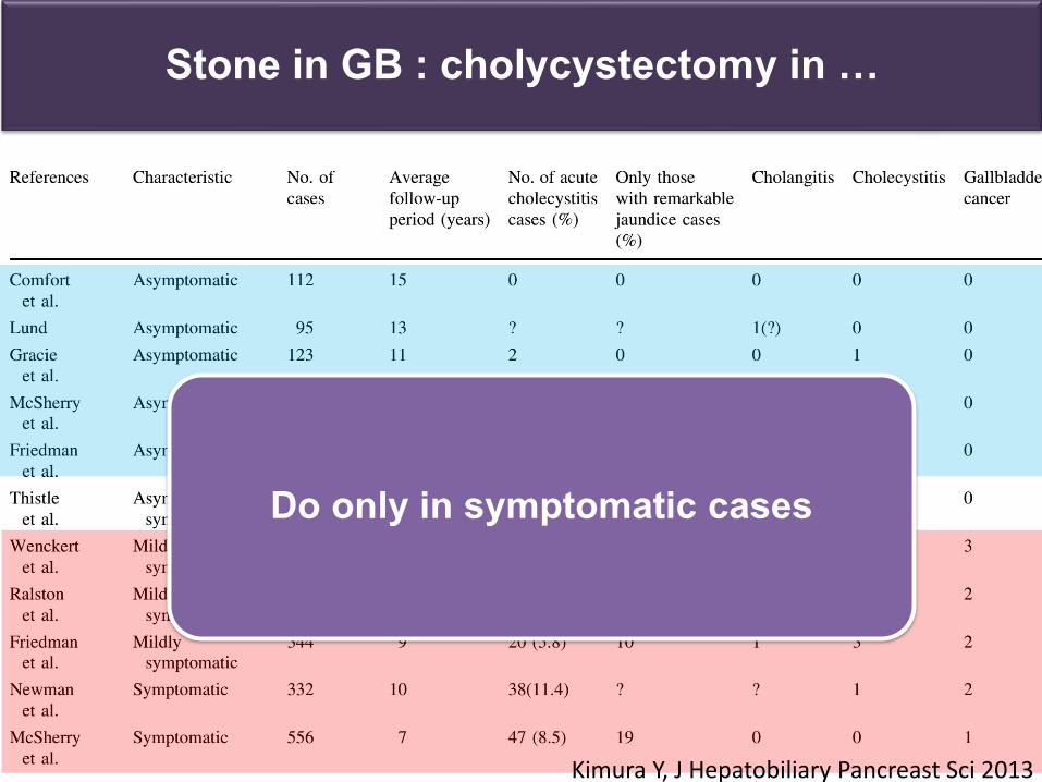
Stone in GB : cholycystectomy in …
Do only in symptomatic cases
Kimura Y, J Hepatobiliary Pancreast Sci 2013
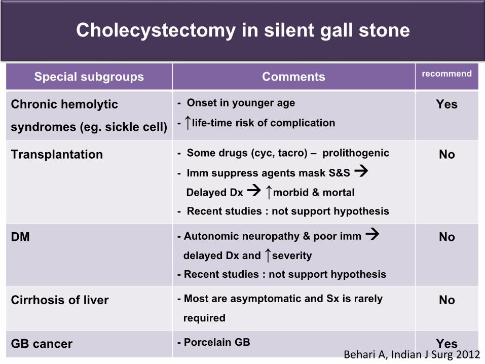
Cholecystectomy in silent gall stone
Special subgroups Comments recommend
Chronic hemolytic syndromes (eg. sickle cell)
- Onset in younger age - ↑life-time risk of complication
Yes
Transplantation - Some drugs (cyc, tacro) – prolithogenic- Imm suppress agents mask S&S
Delayed Dx ↑morbid & mortal - Recent studies : not support hypothesis
No
DM - Autonomic neuropathy & poor immdelayed Dx and ↑severity
- Recent studies : not support hypothesis
No
Cirrhosis of liver - Most are asymptomatic and Sx is rarely required
No
GB cancer - Porcelain GB Yes Behari A, Indian J Surg 2012
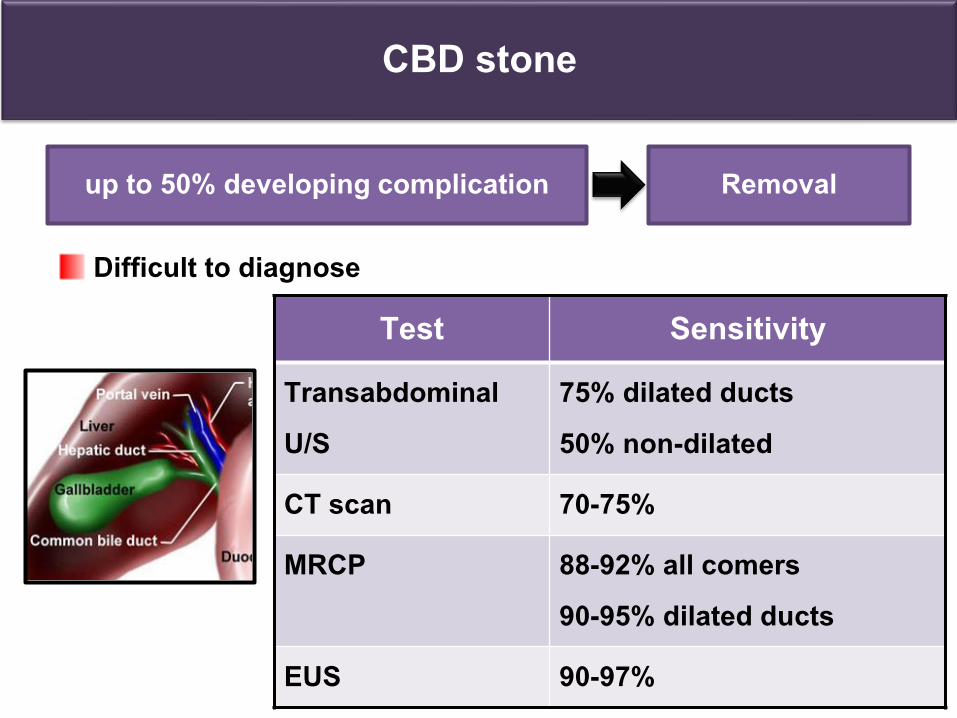
CBD stone
Difficult to diagnose
up to 50% developing complication Removal
Test SensitivityTransabdominalU/S
75% dilated ducts 50% non-dilated
CT scan 70-75%
MRCP 88-92% all comers90-95% dilated ducts
EUS 90-97%
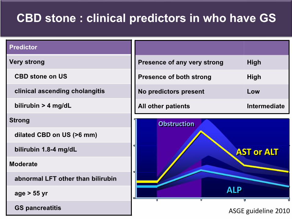
CBD stone : clinical predictors in who have GS
Predictor
Very strong
CBD stone on US
clinical ascending cholangitis
bilirubin > 4 mg/dL
Strong
dilated CBD on US (>6 mm)
bilirubin 1.8-4 mg/dL
Moderate
abnormal LFT other than bilirubin
age > 55 yr
GS pancreatitis
Presence of any very strong High
Presence of both strong High
No predictors present Low
All other patients Intermediate
ASGE guideline 2010
Obstruction
ALP
AST or ALT
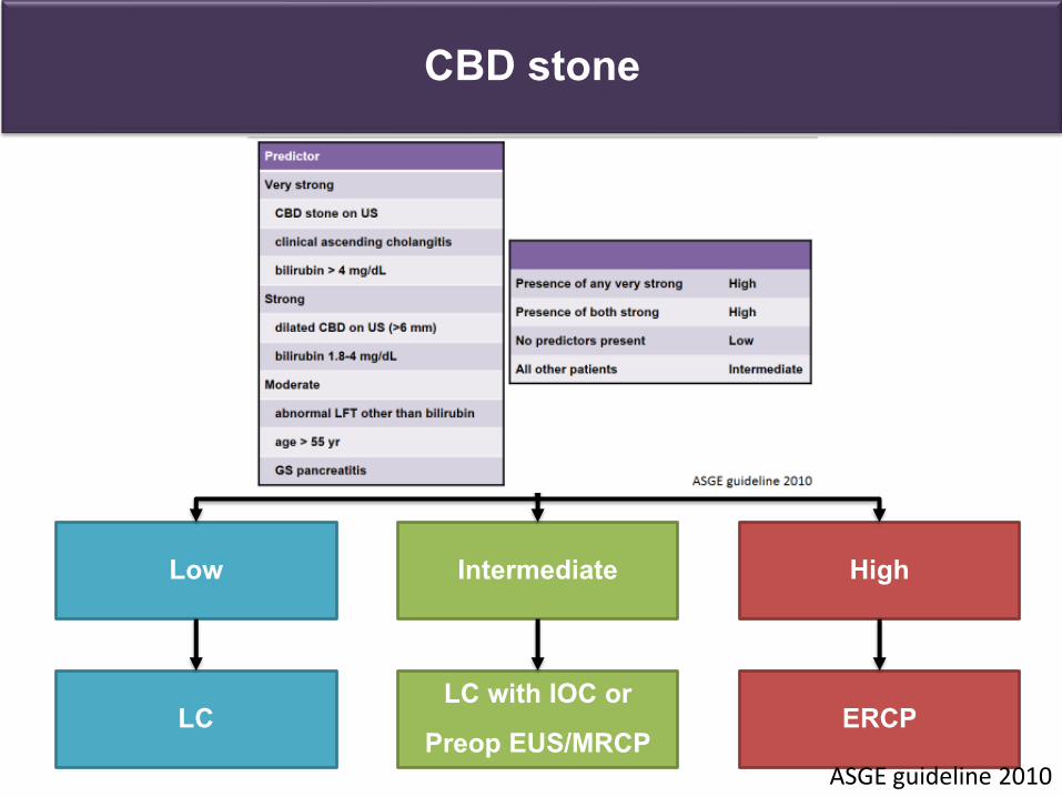
CBD stone
Symptomatic ptwith GS
Likelihood of CBD stone
Low Intermediate High
LC LC with IOC or
Preop EUS/MRCP ERCP
ASGE guideline 2010
Question
A 40 year-old woman presents with acute epigastric pain for 4 hr. LFTs : TB 0.6, DB 0.4, AST 350, ALT 320, ALP 120, A/G 4.4/2.6. U/S reveals gall stone, no biliary duct dilatation What’s the most appropriate next management after controlling her abdominal pain?
1. check for HAV, HBV, and HEV infection 2. LC3. ERCP4. Endoscopic ultrasonography
Acute cholecystitis
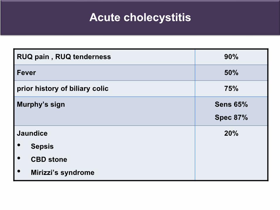
Acute cholecystitis
RUQ pain , RUQ tenderness 90%
Fever 50%
prior history of biliary colic 75%
Murphy’s sign Sens 65%Spec 87%
Jaundice • Sepsis• CBD stone • Mirizzi’s syndrome
20%
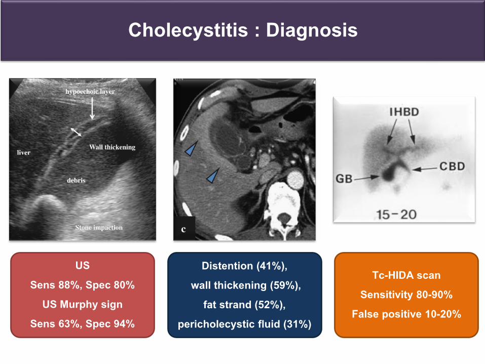
Cholecystitis : Diagnosis
US Sens 88%, Spec 80%
US Murphy sign Sens 63%, Spec 94%
Distention (41%),wall thickening (59%),
fat strand (52%), pericholecystic fluid (31%)
Tc-HIDA scanSensitivity 80-90%
False positive 10-20%
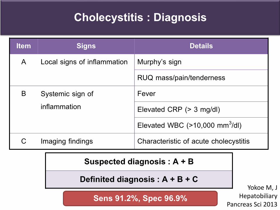
Cholecystitis : Diagnosis
Item Signs Details
A Local signs of inflammation Murphy’s sign
RUQ mass/pain/tenderness
B Systemic sign of inflammation
Fever
Elevated CRP (> 3 mg/dl)
Elevated WBC (>10,000 mm3/dl)
C Imaging findings Characteristic of acute cholecystitis
Suspected diagnosis : A + B
Definited diagnosis : A + B + C
Sens 91.2%, Spec 96.9%Yokoe M, J
HepatobiliaryPancreas Sci 2013
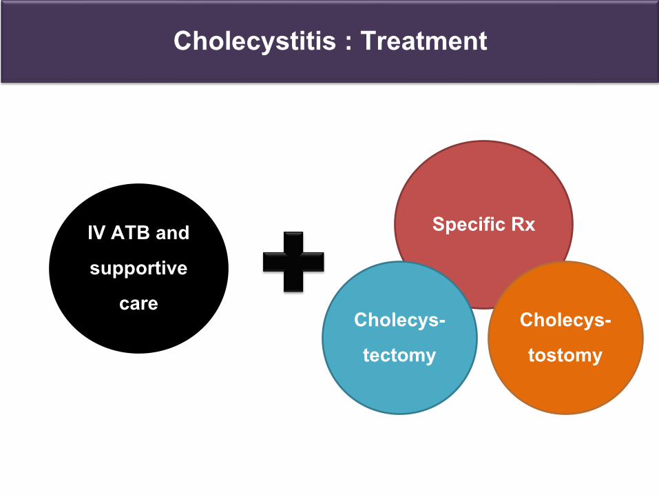
Cholecystitis : Treatment
IV ATB and supportive
care
Specific Rx
Cholecys-tostomy
Cholecys-tectomy
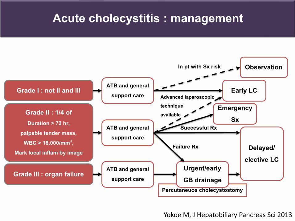
Acute cholecystitis : management
Grade II : 1/4 ofDuration > 72 hr,
palpable tender mass, WBC > 18,000/mm3,
Mark local inflam by image
Grade III : organ failure
Grade I : not II and III
Yokoe M, J Hepatobiliary Pancreas Sci 2013
In pt with Sx risk
Percutaneuos cholecystostomy
ATB and general support care
ATB and general support care
ATB and general support care
Urgent/early GB drainage
Observation
Early LC
Emergency Sx
Delayed/ elective LC
Advanced laparoscopic technique available
Successful Rx
Failure Rx
Ascending cholangitis
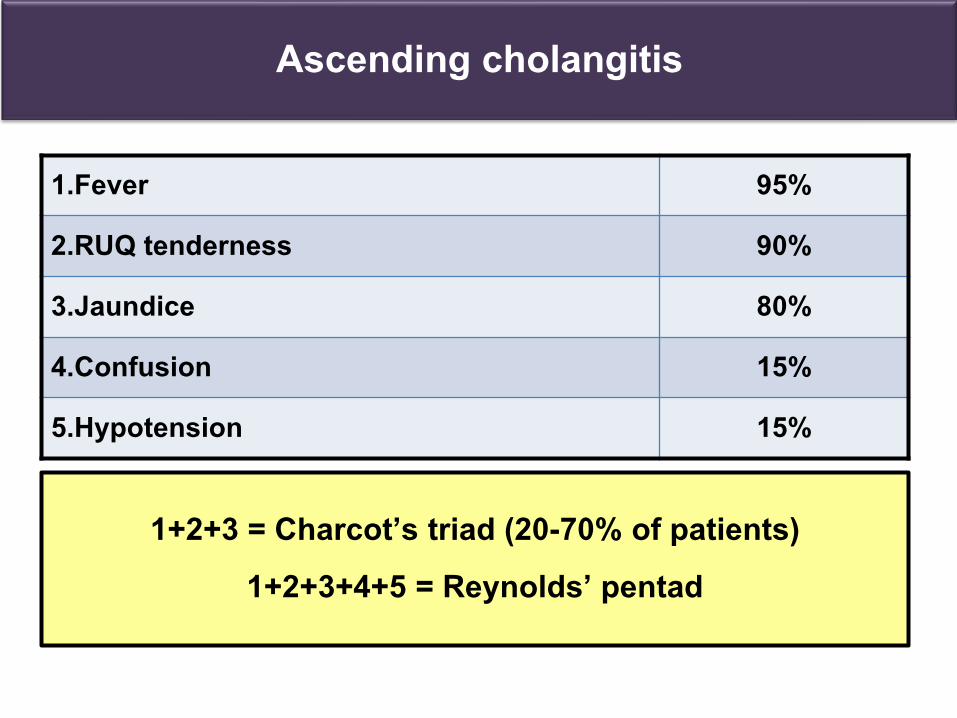
Ascending cholangitis
1.Fever 95%
2.RUQ tenderness 90%
3.Jaundice 80%
4.Confusion 15%
5.Hypotension 15%
1+2+3 = Charcot’s triad (20-70% of patients)1+2+3+4+5 = Reynolds’ pentad
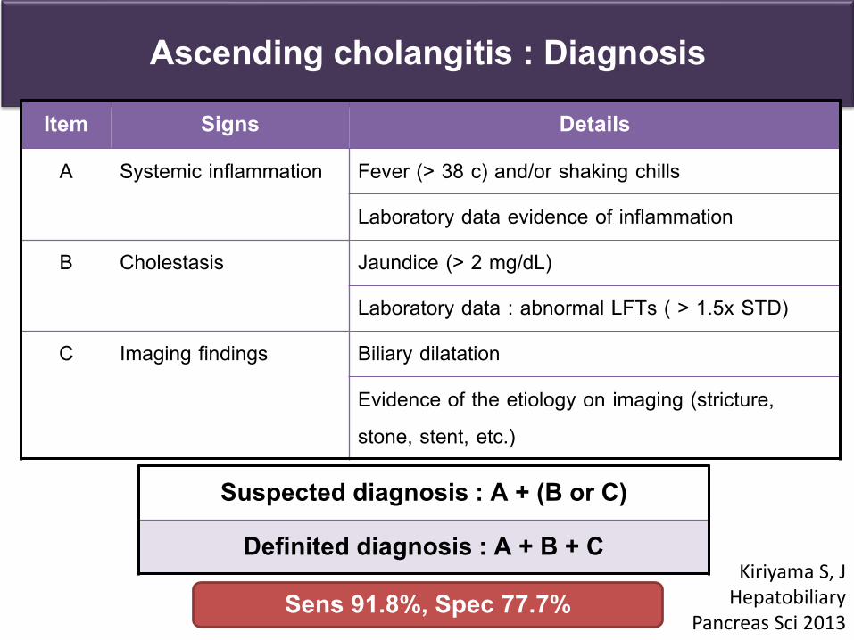
Ascending cholangitis : Diagnosis Item Signs Details
A Systemic inflammation Fever (> 38 c) and/or shaking chills
Laboratory data evidence of inflammation
B Cholestasis Jaundice (> 2 mg/dL)
Laboratory data : abnormal LFTs ( > 1.5x STD)
C Imaging findings Biliary dilatation
Evidence of the etiology on imaging (stricture, stone, stent, etc.)
Suspected diagnosis : A + (B or C)
Definited diagnosis : A + B + C
Sens 91.8%, Spec 77.7%Kiriyama S, J
HepatobiliaryPancreas Sci 2013
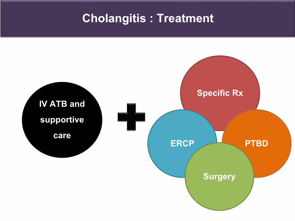
Cholangitis : Treatment
IV ATB and supportive
care
Specific Rx
PTBDERCP
Surgery
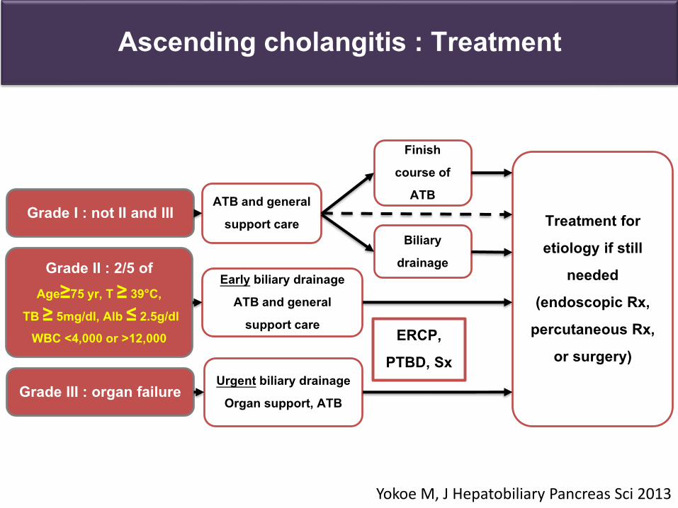
Ascending cholangitis : Treatment
Grade II : 2/5 ofAge≥75 yr, T ≥ 39°C,
TB ≥ 5mg/dl, Alb ≤ 2.5g/dlWBC <4,000 or >12,000
Grade III : organ failure
Grade I : not II and III
Yokoe M, J Hepatobiliary Pancreas Sci 2013
ERCP, PTBD, Sx
ATB and general support care
Early biliary drainageATB and general
support care
Urgent biliary drainage Organ support, ATB
Finish course of
ATB
Biliary drainage
Treatment for etiology if still
needed (endoscopic Rx,
percutaneous Rx, or surgery)
Acute pancreatitis
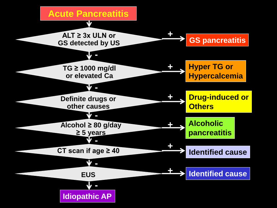
Acute Pancreatitis
Idiopathic AP
ALT ≥ 3x ULN orGS detected by US
TG ≥ 1000 mg/dlor elevated Ca
Alcohol ≥ 80 g/day≥ 5 years
Definite drugs orother causes
CT scan if age ≥ 40
EUS
GS pancreatitis
Hyper TG or
Hypercalcemia
Drug-induced or
Others
Alcoholic
pancreatitis
Identified cause
Identified cause
+
+
+
+
+
+
-
-
-
-
-
-
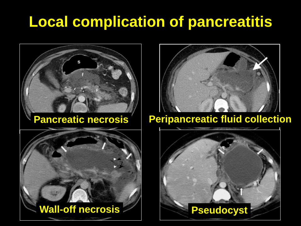
Local complication of pancreatitis
Pancreatic necrosis Peripancreatic fluid collection
Wall-off necrosis Pseudocyst
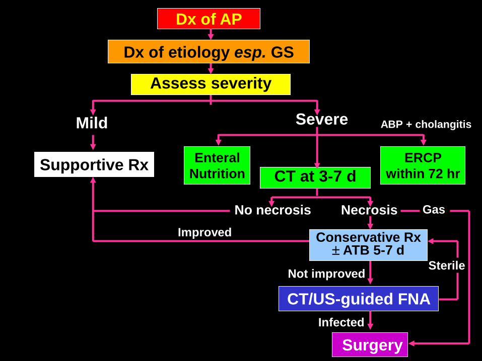
Dx of AP
Mild Severe
CT at 3-7 dSupportive Rx
No necrosis Necrosis
Improved
CT/US-guided FNA
Surgery
ERCP
within 72 hr
Enteral
Nutrition
Gas
Dx of etiology esp. GS
Assess severity
Infected
Sterile
Conservative Rx ATB 5-7 d
Not improved
ABP + cholangitis
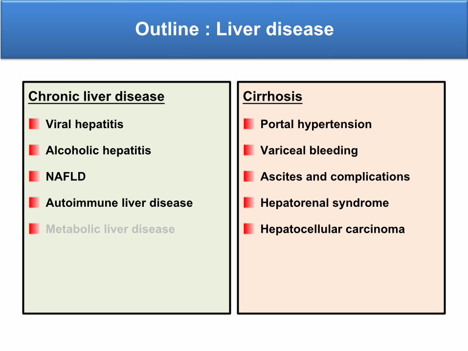
Outline : Liver disease
Chronic liver disease Viral hepatitis
Alcoholic hepatitis
NAFLD
Autoimmune liver disease
Metabolic liver disease
Cirrhosis Portal hypertension
Variceal bleeding
Ascites and complications
Hepatorenal syndrome
Hepatocellular carcinoma
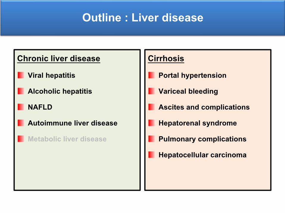
Outline : Liver disease
Chronic liver disease Viral hepatitis
Alcoholic hepatitis
NAFLD
Autoimmune liver disease
Metabolic liver disease
Cirrhosis Portal hypertension
Variceal bleeding
Ascites and complications
Hepatorenal syndrome
Pulmonary complications
Hepatocellular carcinoma
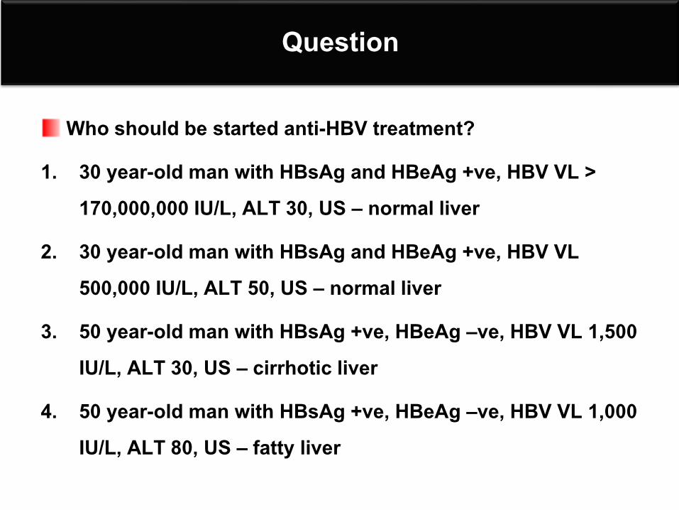
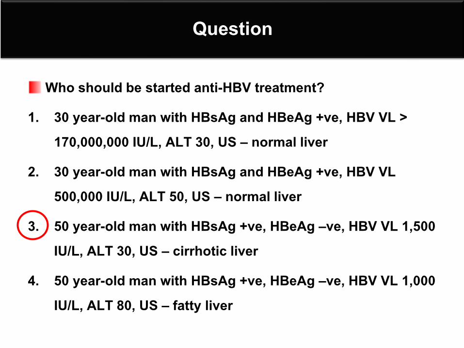
Question
Who should be started anti-HBV treatment?
1. 30 year-old man with HBsAg and HBeAg +ve, HBV VL > 170,000,000 IU/L, ALT 30, US – normal liver
2. 30 year-old man with HBsAg and HBeAg +ve, HBV VL 500,000 IU/L, ALT 50, US – normal liver
3. 50 year-old man with HBsAg +ve, HBeAg –ve, HBV VL 1,500 IU/L, ALT 30, US – cirrhotic liver
4. 50 year-old man with HBsAg +ve, HBeAg –ve, HBV VL 1,000 IU/L, ALT 80, US – fatty liver
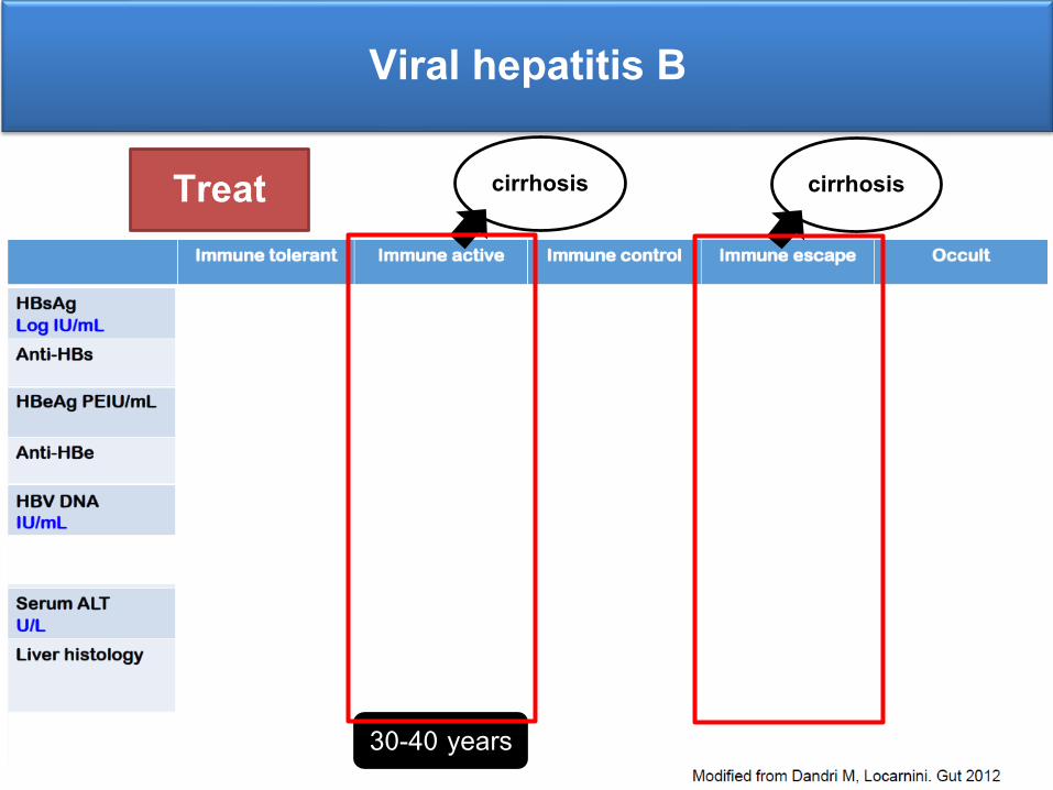
Viral hepatitis B
cirrhosis cirrhosis
30-40 years
Treat
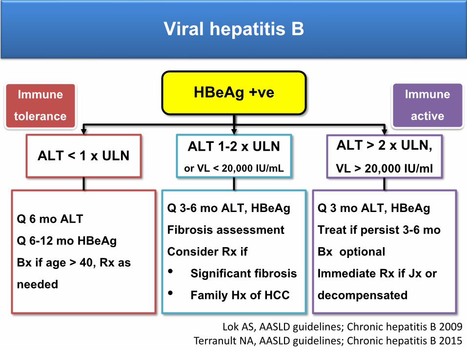
Viral hepatitis B
HBeAg +ve
ALT < 1 x ULN ALT 1-2 x ULN or VL < 20,000 IU/mL
ALT > 2 x ULN, VL > 20,000 IU/ml
Q 6 mo ALTQ 6-12 mo HBeAgBx if age > 40, Rx as needed
Q 3-6 mo ALT, HBeAgFibrosis assessment Consider Rx if • Significant fibrosis• Family Hx of HCC
Q 3 mo ALT, HBeAgTreat if persist 3-6 moBx optional Immediate Rx if Jx or decompensated
Lok AS, AASLD guidelines; Chronic hepatitis B 2009Terranult NA, AASLD guidelines; Chronic hepatitis B 2015
Immune tolerance
Immune active
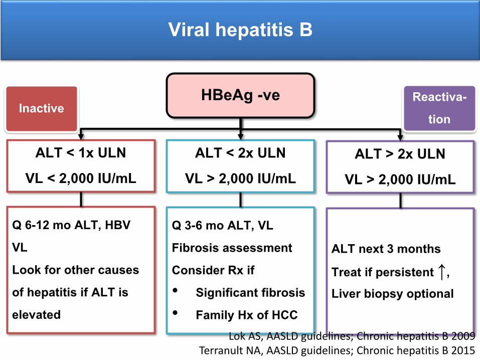
Viral hepatitis B
HBeAg -ve
ALT > 2x ULNVL > 2,000 IU/mL
ALT < 2x ULNVL > 2,000 IU/mL
ALT < 1x ULNVL < 2,000 IU/mL
ALT next 3 months Treat if persistent ↑, Liver biopsy optional
Q 3-6 mo ALT, VL Fibrosis assessment Consider Rx if • Significant fibrosis• Family Hx of HCC
Q 6-12 mo ALT, HBV VLLook for other causes of hepatitis if ALT is elevated
InactiveReactiva-
tion
Lok AS, AASLD guidelines; Chronic hepatitis B 2009Terranult NA, AASLD guidelines; Chronic hepatitis B 2015
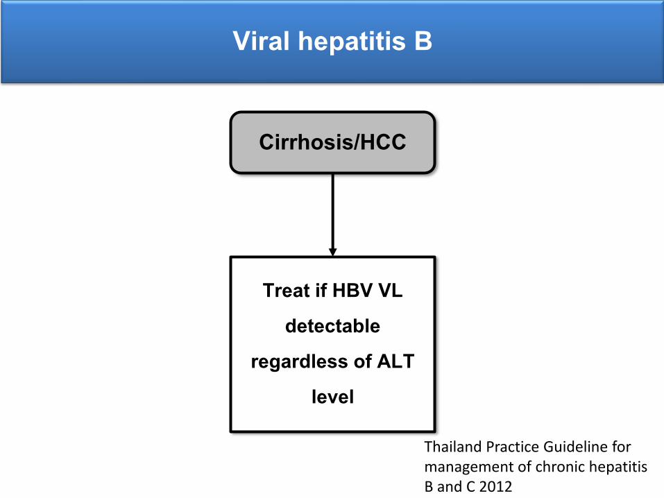
Viral hepatitis B
Cirrhosis/HCC
Treat if HBV VL detectable
regardless of ALT level
Thailand Practice Guideline for management of chronic hepatitis B and C 2012
Question
Who should be started anti-HBV treatment?
1. 30 year-old man with HBsAg and HBeAg +ve, HBV VL > 170,000,000 IU/L, ALT 30, US – normal liver
2. 30 year-old man with HBsAg and HBeAg +ve, HBV VL 500,000 IU/L, ALT 50, US – normal liver
3. 50 year-old man with HBsAg +ve, HBeAg –ve, HBV VL 1,500 IU/L, ALT 30, US – cirrhotic liver
4. 50 year-old man with HBsAg +ve, HBeAg –ve, HBV VL 1,000 IU/L, ALT 80, US – fatty liver
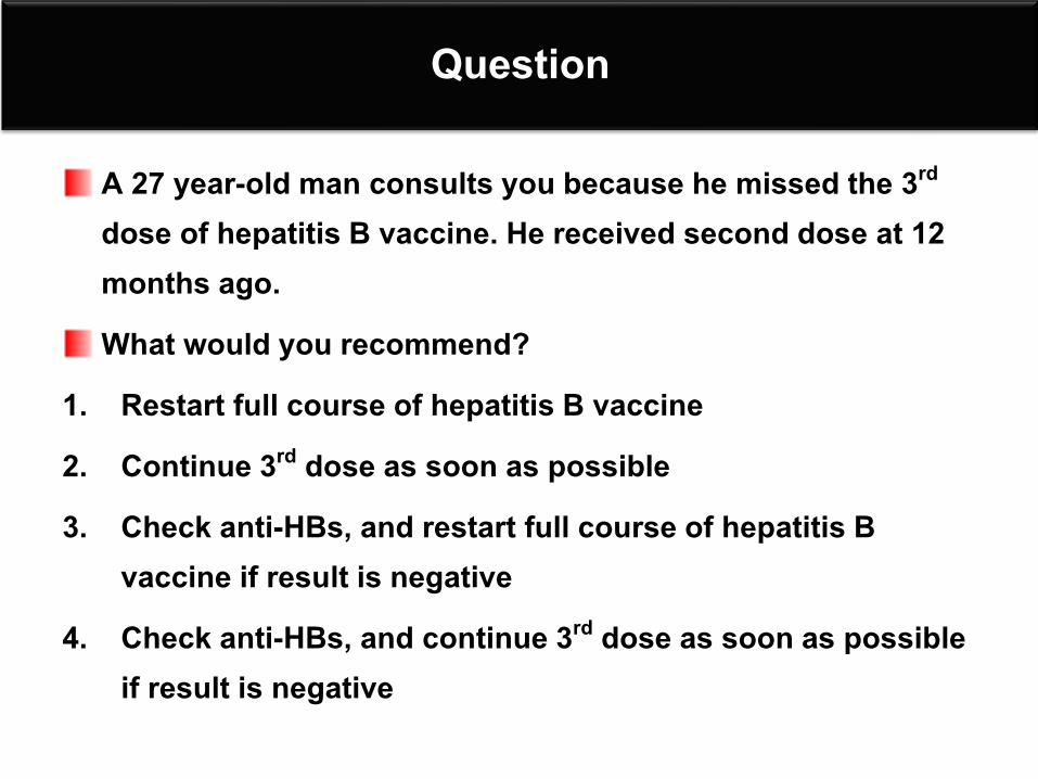
Question
A 27 year-old man consults you because he missed the 3rd
dose of hepatitis B vaccine. He received second dose at 12 months ago. What would you recommend?
1. Restart full course of hepatitis B vaccine 2. Continue 3rd dose as soon as possible 3. Check anti-HBs, and restart full course of hepatitis B
vaccine if result is negative 4. Check anti-HBs, and continue 3rd dose as soon as possible
if result is negative
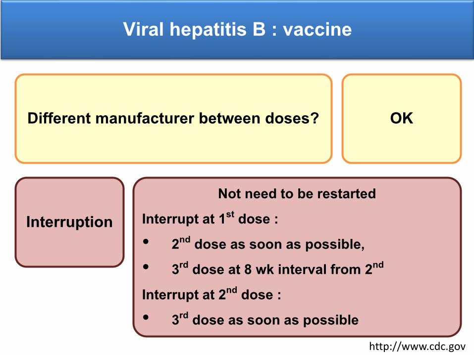
Viral hepatitis B : vaccine
Different manufacturer between doses? OK
Interruption Not need to be restarted
Interrupt at 1st dose : • 2nd dose as soon as possible, • 3rd dose at 8 wk interval from 2nd
Interrupt at 2nd dose : • 3rd dose as soon as possible
http://www.cdc.gov
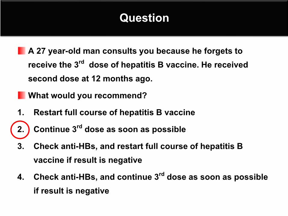
Question
A 27 year-old man consults you because he forgets to receive the 3rd dose of hepatitis B vaccine. He received second dose at 12 months ago. What would you recommend?
1. Restart full course of hepatitis B vaccine 2. Continue 3rd dose as soon as possible 3. Check anti-HBs, and restart full course of hepatitis B
vaccine if result is negative 4. Check anti-HBs, and continue 3rd dose as soon as possible
if result is negative
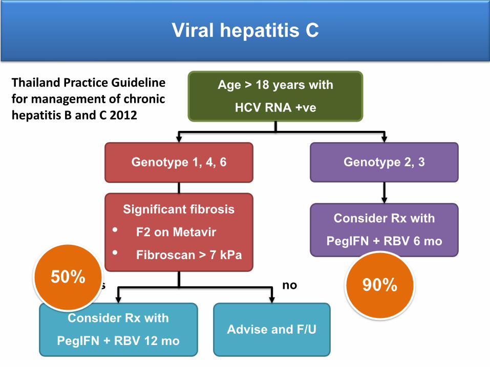
Viral hepatitis C
Age > 18 years with HCV RNA +ve
Genotype 1, 4, 6 Genotype 2, 3
Consider Rx with PegIFN + RBV 6 mo
Significant fibrosis• F2 on Metavir• Fibroscan > 7 kPa
Consider Rx with PegIFN + RBV 12 mo
Advise and F/U
yes no 90%50%
Thailand Practice Guideline for management of chronic hepatitis B and C 2012
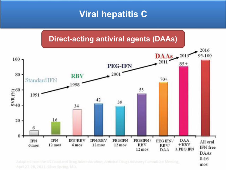
Viral hepatitis C
Direct-acting antiviral agents (DAAs)
Viral hepatitis C
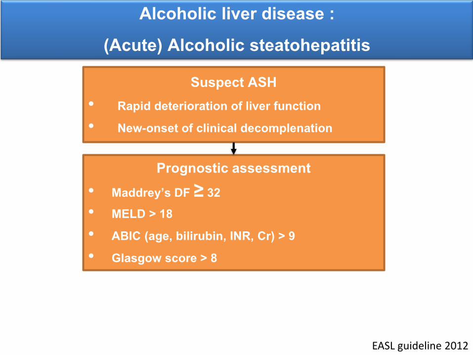
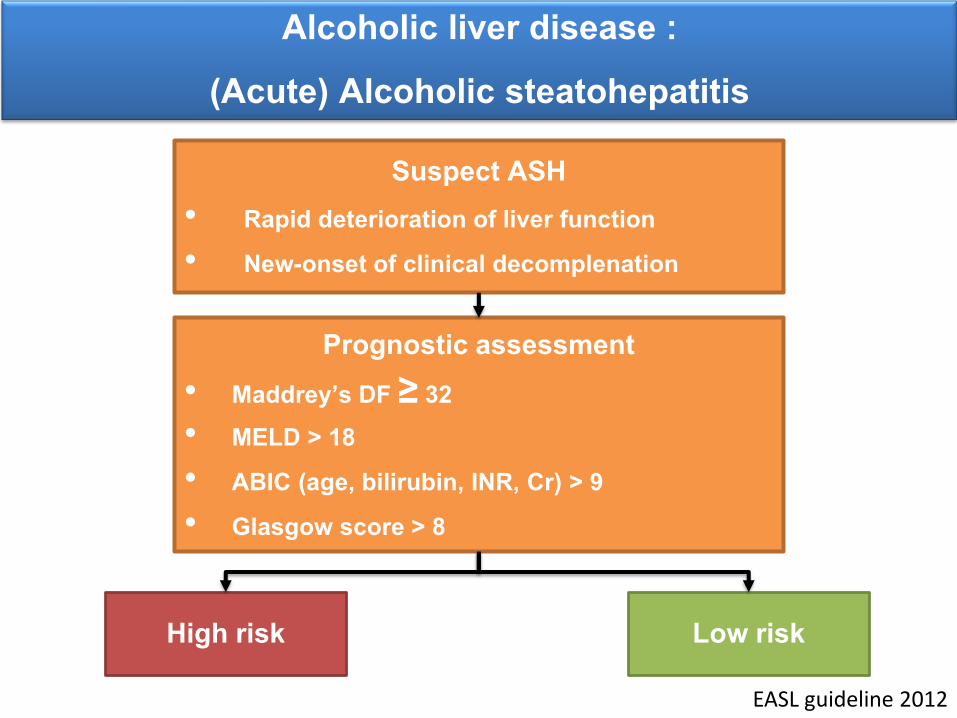
Alcoholic liver disease : (Acute) Alcoholic steatohepatitis
Suspect ASH• Rapid deterioration of liver function • New-onset of clinical decomplenation
Prognostic assessment • Maddrey’s DF ≥ 32• MELD > 18 • ABIC (age, bilirubin, INR, Cr) > 9• Glasgow score > 8
EASL guideline 2012
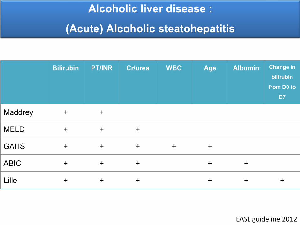
Alcoholic liver disease : (Acute) Alcoholic steatohepatitis
Bilirubin PT/INR Cr/urea WBC Age Albumin Change in bilirubin
from D0 to D7
Maddrey + +
MELD + + +
GAHS + + + + +
ABIC + + + + +
Lille + + + + + +
EASL guideline 2012
Alcoholic liver disease : (Acute) Alcoholic steatohepatitis
Suspect ASH• Rapid deterioration of liver function • New-onset of clinical decomplenation
Prognostic assessment • Maddrey’s DF ≥ 32• MELD > 18 • ABIC (age, bilirubin, INR, Cr) > 9• Glasgow score > 8
High risk Low risk
EASL guideline 2012
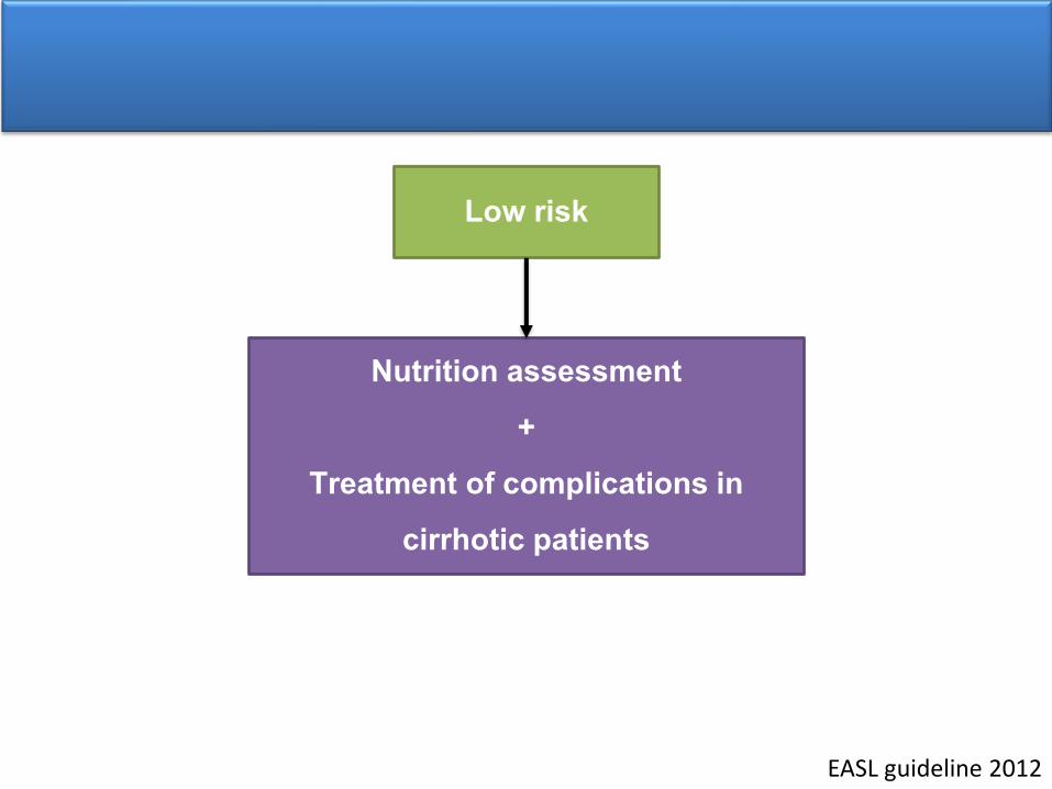
Low risk
Nutrition assessment +
Treatment of complications in cirrhotic patients
EASL guideline 2012
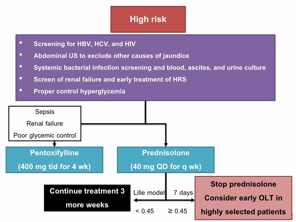
EASL guideline 2012
High risk
• Screening for HBV, HCV, and HIV• Abdominal US to exclude other causes of jaundice • Systemic bacterial infection screening and blood, ascites, and urine culture • Screen of renal failure and early treatment of HRS • Proper control hyperglycemia
Pentoxifylline (400 mg tid for 4 wk)
Prednisolone (40 mg QD for q wk)
Continue treatment 3 more weeks
Stop prednisoloneConsider early OLT in
highly selected patients
Lille model 7 days
< 0.45 ≥ 0.45
SepsisRenal failure
Poor glycemic control
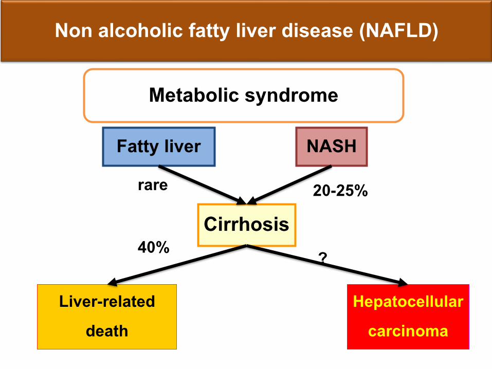
Non alcoholic fatty liver disease (NAFLD)
Cirrhosis
Hepatocellularcarcinoma
Liver-relateddeath
NASH
20-25%
40% ?
Fatty liverrare
Metabolic syndrome
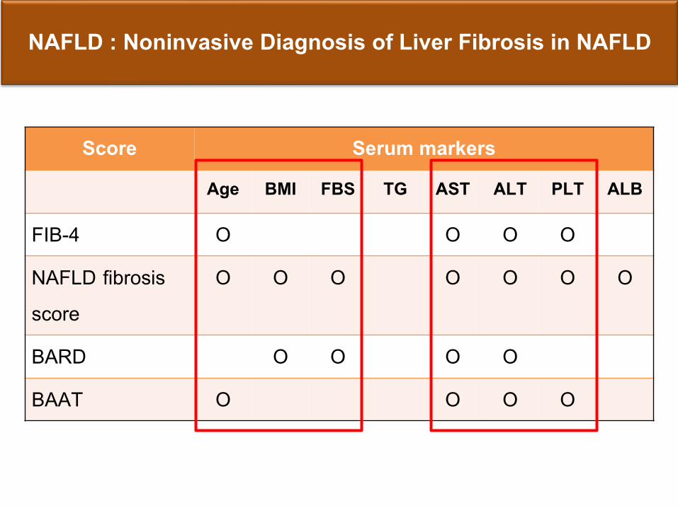
NAFLD : Noninvasive Diagnosis of Liver Fibrosis in NAFLD
Score Serum markersAge BMI FBS TG AST ALT PLT ALB
FIB-4 O O O O
NAFLD fibrosis score
O O O O O O O
BARD O O O O
BAAT O O O O
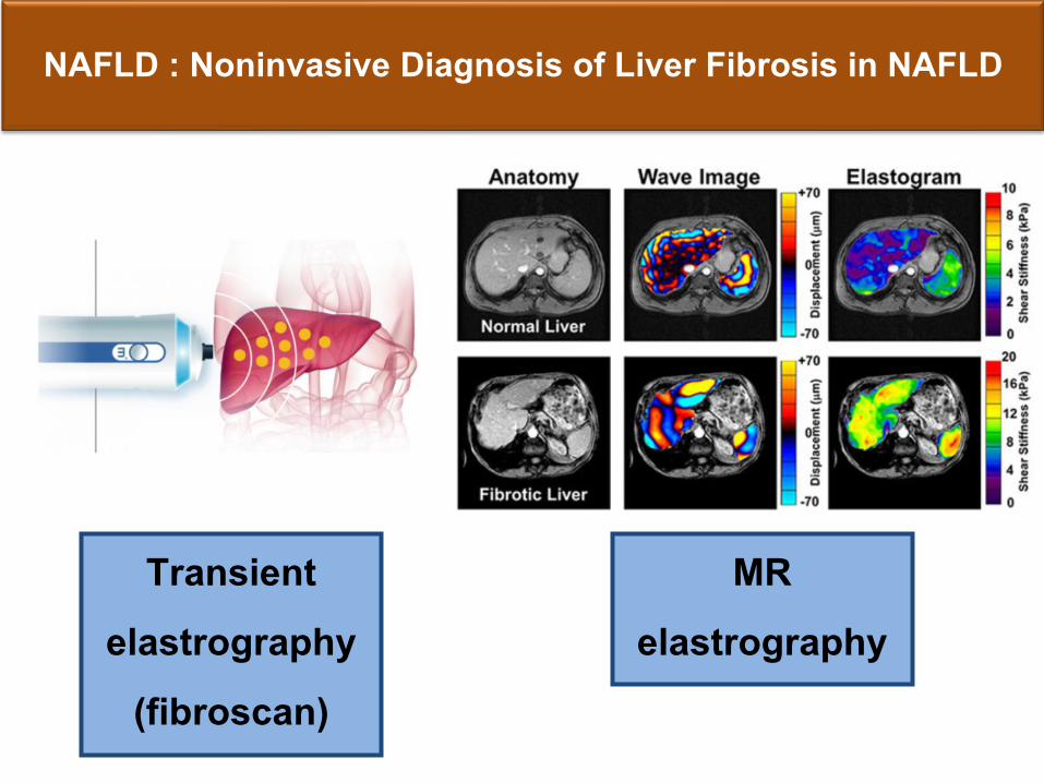
NAFLD : Noninvasive Diagnosis of Liver Fibrosis in NAFLD
Transient elastrography
(fibroscan)
MR elastrography
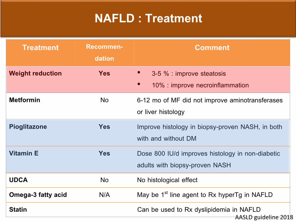
NAFLD : Treatment
Treatment Recommen-dation
Comment
Weight reduction Yes • 3-5 % : improve steatosis• 10% : improve necroinflammation
Metformin No 6-12 mo of MF did not improve aminotransferases or liver histology
Pioglitazone Yes Improve histology in biopsy-proven NASH, in both with and without DM
Vitamin E Yes Dose 800 IU/d improves histology in non-diabetic adults with biopsy-proven NASH
UDCA No No histological effect
Omega-3 fatty acid N/A May be 1st line agent to Rx hyperTg in NAFLD
Statin Can be used to Rx dyslipidemia in NAFLD AASLD guideline 2018
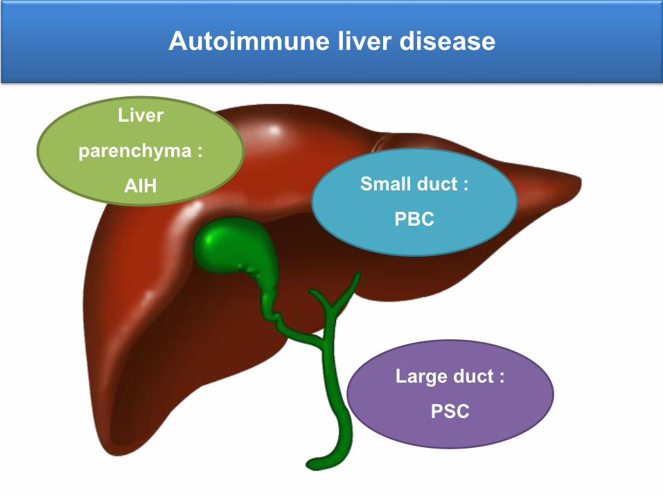
Autoimmune liver disease
Liver parenchyma :
AIH Small duct : PBC
Large duct : PSC
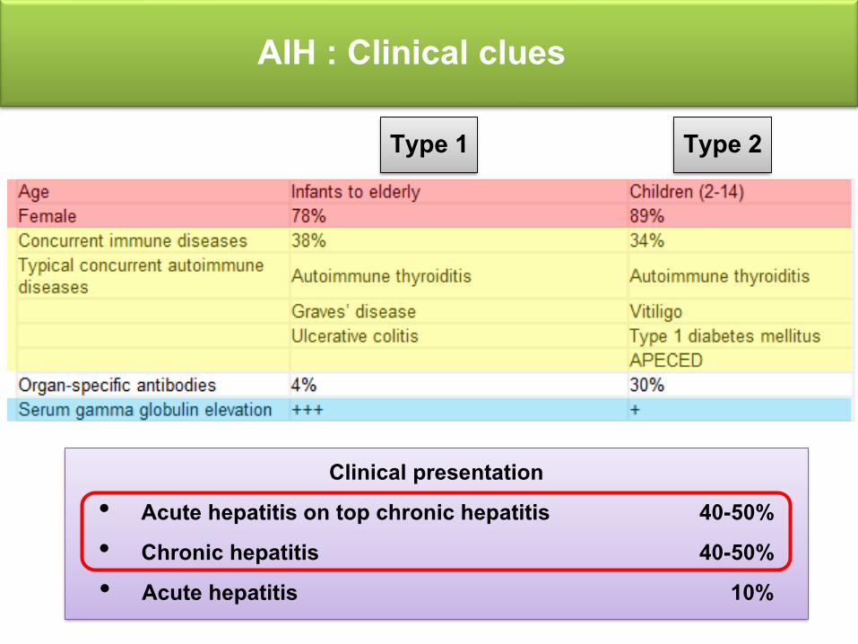
AIH : Clinical clues
Type 1 Type 2
Clinical presentation• Acute hepatitis on top chronic hepatitis 40-50%• Chronic hepatitis 40-50%• Acute hepatitis 10%
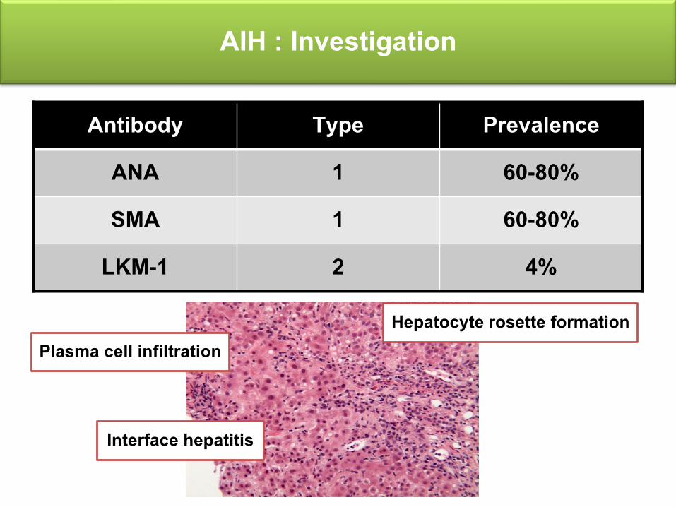
AIH : Investigation
Antibody Type Prevalence ANA 1 60-80%SMA 1 60-80%
LKM-1 2 4%
Plasma cell infiltration
Interface hepatitis
Hepatocyte rosette formation
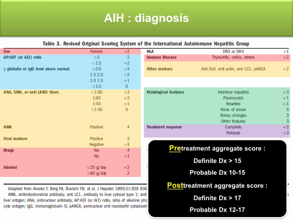
AIH : diagnosis
Pretreatment aggregate score : Definite Dx > 15
Probable Dx 10-15Posttreatment aggregate score :
Definite Dx > 17Probable Dx 12-17
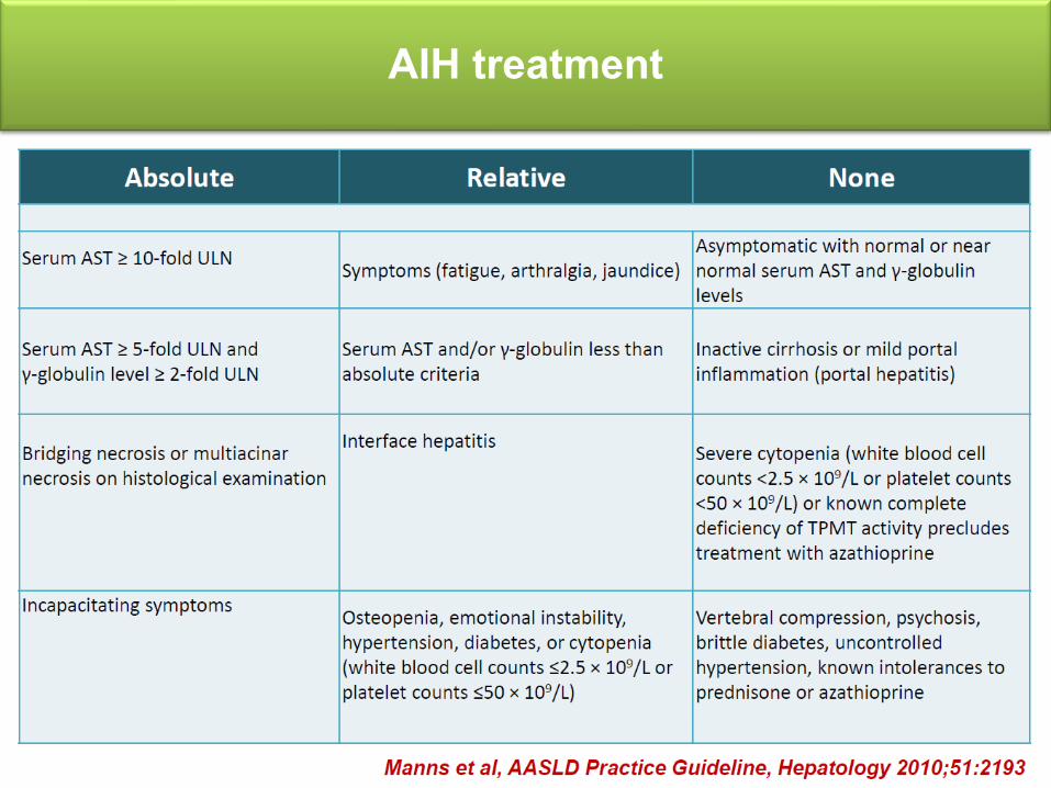
AIH treatment
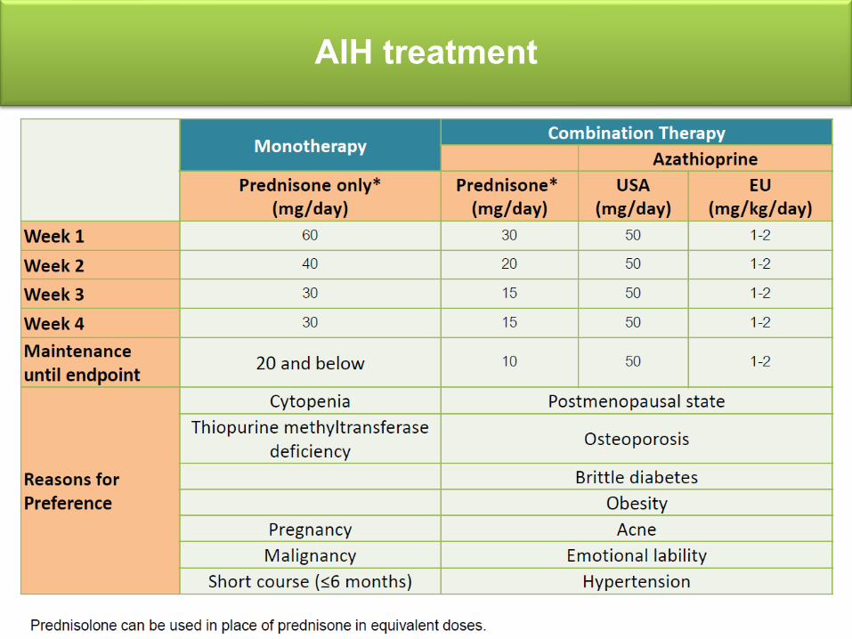
AIH treatment
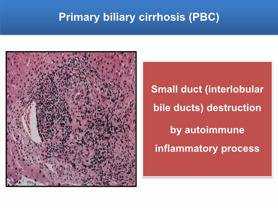
Primary biliary cirrhosis (PBC)
Small duct (interlobular bile ducts) destruction
by autoimmune inflammatory process
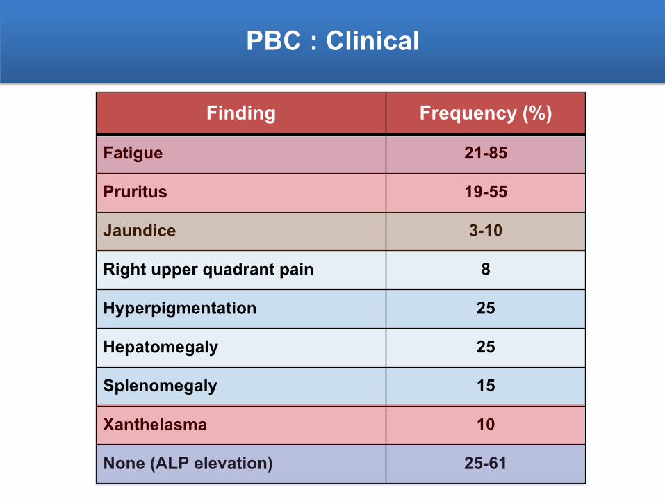
PBC : Clinical
Finding Frequency (%)Fatigue 21-85
Pruritus 19-55
Jaundice 3-10
Right upper quadrant pain 8
Hyperpigmentation 25
Hepatomegaly 25
Splenomegaly 15
Xanthelasma 10
None (ALP elevation) 25-61
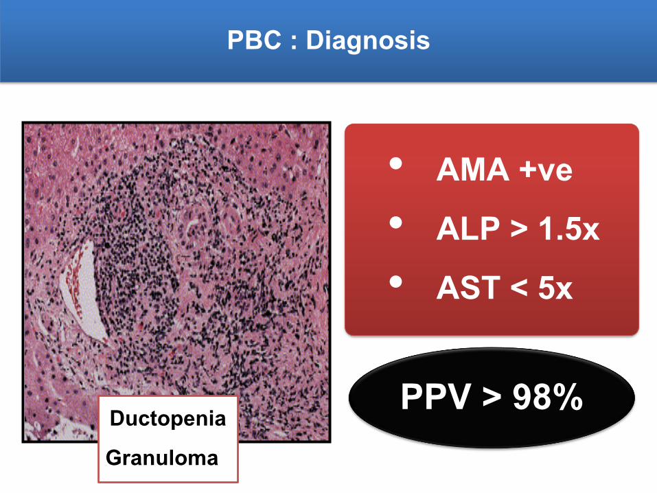
PBC : Diagnosis
• AMA +ve• ALP > 1.5x • AST < 5x
PPV > 98%DuctopeniaGranuloma
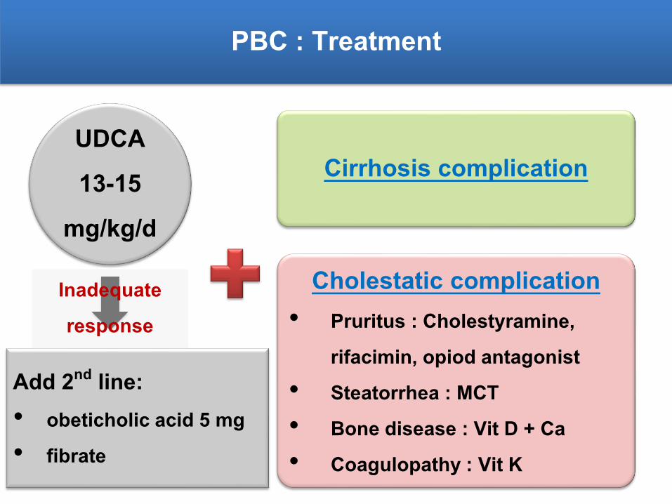
PBC : Treatment
UDCA 13-15
mg/kg/d
Cirrhosis complication
Cholestatic complication • Pruritus : Cholestyramine,
rifacimin, opiod antagonist• Steatorrhea : MCT• Bone disease : Vit D + Ca• Coagulopathy : Vit K
Add 2nd line:• obeticholic acid 5 mg• fibrate
Inadequate response
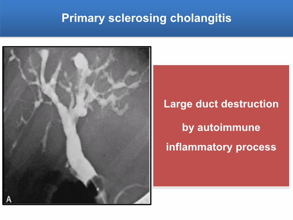
Primary sclerosing cholangitis
Large duct destruction
by autoimmune inflammatory process
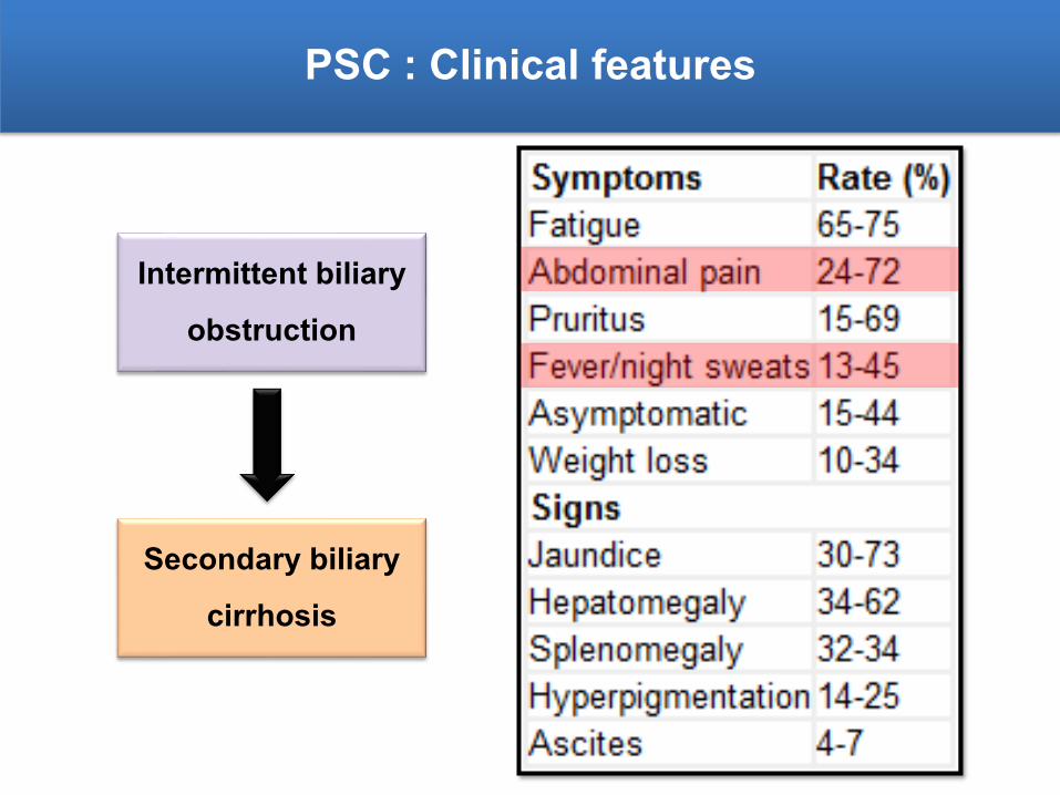
PSC : Clinical features
Intermittent biliary obstruction
Secondary biliary cirrhosis
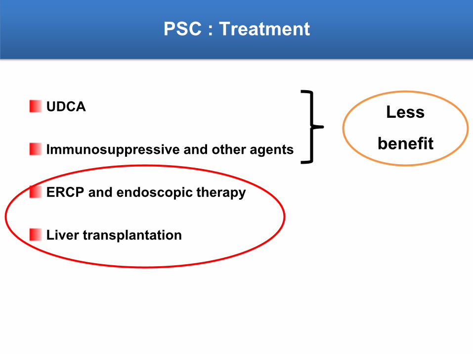
PSC : Treatment
UDCA
Immunosuppressive and other agents
ERCP and endoscopic therapy
Liver transplantation
Less benefit

Outline : Liver disease
Chronic liver disease Viral hepatitis
Alcoholic hepatitis
NAFLD
Autoimmune liver disease
Metabolic liver disease
Cirrhosis Portal hypertension
Variceal bleeding
Ascites and complications
Hepatorenal syndrome
Hepatocellular carcinoma
Question
Which condition can cause varix only at fundus of stomach?
1. Primary biliary cirrhosis
2. Essential thrombocytosis with portal vein thrombosis
3. Chronic pancreatitis
4. Budd-Chiari syndrome
5. Schistosomiasis

Portal hypertension
Left sided portal HT : Isolated fundal varices
Caput medusa : Cirrhosis
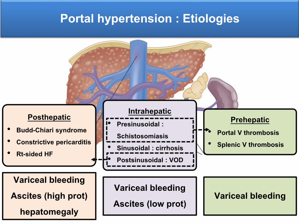
Portal hypertension : Etiologies
Prehepatic• Portal V thrombosis• Splenic V thrombosis
Posthepatic• Budd-Chiari syndrome• Constrictive pericarditis • Rt-sided HF
Intrahepatic • Presinusoidal :
Schistosomiasis• Sinusoidal : cirrhosis • Postsinusoidal : VOD
Variceal bleeding Variceal bleedingAscites (low prot)
Variceal bleedingAscites (high prot)
hepatomegaly
Question
Which condition can cause varix only at fundus of stomach?
1. Primary biliary cirrhosis
2. Essential thrombocytosis with portal vein thrombosis
3. Chronic pancreatitis
4. Budd-Chiari syndrome
5. Schistosomiasis
Variceal bleeding
No varices
Small varicesNo hemorrhage
Med/Large varicesNo hemorrhage
Varicealhemorrhage
Recurrenthemorrhage
8% per year
8% per year
5-15% per year
60%
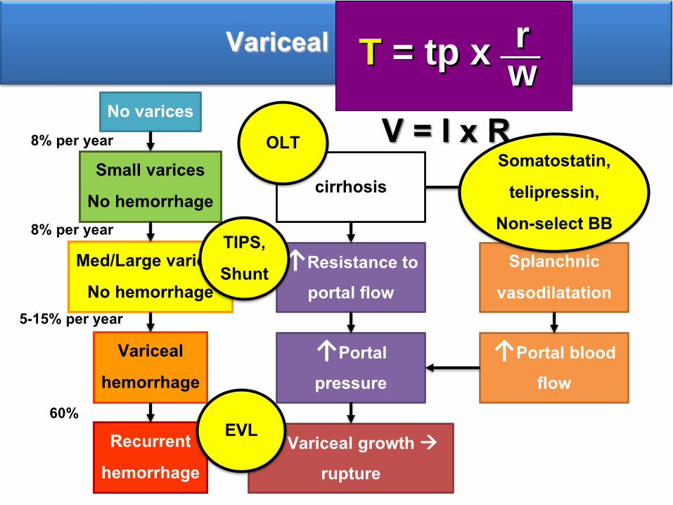
Variceal bleeding
V = I x R
↑Resistance to portal flow
↑Portal pressure
↑Variceal growth rupture
Splanchnic vasodilatation
↑Portal blood flow
cirrhosis↑NO
TIPS, Shunt
EVL
Somatostatin, telipressin,
Non-select BB
OLT
T = tp x rw
AASLD guideline 2017
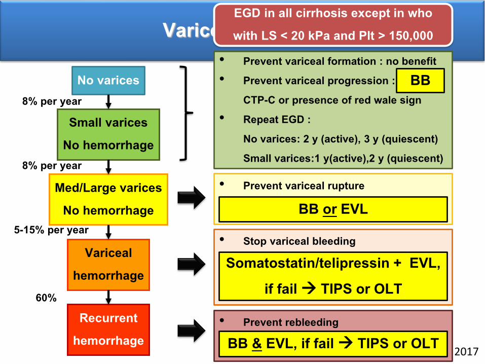
No varices
Small varicesNo hemorrhage
Med/Large varicesNo hemorrhage
Varicealhemorrhage
Recurrenthemorrhage
8% per year
8% per year
5-15% per year
60%
Variceal bleeding• Prevent variceal formation : no benefit • Prevent variceal progression : BB in
CTP-C or presence of red wale sign• Repeat EGD :
No varices: 2 y (active), 3 y (quiescent) Small varices:1 y(active),2 y (quiescent)
BB
• Prevent variceal rupture
BB or EVL
• Stop variceal bleeding
Somatostatin/telipressin + EVL, if fail TIPS or OLT
• Prevent rebleedingBB & EVL, if fail TIPS or OLT
EGD in all cirrhosis except in who with LS < 20 kPa and Plt > 150,000
Ascites
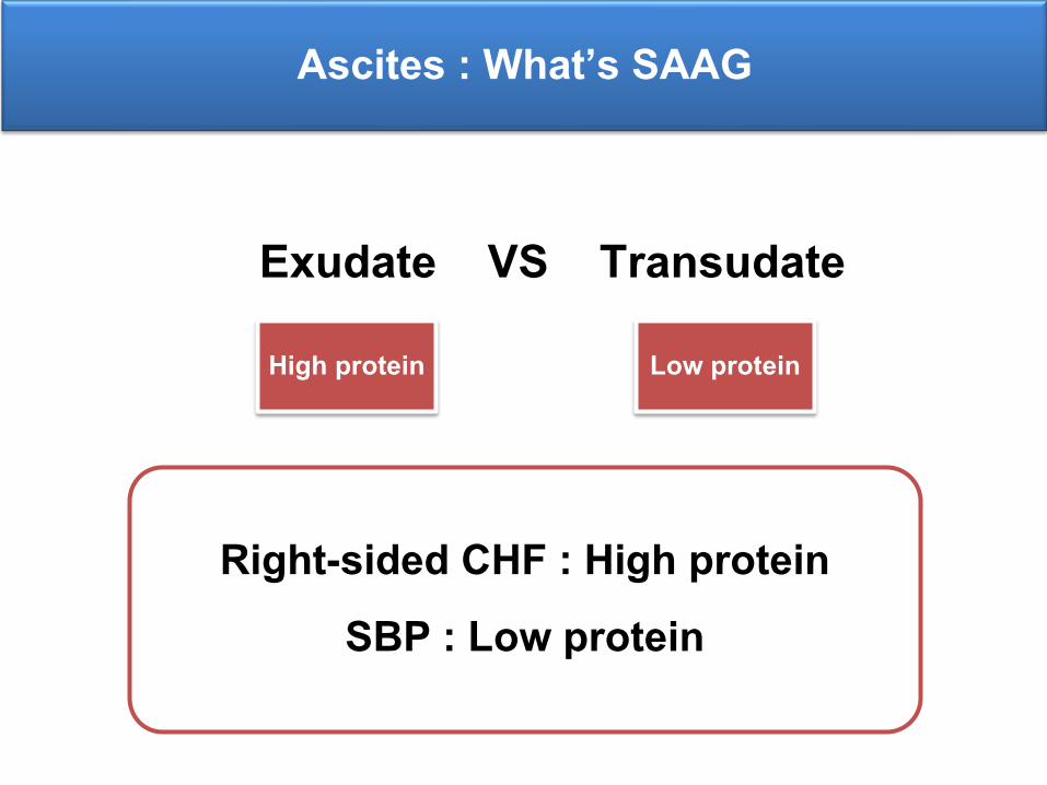
Ascites : What’s SAAG
Exudate VS TransudateHigh protein Low protein
Right-sided CHF : High protein SBP : Low protein
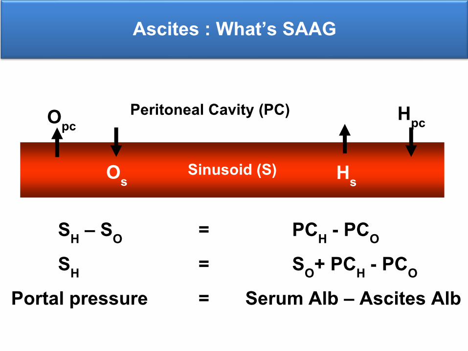
Ascites : What’s SAAG
Sinusoid (S)
Peritoneal Cavity (PC)
Hs
Hpc
Os
Opc
SH – SO = PCH - PCO
SH = SO+ PCH - PCO
Portal pressure = Serum Alb – Ascites Alb
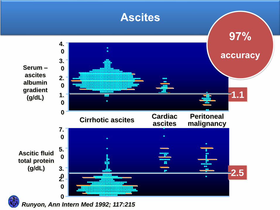
Ascites
Cirrhotic ascitesCardiac ascites
Peritoneal malignancy
1.1
4.
0
3.
0
2.
0
1.
0
0
Serum –
ascites
albumin
gradient
(g/dL)
(75)
Ascitic fluid
total protein
(g/dL)
7.
0
5.
0
3.
02.
0
0
2.5
Runyon, Ann Intern Med 1992; 117:215
97% accuracy
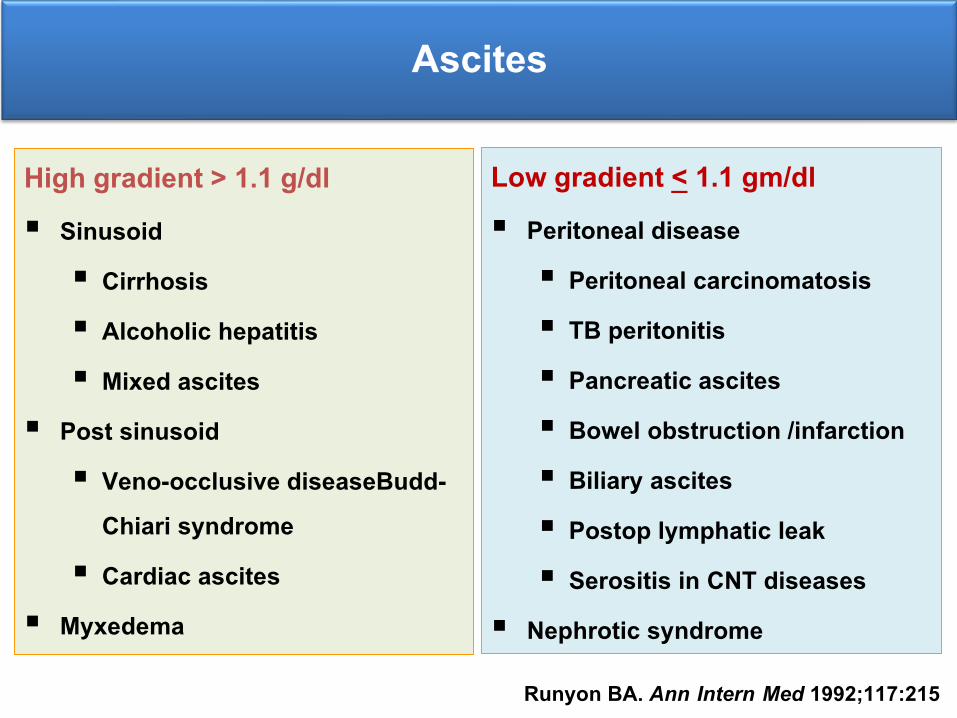
Ascites
Low gradient < 1.1 gm/dl▪ Peritoneal disease▪ Peritoneal carcinomatosis▪ TB peritonitis▪ Pancreatic ascites▪ Bowel obstruction /infarction▪ Biliary ascites▪ Postop lymphatic leak▪ Serositis in CNT diseases
▪ Nephrotic syndrome
High gradient > 1.1 g/dl▪ Sinusoid ▪ Cirrhosis▪ Alcoholic hepatitis▪ Mixed ascites
▪ Post sinusoid ▪ Veno-occlusive diseaseBudd-
Chiari syndrome▪ Cardiac ascites
▪ Myxedema
Runyon BA. Ann Intern Med 1992;117:215
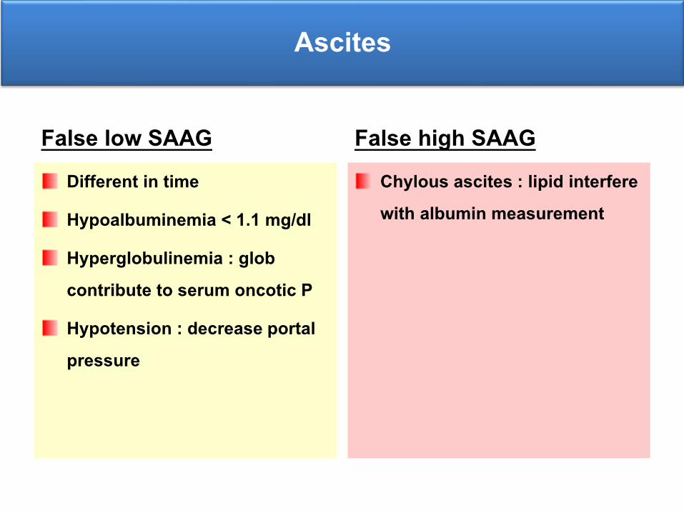
Ascites
False low SAAG Different in time
Hypoalbuminemia < 1.1 mg/dl
Hyperglobulinemia : glob contribute to serum oncotic P
Hypotension : decrease portal pressure
False high SAAG Chylous ascites : lipid interfere with albumin measurement
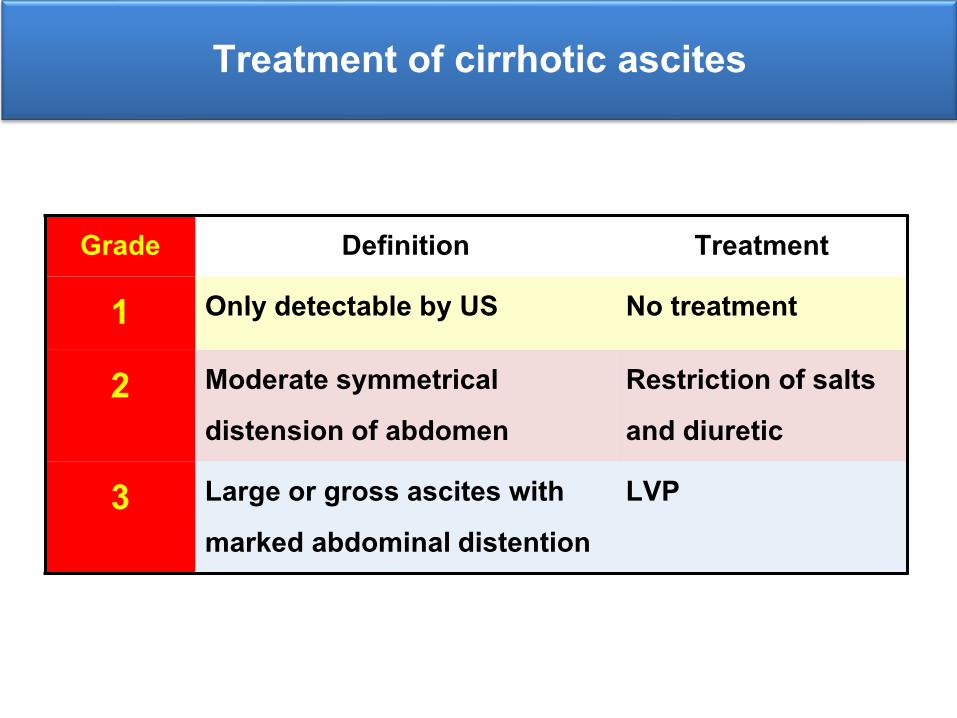
Treatment of cirrhotic ascites
Grade Definition Treatment
1 Only detectable by US No treatment
2 Moderate symmetrical distension of abdomen
Restriction of salts and diuretic
3 Large or gross ascites with marked abdominal distention
LVP
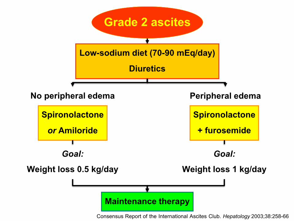
Grade 2 ascites
No peripheral edema
Low-sodium diet (70-90 mEq/day)Diuretics
Maintenance therapy
Spironolactone+ furosemide
Peripheral edema
Spironolactoneor Amiloride
Goal:Weight loss 0.5 kg/day
Goal:Weight loss 1 kg/day
Consensus Report of the International Ascites Club. Hepatology 2003;38:258-66
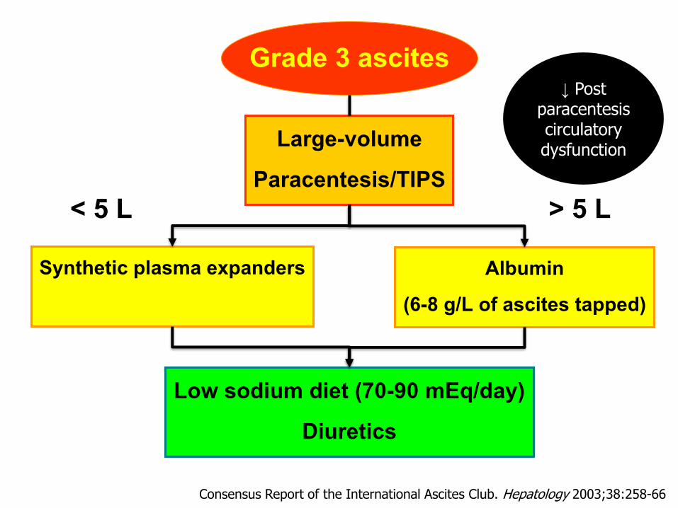
Grade 3 ascites
< 5 L
Large-volumeParacentesis/TIPS
> 5 LSynthetic plasma expanders
Low sodium diet (70-90 mEq/day)Diuretics
Albumin(6-8 g/L of ascites tapped)
Consensus Report of the International Ascites Club. Hepatology 2003;38:258-66
↓ Post paracentesiscirculatory dysfunction
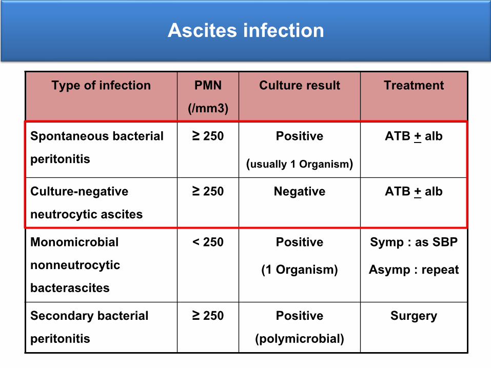
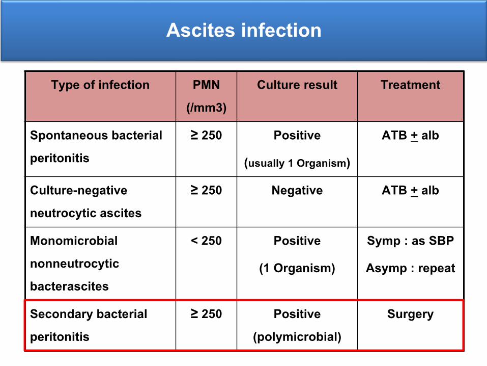
Ascites infection
Type of infection PMN (/mm3)
Culture result Treatment
Spontaneous bacterial peritonitis
≥ 250 Positive
(usually 1 Organism)
ATB + alb
Culture-negative neutrocytic ascites
≥ 250 Negative ATB + alb
Monomicrobial nonneutrocytic bacterascites
< 250 Positive
(1 Organism)
Symp : as SBP
Asymp : repeat
Secondary bacterial peritonitis
≥ 250 Positive (polymicrobial)
Surgery
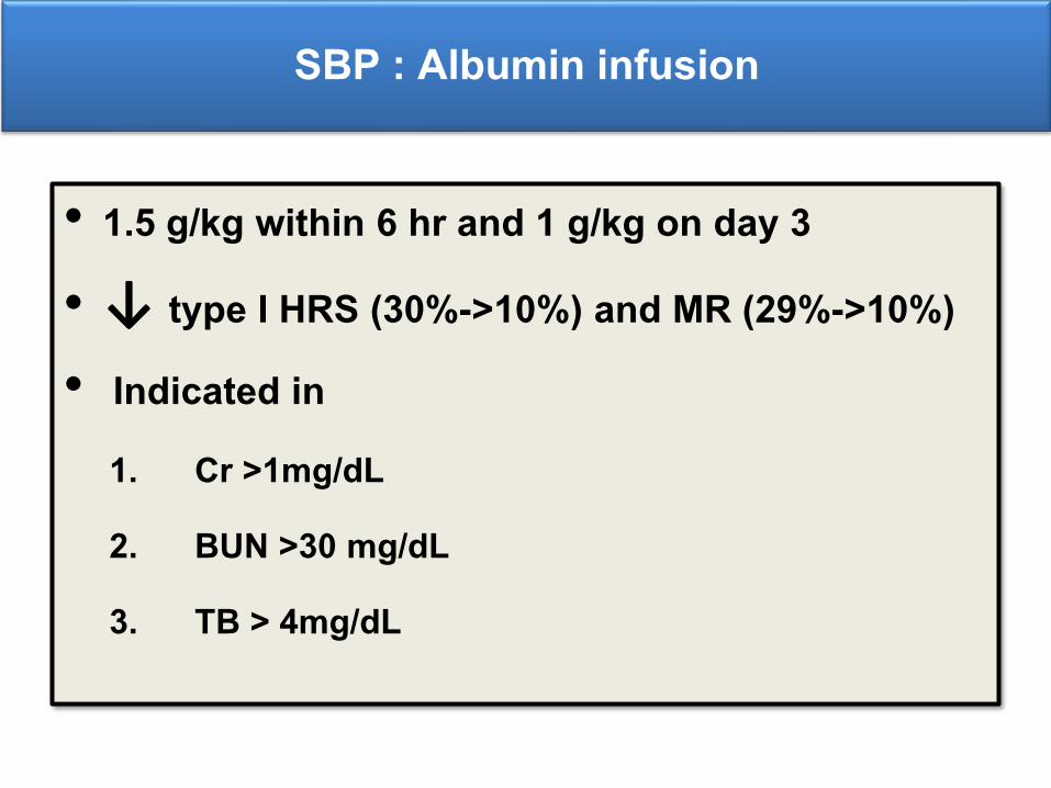
SBP : Albumin infusion
• 1.5 g/kg within 6 hr and 1 g/kg on day 3
• ↓ type I HRS (30%->10%) and MR (29%->10%)• Indicated in
1. Cr >1mg/dL
2. BUN >30 mg/dL
3. TB > 4mg/dL

Ascites infection
Type of infection PMN (/mm3)
Culture result Treatment
Spontaneous bacterial peritonitis
≥ 250 Positive
(usually 1 Organism)
ATB + alb
Culture-negative neutrocytic ascites
≥ 250 Negative ATB + alb
Monomicrobial nonneutrocytic bacterascites
< 250 Positive
(1 Organism)
Symp : as SBP
Asymp : repeat
Secondary bacterial peritonitis
≥ 250 Positive (polymicrobial)
Surgery
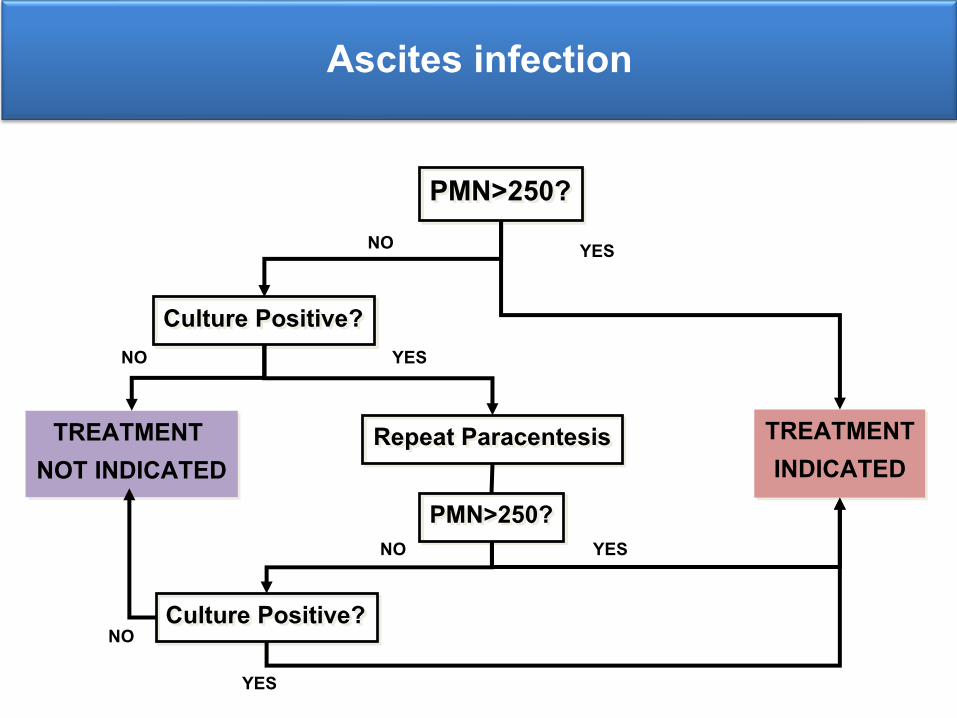
Ascites infection
TREATMENTINDICATED
PMN>250?
Culture Positive?
TREATMENT NOT INDICATED
NO
Repeat Paracentesis
YES
PMN>250?
Culture Positive?
NO
NO
YES
YES
YESNO
Ascites infection
Type of infection PMN (/mm3)
Culture result Treatment
Spontaneous bacterial peritonitis
≥ 250 Positive
(usually 1 Organism)
ATB + alb
Culture-negative neutrocytic ascites
≥ 250 Negative ATB + alb
Monomicrobial nonneutrocytic bacterascites
< 250 Positive
(1 Organism)
Symp : as SBP
Asymp : repeat
Secondary bacterial peritonitis
≥ 250 Positive (polymicrobial)
Surgery
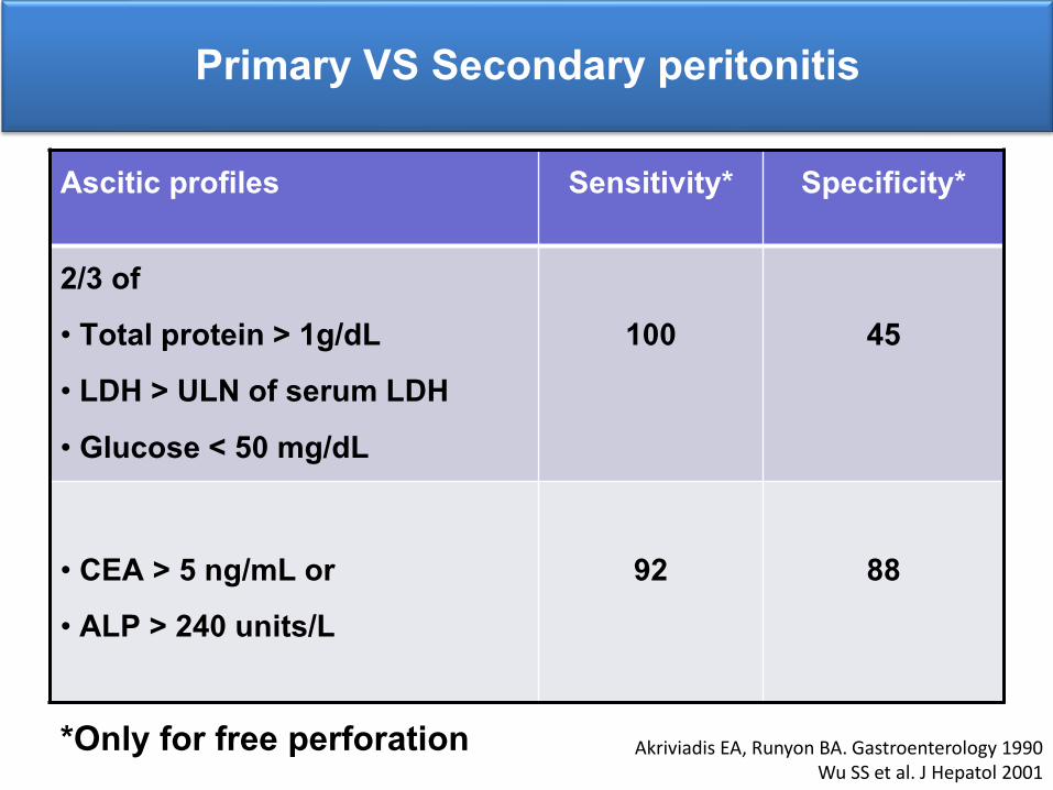
Primary VS Secondary peritonitis
*Only for free perforation
Ascitic profiles Sensitivity* Specificity*
2/3 of• Total protein > 1g/dL• LDH > ULN of serum LDH • Glucose < 50 mg/dL
100 45
• CEA > 5 ng/mL or• ALP > 240 units/L
92 88
Akriviadis EA, Runyon BA. Gastroenterology 1990Wu SS et al. J Hepatol 2001
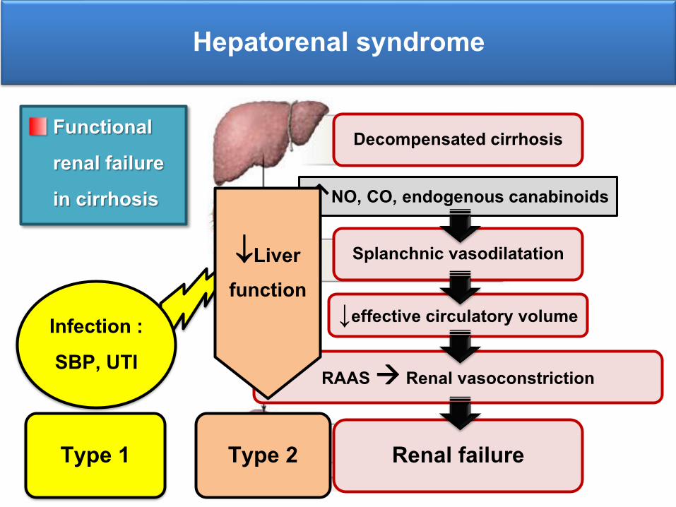
Hepatorenal syndrome
Functional renal failure in cirrhosis ↑NO, CO, endogenous canabinoids
Decompensated cirrhosis
Splanchnic vasodilatation
↓effective circulatory volume
Renal failure
RAAS Renal vasoconstriction
Infection : SBP, UTI
Type 1
Liver function
Type 2
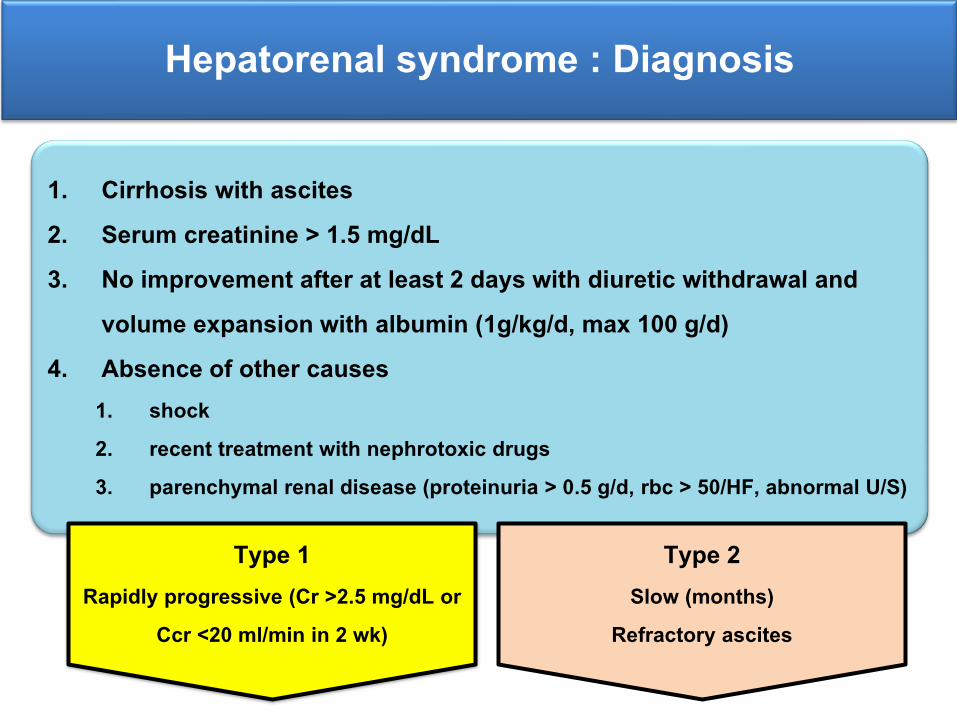
Hepatorenal syndrome : Diagnosis
1. Cirrhosis with ascites2. Serum creatinine > 1.5 mg/dL3. No improvement after at least 2 days with diuretic withdrawal and
volume expansion with albumin (1g/kg/d, max 100 g/d)4. Absence of other causes
1. shock2. recent treatment with nephrotoxic drugs3. parenchymal renal disease (proteinuria > 0.5 g/d, rbc > 50/HF, abnormal U/S)
Type 1 Rapidly progressive (Cr >2.5 mg/dL or
Ccr <20 ml/min in 2 wk)
Type 2 Slow (months)
Refractory ascites
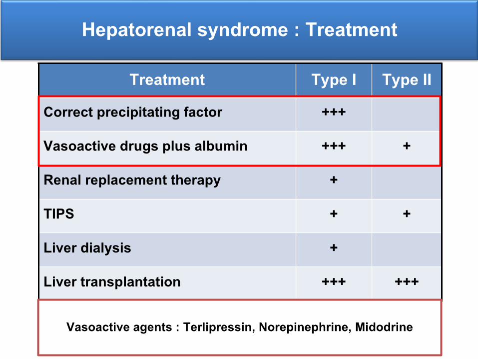
Hepatorenal syndrome : Treatment
Treatment Type I Type IICorrect precipitating factor +++
Vasoactive drugs plus albumin +++ +
Renal replacement therapy +
TIPS + +
Liver dialysis +
Liver transplantation +++ +++
Vasoactive agents : Terlipressin, Norepinephrine, Midodrine

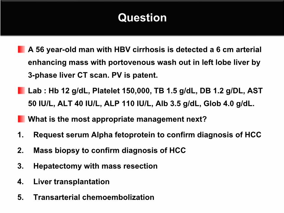
Question
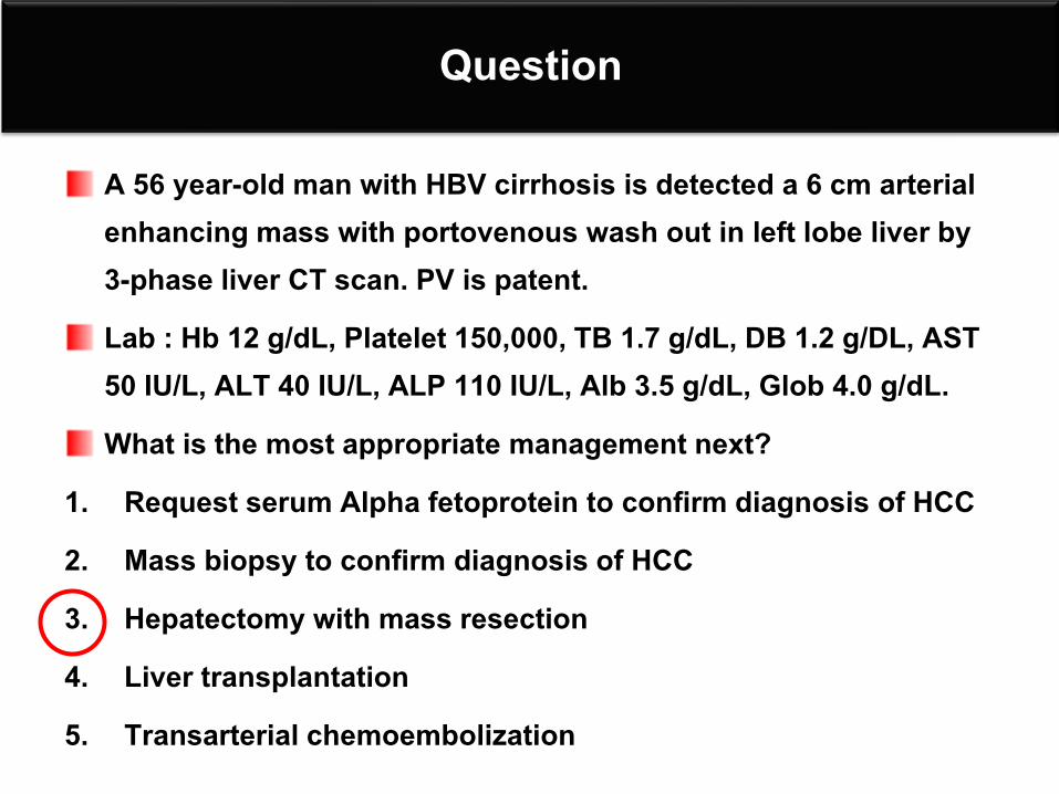
A 56 year-old man with HBV cirrhosis is detected a 6 cm arterial enhancing mass with portovenous wash out in left lobe liver by 3-phase liver CT scan. PV is patent. Lab : Hb 12 g/dL, Platelet 150,000, TB 1.5 g/dL, DB 1.2 g/DL, AST 50 IU/L, ALT 40 IU/L, ALP 110 IU/L, Alb 3.5 g/dL, Glob 4.0 g/dL. What is the most appropriate management next?
1. Request serum Alpha fetoprotein to confirm diagnosis of HCC 2. Mass biopsy to confirm diagnosis of HCC 3. Hepatectomy with mass resection 4. Liver transplantation 5. Transarterial chemoembolization
Hepatocellular carcinoma
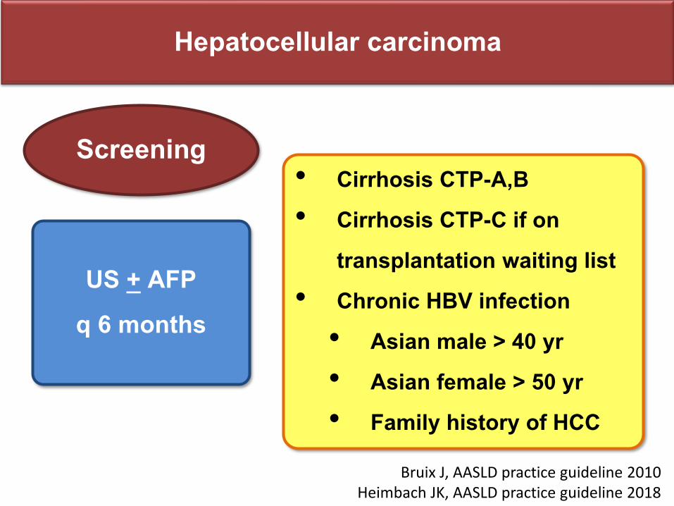
US + AFP q 6 months
• Cirrhosis CTP-A,B• Cirrhosis CTP-C if on
transplantation waiting list • Chronic HBV infection • Asian male > 40 yr • Asian female > 50 yr• Family history of HCC
Screening
Bruix J, AASLD practice guideline 2010Heimbach JK, AASLD practice guideline 2018
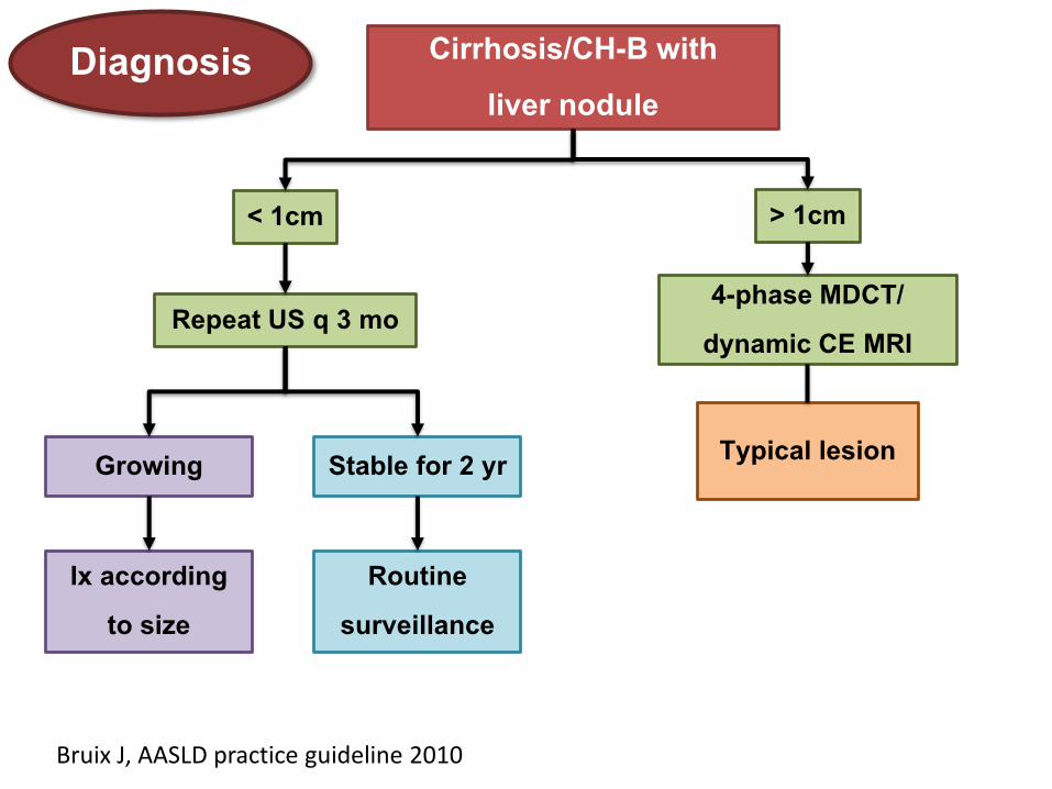
Diagnosis
Bruix J, AASLD practice guideline 2010
Cirrhosis/CH-B with liver nodule
< 1cm
Repeat US q 3 mo
Growing Stable for 2 yr
Ix according to size
Routine surveillance
> 1cm
4-phase MDCT/ dynamic CE MRI
Typical lesion
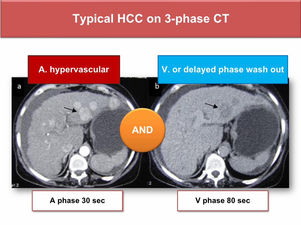
Typical HCC on 3-phase CT
A phase 30 sec V phase 80 sec
V. or delayed phase wash outA. hypervascular
AND
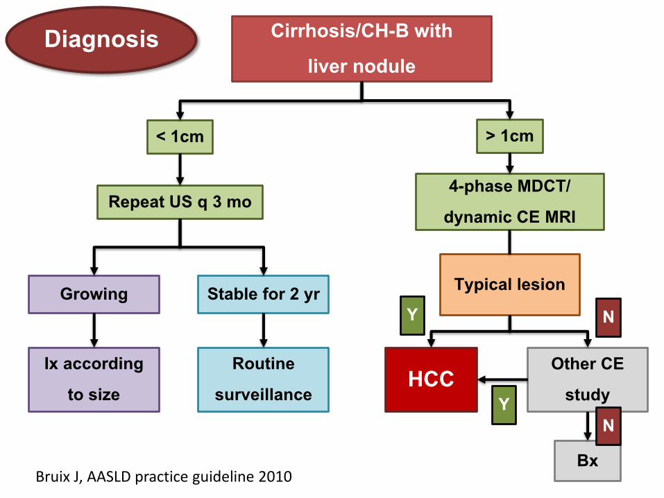
Diagnosis
Bruix J, AASLD practice guideline 2010
Cirrhosis/CH-B with liver nodule
< 1cm
Repeat US q 3 mo
Growing Stable for 2 yr
Ix according to size
Routine surveillance
> 1cm
4-phase MDCT/ dynamic CE MRI
Typical lesion
HCC Other CE study
Bx
Y
YN
N
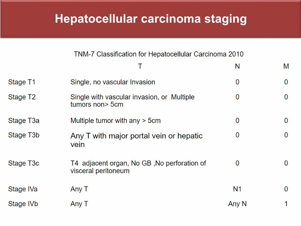
Hepatocellular carcinoma staging
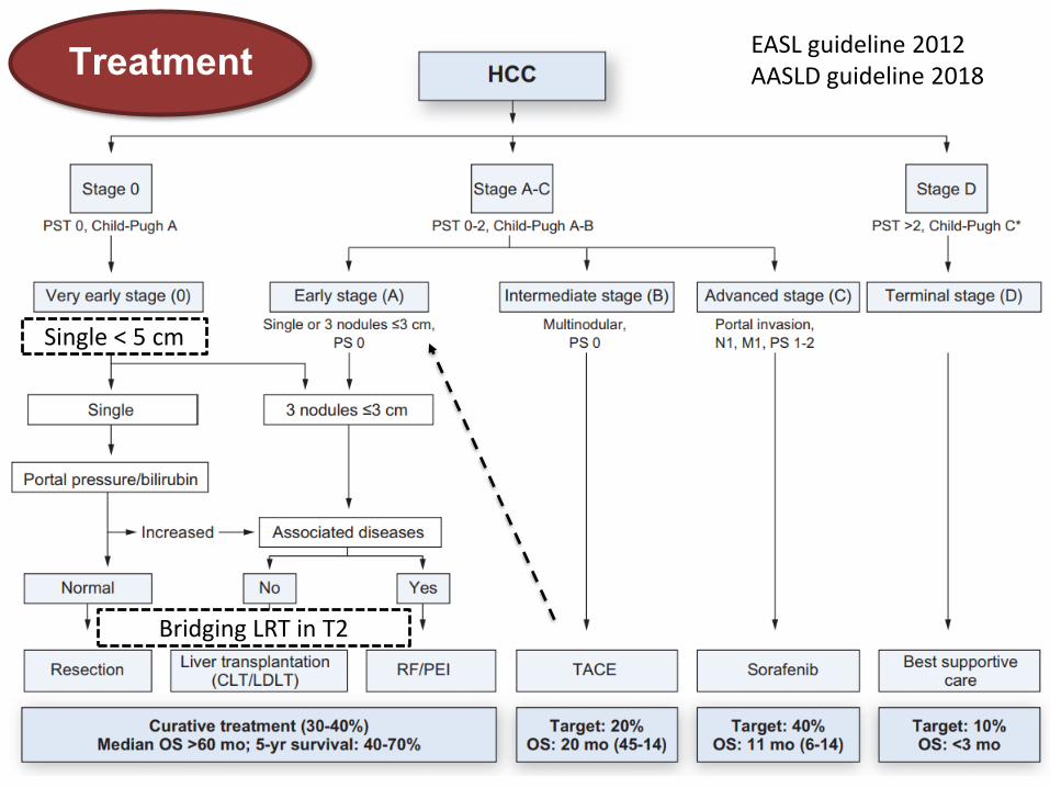
Treatment EASL guideline 2012AASLD guideline 2018
Bridging LRT in T2
Single < 5 cm
Question
A 56 year-old man with HBV cirrhosis is detected a 6 cm arterial enhancing mass with portovenous wash out in left lobe liver by 3-phase liver CT scan. PV is patent. Lab : Hb 12 g/dL, Platelet 150,000, TB 1.7 g/dL, DB 1.2 g/DL, AST 50 IU/L, ALT 40 IU/L, ALP 110 IU/L, Alb 3.5 g/dL, Glob 4.0 g/dL. What is the most appropriate management next?
1. Request serum Alpha fetoprotein to confirm diagnosis of HCC 2. Mass biopsy to confirm diagnosis of HCC 3. Hepatectomy with mass resection 4. Liver transplantation 5. Transarterial chemoembolization

Thank you for your attentions


![Journal of Stem Cell Research & Therapy · Strongyloides stercolaris is endemic in tropical regions, in the United States (US) it is found in the south east [19]. It is usually asymptomatic](https://img.pdfslide.net/doc/110x75/5be5298a09d3f2580c8b482c/journal-of-stem-cell-research-therapy-strongyloides-stercolaris-is-endemic.jpg)


![Hybrid CORDIC 3. ROMless 20180303 - · PDF file3/3/2018 · [23] M. Kuhlmann and K. K. Parhi, "P-CORDIC: A precomputation based rotation CORDIC algorithm," EURASIP J. Appl](https://img.pdfslide.net/doc/110x75/5a9c04cd7f8b9a9c5b8e51cc/hybrid-cordic-3-romless-20180303-23-m-kuhlmann-and-k-k-parhi-p-cordic-a.jpg)