Embed Size (px)
Citation preview
December 15, 2014 ◆ Volume 90, Number 12 www.aafp.org/afp American Family Physician 843
Common Questions About Developmental Dysplasia of the HipJONATHAN C. JACKSON, MD, Uniformed Services University of the Health Sciences, Bethesda, Maryland
MELISSA M. RUNGE, MD, 422nd Medical Squadron, Royal Air Force Croughton, United Kingdom
NATHANIEL S. NYE, MD, 559th Trainee Health Squadron, Joint Base San Antonio-Lackland, Texas
Screening for developmental dys-plasia of the hip (DDH) has been a component of neonatal medical care for more than 70 years.1,2 This arti-
cle reviews current evidence and opinions regarding screening for DDH, with a focus on universal screening in well newborns and a discussion of the current approach to treat-ment of DDH.
DDH denotes an abnormality of the ace-tabulum or femoral head and their congru-ence that presents at birth or in infancy. It is more inclusive than the previous term “con-genital hip dislocation,” because it includes abnormalities other than overt dislocation.
DDH terminology is sometimes confusing (Table 1).2-4 A hip clunk is a significant find-ing. A hip click, on the other hand, is thought to be caused by benign soft-tissue move-ment. It has been shown in multiple studies to be unrelated to DDH and does not require further evaluation.5-7 Sometimes examina-tion findings include a mild sensation of
instability or laxity without frank sublux-ation or dislocation. This finding is often called “mild instability”; this term will be used throughout this article.2-4
Incidence and Risk FactorsDevelopmental dysplasia of the hip is a common musculoskeletal condition in newborns. The reported prevalence of hip instability on physical examination at birth ranges from 1.6 to 28.5 per 1,000 infants, but the prevalence of persistent abnormalities after the first few days of life, as reported in a meta-analysis of several studies in American and European populations, is 1.3 per 1,000.2 Studies using ultrasonography have found that about 5% of newborns have some radio-graphic abnormality of the hip, although many of these abnormalities are undetect-able on physical examination.8 Rarely, DDH may develop or worsen after the neonatal period.9 DDH is associated with early-onset osteoarthritis of the hip in adulthood.
Developmental dysplasia of the hip is a common musculoskeletal condition in newborns. Infants with developmen-tal dysplasia of the hip, whether treated or untreated, have a higher incidence of early-onset hip osteoarthritis in adulthood. Evidence to support universal screening by physical examination or ultrasonography is limited and often conflicting. The U.S. Preventive Services Task Force found insufficient evidence that screening for developmental dysplasia of the hip prevents adverse outcomes. Physical examination screening is recommended by the American Academy of Pediatrics and the Pediatric Orthopaedic Society of North America. These organizations recommend use of the Ortolani and Barlow maneuvers to screen infants up to three months of age. Several recent studies support starting assessment for limited hip abduction at eight weeks of age, which is the most sensitive test for developmen-tal dysplasia of the hip from this age on. Infants with overtly dislocated or dislocatable hips should be referred to an orthopedist on a priority basis at the time of diagnosis. Infants with equivocal hip examination findings at birth can be reexamined in two weeks. If there is subluxation or dislocation at the follow-up examination, referral should be made at that time. If the examination findings are still equivocal, the infant can undergo ultrasonography of the hips or be reexamined every few weeks through the first six weeks of life. Although equivocal findings commonly resolve spontaneously, infants with persistent equivocal findings of developmental dysplasia of the hip longer than six weeks should be evaluated by an orthopedist. Treatment generally involves flexion-abduction splinting. The benefits of treat-ment are unclear, and there are risks to treatment, most notably an increased occurrence of avascular necrosis of the femoral head. (Am Fam Physician. 2014;90(12):843-850. Copyright © 2014 American Academy of Family Physicians.)
CME This clinical content conforms to AAFP criteria for continuing medical edu-cation (CME). See CME Quiz Questions on page 830.
Author disclosure: No rel-evant financial affiliations.
▲
Patient information: A handout on this topic, written by the authors of this article, is available at http://www.aafp.org/afp/ 2014/1215/843-s1.html.
Downloaded from the American Family Physician website at www.aafp.org/afp. Copyright © 2014 American Academy of Family Physicians. For the private, noncom-mercial use of one individual user of the website. All other rights reserved. Contact [email protected] for copyright questions and/or permission requests.
Developmental Dysplasia of the Hip
844 American Family Physician www.aafp.org/afp Volume 90, Number 12 ◆ December 15, 2014
Some of the strongest risk factors for DDH are breech position, female sex, and first gestation (with odds ratios in one study of 6.0, 4.3, and 2.7, respectively).10 The inci-dence of clinically unstable hips at birth among infants
in the breech position may exceed 10%.11 Other risk factors and associated findings include family history of DDH, oligohy-dramnios, large birth weight for gestational age, metatarsus adductus, and torticollis, among others. Why these risk factors are associated with DDH is not well understood, although limited intrauterine space and fetal position may be involved.2
ScreeningWHAT METHODS ARE USED FOR SCREENING?
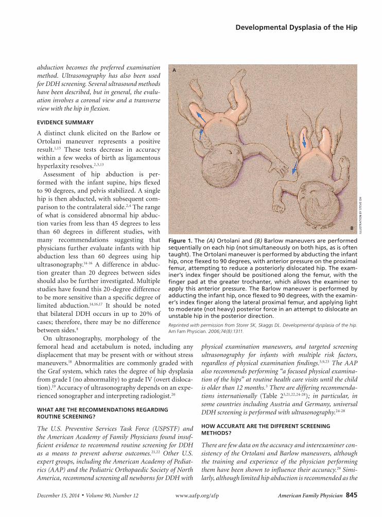
The Ortolani (reducing a dislocated hip) and Barlow (dislocating an unstable hip) maneu-
vers are the physical examination tests most commonly per-formed for detection of DDH in early infancy (Figure 112). By two to three months of age, the Barlow and Ortolani maneuvers are less useful and assessment for limited hip
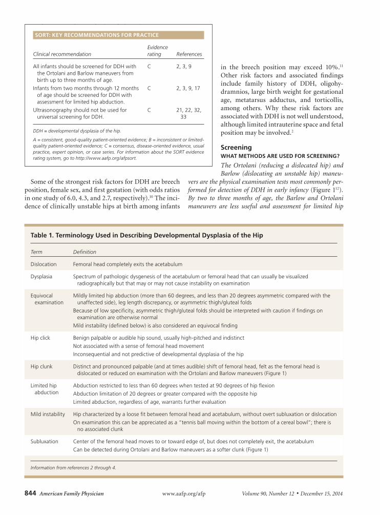
Table 1. Terminology Used in Describing Developmental Dysplasia of the Hip
Term Definition
Dislocation Femoral head completely exits the acetabulum
Dysplasia Spectrum of pathologic dysgenesis of the acetabulum or femoral head that can usually be visualized radiographically but that may or may not cause instability on examination
Equivocal examination
Mildly limited hip abduction (more than 60 degrees, and less than 20 degrees asymmetric compared with the unaffected side), leg length discrepancy, or asymmetric thigh/gluteal folds
Because of low specificity, asymmetric thigh/gluteal folds should be interpreted with caution if findings on examination are otherwise normal
Mild instability (defined below) is also considered an equivocal finding
Hip click Benign palpable or audible hip sound, usually high-pitched and indistinct
Not associated with a sense of femoral head movement
Inconsequential and not predictive of developmental dysplasia of the hip
Hip clunk Distinct and pronounced palpable (and at times audible) shift of femoral head, felt as the femoral head is dislocated or reduced on examination with the Ortolani and Barlow maneuvers (Figure 1)
Limited hip abduction
Abduction restricted to less than 60 degrees when tested at 90 degrees of hip flexion
Abduction limitation of 20 degrees or greater compared with the opposite hip
Limited abduction, regardless of age, warrants further evaluation
Mild instability Hip characterized by a loose fit between femoral head and acetabulum, without overt subluxation or dislocation
On examination this can be appreciated as a “tennis ball moving within the bottom of a cereal bowl”; there is no associated clunk
Subluxation Center of the femoral head moves to or toward edge of, but does not completely exit, the acetabulum
Can be detected during Ortolani and Barlow maneuvers as a softer clunk (Figure 1)
Information from references 2 through 4.
SORT: KEY RECOMMENDATIONS FOR PRACTICE
Clinical recommendationEvidence rating References
All infants should be screened for DDH with the Ortolani and Barlow maneuvers from birth up to three months of age.
C 2, 3, 9
Infants from two months through 12 months of age should be screened for DDH with assessment for limited hip abduction.
C 2, 3, 9, 17
Ultrasonography should not be used for universal screening for DDH.
C 21, 22, 32, 33
DDH = developmental dysplasia of the hip.
A = consistent, good-quality patient-oriented evidence; B = inconsistent or limited-quality patient-oriented evidence; C = consensus, disease-oriented evidence, usual practice, expert opinion, or case series. For information about the SORT evidence rating system, go to http://www.aafp.org/afpsort.
Developmental Dysplasia of the Hip
December 15, 2014 ◆ Volume 90, Number 12 www.aafp.org/afp American Family Physician 845
abduction becomes the preferred examination method. Ultrasonography has also been used for DDH screening. Several ultrasound methods have been described, but in general, the evalu-ation involves a coronal view and a transverse view with the hip in flexion.
EVIDENCE SUMMARY
A distinct clunk elicited on the Barlow or Ortolani maneuver represents a positive result.1,13 These tests decrease in accuracy within a few weeks of birth as ligamentous hyperlaxity resolves.2,3,13
Assessment of hip abduction is per-formed with the infant supine, hips flexed to 90 degrees, and pelvis stabilized. A single hip is then abducted, with subsequent com-parison to the contralateral side.2,4 The range of what is considered abnormal hip abduc-tion varies from less than 45 degrees to less than 60 degrees in different studies, with many recommendations suggesting that physicians further evaluate infants with hip abduction less than 60 degrees using hip ultrasonography.14-16 A difference in abduc-tion greater than 20 degrees between sides should also be further investigated. Multiple studies have found this 20-degree difference to be more sensitive than a specific degree of limited abduction.14,16,17 It should be noted that bilateral DDH occurs in up to 20% of cases; therefore, there may be no difference between sides.4
On ultrasonography, morphology of the femoral head and acetabulum is noted, including any displacement that may be present with or without stress maneuvers.18 Abnormalities are commonly graded with the Graf system, which rates the degree of hip dysplasia from grade I (no abnormality) to grade IV (overt disloca-tion).19 Accuracy of ultrasonography depends on an expe-rienced sonographer and interpreting radiologist.20
WHAT ARE THE RECOMMENDATIONS REGARDING ROUTINE SCREENING?
The U.S. Preventive Services Task Force (USPSTF) and the American Academy of Family Physicians found insuf-ficient evidence to recommend routine screening for DDH as a means to prevent adverse outcomes.21,22 Other U.S. expert groups, including the American Academy of Pediat-rics (AAP) and the Pediatric Orthopaedic Society of North America, recommend screening all newborns for DDH with
physical examination maneuvers, and targeted screening ultrasonography for infants with multiple risk factors, regardless of physical examination findings.3,9,23 The AAP also recommends performing “a focused physical examina-tion of the hips” at routine health care visits until the child is older than 12 months.3 There are differing recommenda-tions internationally (Table 23,21,22,24-28); in particular, in some countries including Austria and Germany, universal DDH screening is performed with ultrasonography.24-28
HOW ACCURATE ARE THE DIFFERENT SCREENING METHODS?
There are few data on the accuracy and interexaminer con-sistency of the Ortolani and Barlow maneuvers, although the training and experience of the physician performing them have been shown to influence their accuracy.29 Simi-larly, although limited hip abduction is recommended as the
Figure 1. The (A) Ortolani and (B) Barlow maneuvers are performed sequentially on each hip (not simultaneously on both hips, as is often taught). The Ortolani maneuver is performed by abducting the infant hip, once flexed to 90 degrees, with anterior pressure on the proximal femur, attempting to reduce a posteriorly dislocated hip. The exam-iner’s index finger should be positioned along the femur, with the finger pad at the greater trochanter, which allows the examiner to apply this anterior pressure. The Barlow maneuver is performed by adducting the infant hip, once flexed to 90 degrees, with the examin-er’s index finger along the lateral proximal femur, and applying light to moderate (not heavy) posterior force in an attempt to dislocate an unstable hip in the posterior direction.
Reprinted with permission from Storer SK, Skaggs DL. Developmental dysplasia of the hip. Am Fam Physician. 2006;74(8):1311.
A
B
ILLU
STR
ATI
ON
BY
STE
VE
OH
Developmental Dysplasia of the Hip
846 American Family Physician www.aafp.org/afp Volume 90, Number 12 ◆ December 15, 2014
best assessment method after two months of age, evidence to support this recommendation is mixed.2,3,9,14,16,17,30 Perform-ing screening ultrasonography finds more abnormalities, thus leading to increased diagnosis of DDH.31,32
EVIDENCE SUMMARY
A USPSTF review of seven studies that measured the accuracy of the Ortolani and Barlow maneuvers using ultrasonography as the reference standard found wide-ranging sensitivities.33 Even when abnormalities are found with these maneuvers, more than one-half of patients will be determined to have normal hips within one month on repeat examination or ultrasonography.29
One study that included nearly 700 infants older than three months found limited hip abduction to have sen-sitivity of 69% and specificity of 54% compared with ultrasonography.16 Another study on unilateral limited hip abduction in infants older than eight weeks demon-strated 78% sensitivity and 93% specificity vs. an ultra-sound standard.17 Regarding universal ultrasonography, one study showed that screening led to improved clini-cal outcomes and cost savings.31 However, in a Cochrane review, universal ultrasonography (vs. clinical examina-tion alone) resulted in a higher rate of detected DDH and a higher rate of treatment, but it did not reduce the rate of missed (late-diagnosed) DDH or the need
Table 2. Screening Recommendations for Developmental Dysplasia of the Hip
Organization
Universal physical examination
Ultrasonography in high-risk infants
Universal ultrasonography
Quality of evidenceFor Against For Against For Against
United States
American Academy of Family Physicians (2006)22
— — — — — — Insufficient evidence to make a recommendation
American Academy of Pediatrics (2000)3
X — X — — X Good evidence regarding universal physical examination and targeted ultrasonography; fair evidence regarding universal ultrasonography
U.S. Preventive Services Task Force (2006)21
— — — — — — Insufficient evidence to make a recommendation
International
Austrian Society for Child and Adolescent Healthcare (2009)24
X — — X X — Evidence not specified in recommendation guidelines
Canadian Task Force on Preventive Health Care (2001)25
X — — X — X Fair evidence regarding universal physical examination, universal ultrasonography, and targeted ultrasonography
German Society for Orthopaedics and Orthopaedic Surgery (2002)26
X — — X X — Evidence not specified in recommendation guidelines
South Australian Perinatal Practice Guidelines (2010)27
X — — X — X Guidelines based on recommendations from Canadian Task Force and studies conducted in Australia
United Kingdom National Screening Committee (2004)28
X — X — — X Evidence rated fair (cohort and case-control studies)
Information from references 3, 21, 22, and 24 through 28.
Developmental Dysplasia of the Hip
December 15, 2014 ◆ Volume 90, Number 12 www.aafp.org/afp American Family Physician 847
for surgery.32 This is likely because screening ultraso-nography in infants detects many mild abnormalities of the hip joint, nearly all of which resolve spontane-ously.3 Ultrasonography should not be used for univer-sal screening.21,22,33
IS THERE EVIDENCE INDICATING THAT SCREENING RESULTS IN SUPERIOR OUTCOMES COMPARED WITH NO SCREENING?
Despite inconclusive evidence, the current standard of care is based on a recommendation from the AAP to perform physical examination screening.3
EVIDENCE SUMMARY
The USPSTF gives DDH screening, whether by physical examination or ultrasonography, an “I” rating (insuffi-cient evidence to recommend for or against screening).21 The American Academy of Family Physicians endorses this “I” rating.22
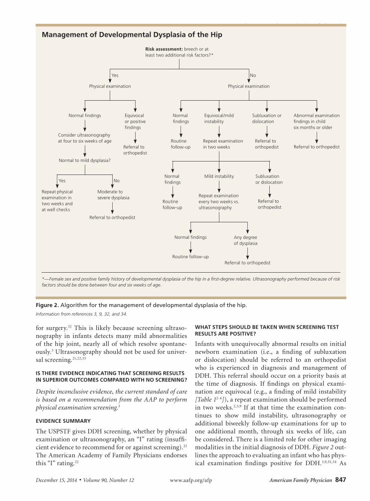
WHAT STEPS SHOULD BE TAKEN WHEN SCREENING TEST RESULTS ARE POSITIVE?
Infants with unequivocally abnormal results on initial newborn examination (i.e., a finding of subluxation or dislocation) should be referred to an orthopedist who is experienced in diagnosis and management of DDH. This referral should occur on a priority basis at the time of diagnosis. If findings on physical exami-nation are equivocal (e.g., a finding of mild instability [Table 12-4]), a repeat examination should be performed in two weeks.2,3,9 If at that time the examination con-tinues to show mild instability, ultrasonography or additional biweekly follow-up examinations for up to one additional month, through six weeks of life, can be considered. There is a limited role for other imaging modalities in the initial diagnosis of DDH. Figure 2 out-lines the approach to evaluating an infant who has phys-ical examination findings positive for DDH.3,9,32,34 As
Management of Developmental Dysplasia of the Hip
Figure 2. Algorithm for the management of developmental dysplasia of the hip.
Information from references 3, 9, 32, and 34.
Risk assessment: breech or at least two additional risk factors?*
Physical examination
Normal findings
Equivocal/mild instability
Subluxation or dislocation
Repeat examination in two weeks
Referral to orthopedist
Routine follow-up
Abnormal examination findings in child six months or older
Normal findings
Mild instability Subluxation or dislocation
Routine follow-up
Repeat examination every two weeks vs. ultrasonography
Routine follow-up
Any degree of dysplasia
No
Physical examination
Normal findings Equivocal or positive findings
Consider ultrasonography at four to six weeks of age
Referral to orthopedist
Referral to orthopedist
Normal to mild dysplasia?
Moderate to severe dysplasia
Repeat physical examination in two weeks and at well checks
No
Yes
Yes
Normal findings
Referral to orthopedist
Referral to orthopedist
Referral to orthopedist
*—Female sex and positive family history of developmental dysplasia of the hip in a first-degree relative. Ultrasonography performed because of risk factors should be done between four and six weeks of age.
Developmental Dysplasia of the Hip
848 American Family Physician www.aafp.org/afp Volume 90, Number 12 ◆ December 15, 2014
mentioned previously, most studies indicate DDH resolves spontaneously over a period of weeks.
EVIDENCE SUMMARY
Although controversial as a screening tool, ultraso-nography has good negative predictive value (as high as 90%) for disproving a diagnosis of DDH.35-37 Plain radiography is inappropriate in infants younger than six months because the neonatal hip is primarily cartilagi-nous and because of radiation exposure. It is, however, sometimes used as an adjunct following treatment in an older child.2 Although not used for initial diagnosis, computed tomography and magnetic resonance imaging can be used to follow treatment if an infant is in a cast, making ultrasonography ineffective.38
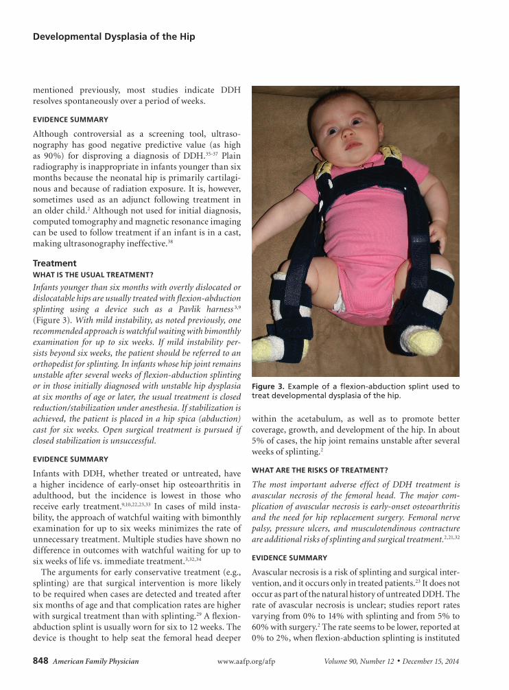
TreatmentWHAT IS THE USUAL TREATMENT?
Infants younger than six months with overtly dislocated or dislocatable hips are usually treated with flexion-abduction splinting using a device such as a Pavlik harness 3,9 (Figure 3). With mild instability, as noted previously, one recommended approach is watchful waiting with bimonthly examination for up to six weeks. If mild instability per-sists beyond six weeks, the patient should be referred to an orthopedist for splinting. In infants whose hip joint remains unstable after several weeks of flexion-abduction splinting or in those initially diagnosed with unstable hip dysplasia at six months of age or later, the usual treatment is closed reduction/stabilization under anesthesia. If stabilization is achieved, the patient is placed in a hip spica (abduction) cast for six weeks. Open surgical treatment is pursued if closed stabilization is unsuccessful.
EVIDENCE SUMMARY
Infants with DDH, whether treated or untreated, have a higher incidence of early-onset hip osteoarthritis in adulthood, but the incidence is lowest in those who receive early treatment.9,10,22,23,33 In cases of mild insta-bility, the approach of watchful waiting with bimonthly examination for up to six weeks minimizes the rate of unnecessary treatment. Multiple studies have shown no difference in outcomes with watchful waiting for up to six weeks of life vs. immediate treatment.3,32,34
The arguments for early conservative treatment (e.g., splinting) are that surgical intervention is more likely to be required when cases are detected and treated after six months of age and that complication rates are higher with surgical treatment than with splinting.29 A flexion-abduction splint is usually worn for six to 12 weeks. The device is thought to help seat the femoral head deeper
within the acetabulum, as well as to promote better coverage, growth, and development of the hip. In about 5% of cases, the hip joint remains unstable after several weeks of splinting.2
WHAT ARE THE RISKS OF TREATMENT?
The most important adverse effect of DDH treatment is avascular necrosis of the femoral head. The major com-plication of avascular necrosis is early-onset osteoarthritis and the need for hip replacement surgery. Femoral nerve palsy, pressure ulcers, and musculotendinous contracture are additional risks of splinting and surgical treatment.2,21,32
EVIDENCE SUMMARY
Avascular necrosis is a risk of splinting and surgical inter-vention, and it occurs only in treated patients.23 It does not occur as part of the natural history of untreated DDH. The rate of avascular necrosis is unclear; studies report rates varying from 0% to 14% with splinting and from 5% to 60% with surgery.2 The rate seems to be lower, reported at 0% to 2%, when flexion-abduction splinting is instituted
Figure 3. Example of a flexion-abduction splint used to treat developmental dysplasia of the hip.
Developmental Dysplasia of the Hip
December 15, 2014 ◆ Volume 90, Number 12 www.aafp.org/afp American Family Physician 849
early in infancy.9,32,39 Rates of avascular necrosis increase with delayed diagnosis and surgical treatment.9,23
ControversiesBecause screening for DDH was widely implemented before research had been done to determine the bene-fits, it would now be difficult from ethical and practical standpoints to conduct studies that would definitively determine if screening improves outcomes. Because of the risk of avascular necrosis following treatment and the high likelihood of spontaneous resolution of mild forms of DDH, some researchers have questioned the utility of screening.21,22,28 There are no clinical studies to determine if abandoning screening is a viable approach, but a decision analysis showed that screening with physi-cal examination, compared with no screening or uni-versal ultrasonography screening, would result in fewer adults having osteoarthritis of the hip at 60 years of age.23
Although the benefits and risks of screening and treatment are unclear, physical examination screen-ing of newborn hips remains the standard of care, and ethical barriers and medicolegal risks of not screening will likely keep the practice in place for the foreseeable future. Rather than answering the question of whether screening should be performed, ongoing research will likely focus on approaches to screening and management that will decrease the number of children unnecessarily treated for DDH without increasing morbidity from the condition.2,9,20,29
Data Sources: We searched PubMed (U.S. National Library of Medi-cine, National Institutes of Health) using the MeSH term congenital hip dislocation combined with epidemiology, screening, or treatment. We reviewed all pertinent articles from these queries. We reviewed refer-ence lists from key articles. We used the AAFP-recommended resources for evidence-based continuing medical education and searched these resources/databases for hip dysplasia. We specifically used the Agency for Healthcare Research and Quality, U.S. Preventive Services Task Force, Canadian Task Force on Preventive Health Care, Cochrane database, and UpToDate. Search dates: March through November 2012, and May 2014.
The authors thank David Stewart, MD, Children’s Bone and Spine Surgery, Las Vegas, Nev., for his generous contributions in supporting and reviewing this article.
This article represents the views of the authors and does not represent the views of the U.S. Air Force, the Defense Department, or the U.S. government.
The AuthorsJONATHAN C. JACKSON, MD, is an assistant professor of family medicine at the Uniformed Services University of the Health Sciences in Bethesda, Md., and a staff physician with the 10th Aerospace Medicine Squadron, U.S. Air Force Academy, Colo.
MELISSA M. RUNGE, MD, is a staff physician with the 422nd Medical Squadron, Royal Air Force Croughton, United Kingdom.
NATHANIEL S. NYE, MD, is a staff physician with the 559th Trainee Health Squadron, Joint Base San Antonio-Lackland, Tex.
At the time this manuscript was written, all authors were part of the Nellis Family Medicine Residency at Mike O’Callaghan Federal Medical Center, Nellis Air Force Base, Nev.
Address correspondence to Jonathan C. Jackson, MD, Uniformed Ser-vices University of the Health Sciences, 4301 Jones Bridge Rd., Bethesda, MD 20814 (e-mail: [email protected]). Reprints are not available from the authors.
REFERENCES
1. Ortolani M. Congenital hip dysplasia in the light of early and very early diagnosis. Clin Orthop Relat Res. 1976;(119):6-10.
2. Dezateux C, Rosendahl K. Developmental dysplasia of the hip. Lancet. 2007;369(9572):1541-1552.
3. American Academy of Pediatrics. Clinical practice guideline: early detection of developmental dysplasia of the hip. Committee on Quality Improvement, Subcommittee on Developmental Dysplasia of the Hip. Pediatrics. 2000;105(4 pt 1):896-905.
4. Novacheck TF. Developmental dysplasia of the hip. Pediatr Clin North Am. 1996;43(4):829-848.
5. Kane TP, Harvey JR, Richrds RH, Burby NG, Clarke NM. Radiological out-come of innocent infant hip clicks. J Pediatr Orthop B. 2003;12(4): 259-263.
6. Kamath S, Bramley D, Richards RH, Burby NG, Clarke NM. Is ‘clicky hip’ a risk factor in developmental dysplasia of the hip? Scott Med J. 2005;50(2):56-58.
7. Bond CD, Hennrikus WL, DellaMaggiore ED. Prospective evaluation of newborn soft-tissue hip “clicks” with ultrasound. J Pediatr Orthop. 1997;17(2):199-201.
8. Peled E, Eidelman M, Katzman A, Bialik V. Neonatal incidence of hip dysplasia: ten years of experience. Clin Orthop Relat Res. 2008;466(4): 771-775.
9. Schwend RM, Schoenecker P, Richards BS, Flynn JM, Vitale M; Pedi-atric Orthopaedic Society of North America. Screening the newborn for developmental dysplasia of the hip: now what do we do? J Pediatr Orthop. 2007;27(6):607-610.
10. Stein-Zamir C, Volovik I, Rishpon S, Sabi R. Developmental dysplasia of the hip: risk markers, clinical screening and outcome. Pediatr Int. 2008;50(3):341-345.
11. Imrie M, Scott V, Stearns P, Bastrom T, Mubarak SJ. Is ultrasound screening for DDH in babies born breech sufficient? J Child Orthop. 2010;4(1):3-8.
12. Storer SK, Skaggs DL. Developmental dysplasia of the hip. Am Fam Phy-sician. 2006;74(8):1310-1316.
13. Barlow TG. Early diagnosis and treatment of congenital dislocation of the hip. Proc R Soc Med. 1963;56:804-806.
14. Roposch A, Liu LQ, Hefti F, Clarke NM, Wedge JH. Standardized diag-nostic criteria for developmental dysplasia of the hip in early infancy. Clin Orthop Relat Res. 2011;469(12):3451-3461.
15. Stoffelen D, Urlus M, Molenaers G, Fabry G. Ultrasound, radiographs, and clinical symptoms in developmental dislocation of the hip: a study of 170 patients. J Pediatr Orthop B. 1995;4(2):194-199.
16. Castelein RM, Korte J. Limited hip abduction in the infant. J Pediatr Orthop. 2001;21(5):668-670.
17. Choudry Q, Goyal R, Paton RW. Is limitation of hip abduction a useful clinical sign in the diagnosis of developmental dysplasia of the hip? Arch Dis Child. 2013;98:862-866.
18. American Institute of Ultrasound in Medicine. AIUM practice guideline for the performance of an ultrasound examination for detection and assessment of developmental dysplasia of the hip. J Ultrasound Med. 2013;32(7):1307-1313. http://www.aium.org/resources/guidelines/hip.pdf. Accessed April 16, 2014.
Developmental Dysplasia of the Hip
850 American Family Physician www.aafp.org/afp Volume 90, Number 12 ◆ December 15, 2014
19. Graf R. The diagnosis of congenital hip-joint dislocation by the ultrasonic Combound treatment. Arch Orthop Trauma Surg. 1980;97(2):117-133.
20. Tamai J. Hip ultrasounds: where do we go from here? J Pediatr. 2012; 160(2):189-190.
21. U.S. Preventive Services Task Force. Screening for developmental dys-plasia of the hip: recommendation statement. Pediatrics. 2006;117(3): 898-902.
22. American Academy of Family Physicians. Recommendations for clini-cal preventive services: dysplasia (developmental) of the hip in infants. http:/ /www.aafp.org /patient-care /clinical-recommendations /all /dysplasia.html. Accessed April 16, 2014.
23. Mahan ST, Katz JN, Kim YJ. To screen or not to screen? A decision analy-sis of the utility of screening for developmental dysplasia of the hip. J Bone Joint Surg Am. 2009;91(7):1705-1719.
24. Austrian Society for Child and Adolescent Healthcare. Counsel to Aus-trian parents on hip ultrasound [in German]. http://www.docs4you.at/Content.Node/Vorsorgemedizin/Baby/saeuglings-hueftultraschall.php. Accessed January 14, 2013.
25. Patel H; Canadian Task Force on Preventive Health Care. Preventive health care, 2001 update: screening and management of developmen-tal dysplasia of the hip in newborns. CMAJ. 2001;164(12):1669-1677.
26. Guidelines of the German Society for Orthopaedics and Orthopae-dic Surgery and the Professional Association of Doctors for Ortho-paedics. Hip dysplasia [in German]. http://www.dgooc.de/leitlinien/ nicht-aktualisierte-leitlinien. Accessed January 14, 2013.
27. Government of South Australia. South Australian Perinatal Practice Guidelines. Neonatal hip screening and management of developmental dysplasia of the hip. March 2010. http://www.health.sa.gov.au/PPG/Default.aspx?tabid=252. Accessed January 14, 2013.
28. United Kingdom National Screening Committee. Child Health Sub-Group Report: dysplasia of the hip. September 2004. http://www.screening.nhs.uk/policydb_download.php?doc=34. Accessed January 14, 2013.
29. Sewell MD, Eastwood DM. Screening and treatment in developmen-tal dysplasia of the hip-where do we go from here? Int Orthop. 2011; 35(9):1359-1367.
30. Elbourne D, Dezateux C, Arthur R, et al.; UK Collaborative Hip Trial Group. Ultrasonography in the diagnosis and management of developmental hip dysplasia (UK Hip Trail): clinical and economic results of a multicen-tre randomised controlled trial. Lancet. 2002;360(9350):2009-2017.
31. Thaler M, Biedermann R, Lair J, Krismer M, Landauer F. Cost-effectiveness of universal ultrasound screening compared with clinical examination alone in the diagnosis and treatment of neonatal hip dys-plasia in Austria. J Bone Joint Surg Br. 2011;93(8):1126-1130.
32. Shorter D, Hong T, Osborn DA. Screening programmes for developmen-tal dysplasia of the hip in newborn infants. Cochrane Database Syst Rev. 2011;(9):CD004595.
33. Shipman SA, Helfand M, Moyer VA, Yawn BP. Screening for develop-mental dysplasia of the hip: a systematic literature review for the US Preventive Services Task Force. Pediatrics. 2006;117(3):e557-e576.
34. Rosendahl K, Dezateux C, Fosse KR, et al. Immediate treatment versus sonographic surveillance for mild hip dysplasia in newborns. Pediatrics. 2010;125(1):e9-e16.
35. Clarke NM, Reading IC, Corbin C, Taylor CC, Bochmann T. Twenty years experience of selective secondary ultrasound screening for congenital dislocation of the hip. Arch Dis Child. 2012;97(5):423-429.
36. Pillai A, Joseph J, McAuley A, Bramley D. Diagnostic accuracy of static graf technique of ultrasound evaluation of infant hips for developmen-tal dysplasia. Arch Orthop Trauma Surg. 2011;131(1):53-58.
37. Clarke NM, Harcke HT, McHugh P, Lee MS, Borns PF, MacEwen GD. Real-time ultrasound in the diagnosis of congenital dislocation and dys-plasia of the hip. J Bone Joint Surg Br. 1985;67(3):406-412.
38. American College of Radiology. ACR Appropriateness Criteria: develop-mental dylsplasia of the hip—child. http://www.acr.org/~/media/ACR/Documents/AppCriteria /Diagnostic /DevelopmentalDysplasiaOfHip Child.pdf. Accessed April 16, 2014.
39. Cashman JP, Round J, Taylor G, Clarke NM. The natural history of devel-opmental dysplasia of the hip after early supervised treatment in the Pavlik harness. A prospective, longitudinal follow-up. J Bone Joint Surg Br. 2002;84(3):418-425.


![Developmental Dysplasia of the Hip: [Print] - eMedicine … · 2017-07-13 · emedicine.medscape.com eMedicine Specialties > Orthopedic Surgery > Hip Developmental Dysplasia of the](https://img.pdfslide.net/doc/110x75/5e8dfff173e63d53604f5cb5/developmental-dysplasia-of-the-hip-print-emedicine-2017-07-13-emedicinemedscapecom.jpg)