Embed Size (px)
Citation preview
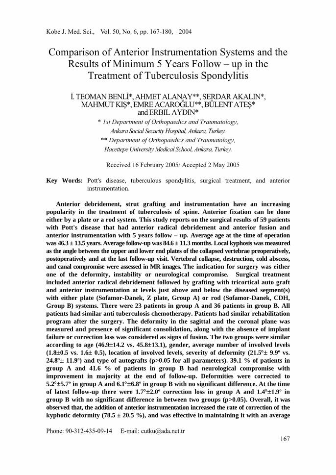
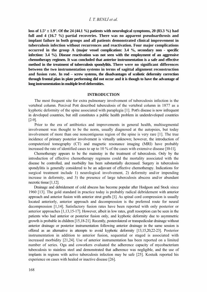
Kobe J. Med. Sci., Vol. 50, No. 6, pp. 167-180, 2004
Phone: 90-312-435-09-14 E-mail: [email protected]
167
Comparison of Anterior Instrumentation Systems and the Results of Minimum 5 Years Follow – up in the
Treatment of Tuberculosis Spondylitis
İ. TEOMAN BENLİ*, AHMET ALANAY**, SERDAR AKALIN*, MAHMUT KIŞ*, EMRE ACAROĞLU**, BÜLENT ATEŞ*
and ERBIL AYDIN* * 1st Department of Orthopaedics and Traumatology,
Ankara Social Security Hospital, Ankara, Turkey. ** Department of Orthopaedics and Traumatology,
Hacettepe University Medical School, Ankara, Turkey.
Received 16 February 2005/ Accepted 2 May 2005
Key Words: Pott's disease, tuberculous spondylitis, surgical treatment, and anterior instrumentation.
Anterior debridement, strut grafting and instrumentation have an increasing
popularity in the treatment of tuberculosis of spine. Anterior fixation can be done either by a plate or a rod system. This study reports on the surgical results of 59 patients with Pott's disease that had anterior radical debridement and anterior fusion and anterior instrumentation with 5 years follow – up. Average age at the time of operation was 46.3 ± 13.5 years. Average follow-up was 84.6 ± 11.3 months. Local kyphosis was measured as the angle between the upper and lower end plates of the collapsed vertebrae preoperatively, postoperatively and at the last follow-up visit. Vertebral collapse, destruction, cold abscess, and canal compromise were assessed in MR images. The indication for surgery was either one of the deformity, instability or neurological compromise. Surgical treatment included anterior radical debridement followed by grafting with tricortical auto graft and anterior instrumentation at levels just above and below the diseased segment(s) with either plate (Sofamor-Danek, Z plate, Group A) or rod (Sofamor-Danek, CDH, Group B) systems. There were 23 patients in group A and 36 patients in group B. All patients had similar anti tuberculosis chemotherapy. Patients had similar rehabilitation program after the surgery. The deformity in the sagittal and the coronal plane was measured and presence of significant consolidation, along with the absence of implant failure or correction loss was considered as signs of fusion. The two groups were similar according to age (46.9±14.2 vs. 45.8±13.1), gender, average number of involved levels (1.8±0.5 vs. 1.6± 0.5), location of involved levels, severity of deformity (21.5°± 9.9º vs. 24.8°± 11.9º) and type of autografts (p>0.05 for all parameters). 39.1 % of patients in group A and 41.6 % of patients in group B had neurological compromise with improvement in majority at the end of follow-up. Deformities were corrected to 5.2°±5.7º in group A and 6.1°±6.8º in group B with no significant difference. At the time of latest follow-up there were 1.7°±2.0º correction loss in group A and 1.4°±1.9º in group B with no significant difference in between two groups (p>0.05). Overall, it was observed that, the addition of anterior instrumentation increased the rate of correction of the kyphotic deformity (78.5 ± 20.5 %), and was effective in maintaining it with an average
İ. T. BENLİ et al.
168
loss of 1.5° ± 1.9°. Of the 24 (44.1 %) patients with neurological symptoms, 20 (83.3 %) had full and 4 (16.7 %) partial recoveries. There was no apparent pseudoarthrosis and implant failure in both groups and all patients demonstrated clinical improvement in tuberculosis infection without recurrences and reactivation. Four major complications occurred in the group A (major vessel complication: 3.4 %, secondary non - specific infection: 3.4 %). Disease reactivation was not seen with the employment of an aggressive chemotherapy regimen. It was concluded that anterior instrumentation is a safe and effective method in the treatment of tuberculosis spondylitis. There were no significant differences between the two instrumentation systems in terms of sagittal alignment reconstruction and fusion rate. In rod – screw systems, the disadvantages of scoliotic deformity correction through frontal plan in plate performing did not occur and it is though to have the advantage of long instrumentation in multiple level deformities.
INTRODUCTION The most frequent site for extra pulmonary involvement of tuberculosis infection is the
vertebral column. Percival Pott described tuberculosis of the vertebral column in 1877 as a kyphotic deformity of the spine associated with paraplegia [1]. Pott's disease is now infrequent in developed countries, but still constitutes a public health problem in underdeveloped countries [2-9].
Prior to the era of antibiotics and improvements in general health, multisegmental involvement was thought to be the norm, usually diagnosed at the autopsies, but today involvement of more than one noncontiguous region of the spine is very rare [1]. The true incidence of primary posterior involvement is virtually unknown; however, the introduction of computerized tomography (CT) and magnetic resonance imaging (MRI) have probably increased the rate of identified cases to up to 10 % of the cases with extensive disease [10-11].
Chemotherapy appears to be the mainstay in the treatment of tuberculosis. Only by the introduction of effective chemotherapy regimens could the mortality associated with the disease be controlled, and morbidity has been substantially decreased. Surgery in tuberculosis spondylitis is generally considered to be an adjuvant of effective chemotherapy. Indications for surgical treatment include 1) neurological involvement, 2) deformity and/or impending increase in deformity, and 3) the presence of large tuberculosis abscess and/or abundant necrotic tissue [1,12].
Drainage and debridement of cold abscess has become popular after Hodgson and Stock since 1960 [13]. The gold standard in practice today is probably radical debridement with anterior approach and anterior fusion with anterior strut grafts [1]. As spinal cord compression is usually located anteriorly, anterior approach and decompression is the preferred route for neural decompression [1,14]. Satisfactory fusion rates have been reported with only posterior or anterior approaches [1,13,15-17]. However, albeit in low rates, graft resorption can be seen in the patients who had anterior or posterior fusion only, and kyphotic deformity due to asymmetric growth is probable in children [15,18-21]. Recently, posterolateral or transpedicular drainage without anterior drainage or posterior instrumentation following anterior drainage in the same session is offered as an alternative in attempts to avoid kyphotic deformity [13,15,20,22-23]. Posterior instrumentation in addition to anterior fusion, sequential or staged is associated with increased morbidity [21,24]. Use of anterior instrumentation has been reported on a limited number of series. Oga and coworkers evaluated the adherence capacity of mycobacterium tuberculosis to stainless steel and demonstrated that adherence was negligible, and the use of implants in regions with active tuberculosis infection may be safe [25]. Kostuik reported his experience on cases with healed or inactive disease [26].
ANTERIOR INSTRUMENTATION IN TUBERCULOSIS SPONDYLITIS
169
The successful results provided by the posterior instrumentation enabled the surgeons to instrument the spinal column anteriorly after radical debridement of tuberculous spondylitis. Few studies have demonstrated satisfactory results by anterior instrumentation providing several advantages [3-5,8-9]. Anterior fixation and stabilisation after removing the diseased vertebral bodies offers the advantages of enabling the patients to mobilise early without a need for external immobilisation. Additional advantages have been suggested as performing a single approach and saving more motion segments.
Many different types of instrumentation systems are available to fix the spinal column after corpectomy and reconstruction with any types of strut grafts [27-36]. The two major types are anterior plate and rod systems. Both systems have been used in the treatment of several diseases such as tumors, fractures and infections. Several advantages and disadvantages of both systems have been shown by invitro biomechanical studies [28,34-36]. Briefly, dual rod design has been suggested to offer greater adjustability and lower rigidity while plate systems were believed to be stiffer but may lead higher risk of pseudoarthrosis and device related osteopenia [37]. However Faro found that there was greater stiffness in all direction of bending for the anterior dual rod construct compared with the Z-plate constructs [38]. A current biomechanical study comparing each implant types has demonstrated no difference between the systems in terms of load sharing and stiffness [39]. According to our knowledge, there is not yet any published clinical study in the English literature comparing both systems in the treatment of a single disease affecting the stability of spinal column.
In this study, the clinical and radiographic five years follow-up results of 59 patients with active tuberculosis spondylitis treated by anterior instrumentation was evaluated. In addition to this, the safety and efficacy of anterior plate systems vs. dual rod systems in the treatment of tuberculosis of spine was compared.
PATIENTS AND METHODS
Fifty-nine patients with a minimum follow-up of 5 years were included in this study. All were 19 year - old or older, the average age at the time of operation was 46.3 ± 13.5. Average follow-up was 84.6 ± 11.3 (60 - 115) months. Almost all patients had general tuberculosis symptoms like weight loss, moderate fever, and fatigue and predominantly back pain in their histories. Twenty-four patients (40.7 %) presented with partial or complete neural deficits. Four patients (6.8 %) had active pulmonary system tuberculosis and two patients (3.4 %) had active gastrointestinal tuberculosis at the time of presentation. Overall, 6 patients (10.2 %) had active primary tuberculosis. The patients were assessed clinically and radiological following hospitalization. In general these patients were notable for mild increases in ESR and relative lymphocytosis. Radiology revealed kyphotic deformities due to vertebral collapse and paravertebral abscess formation. The levels of involvement, numbers of affected levels, and local kyphosis angles were assessed radiological and the sagittal contours were measured independently for cervical, thoracic, thoracolumbar or lumbar segments. Twenty-three patients had Tc99m bone scans, which were suggestive of infection prior to referral to our center. We evaluated all patients with computerized tomography (CT) and magnetic resonance imaging (MRI). CT scans mostly revealed bony destruction and the lack of any soft tissue masses. Vertebral collapse, destruction, paravertebral and/or psoas abscess, and spinal cord compression due to abscess or bony debris were evidenced on MRI. Fifty -seven (96.6 %) patients were diagnosed with tuberculosis spondylitis pre-operatively in this prospective series. Only 2 (3.4 %) patients were misdiagnosed initially as their radiology was notable for soft tissue masses, but were treated by anterior vertebrectomies along with anterior instrumentation anyway.
İ. T. BENLİ et al.
170
All patients, with the exception of those who had recently developed or demonstrated progressive neurologic deficits that necessitated urgent decompression, underwent three drugs antituberculous chemotherapy prior to surgery for three weeks. Presence of a large abscess, vertebral collapse and spinal instability, local kyphotic deformity with impending increase and neurological compromise constituted the surgical indications. No special indication was determined in the selection of the instrumentation type. It was chosen randomly.
Access was gain to involved vertebrae by means of thoracotomy in the thoracic region and thoracophrenolumbotomy in the thoracolumbar and lumbar region. The abscess was drained if present and neural decompression was done with complete corpectomy of the destroyed vertebra. Anterior fusion was accomplished with tricortical autologous graft from the iliac crest and at least three costal parts obtained by the thoracotomy. Anterior instrumentation at the levels just above and below the fused segments was performed using titanium Z-plate and University plate instrumentation [27-28] in 23 patients and Cotrel-Dubousset-Hopf (CDH) instrumentation [29-30] in 36 patients. After radical debridement, two bolt screws were placed to the vertebrae above and below the corpectomy area in patients treated with anterior titanium plate instrumentation (Z – plate : 21, University plate : 2). Afterwards, anterior plate was placed to these bolt screws and security bolts were screwed tightly, and then spongious screws were fixed. In the thoracic region titanium thoracic plates and in the lumbar region standard titanium plates were used. In the patients treated with CDH, vertebral implants were placed first by two spongious screws and the rod was bent according to the curve (6mm) were inserted in plates. Derotation maneuver followed distraction. The other rod (4mm) and drawers were placed and locking screws screwed. Average operation time was 1.5 ± 0.9 hours. For all patients in this study, SSEP and "Transcranial cortical magnetic stimulation-motor evoked potentials" (TkMMEP) were combined for intra-operative neurologic monitorization. The mean blood loss was 660 ± 125 cc and an average of 2.3 ± 1.2 units of banked blood was transfused.
Patients were allowed to turn in bed on the first postoperative day, sitting on the second and were encouraged to stand and walk on the third postoperative day. Active rehabilitation was started immediately for the patients who had neural deficits. No cast immobilization or bracing was used in patients with satisfactory bone quality. Light braces were used for four months in 13 osteoporotic patients.
Diagnosis was confirmed by histopathological examination. Postoperative chemotherapy regimen consisted of rifampicin (R) 15 mg/kg (maximum 600 mg/day), isoniasid (INH) 6 mg/kg (maximum 300 mg/day), and streptomycin 1 gr. daily for a month. Thereafter, ethambutol (EMB) 25 mg/kg/day (maximum 2.5 gr/day) replaced streptomycin for six months. EMB, in turn, was replaced by Piranizamid 25 mg/kg daily (maximum 2 gr/day) on the 6th month, and chemotherapy was discontinued after one year.
Patients with neural deficits were graded according to the Frankel's scale [40]. Local kyphosis was measured as the angle between the upper and lower end plates of the collapsed levels postoperatively and at the last follow-up visit. These values were corrected by subtracting 5° per level from the thoracic values and by adding 10° per level to the lumbar angles. On lateral X-rays, sagittal contours between T2 to T12 and LI to L5 vertebra were measured by Cobb method. Normal thoracic physiological kyphosis and physiological lumbar lordosis were accepted as 30° to 50° and 40° to 60° respectively [41]. All measurement was made in cooperation with radiologist. Sagittal contours were given (+) and (-) angle values if they had kyphotic and lordotic patterns, respectively.
ANTERIOR INSTRUMENTATION IN TUBERCULOSIS SPONDYLITIS
171
Presence of significant consolidation, along with the absence of implant failure or correction loss and pain relief was considered as signs of fusion. At the last follow-up, implant failure and other complications were also evaluated.
The last follow - up visits were made in November 2004 and patients who had more than 5 years follow - up were included in the study. The tests used for statistical assessment were "Wilcoxon Matched - Pair Signed - ranks" and “Difference of paired samples t – test” (SPSS for MS Windows) with confidence interval of 95 %.
RESULTS
- General Assessment: The most commonly affected vertebra was L1 with 18 cases (30.5 %). Overall,
involvement was seen in 1.7 ± 0.5 levels, and an average of 2.3 ± 0.6 mobile segments was included in the instrumentation and fusion area.
All patients in this series could be followed closely because of the constitution of the social security system, so no patients were lost to follow-up. The preoperative local kyphosis angle was 23.5° ± 11.2° (10° - 80°), and was corrected to 5.7° ± 6.3° (0° - 22°) with a correction rate of 78.5 ± 20.5 % (42.3 - 100 %) for the entire study population (Table-1). This correction rate was statistically significant (t: 20.5, p < 0.05). On average, 1.5° ± 1.9° of correction loss was noted in local kyphosis angle and the final correction was measured to be 72.0 ± 21.8 %. - Neural Assessment:
Of the 24 (40.7 %) patients with neuralgic deficits; 4 were Frankel A, 3 Frankel B, 10 Frankel C and 7 Frankel D. Postoperatively 4 patients (16.7 %) had partial recovery; two patients with Frankel A and one patient with Frankel B involvement improved to Frankel D, while one patient with Frankel A improved to Frankel C. The remaining 20 patients (83.3 %) had complete recoveries. - Comparison of anterior instrumentation systems:
The two groups were similar according to age (46.9 ± 14.2 vs. 45.8 ± 13.1), gender, average number of involved levels (1.8±0.5 vs. 1.6± 0.5), location of involved levels, severity of deformity (21.5°± 9.9º vs. 24.8°± 11.9º) type of autografts, incidence (39.1 % vs. 41.6%) and recovery rate of neurological compromise, operation times and bleeding volumes (Table-2) (p > 0.05).
Deformities were corrected to 5.2° ± 5.7º in group A and 6.1° ± 6.8º in group B with no significant difference. At the time of latest follow-up there were 1.7° ± 2.0º correction loss in group A and 1.4° ± 1.9º in group B with no significant difference in between two groups. There were also no differences in both postoperative and final correction rates statistically (p>0.05) (Table-1).
In the CDH group, an average of 8.4° ± 4.6° of scoliosis was seen in 16 patients and was completely corrected postoperatively. In 5 patients with Z-plate instrumentation, mean of 6.7° ± 5.4° of scoliosis was seen, in one patient was corrected to 5° and persisted, in 3 patients it was entirely corrected and in the remaining one patient increased from 6° to 10°. Additionally, in 5 patients without any scoliotic deformity preoperatively, an average of 5.2° ± 2.6° of scoliotic curves occurred, so postoperatively 7 patients (19.4 %) of this group had scoliotic curves.
There was no apparent pseudoarthrosis and implant failures in both groups and all patients demonstrated clinical improvement in tuberculosis infection without recurrences and reactivation. - Complications:
Both groups demonstrated no apparent pseudoarthrosis or implant failures at the latest follow-up. Furthermore, both groups demonstrated clinical healing of the tuberculosis
İ. T. BENLİ et al.
172
infection. No recurrences, reactivation or draining sinuses were encountered. Four major complications occurred in group A. One patient with T9 involvement developed aortic aneurysm, which was diagnosed at the 20th postoperative day, and the patient was immediately operated. The other patient had L4 - L5 destruction and the iliac vein which had adhered to the L5 corpus was torn during the dissection, and had to be repaired using a Teflon graft during operation. Secondary late non-specific infection developed in two patients, whose implants were removed at 14th and 16th postoperative months and the infected area was debrided. Both were confirmed to be positive for acid-fast staining on retrospective evaluation, and it was considered that the staphylococcal infections in these patients were secondary. Fortunately, both could be treated by debridement and IV antibiotics (sulbactam ampicillin 1 gr. two times daily, six weeks).
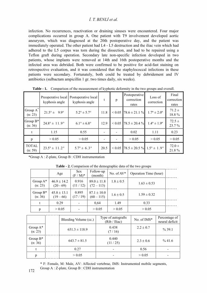
Table - 1. Comparison of the measurement of kyphotic deformity in the two groups and overall.
Preoperative local
kyphosis anglePostoperative local
kyphosis angle t p
Postoperativecorrection
rates
Loss of correction
Final correction
rates Group A*
(n: 23) 21.5° ± 9.9° 5.2° ± 5.7° 11.8 < 0.05 78.6 ± 21.1 % 1.7º ± 2.0º 71.2 ± 18.8 %
Group B* (n: 36) 24.8° ± 11.9° 6.1° ± 6.8° 12.9 < 0.05 78.5 ± 20.4 % 1.4º ± 1.9º 72.5 ±
23.7 %
t 1.15 0.55 - - 0.02 1.11 0.23
p > 0.05 > 0.05 - - > 0.05 > 0.05 > 0.05
TOTAL (n: 59) 23.5° ± 11.2° 5.7° ± 6.3° 20.5 < 0.05 78.5 ± 20.5 % 1.5° ± 1.9° 72.0 ±
21.8 %
*Group A : Z-plate, Group B : CDH instrumentation
Table - 2. Comparison of the demographic data of the two groups
Age Sex (F / M)*
Follow-up(month) No. of AV* Operation Time (hour)
Group A* (n: 23)
46.9 ± 14.2(20 - 69)
0.916 (11 / 12)
89.0 ± 11.8(72 – 113)
1.8 ± 0.5 1.63 ± 0.53
Group B* (n: 36)
45.8 ± 13.1(19 – 66)
0.895 (17 / 19)
87.1 ± 10.0(60 – 115) 1.6 ± 0.5 1.59 ± 0.32
t 0.29 - 0,64 1.49 0.33
p > 0.05 - > 0.05 > 0.05 > 0.05
Bleeding Volume (cc.) Type of autografts (Rib / Iliac) No. of IMS* Percentage of
neural deficit Group A*
(n: 23)
651.3 ± 118.9 0.438 (7 / 16)
2.2 ± 0.7 % 39.1
Group B* (n: 36)
643.7 ± 81.5 0.440
(11 / 25) 2.3 ± 0.6 % 41.6
t 0.27 - 0.56 -
p > 0.05 - > 0.05 -
* F: Female, M: Male, AV: Affected vertebrae, IMS: Instrumented mobile segments,Group A : Z-plate, Group B : CDH instrumentation
ANTERIOR INSTRUMENTATION IN TUBERCULOSIS SPONDYLITIS
173
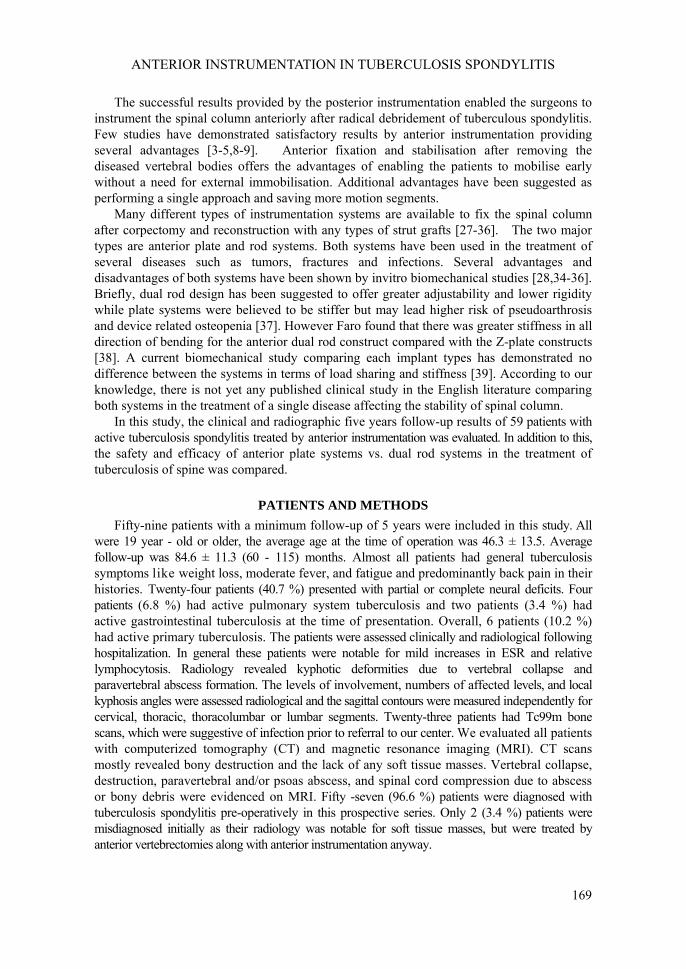
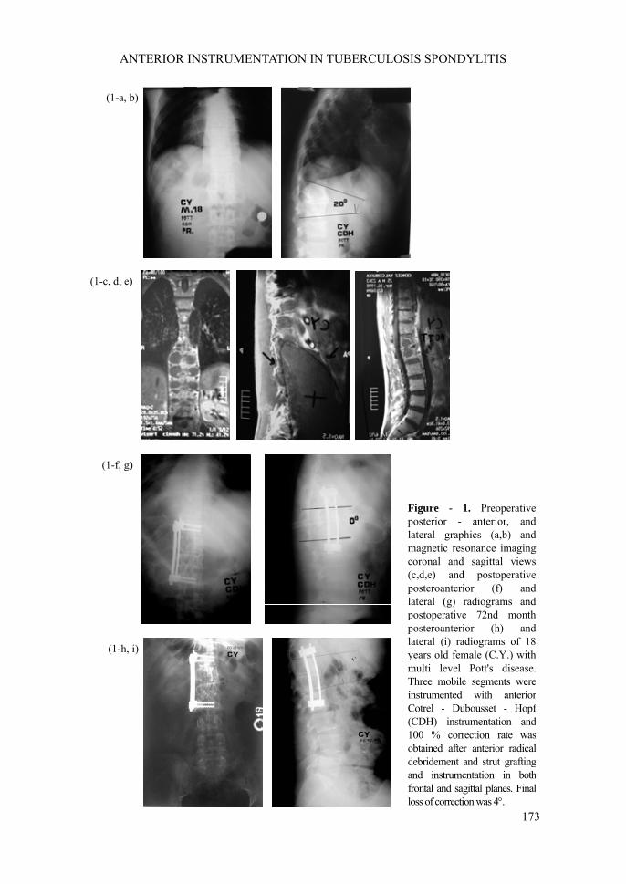
Figure - 1. Preoperativeposterior - anterior, andlateral graphics (a,b) andmagnetic resonance imagingcoronal and sagittal views(c,d,e) and postoperativeposteroanterior (f) andlateral (g) radiograms andpostoperative 72nd monthposteroanterior (h) andlateral (i) radiograms of 18years old female (C.Y.) withmulti level Pott's disease.Three mobile segments wereinstrumented with anteriorCotrel - Dubousset - Hopf(CDH) instrumentation and100 % correction rate wasobtained after anterior radicaldebridement and strut graftingand instrumentation in bothfrontal and sagittal planes. Finalloss of correction was 4°.
(1-a, b)
(1-c, d, e)
(1-f, g)
(1-h, i)
İ. T. BENLİ et al.
174
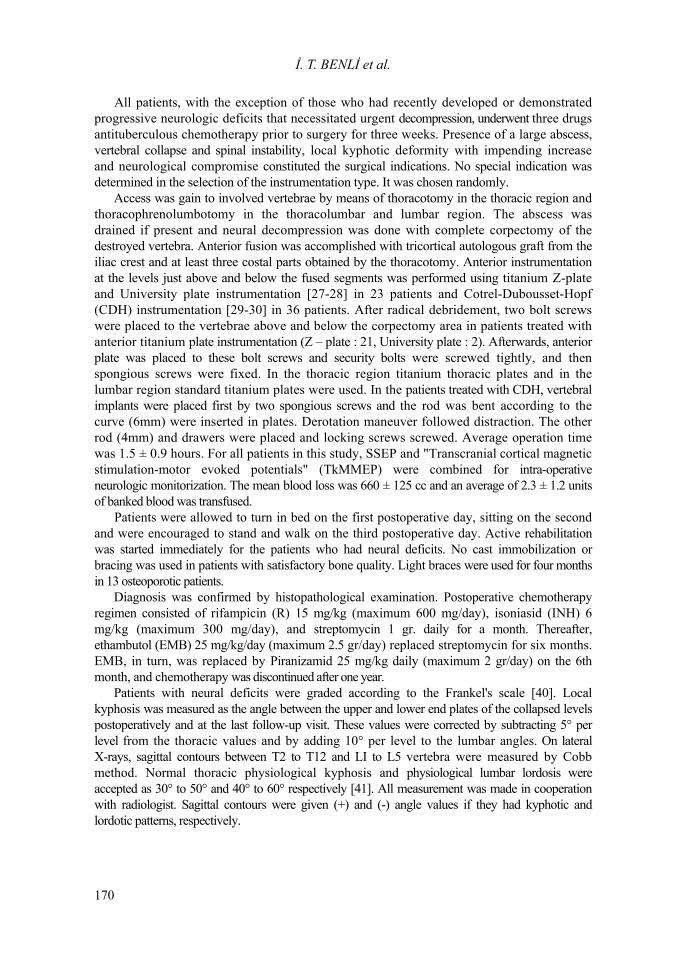
2-a, b
2-c, d
2-e, f
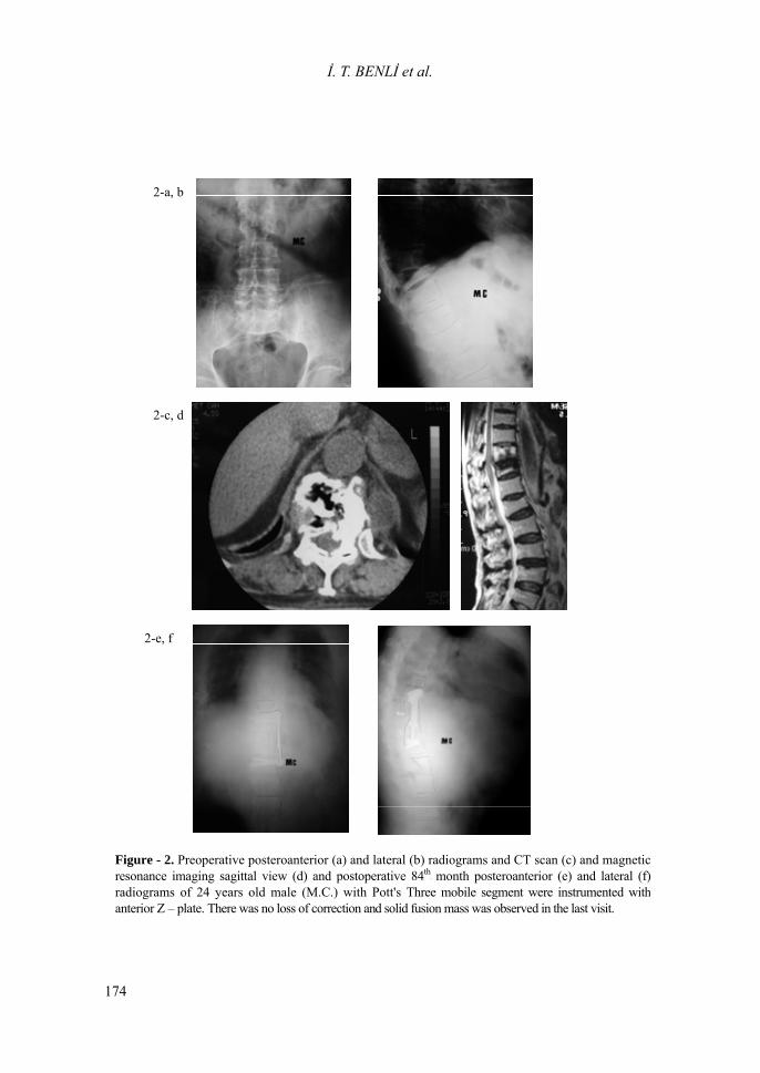
Figure - 2. Preoperative posteroanterior (a) and lateral (b) radiograms and CT scan (c) and magneticresonance imaging sagittal view (d) and postoperative 84P
thP month posteroanterior (e) and lateral (f)
radiograms of 24 years old male (M.C.) with Pott's Three mobile segment were instrumented withanterior Z – plate. There was no loss of correction and solid fusion mass was observed in the last visit.
ANTERIOR INSTRUMENTATION IN TUBERCULOSIS SPONDYLITIS
175
DISCUSSION Indications for surgery in spinal tuberculosis are reported to include the presence of a
large paraspinal abscess, the presence of severe bone destruction and kyphotic deformity, neurological deficit with spinal cord compression, and lack of response to conservative treatment [23]. Posterior fusion had been the standard surgical procedure for the limited correction and prevention of progression of deformity in many centers before the safe and liberal use of anterior spinal surgery became feasible. However, posterior fusion does not appear to alter the natural course of the disease process, pseudoarthrosis and bending of the fusion mass very frequently leads to substantial increase of the kyphotic deformity [2,15,42].
Anterior debridement without fusion in the treatment of spinal tuberculosis has been evaluated in MRC studies performed in Hong Kong and Bulawayo, demonstrating that the magnitude and the rate of progression of the kyphotic deformity was similar in patients who had no surgery, and were significantly inferior compared to anterior debridement and fusion [43]. Longitudinal follow-up of the same group of patients revealed that bony fusion occurred later in those who had anterior grafting compared to only debridement, but the rates of fusion were similar at five years [44]. Over ten years, debridement group exhibited mean increases in kyphosis of 9.8° for thoracic and thoracolumbar lesions and 7.6° for lumbar lesions, compared to minor changes in the fusion group [45]. Upadhyay and coworkers reported the latest follow-up of the same group of patients, concluding that the debridement group demonstrated increases in kyphotic deformity for up to six months. Therefore, adult patients demonstrated an arrest in progression, while some spontaneous correction of the deformity occurred in the pediatric patients [42,46-48]. Aksoy et al. reported a series of 100 patients either with posterior or anterior fusion only and demonstrated that kyphotic deformity developed less frequently after anterior fusion [2]. Rajasekaren and Soundarapendian reported 59 % kyphotic deformity with anterior fusion [20].
With anterior debridement and fusion, the MRC trials demonstrated that an increase in kyphotic deformity occurred in only 17 % of patients compared to 39 % of patients treated with chemotherapy. In contrast to patients treated with only anterior debridement, the progression of the kyphotic deformity was considerably less, especially during the first six months of the treatment [24,43-45]. Kyphotic deformity did not significantly increase in these patients after six months regardless of the treatment method. In another study 59 % of patients had favorable results (excellent or good), 19 % were rated as fair, and 22 % as poor [20].
The necessity of prolonged immobilization following anterior procedures, and the relatively high rates of progression of kyphosis frequently related to the problems with strut grafts prompted the idea that tuberculosis spondylitis may be stabilized by posterior instrumentation [1,15]. Oga and coworkers obtained good clinical results but the instrumentation was extended to an alarming average of 8.5 levels, in spite of the fact that 3.5 levels on average were involved by the disease [25]. Moon et al reported very good rates of correction and good maintenance of correction for both children and adults, fusion occurred in four months in single level spondylodesis cases and in six months in two-level [49].
Several studies have demonstrated satisfactory results using posterior instrumentation along with anterior debridement and fusion [11-12,24-25]. Güven et al. reported a series of 10 cases with posterior instrumentation, in which there was a 3.4° loss in the correction of local kyphosis [50]. Domaniç et al. reported that in their series with anterior debridement, correction of the kyphosis was more successful in-patients who had additional posterior CD instrumentation [6]. Yau and Fukuta and their collabrates reported higher success rates with anterior fusion and posterior instrumentation in the same session [17,51]. In our recent article, 72 adult patients with different surgical procedures were assessed. Eight patients had only anterior debridement and fusion, with 8.6 % correction rate and average 23.6° correction loss during follow-up, compared to
İ. T. BENLİ et al.
176
76.8 % average correction and 2.5° correction loss in 11 patients who had posterior instrumentation following anterior radical surgery [4].
Anterior instrumentation in active tuberculosis infection is a relatively new concept, and the results of this study should probably be compared to those achieved with other modalities of surgical treatment as well as other reports on anterior instrumentation.
Kostuik reported a series of 79 patients with anterior decompression and anterior internal fixation in 1983, among whom 51 had neuralgic deficits. He reported two patients developing deformity because of spinal tuberculosis [26]. There has been very limited experience with anterior instrumentation following anterior radical debridement and fusion, especially on the early cases with active disease [3-4]. This reluctance so far probably arises from the presumption that placing the instrumentation in an area with active infection would be prone to complications like disease reactivation or secondary infection [52]. The results of the present study demonstrated that, anterior instrumentation in the presence of active infection does not cause any major complications, probably because of the poor adherence capacity of the tuberculosis bacilli to metals [25].
There are few studies analysing the efficacy and safety of anterior instrumentation [3-5,8-9]. Anterior fixation and stabilisation after removing the diseased vertebral bodies offered the advantages of enabling the patients to mobilise early without a need for external immobilisation. Additional advantages have been suggested as performing a single approach and saving more motion segments.
In the present study, the results of 59 patients with minimum 5 years of follow - up were evaluated and our correction rates in local kyphosis angle (78.5 ± 20.5 %), our correction loss at the last control visit (1.5° ± 1.9°) and our final correction rate (72.0 ± 21.8 %) were compatible with the results reported in the literature. Evaluation of the effect on sagittal global contours showed a statistically significant correction rate in thoracic, thoracolumbar and lumbar regions and also correction loss rates at the last control visit were very low. It is noted that application of a distraction for correction of the local kyphosis deformity in thoracic region resulted in a decrease in the global kyphosis angle but though this effect neither hypokyphosis nor lordosis was noted in the thoracic region. It played a positive role in the lumbar region by increasing lordosis.
Ozdemir and colleagues reported 96 % of fusion rate with 6° of average correction loss in 28 patients treated with anterior fusion with allograft and anterior instrumentation (9). Govender reported allografts incorporated late but latter than autologous costa grafts in patients treated with anterior fusion and anterior instrumentation [53]. In our study, we used at least 3 autologous costa grafts on the thoracic and thoracolumbar region and iliac grafts in the lumbar region and any problems such as graft resorption or other were not noted. Implant failure and pseudoarthrosis were not noted and a solid fusion mass was obtained in all patients. Tuberculosis reactivation was not noted.
Therefore, based on our results and those reported with the use of posterior instrumentation, it can be stated that instrumentation in active tuberculosis spondylitis can be performed safely with few complications, and is effective in obtaining and maintaining the correction of the deformity as well as obviating the need for external support. The two major advantages of anterior instrumentation over posterior are the ability to perform the operation with a single approach, and to prevent the inclusion of unnecessarily large number of levels into fusion. In this study, an average of 2.3 ± 0.6 (1-4) mobile segments were instrumented in the whole vertebral column.
It should be noted that all patients included in this study have undergone a very aggressive alternating three drug antibiotic regimen for twelve months, which has been the standard protocol in our center over the years, contrary to recent reports suggesting that shorter and less aggressive chemotherapy may be as effective [24,48,54].
ANTERIOR INSTRUMENTATION IN TUBERCULOSIS SPONDYLITIS
177
Although there is a large array of devices available for internal fixation of the anterior column, most systems are designed as either a plate or a rod system. There are several invitro biomechanical studies evaluating the properties of both systems. Prior publications have reported on the biomechanical characteristics of many anterior spinal instrumentation systems. In general, the results of previous studies demonstrated that dual rod designs may offer greater adjustability and control over screw placement and increased load sharing, but possibly at the expense of rigidity. Plate systems are designed to be stiffer and less prone to fatigue failure, but there are theoretical concerns and unanswered questions regarding the risk of pseudoarthrosis and device-related osteopenia with very rigid spinal implants [31-36]. However Faro found that there was greater stiffness in all direction of bending for the anterior dual rod construct compared with the Z-plate constructs [38]. In a recent biomechanical study, any correlation between load sharing and implant style (rod vs. plate) was not found. According to Brodke, stiffness results of the instrumentation was noted to vary with the graft in place rather than by plate/ rod style. Brodke also addressed the importance of graft in the overall contruct stiffness [39].
To our knowledge, there is not yet any clinical study comparing the efficacy of both anterior instrumentation systems in the English literature. Our findings support the results of the biomechanical study by Brodke et al. demonstrating no superiority of one system to the other in terms of restoration of the sagittal plane, rate of failure and rate of fusion. We think that, the differences between these instrumentation systems are likely to be less significant than other factors that influence clinical outcomes, such as bone quality, alignment, and the quality of the graft-vertebral body interface. Thus, it seems reasonable to conclude that because either the plate or the rod instrumentation provides similar clinical outcome, ease of use and surgeon familiarity with a particular system may be more important than the material capabilities of each particular implant.
Considering the coronal plane alignment we found that correction of deformity in this plane may be less effective with the use of plate systems. Moreover, one can create a malalignment in the coronal plane when plates are used. The disadvantage of manipulation of the plates to control the coronal plane deformity when compared to rods may be accounted for this problem. However, mild residual coronal plane deformities did not result with significant clinical problems for the patients. Another advantage of double rod screw system is thought to be the ability of effectiveness treating the deformity in multilevel tuberculosis patients compared with anterior plate screw system as posterior instrumentation.
The results of the current study are parallel to previous studies in terms of efficacy and safety. We believe anterior instrumentation may be the ideal stabilization method for tbc spondylitis by providing the advantages of less segment fusion, single approach and obviating the need for external immobilization.
Major drawbacks of this study are its retrospective character despite the evaluation of a prospectively collected data, and lack of randomization of the patients for either implant group. However it is the only study clinically comparing the anterior rod and plate systems in the treatment of a single disease causing sagittal plane malalignment along with vertebral body destruction. The study demonstrated that anterior instrumentation was a safe and effective method in the treatment of tuberculosis independent of the type of anterior instrumentation.
REFERENCES 1. Slucky AV, and Eismont FJ. Spinal infections. In: Bridwell KH, DeWald RL, Eds., The
Textbook of Spinal Surgery, Philadelphia, Lippincott - Raven Publishers. 1997 ; 2141-2183.
İ. T. BENLİ et al.
178
2. Aksoy MC, Acaroglu RE, Tokgozoglu AM, Ozdemir N, and Surat A. Retrospective evaluation of treatment methods in tuberculosis spondylitis. Hacettepe J Orthop Surg 1995 ; 5:207-209.
3. Benli IT , Aydın E, Kis M, Akalın S, Tuzuner M, and Baz AB. The results of anterior instrumentation in vertebral tuberculosis. J Turkish Spine Surg 1996 ; 7(3):98-101.
4. Benli IT, Kis M, Akalın S, Citak M, Kanevetci S, and Duman E. The results of anterior radical debridement and anterior instrumentation in Pott's disease and comparison with other surgical techniques. Kobe J Med Sci 2000 ; 46 : 39 - 68.
5. Benli IT, Acaroglu E, Akalin S, Kis M, Duman E, and Un A. Anterior radical debridement and anterior instrumentation in tubercolous spondylitis. Eur Spine J 2003 ; 12 : 224 – 234.
6. Domanic U, Hamzaoglu A, Sar C, and Yavuzer Y. Posterior fusion and instrumentation after anterior radical debridement and fusion in the surgical treatment of Pott's disease. J Turkish Spine Surg 1993 ; 4(1): 16-19.
7. Korkusuz F, Islam C, and Korkusuz Z. Prevention of postoperative late kyphosis in Pott's disease by anterior decompession and intervertebral grafting. World J Surg 1997 ; 21(5) : 524-528.
8. Yilmaz C, Selek HY, Gurkan I, Erdemli B, and Korkusuz Z. Anterior instrumentation for the treatment of spinal tuberculosis. J Bone Joint Surg 1999 ; 81-A (9): 1261-1267.
9. Ozdemir HM, Us AK, and Ogun T. The rol of anterior spinal instrumentation and allograft fibula for the treatment of Pott’s disease. Spine 2003 ; 28 (5) : 474 – 479.
10. Arthornthurasook A, and Chongpieboonpatana A. Spinal tuberculosis with posterior element involvement. Spine 1990; 15:191-194.
11. Tuli SM. Current concept. Severe kyphotic deformity in tuberculosis of the spine. Int Orthop 1995 ; 19 : 327-331.
12. Moon M S. Spine update : tuberculosis of the spine. Spine 1997 ; 22 (15) : 1791-1797. 13. Hodgson AR, Stock FE, Forg HSY, and Ong GB. Anterior spinal fusion : the operative
approach and pathological findings in 412 patients with Pott's disease of the spine. Br J Surg 1960 ; 48: 172-178.
14. Hsu LC, Cheng CL, and Leong JC. Pott's paraplegia of late onset: The cause of compression and results after anterior decompression. J Bone Joint Surg 1988 ; 70-B : 534-538.
15. Hodgson AR, and Stock FE. Anterior spinal fusion. A preliminary communication on the radical treatment of Pott's disease and Pott paraplegia. Clin Orthop 1994 ; 300: 16-23.
16. Kempf HBS, Jackson JW, Jeremiah JD, and Cook J. Anterior fusion of the spine for infective lesions in adults. J Bone Joint Surg 1973 ; 55-B : 715-734.
17. Yau ACMC, Hsu LCS, O'Brein JP, and Hodgson AR. Tuberculosis kyphosis : correction with spinal osteotomy halopelvis distraction and anterior and posterior fusion. J Bone Joint Surg 1974 ; 56-A : 1419-1434.
18. Moula T, Fowles JV, Kassab MT, and Sliman N. Pott's paraplegia : a clinical review of operative and conservative treatment in 63 adults and children. Int Orthop 1981 ; 5(1) : 23-29.
19. Nemir RL, and Krasinski K. Tuberculosis in children and adolescents in the 1980s. Pediatr Infect Dis J 1988 ; 7 (6): 375-379.
20. Rajasekaran S, and Soundarapandian S. Progression of kyphosis in tuberculosis of the spine treated by anterior arthrodesis. J Bone Joint Surg 1989 ; 71-A : 1314-1323.
21. Schulitz KP, Kothe R, and Leong JCY, Wehling P. Growth changes of solid fusion kyphotic bloc after surgery for tuberculosis. Spine 1997 ; 22 (10): 1150-1155.
ANTERIOR INSTRUMENTATION IN TUBERCULOSIS SPONDYLITIS
179
22. Loembe PM. Medical - surgical treatment of progressive tuberculous (Pott's) paraplegia in Gabon. Paraplegia 1995 ; 33(10): 579-584.
23. Rezai AR, Lee M, Cooper PR, Errico TJ, and Koslow M. Modern management of spinal tuberculosis. Neurosurgery 1995 ; 36 (1): 87-97.
24. Medical Research Council Working Party on Tuberculosis of the Spine. A 15 - year assessment of controlled trials of the management of tuberculosis of the spine in Korea and Hong Kong. Thirteenth Report of the Medical Research Council Working Party on Tuberculosis of the Spine. J Bone Joint Surg 1998 ; 80-B(3): 456-462.
25. Oga M, Arizono T, Takasita M, and Sugioka Y. Evaluation of the risk of instrumentation as a foreign body in spinal tuberculosis: Clinical and biologic study. Spine 1993 ; 18 : 1890-1894.
26. Kostuik JP. Anterior spinal cord decompression for lesions of the thoracic and lumbar spine: Techniques, new methods of internal fixation. Spine 1983 ; 8:512-531.
27. Aydın E, Solak AS, Tuzuner MM, Benli IT, and Kis M. Z-plate instrumentation in thoracolumbar spinal fractures. Bull Hosp Jt Dis 1999 ; 58 (2) : 92-97.
28. Lim TH, An HS, Hong JH, Ahn JY, You JW, Eck J, and McGrady LM. Bomechanical evaluation of anterior and posterior fixations in an unstable calf spine model. Spine 1997 ; 22(3) : 261-266.
29. Hopf C, Eysel P, and Dubousset J. CDH : Preliminary report on new anterior spinal instrumentation. Eur Spine J 1995 ; 4: 194-199.
30. Benli IT, Akalin S, Kis M, Citak M, Kurtulus B, and Duman E. The results of anterior fusion and Cotrel – Dubousset – Hopf instrumentation in idiopathic scoliosis. Eur Spine J 2000 ; 9(6) : 5005-515.
31. McCullen G, Vaccaro AR, and Garfin SR. Thoracic and lumbar trauma: rationale for selecting the appropriate fusion technnique. Orthop Clin North Am 1998; 29: 813-28.
32. Dunn HK. Anterior stabilization of thoracolumbar injuries. Clin Orthop 1984; 189: 116-24.
33. Kaneda K, Abumi K, and Fujiya M. Burst fractures with neurologic deficits of the thoracolumbar-lumbar spine. Results of anterior decompression and stabilization with anterior instrumentation. Spine 1984; 9: 788-95.
34. Dick JC, Brodke DS, Zdeblick TA, et al. Anterior instrumentation of the thoracolumbar spine. Spine 1997; 22: 744-50.
35. Harris, MB, Thomas KA, Igram CM, et al. The effect of anterior thoracolumbar plate application on the compressive loading of the strut graft. Spine 1996; 21: 1487-93.
36. Shimamoto N, Kotani Y, Shono Y, et al. Biomechanical evaluation of anterior spinal instrumentation systems for scoliosis: in vitro fatigue simulation. Spine 2001; 26: 2701-8.
37. Carl AL, Tranmer BI, and Sachs BL. Anterolateral dynamized instrumentation and fusion for unstable thoracolumbar and lumbar burst fractures. Spine 1997; 22: 686-90.
38. Faro FD, White KK, Ahn JS, Oka RS, Mahar AT, Bawa M, Farmsworth CL, Garfin SR, and Newton PO. Biomechanical analysis of anterior instrumentation for lumbar corpectomy. Spine 2003 ; 28 (22) : E468-471.
39. Brodke DS, Gollogly S, and Bachus K et al. Anterior Thoracolumbar Instrumentation: Stiffness and Load Sharing Characteristics of Plate and Rod Systems. Spine 2003;28:1794-1801
40. Frankel HL, Hancock DO, Hyslop G, Melzah J, Michaelis LS, Ungar GH, Vernon JD, and Walsh JJ. The value of postural reduction in the initial management of closed injuries of the spine with paraplegia and tetraplegia. Paraplegia 1969 ; 7:179-192.
İ. T. BENLİ et al.
180
41. Benhardt, M. Normal spinal anatomy: normal sagittal plane alignment. In: Bridwell, K.H., DeWald, R.L., eds. The Text Book of Spinal Surgery, Philadelphia, Lippincott - Raven Publishers, 1997 : 188-189 .
42. Upadhyay SS, Saji MJ, Sell P, and Yau ACMC. The effect of age on the change in deformity after radical resection and anterior arthrodesis for tuberculosis of the spine. J Bone Joint Surg 1994 ; 76-A : 701-708.
43. Medical Research Council Working Party on Tuberculosis of the Spine. A controlled trial of anterior spinal fusion and debridement in the surgical management of tuberculosis of the spine in patients on standard chemotherapy. A study in Hong Kong. British J Surg 1974 ; 61 : 853-866.
44. Medical Research Council Working Party on Tuberculosis of the Spine. Five-year assessments of controlled trials of ambulatory treatment, debridement and anterior spinal fusion in the management of tuberculosis of the spine: Studies in Bulawayo (Rhodesia) and in Hong Kong. J Bone Joint Surg 1978 ; 60-B : 163-177.
45. Medical Research Council Working Party on Tuberculosis of the Spine. A ten-year assessment of a controlled trial comparing debridement and anterior spinal fusion in the management of tuberculosis of the spine in patients on standard chemotherapy in Hong Kong. J Bone Joint Surg 1982 ; 64-B : 393-398.
46. Upadhyay SS, Saji MJ, Sell P, Sell B, and Hsu LCS. Spinal deformity after childhood surgery for tuberculosis of the spine. A comparison of radical surgery and debridement. J Bone Joint Surg 1994 ; 76-B : 91-98.
47. Upadhyay SS, Sell P, Saji MJ, Sell B, and Hsu LC. Surgical management of spinal tuberculosis in adults: Hong Kong operation compared with debridement surgery for short and long term outcome of deformity. Clin Orthop Rel Res 1994 ; 302 : 173-182.
48. Upadhyay SS, Saji MJ, and Yau ACMC. Duration of antituberculous chemotherapy in conjunction with radical surgery in the management of spinal tuberculosis. Spine 1996 ; 21: 1898-1903.
49. Moon MS, Woo YK, Lee KS, Ha KY, Kim SS, and Sun DH. Posterior instrumentation and anterior interbody fusion for tuberculosis kyphosis of dorsal and lumbar spines. Spine 1995 ; 20 : 1910-1916.
50. Guven O, Kumano K, Yasin S, Karahan M, and Tsuji S. A single stage posterior approach and rigid fixation for preventing kyphosis in the treatment of spinal tuberculosis. Spine 1994 ; 19:1039-1043.
51. Fukuta S, Miyamoto K, Masuda T, Hosoe H, Kodoma H, Nishimato H, Sakaeda H, and Shimizu K. Two stage (posterior and anterior) surgical treatment using posterior spinal instrumentation for pyogenic and tuberculotic spondylitis. Spine 2003 ; 28 (15) : 302 – 308.
52. Kim NH, Lee DHM, Choi CH, and Park SJ. The comparison of the fusion rate in anterior interbody fusion between noninfectious and infectious disease of the spine. J Turkish Spine Surg 1994 ; 5(2): 49-58.
53. Govender S. The outcome of allografts and anterior instrumentation in spinal tuberculosis. Clin Orthop 2002 ; 398 : 60 – 66.
54. Medical Research Council Working Party on Tuberculosis of the Spine. Five-year assessment of controlled trials of short - course chemotherapy regimens of 6, 9 or 18 months' duration for spinal tuberculosis in patients ambulatory from the start or undergoing radical surgery. Fourteenth report of the Medical Research Council Working Party on Tuberculosis of the Spine. Int Orthop 1999 ; 23(2):73-81.