Embed Size (px)
Citation preview
INFECTION AND IMMUNITY, Feb. 2002, p. 878–888 Vol. 70, No. 20019-9567/02/$04.00�0 DOI: 10.1128/IAI.70.2.878–888.2002Copyright © 2002, American Society for Microbiology. All Rights Reserved.
Comparison of Biofilms Formed by Candida albicansand Candida parapsilosis on Bioprosthetic Surfaces
D. M. Kuhn,1,2 J. Chandra,1,3 P. K. Mukherjee,1,3 and M. A. Ghannoum1,3*Case Western Reserve University1 and Division of Infectious Diseases, Department of Medicine,2 and Department of Dermatology,3
University Hospitals of Cleveland, Cleveland, Ohio 44106
Received 8 June 2001/Returned for modification 9 October 2001/Accepted 30 October 2001
Little is known about fungal biofilms, which may cause infection and antibiotic resistance. In this study,biofilm formation by different Candida species, particularly Candida albicans and C. parapsilosis, was evaluatedby using a clinically relevant model of Candida biofilm on medical devices. Candida biofilms were allowed toform on silicone elastomer and were quantified by tetrazolium (XTT) and dry weight (DW) assays. Formedbiofilm was visualized by using fluorescence microscopy and confocal scanning laser microscopy with Cal-cofluor White (Sigma Chemical Co., St. Louis, Mo.), concanavalin A-Alexafluor 488 (Molecular Probes,Eugene, Oreg.), and FUN-1 (Molecular Probes) dyes. Although minimal variations in biofilm productionamong invasive C. albicans isolates were seen, significant differences between invasive and noninvasive isolates(P < 0.001) were noted. C. albicans isolates produced more biofilm than C. parapsilosis, C. glabrata, and C.tropicalis isolates, as determined by DW assays (P was <0.001 for all comparisons) and microscopy. Interest-ingly, noninvasive isolates demonstrated a higher level of XTT activity than invasive isolates. On microscopy,C. albicans biofilms had a morphology different from that of other species, consisting of a basal blastosporelayer with a dense overlying matrix composed of exopolysaccharides and hyphae. In contrast, C. parapsilosisbiofilms had less volume than C. albicans biofilms and were comprised exclusively of clumped blastospores.Unlike planktonically grown cells, Candida biofilms rapidly (within 6 h) developed fluconazole resistance (MIC,>128 �g/ml). Importantly, XTT and FUN-1 activity showed biofilm cells to be metabolically active. Inconclusion, our data show that C. albicans produces quantitatively larger and qualitatively more complexbiofilms than other species, in particular, C. parapsilosis.
Biofilms represent the most prevalent type of microbialgrowth in nature and are crucial to the development of clinicalinfections (14, 34). In the latter setting, they serve as a nidusfor disease and are associated with high-level antibiotic resis-tance of the associated organisms (29). Although bacterialbiofilms involving organisms such as Pseudomonas have beenwell characterized (14, 34), the study of fungal biofilms is stillin its infancy.
Candida is the fourth most common cause of bloodstreaminfections in hospitalized patients (5). Up to 40% of patientswith Candida strains isolated from intravenous catheters haveunderlying fungemia (1, 32), and the mortality rate of patientswith catheter-related candidemia approaches 40% (32). WhileCandida albicans is the most commonly isolated fungal species,other species are being isolated with increasing frequency (26,36). In several studies, C. parapsilosis has become the secondmost commonly isolated fungal organism (25, 35). This speciesis of special concern in critically ill neonates, in whom it isknown to be associated with the use of central lines and par-enteral nutrition (42, 43, 50).
Candidiasis associated with intravenous lines and biopros-thetic devices is problematic, since these devices can act assubstrates for biofilm growth. Antifungal therapy alone is in-sufficient for cure; affected devices generally need to be re-moved (30, 40). Removal of these devices has serious implica-
tions in the setting of heart valves, joint prostheses, and centralnervous system shunts. Until recently, the reason for the needfor device removal has been a mystery. However, our labora-tories and others have demonstrated nearly total resistance ofbiofilm-associated organisms to antifungal agents (2, 10, 11,21).
Previous Candida biofilm model systems have had numerouslimitations. In some studies, common pathogenic yeast isolatesor clinically relevant materials were not used (41). In otherstudies, experiments were performed with sections of cathetermaterial, which are difficult to work with, quantify, or image indetail (20). Thus, to study Candida biofilms representative ofthose formed on biomedical materials, it was necessary todevelop a new, more physiologically relevant model of biofilmformation on intravascular catheters and indwelling biopros-thetic devices.
Our goal was to design a model enabling detailed quantita-tion and confocal microscopic analysis of biofilms. In thisstudy, we developed such a model and used it to comparebiofilms from clinical C. albicans strains obtained from bothdifferent body sites and different types of infections. We alsocompared biofilm production by various Candida species. Inparticular, we compared C. albicans with C. parapsilosis be-cause of the increasing prevalence, importance as an invasivepathogen (especially in neonatal disease), and association withintravenous lines of the latter (42, 43, 50). Given the preemi-nence of C. albicans in clinical infections, we hypothesized thatstrains of this species isolated from sites of invasive disease(i.e., obtained from normally sterile sites) would be betterbiofilm formers than those obtained from nonsterile sites. We
* Corresponding author. Mailing address: Center for Medical My-cology, University Hospitals of Cleveland, 11100 Euclid Ave., Cleve-land, OH 44106. Phone: (216) 844-8580. Fax: (216) 844-1076. E-mail:[email protected].
878
on January 5, 2020 by guesthttp://iai.asm
.org/D
ownloaded from
also postulated that C. albicans would form more biofilms thannon-C albicans species. Finally, to determine if the model hadfunctional relevance, we examined the fluconazole susceptibil-ity of C. albicans biofilms.
MATERIALS AND METHODS
Organisms. The various Candida isolates (C. albicans, C. parapsilosis, C. gla-brata, and C. tropicalis) used in this study were obtained by subculturing clinicalspecimens from the microbiology laboratory at the University Hospitals of Cleve-land. Species identification was performed by using routine germ tube and APItesting methods. C. albicans strain M61 was obtained at University Hospitals ofCleveland from an intravascular line culture, and C. parapsilosis strain A71 wasobtained from a sputum culture. C. albicans strain GDH2346 was obtained froma patient with documented denture stomatitis (obtained from L. Julia Douglas,University of Glasgow, Glasgow, United Kingdom) and was previously shown toproduce biofilms (11).
Medium and growth conditions. All Candida strains were grown in yeastnitrogen base (YNB) medium (Difco Laboratories, Detroit, Mich.) supple-mented with 50 mM glucose (11). Fifty milliliters of medium (in 250-ml Erlen-meyer flasks) was inoculated with Candida from fresh Sabouraud dextrose agarplates (Difco) and incubated for 24 h at 37°C in an orbital water bath shaker at60 rpm. Cells were harvested and washed twice with 0.15 M phosphate-bufferedsaline (PBS; pH 7.2, Ca2�- and Mg2�-free). Cells were resuspended in 10 ml ofPBS, counted after serial dilution, standardized, and used immediately.
Substrate material. Silicone elastomer (SE) sheets were obtained from Car-diovascular Instrument Corp., Wakefield, Mass. This material was chosen due toits similarity to material used in indwelling devices, its availability as flat medical-grade sheeting (which is not the case for materials such as catheter-grade poly-vinyl chloride), and the documented ability of SE to promote Candida biofilmformation (20). A flat profile facilitates quantitation and imaging of biofilms. Inaccordance with the manufacturer’s instructions, the material was cleaned bywashing in hand soap and water, rinsed with distilled water, and autoclaved. Flatcircular disks, 1.5 cm in diameter, were obtained by cutting with a cork borer.
Biofilm formation. SE disks were placed in 12-well tissue culture plates (Bec-ton Dickinson, Franklin Lakes, N.J.) and incubated in fetal bovine serum (FBS)for 24 h at 37°C on a rocker table (Bellco Glass Inc., Vineland, N.J.) (pretreat-ment phase) (10, 11). The rocker table was used to provide quasi-linear mediumflow over the surface of the disks. The disks were then moved to new plates andwashed with PBS to remove residual FBS. To ensure uniform biofilm formationon disks, we immersed them in a Candida cell suspension. Three milliliters ofstandardized cell suspension, containing 107 blastospores/ml, was added to thewells, and the disks were incubated for 90 min at 37°C on a rocker table(adhesion phase). The disks were gently agitated and transferred to new plates toensure the removal of nonadherent cells. The disks were then immersed in YNBmedium with 50 mM glucose and incubated for 48 h at 37°C on a rocker table(biofilm formation phase). For controls, disks were processed in identical fash-ion, except that no Candida cells were added. All assays were carried out inquadruplicate and on different days.
Quantitation of biofilms. Quantitation of Candida biofilms was performed asdescribed previously (11) by using both a biochemical assay, i.e., the 2,3-bis(2-methoxy-4-nitro-5-sulfophenyl)-5-[(phenylamino)carbonyl]-2H-tetrazolium hy-droxide (XTT; Sigma Chemical Co., St. Louis, Mo.) reduction assay (20), and dryweight (DW) measurements. XTT is reduced by mitochondrial dehydrogenaseinto a water-soluble formazan product that is measured spectrophotometrically.Following the biofilm formation phase, SE disks containing C. albicans biofilmswere transferred to new 12-well tissue culture plates containing 3 ml of PBS perwell. Fifty microliters of XTT salt solution (1 mg/ml in PBS) and 4 �l ofmenadione solution (1 mM in acetone; Sigma) were added to each well. Theplates were incubated at 37°C for 5 h, and then the medium was removed andcentrifuged for 5 min at 6,000 � g to pellet any suspended cells or debris. XTTformazan in the supernatant was measured at 492 nm by using a spectropho-tometer (Genesys 5; Spectronic Instruments, Rochester, N.Y.).
DW measurements represent total biofilm mass, including fungal cells andextracellular matrix (for details of this technique, see references 11 and 20).Briefly, biofilms were scraped off the surface of the disks by using a cell scraper(Becton Dickinson), and both disks and scrapers were rinsed with PBS to removeresidual biofilms. The material was filtered by using a 0.45-�m-pore-size Milli-pore filter, dried in an incubator at 37°C for 48 h, and weighed.
Wet weight (WW) measurements represent the entire, hydrated mass of bio-film. After biofilm formation, preweighed SE disks with attached biofilm were
removed from culture plates, carefully side blotted to remove excess mediumwithout disrupting the biofilm, and weighed.
FM. Evaluation of gross biofilm morphology was carried out by using fluores-cence microscopy (FM). Intact biofilms on SE disks were transferred to micro-scope slides and stained with 1 drop of Calcofluor White (0.05% [vol/vol];Sigma). The samples were examined on a ZVS-47E Axioskop (Carl Zeiss Inc.,Thornwood, N.Y.) platform (11) at a magnification of �10 to �20 by using aZeiss short-arc mercury lamp with excitation at 395 to 440 nm, beam splitting at460 nm, and emission at 470 nm. Images were captured by using Zeiss Axiovisionv3.0.6 software.
Confocal microscopy. Biofilm staining and confocal scanning laser microscopy(CSLM) were performed as described previously (10). FUN-1 (MolecularProbes, Eugene, Oreg.) is a fluorescent dye taken up by fungal cells; in thepresence of metabolic viability, it is converted from a diffuse yellow cytoplasmicstain to red, rod-like collections. Concanavalin A-Alexafluor 488 conjugate(CAAF; Molecular Probes) selectively binds to polysaccharides including �-man-nopyranosyl and �-glucopyranosyl residues and gives green fluorescence. Fol-lowing biofilm formation, disks were removed and transferred to new 12-wellplates. FUN-1 (10 �M) from a 10 mM stock and 25 �g of CAAF/ml from a5-mg/ml stock were mixed in 4 ml of PBS. This mixture was added to wellscontaining biofilm disks. The plates were then incubated for 45 min at 37°C. Thedisks were removed from the wells, placed in 35-mm glass-bottom microwelldishes (MatTek Corp., Ashland, Mass.), and observed by using a Zeiss Axiovert100 M confocal scanning laser microscope (with a rhodamine-fluorescein iso-thiocyanate protocol and with excitation at 543 [HeNe laser] and 488 nm [argonlaser], beam splitting at 488 and 543 nm, and emission at 560 and 505 nm forFUN-1 [red] and CAAF [green], respectively). The lenses used included ZeissAchroplan 20x/0.5 and C-Apochromat 40x/1.2 water immersion objectives. Im-ages were captured and processed by using Zeiss LSM510 v2.8 and Photoshopv5.5 (Adobe Systems, Inc., San Jose, Calif.) software.
Antifungal susceptibility. Fluconazole was obtained from Pfizer Pharmaceu-ticals Group (New York, N.Y.). We used a published method to determine theantifungal susceptibility of biofilm-grown Candida (11, 21). Briefly, followingbiofilm formation, disks were gently agitated and transferred to new cultureplates to remove non-biofilm-associated cells. YNB medium (3 ml) containingdifferent concentrations of fluconazole was added to each well to produce finalconcentrations of 0.5 to 128 �g/ml. Biofilm activity at 48 h was determined byusing the XTT assay as described above. The antifungal concentration whichcaused a 50% reduction in the metabolic activity of the biofilm compared withthe activity of the control (incubated in the absence of drug) was then deter-mined by using the XTT colorimetric results (21). As shown previously, when thisassay is used, the 50% reduction in metabolic activity is equivalent to the MIC atwhich 50% of tested isolates are inhibited (MIC50), as determined by the NCCLSM27-A method (11, 23, 31). The antifungal susceptibility of planktonically growncells to fluconazole was determined by using both the established XTT assay (23)and the NCCLS M27-A method (31) to ensure validity. Fluconazole resistancewas defined as an MIC of �64 (41).
Statistical analysis. Each experiment was performed in quadruplicate on atleast two separate days; data shown in the figures are from one representativeexperiment. Comparative results for different isolates were normalized to C.albicans strain M61 which, by definition, was considered to have 100% activity.Statistical analysis, including analysis of variance post hoc analysis with theBonferroni-Dunn calculation, was performed by using StatView v5.0.1 software(SAS Institute, Cary, N.C.). Significance levels for P values are given in the figurelegends.
RESULTS
Optimization of biofilm growth on SE disks. Preliminaryexperiments showed that preincubation of SE disks with FBSconsistently produced more abundant biofilm than incubationof disks with PBS (data not shown). In previous studies, mi-croliter amounts of cell suspensions were used for the adhesionphase of biofilm formation on catheter material (11, 20). Wefound it difficult to obtain consistent results with such amethod, likely because of the highly hydrophobic surface of theSE disks. As a result, we immersed disks in 3 ml of cell sus-pension during the adhesion phase. We measured biofilmgrowth by using the XTT assay. When confined to a singlespecies or strain, this method correlates with the more labori-
VOL. 70, 2002 BIOFILM FORMATION BY C. ALBICANS AND C. PARAPSILOSIS 879
on January 5, 2020 by guesthttp://iai.asm
.org/D
ownloaded from
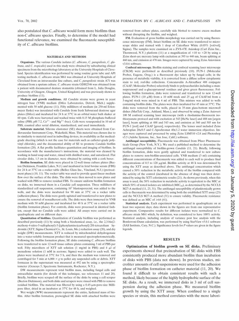
ous biofilm DW measurement method (11). Figure 1 illustratesbiofilm growth obtained with C. albicans strains M61 andGDH2346 to determine optimal adhesion time, inoculum con-centration, and duration of growth. The latter strain was usedto provide a reference to an established dental biofilm modelsystem (10, 11). As shown in Fig. 1A, adhesion under staticconditions for 90 min produced maximal subsequent biofilmformation at 48 h. Figure 1B shows that biofilm formation byboth M61 and GDH2346 on SE appeared to reach a plateau by48 h, regardless of inoculum size. The XTT, DW, and FManalyses revealed that the inoculum size of 107 blastosporesproduced the most abundant and stable mature biofilms. Asmeasured by the DW method at 48 h and under optimal con-ditions, 3 to 4 mg of biofilm was produced per disk.
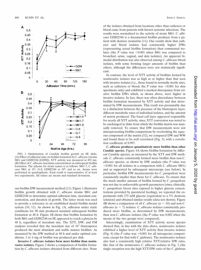
Invasive C. albicans isolates form more biofilm than nonin-vasive isolates. Figure 2 shows a comparison of biofilm forma-tion by C. albicans isolates obtained from different sites. None
of the isolates obtained from locations other than catheters orblood came from patients with known systemic infections. Theresults were normalized to the activity of strain M61. C. albi-cans GDH2346 is a documented biofilm producer from a pa-tient with denture stomatitis (11). Our results show that cath-eter and blood isolates had consistently higher DWs(representing actual biofilm formation) than commensal iso-lates (the P value was �0.001 when M61 was compared tobronchial, urine, vaginal, and skin isolates). An apparent bi-modal distribution was also observed among C. albicans bloodisolates, with some forming larger amounts of biofilm thanothers, although the differences were not statistically signifi-cant.
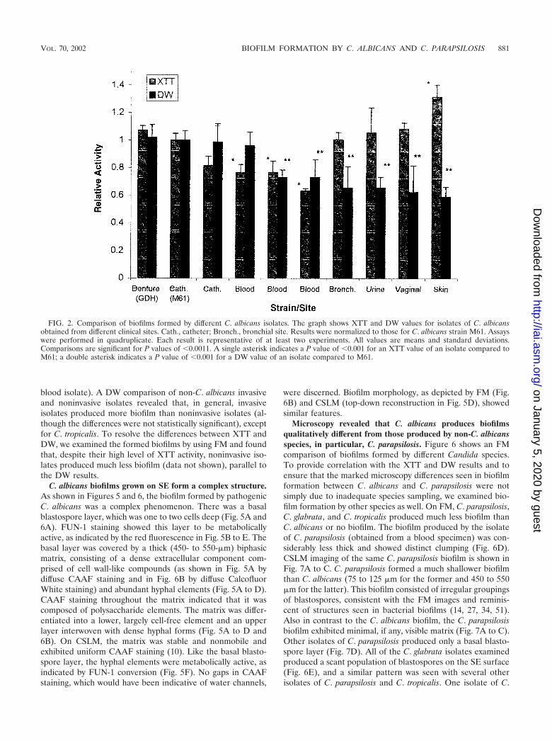
In contrast, the level of XTT activity of biofilms formed bynoninvasive isolates was as high as or higher than that seenwith invasive isolates (i.e., those found in normally sterile sites,such as catheters or blood; the P value was �0.001 for skinspecimens only) and exhibited a marked discrepancy from rel-ative biofilm DWs which, as shown above, were higher ininvasive isolates. In fact, there was often discordance betweenbiofilm formation measured by XTT activity and that deter-mined by DW measurements. This result was presumably dueto a distinction between the presence of the blastospore layer,different metabolic rates of individual isolates, and the amountof matrix produced. The basal cell layer appeared responsiblefor nearly all XTT activity, since XTT conversion was noted tobe unchanged in disks from which the biofilm matrix was man-ually removed. To ensure that DW measurements were notmisrepresenting biofilm comparisons by overlooking the aque-ous component of the matrix (52), we compared DW and WWand found them to be well correlated (Fig. 3), with a correla-tion coefficient of 0.997.
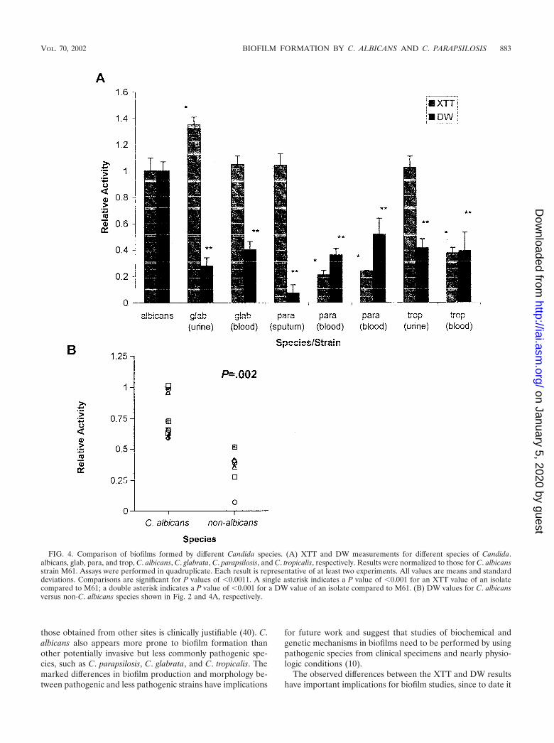
C. albicans produces quantitatively more biofilm than otherCandida species. Figure 4A shows biofilm formation by differ-ent Candida species, as measured by the XTT and DW meth-ods. C. albicans consistently formed more biofilm than non-C.albicans species, as shown by DW analysis (the P value was�0.001 for all isolates in a comparison with C. albicans M61)and as supported by subsequent microscopy (see below). Inparticular, biofilm DW measurements for C. parapsilosis wereconsistently smaller than those for C. albicans. To ensure thatthe much smaller amount of biofilm formed by C. parapsilosiswas not due to unfavorable growth parameters (since clinically,C. parapsilosis favors sites exposed to higher glucose concen-trations provided by parenteral feeding), we repeated the ex-periments with 275 mM glucose (equivalent to a 5% dextrosesolution) and obtained similar results (data not shown). Figure4B shows a comparison of all C. albicans (n � 10) and non-C.albicans (n � 7) isolates. C. albicans isolates consistently pro-duced more biofilm, as determined by DW measurements,than non-C. albicans isolates (the P value was 0.002 when themeans of the two groups were compared).
Interestingly, examination of XTT activity across speciesshowed that, in line with the data above, noninvasive isolatesexhibited a higher level of XTT activity than invasive isolates(Fig. 4) (the P value was �0.001 for all intraspecies compari-sons, except for that with C. glabrata). The noninvasive isolatesalso had a consistently high relative XTT/relative DW ratio,like that of the noninvasive C. albicans isolates in Fig. 2 (thesingle exception was the high XTT/DW ratio of the C. glabrata
FIG. 1. Optimization of Candida biofilm growth on SE disks.(A) Effect of adhesion time on biofilm formation by C. albicans (strainsM61 and GDH2346 [GDH]). XTT activity was measured at 492 nm.(B) Effect of C. albicans inoculum concentration on subsequent biofilmformation. The scheme for strain names is as follows: M61-7, C. albi-cans M61 at 107; G-8, GDH2346 at 108; and so on. Assays wereperformed in quadruplicate. Each result is representative of at leasttwo experiments. All values are means and standard deviations.
880 KUHN ET AL. INFECT. IMMUN.
on January 5, 2020 by guesthttp://iai.asm
.org/D
ownloaded from
blood isolate). A DW comparison of non-C. albicans invasiveand noninvasive isolates revealed that, in general, invasiveisolates produced more biofilm than noninvasive isolates (al-though the differences were not statistically significant), exceptfor C. tropicalis. To resolve the differences between XTT andDW, we examined the formed biofilms by using FM and foundthat, despite their high level of XTT activity, noninvasive iso-lates produced much less biofilm (data not shown), parallel tothe DW results.
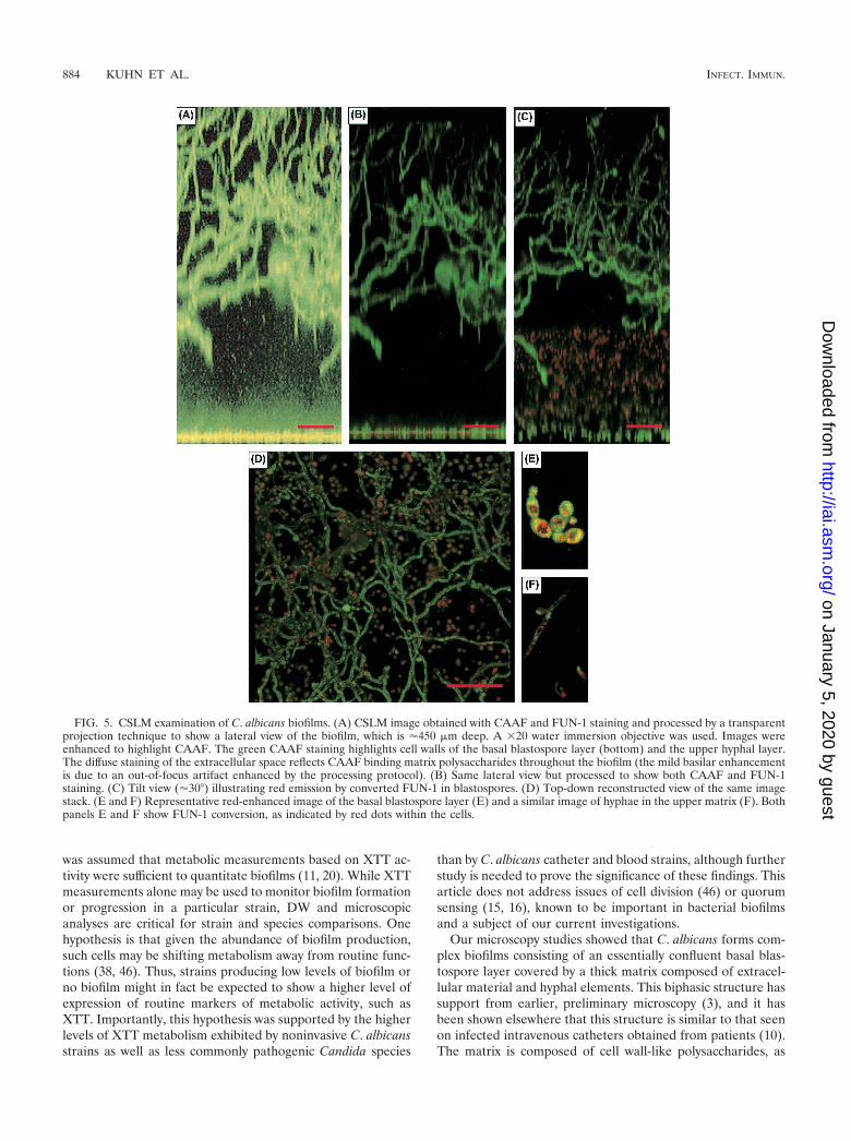
C. albicans biofilms grown on SE form a complex structure.As shown in Figures 5 and 6, the biofilm formed by pathogenicC. albicans was a complex phenomenon. There was a basalblastospore layer, which was one to two cells deep (Fig. 5A and6A). FUN-1 staining showed this layer to be metabolicallyactive, as indicated by the red fluorescence in Fig. 5B to E. Thebasal layer was covered by a thick (450- to 550-�m) biphasicmatrix, consisting of a dense extracellular component com-prised of cell wall-like compounds (as shown in Fig. 5A bydiffuse CAAF staining and in Fig. 6B by diffuse CalcofluorWhite staining) and abundant hyphal elements (Fig. 5A to D).CAAF staining throughout the matrix indicated that it wascomposed of polysaccharide elements. The matrix was differ-entiated into a lower, largely cell-free element and an upperlayer interwoven with dense hyphal forms (Fig. 5A to D and6B). On CSLM, the matrix was stable and nonmobile andexhibited uniform CAAF staining (10). Like the basal blasto-spore layer, the hyphal elements were metabolically active, asindicated by FUN-1 conversion (Fig. 5F). No gaps in CAAFstaining, which would have been indicative of water channels,
were discerned. Biofilm morphology, as depicted by FM (Fig.6B) and CSLM (top-down reconstruction in Fig. 5D), showedsimilar features.
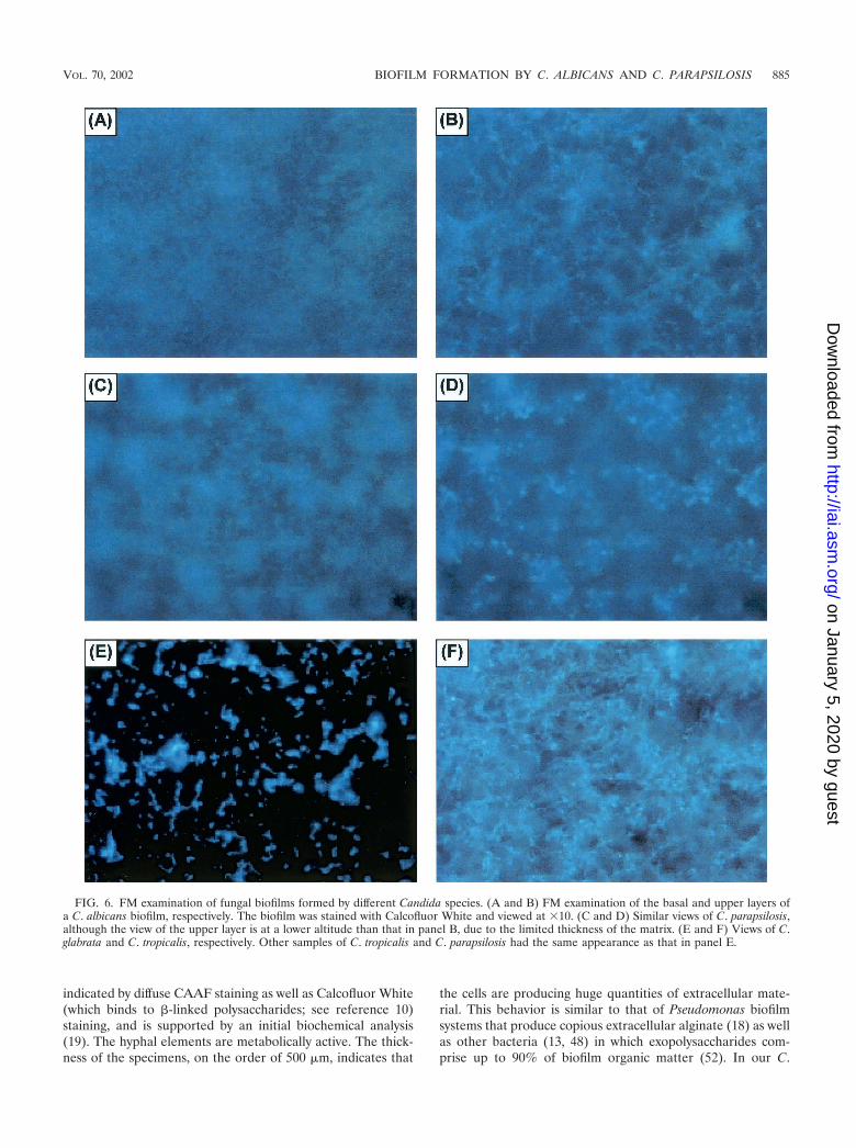
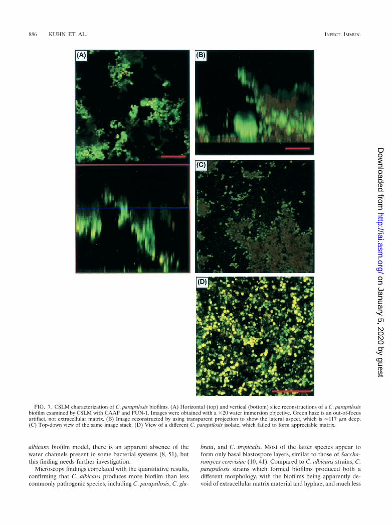
Microscopy revealed that C. albicans produces biofilmsqualitatively different from those produced by non-C. albicansspecies, in particular, C. parapsilosis. Figure 6 shows an FMcomparison of biofilms formed by different Candida species.To provide correlation with the XTT and DW results and toensure that the marked microscopy differences seen in biofilmformation between C. albicans and C. parapsilosis were notsimply due to inadequate species sampling, we examined bio-film formation by other species as well. On FM, C. parapsilosis,C. glabrata, and C. tropicalis produced much less biofilm thanC. albicans or no biofilm. The biofilm produced by the isolateof C. parapsilosis (obtained from a blood specimen) was con-siderably less thick and showed distinct clumping (Fig. 6D).CSLM imaging of the same C. parapsilosis biofilm is shown inFig. 7A to C. C. parapsilosis formed a much shallower biofilmthan C. albicans (75 to 125 �m for the former and 450 to 550�m for the latter). This biofilm consisted of irregular groupingsof blastospores, consistent with the FM images and reminis-cent of structures seen in bacterial biofilms (14, 27, 34, 51).Also in contrast to the C. albicans biofilm, the C. parapsilosisbiofilm exhibited minimal, if any, visible matrix (Fig. 7A to C).Other isolates of C. parapsilosis produced only a basal blasto-spore layer (Fig. 7D). All of the C. glabrata isolates examinedproduced a scant population of blastospores on the SE surface(Fig. 6E), and a similar pattern was seen with several otherisolates of C. parapsilosis and C. tropicalis. One isolate of C.
FIG. 2. Comparison of biofilms formed by different C. albicans isolates. The graph shows XTT and DW values for isolates of C. albicansobtained from different clinical sites. Cath., catheter; Bronch., bronchial site. Results were normalized to those for C. albicans strain M61. Assayswere performed in quadruplicate. Each result is representative of at least two experiments. All values are means and standard deviations.Comparisons are significant for P values of �0.0011. A single asterisk indicates a P value of �0.001 for an XTT value of an isolate compared toM61; a double asterisk indicates a P value of �0.001 for a DW value of an isolate compared to M61.
VOL. 70, 2002 BIOFILM FORMATION BY C. ALBICANS AND C. PARAPSILOSIS 881
on January 5, 2020 by guesthttp://iai.asm
.org/D
ownloaded from
tropicalis produced a thin (�30-�m) layer of matrix-encasedhyphae (Fig. 6F); interestingly, however, there was no visiblebasal blastospore layer.
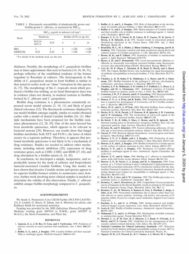
C. albicans biofilms rapidly develop fluconazole resistance.Bacterial and fungal biofilms exhibit antimicrobial drug resis-tance (2, 7, 10, 11). We selected two representative C. albicansstrains (M61 and GDH2346) known to form mature biofilm inour model system and tested their susceptibility to fluconazole.As shown in Table 1, both GDH2346 and M61 biofilms devel-oped fluconazole resistance (MIC50, �128 �g/ml) in a rapidtemporal manner, and the process was complete by 6 h. Thesame organisms grown under planktonic conditions retainedtheir fluconazole susceptibility; the MIC50 was �1 �g/ml.
Metabolic quiescence has been proposed as a mechanism ofbiofilm antimicrobial resistance in bacteria (28) and could ac-count for the phenomenon in Candida biofilms. While ourexperiments were not focused on resistance mechanisms, it isimportant to note that C. albicans cells grown in biofilm ac-tively metabolized XTT. Furthermore, Fig. 5B to F show thatC. albicans cells in biofilms metabolized FUN-1, a processwhich occurred by a biochemical pathway different from that ofXTT, confirming that these cells were metabolically active.
DISCUSSION
We developed a novel system for modeling the behavior ofbiofilms produced by pathogenic fungi and used it to comparethe abilities of different C. albicans strains and non-C. albicansspecies to form biofilms. The advantages of this system aremultiple. The growth system is simple and does not requireelaborate flow chamber devices, which are costly, complicated,and labor-intensive. Medium circulation provided by rockertable motion induces quasi-linear flow stimulating biofilm
growth, similar to the Calgary Biofilm Device (9). The en-hancement of biofilm formation by FBS preincubation is sim-ilar to the effects of saliva shown in a previously describeddenture biofilm model (11) and thereby mimics physiologicconditions. The use of flat sheeting of a medically relevantsubstrate is new, allows easy quantitation and, for the first time,produces reproducible imaging of intact, living, and nondehy-drated specimens by various microscopic techniques, includingCSLM. Moreover, this system allows for the high-volume pro-duction of biofilms (gram amounts), which will facilitate futuregenetic and proteomic studies aimed at understanding the bi-ology and drug resistance of biofilms.
The choice of substrate is not trivial, since prior studiesindicated that chemical composition, degree of hydrophobicity,and the presence of sera, protein, or amino acids affect Can-dida binding to surfaces (11, 17, 22, 33, 49). Other studies haveshown that SE (the material used in the current study) allowsbetter biofilm formation than other substrates (20), and ourown unpublished observations support this finding. Finally, it iswell documented that different substrates induce separate setsof structural genes critical for attachment in bacterial systems(39, 47). Similar work needs to be conducted for Candidaspecies.
Our quantitative results are in contrast to those of mostprior studies, which have reported few consistent differences inbiofilm activity between strains or species (20), and indicatethat more invasive isolates (i.e., those obtained from normallysterile sites, such as catheters or blood) of C. albicans have anincreased ability to form biofilms. Only one other study hassuggested a similar phenomenon for C. parapsilosis (37).Whether this finding reflects innate virulence or up-regulationof virulence following invasion remains to be determined. Dis-tinguishing blood and catheter isolates as invasive compared to
FIG. 3. Correlation of DW and WW measurements. The graph shows DW and WW measurements for different Candida isolates. CA, CG, andCP, C. albicans, C. glabrata, and C. parapsilosis, respectively. Results were normalized to those for C. albicans strain M61. Assays were performedin quadruplicate. Each result is representative of at least two experiments. All values are means and standard deviations. CC, correlation coefficientfor the comparison of DW and WW measurements for all five isolates.
882 KUHN ET AL. INFECT. IMMUN.
on January 5, 2020 by guesthttp://iai.asm
.org/D
ownloaded from
those obtained from other sites is clinically justifiable (40). C.albicans also appears more prone to biofilm formation thanother potentially invasive but less commonly pathogenic spe-cies, such as C. parapsilosis, C. glabrata, and C. tropicalis. Themarked differences in biofilm production and morphology be-tween pathogenic and less pathogenic strains have implications
for future work and suggest that studies of biochemical andgenetic mechanisms in biofilms need to be performed by usingpathogenic species from clinical specimens and nearly physio-logic conditions (10).
The observed differences between the XTT and DW resultshave important implications for biofilm studies, since to date it
FIG. 4. Comparison of biofilms formed by different Candida species. (A) XTT and DW measurements for different species of Candida.albicans, glab, para, and trop, C. albicans, C. glabrata, C. parapsilosis, and C. tropicalis, respectively. Results were normalized to those for C. albicansstrain M61. Assays were performed in quadruplicate. Each result is representative of at least two experiments. All values are means and standarddeviations. Comparisons are significant for P values of �0.0011. A single asterisk indicates a P value of �0.001 for an XTT value of an isolatecompared to M61; a double asterisk indicates a P value of �0.001 for a DW value of an isolate compared to M61. (B) DW values for C. albicansversus non-C. albicans species shown in Fig. 2 and 4A, respectively.
VOL. 70, 2002 BIOFILM FORMATION BY C. ALBICANS AND C. PARAPSILOSIS 883
on January 5, 2020 by guesthttp://iai.asm
.org/D
ownloaded from
was assumed that metabolic measurements based on XTT ac-tivity were sufficient to quantitate biofilms (11, 20). While XTTmeasurements alone may be used to monitor biofilm formationor progression in a particular strain, DW and microscopicanalyses are critical for strain and species comparisons. Onehypothesis is that given the abundance of biofilm production,such cells may be shifting metabolism away from routine func-tions (38, 46). Thus, strains producing low levels of biofilm orno biofilm might in fact be expected to show a higher level ofexpression of routine markers of metabolic activity, such asXTT. Importantly, this hypothesis was supported by the higherlevels of XTT metabolism exhibited by noninvasive C. albicansstrains as well as less commonly pathogenic Candida species
than by C. albicans catheter and blood strains, although furtherstudy is needed to prove the significance of these findings. Thisarticle does not address issues of cell division (46) or quorumsensing (15, 16), known to be important in bacterial biofilmsand a subject of our current investigations.
Our microscopy studies showed that C. albicans forms com-plex biofilms consisting of an essentially confluent basal blas-tospore layer covered by a thick matrix composed of extracel-lular material and hyphal elements. This biphasic structure hassupport from earlier, preliminary microscopy (3), and it hasbeen shown elsewhere that this structure is similar to that seenon infected intravenous catheters obtained from patients (10).The matrix is composed of cell wall-like polysaccharides, as
FIG. 5. CSLM examination of C. albicans biofilms. (A) CSLM image obtained with CAAF and FUN-1 staining and processed by a transparentprojection technique to show a lateral view of the biofilm, which is �450 �m deep. A �20 water immersion objective was used. Images wereenhanced to highlight CAAF. The green CAAF staining highlights cell walls of the basal blastospore layer (bottom) and the upper hyphal layer.The diffuse staining of the extracellular space reflects CAAF binding matrix polysaccharides throughout the biofilm (the mild basilar enhancementis due to an out-of-focus artifact enhanced by the processing protocol). (B) Same lateral view but processed to show both CAAF and FUN-1staining. (C) Tilt view (�30°) illustrating red emission by converted FUN-1 in blastospores. (D) Top-down reconstructed view of the same imagestack. (E and F) Representative red-enhanced image of the basal blastospore layer (E) and a similar image of hyphae in the upper matrix (F). Bothpanels E and F show FUN-1 conversion, as indicated by red dots within the cells.
884 KUHN ET AL. INFECT. IMMUN.
on January 5, 2020 by guesthttp://iai.asm
.org/D
ownloaded from
indicated by diffuse CAAF staining as well as Calcofluor White(which binds to -linked polysaccharides; see reference 10)staining, and is supported by an initial biochemical analysis(19). The hyphal elements are metabolically active. The thick-ness of the specimens, on the order of 500 �m, indicates that
the cells are producing huge quantities of extracellular mate-rial. This behavior is similar to that of Pseudomonas biofilmsystems that produce copious extracellular alginate (18) as wellas other bacteria (13, 48) in which exopolysaccharides com-prise up to 90% of biofilm organic matter (52). In our C.
FIG. 6. FM examination of fungal biofilms formed by different Candida species. (A and B) FM examination of the basal and upper layers ofa C. albicans biofilm, respectively. The biofilm was stained with Calcofluor White and viewed at �10. (C and D) Similar views of C. parapsilosis,although the view of the upper layer is at a lower altitude than that in panel B, due to the limited thickness of the matrix. (E and F) Views of C.glabrata and C. tropicalis, respectively. Other samples of C. tropicalis and C. parapsilosis had the same appearance as that in panel E.
VOL. 70, 2002 BIOFILM FORMATION BY C. ALBICANS AND C. PARAPSILOSIS 885
on January 5, 2020 by guesthttp://iai.asm
.org/D
ownloaded from
albicans biofilm model, there is an apparent absence of thewater channels present in some bacterial systems (8, 51), butthis finding needs further investigation.
Microscopy findings correlated with the quantitative results,confirming that C. albicans produces more biofilm than lesscommonly pathogenic species, including C. parapsilosis, C. gla-
brata, and C. tropicalis. Most of the latter species appear toform only basal blastospore layers, similar to those of Saccha-romyces cerevisiae (10, 41). Compared to C. albicans strains, C.parapsilosis strains which formed biofilms produced both adifferent morphology, with the biofilms being apparently de-void of extracellular matrix material and hyphae, and much less
FIG. 7. CSLM characterization of C. parapsilosis biofilms. (A) Horizontal (top) and vertical (bottom) slice reconstructions of a C. parapsilosisbiofilm examined by CSLM with CAAF and FUN-1. Images were obtained with a �20 water immersion objective. Green haze is an out-of-focusartifact, not extracellular matrix. (B) Image reconstructed by using transparent projection to show the lateral aspect, which is �117 �m deep.(C) Top-down view of the same image stack. (D) View of a different C. parapsilosis isolate, which failed to form appreciable matrix.
886 KUHN ET AL. INFECT. IMMUN.
on January 5, 2020 by guesthttp://iai.asm
.org/D
ownloaded from
thickness. Notably, the morphology of C. parapsilosis biofilmsmay at times approximate that seen in bacteria (14, 34, 46, 51),perhaps reflective of the established tendency of the formerorganism to flocculate in cultures. The heterogeneity in theability of C. parapsilosis strains to form biofilms is similar tothat noted in earlier work on “slime” formation by this species(6, 37). The morphology of the C. tropicalis strain which pro-duced a biofilm was striking, as no basal blastospore layer wasin evidence (data not shown), in marked contrast to biofilmsformed by C. albicans and C. parapsilosis.
Biofilm drug resistance is a phenomenon consistently ex-pressed across model systems (2, 10, 11) and likely of greatclinical relevance (12). The fluconazole resistance displayed byour model biofilms emerges rapidly, similar to results obtainedearlier with a model of dental Candida biofilm (10, 11). Mul-tiple mechanisms have been proposed for the biofilm resis-tance phenomenon (12, 28, 29). One of the most favored hasbeen metabolic quiescence, which appears to be relevant inbacterial systems (29). However, our results show that fungalbiofilms metabolize both XTT and FUN-1, the latter of whichoccurs via a separate pathway. Therefore, it appears that pro-found metabolic quiescence is not likely to be a mechanism ofdrug resistance. Studies are needed to address other mecha-nisms, including mitotic inhibition (29); expression of drugresistance genes, such as CDR1, CDR2, and MDR (27, 44); anddrug absorption by a biofilm matrix (4, 24, 45).
In conclusion, we developed a simple, inexpensive, and re-producible system for the study of catheter and bioprostheticmaterial-associated Candida biofilms. Using this model, wehave shown that invasive Candida strains and species appear tobe superior biofilm formers relative to noninvasive ones; how-ever, further work involving more clinical samples is needed todetermine the validity of this observation. Finally, C. albicansexhibits unique biofilm morphology compared to C. parapsilo-sis.
ACKNOWLEDGMENTS
We thank A. Niemenen’s Core CSLM Facility (NCI P30 CA43703–12); S. Leidich, G. Reyes, N. Isham, and A. Morrisey for advice; andKathleen Smith for secretarial assistance.
Funding was provided by the NIH (Infectious Disease/GeographicMedicine training grant AI07024 to D.M.K.; grant AI35097 toM.A.G.), the Steris Foundation, and Pfizer Inc.
REFERENCES
1. Anaissie, E. J., J. H. Rex, O. Uzon, and S. Vartivarian. 1998. Predictors ofadverse outcome in cancer patients with candidemia. Am. J. Med. 104:238–245.
2. Baillie, G. S., and L. J. Douglas. 1999. Candida biofilms and their suscepti-bility to antifungal agents. Methods Enzymol. 310:644–656.
3. Baillie, G. S., and L. J. Douglas. 1999. Role of dimorphism in the develop-ment of Candida albicans biofilms. J. Med. Microbiol. 48:671–679.
4. Baillie, G. S., and L. J. Douglas. 2000. Matrix polymers of Candida biofilmsand their possible role in biofilm resistance to antifungal agents. J. Antimi-crob. Chemother. 46:397–403.
5. Banejee, S. N., G. T. Emori, D. H. Culver, R. P. Gaynes, W. R. Jarvis, T.Horan, J. R. Edwards, et al. 1991. Secular trends in nosocomial primarybloodstream infections in the United States, 1980–1989. Am. J. Med.91(Suppl. 3B):86–89.
6. Branchini, M. L., M. A. Pfaller, J. Rhine-Chalberg, T. Frempong, and H. D.Isenberg. 1994. Genotypic variation and slime production among blood andcatheter isolates of Candida parapsilosis. J. Clin. Microbiol. 32:452–456.
7. Brown, M. R., and P. Gilbert. 1993. Sensitivity of biofilms to antimicrobialagents. J. Appl. Bacteriol. 74(Suppl.):87S-97S.
8. Bryers, J. D., and F. Drummond. 1998. Local macromolecule diffusion co-efficients in structurally non-uniform bacterial biofilms using fluorescencerecovery after photobleaching (FRAP). Biotechnol. Bioeng. 60:462–473.
9. Ceri, H., M. E. Olson, C. Stremick, R. R. Read, D. Morck, and A. Buret.1999. The Calgary Biofilm Device: new technology for rapid determinationof antibiotic susceptibilities in bacterial biofilms. J. Clin. Microbiol. 37:1771–1776.
10. Chandra, J., D. M. Kuhn, P. K. Mukherjee, L. L. Hoyer, and M. A. Ghan-noum. 2001. Biofilm formation by the pathogen C. albicans–-development,architecture, and drug resistance. J. Bacteriol. 183:5385–5394.
11. Chandra, J., P. K. Mukherjee, S. D. Leidich, F. F. Faddoul, L. L. Hoyer, L. J.Douglas, and M. A. Ghannoum. 2001. Antifungal resistance of Candidabiofilms formed on denture acrylic in vitro. J. Dent. Res. 80:903–908.
12. Costerton, J. W., P. S. Stewart, and E. P. Greenberg. 1999. Bacterial biofilms:a common cause of persistent infections. Science 284:1318–1322.
13. Danese, P. N., L. A. Pratt, and R. Kolter. 2000. Exopolysaccharide produc-tion is required for development of Escherichia coli K-12 biofilm architec-ture. J. Bacteriol. 182:3593–3596.
14. Davey, M. E., and G. A. O’Toole. 2000. Microbial biofilms: from ecology tomolecular genetics. Microbiol. Mol. Biol. Rev. 64:847–867.
15. Davies, D. G., M. R. Parsek, J. P. Pearson, B. H. Iglewski, J. W. Costerton,and E. P. Greenberg. 1998. The involvement of cell-to-cell signals in thedevelopment of a bacterial biofilm. Science 280:295–298.
16. De Kievit, T. R., R. Gillis, S. Marx, C. Brown, and B. H. Iglewski. 2001.Quorum-sensing genes in Pseudomonas aeruginosa biofilms: their role andexpression patterns. Appl. Environ. Microbiol. 67:1865–1873.
17. El-Azizi, M., and N. Khardori. 1999. Factors influencing adherence of Can-dida spp. to host tissues and plastic surfaces. Indian J. Exp. Biol. 37:941–951.
18. Gacesa, P. 1998. Bacterial alginate biosynthesis–-recent progress and futureprospects. Microbiology 144:1133–1143.
19. Hawser, S. P., G. S. Baillie, and L. J. Douglas. 1998. Production of extra-cellular matrix by Candida albicans biofilms. J. Med. Microbiol. 47:253–256.
20. Hawser, S. P., and L. J. Douglas. 1994. Biofilm formation by Candida specieson the surface of catheter materials in vitro. Infect. Immun. 62:915–921.
21. Hawser, S. P., and L. J. Douglas. 1995. Resistance of Candida albicansbiofilms to antifungal agents in vitro. Antimicrob. Agents Chemother. 39:2128–2131.
22. Hawser, S. P., and K. Islam. 1998. Binding of C. albicans to immobilizedamino acids and bovine serum albumen. Infect. Immun. 66:140–144.
23. Hawser, S. P., H. Norris, C. J. Jessup, and M. A. Ghannoum. 1998. Com-parison of a 2,3-bis(2-methoxy-4-nitro-5-sulfophenyl)-5-[(phenylamino)car-bonyl]-2H-tetrazolium hydroxide (XTT) colorimetric method with the stan-dardized National Committee for Clinical Laboratory Standards method oftesting clinical yeast isolates for susceptibility to antifungal agents. J. Clin.Microbiol. 36:1450–1452.
24. Hoyle, B. D., J. Jass, and J. W. Costerton. 1990. The biofilm glycocalyx as aresistance factor. J. Antimicrob. Chemother. 26:1–5.
25. Kremery, V., and G. Kovacicova, Jr. 2000. Longitudinal 10-year prospectivesurvey of fungemia in the Slovak Republic: trends in etiology in 310 episodes.Slovak Fungaemic Group. Diagn. Microbiol. Infect. Dis. 36:7–11.
26. Kremery, V., Jr., M. Mrazova, A. Kunora, E. Grey, J. Mardiak, L. Jurga, A.Sabo, et al. 1999. Nosocomial candidemias due to species other than Can-dida albicans in cancer patients. Aetiology, risk factors, and outcome of 45episodes within 10 years in a single cancer institution. Support Care Cancer7:428–431.
27. Kuchma, S. L., and G. A. O’Toole. 2000. Surface-induced and biofilm-induced changes in gene expression. Curr. Opin. Biotechnol. 11:429–433.
28. Lewis, K. 2001. Riddle of biofilm resistance. Antimicrob. Agents Chemother.45:999–1007.
29. Mahmoud, T. F., and G. A. O’Toole. 2001. Mechanisms of biofilm resistanceto antimicrobial agents. Trends Microbiol. 9:34–39.
30. Mermel, L. A., B. M. Farr, R. J. Sherertz, I. I. Raad, N. O’Grady, J. S.Harris, and O. E. Craven. 2001. Guidelines for the management of intra-vascular catheter-related infections. Clin. Infect. Dis. 32:1249–1272.
31. National Committee for Clinical Laboratory Standards. 1997. Referencemethod for broth dilution antifungal susceptibility testing of yeasts. M27-A.National Committee for Clinical Laboratory Standards, Wayne, Pa.
32. Nguyen, M. H., J. E. Peacock Jr., D. C. Tanner, A. J. Morris, M. L. Nguyen,
TABLE 1. Fluconazole susceptibility of planktonically grown andbiofilm-grown C. albicans, as measured by MIC50
Strain
MIC50 (�g/ml) in indicated cell typea:
Planktonic Biofilm (XTT) at age (h):
NCCLSM27-A XTT 0 6 24
M61 1 1 4 �128 �128GDH2346 0.25 0.25 4 �128 �128
a For details of the methods used, see the text.
VOL. 70, 2002 BIOFILM FORMATION BY C. ALBICANS AND C. PARAPSILOSIS 887
on January 5, 2020 by guesthttp://iai.asm
.org/D
ownloaded from
D. R. Snydman, M. M. Wagener, et al. 1995. Therapeutic approaches inpatients with candidemia: evaluation in a multicenter, prospective, observa-tional study. Arch. Intern. Med. 155:2429–2435.
33. Nikawa, H., H. Nishimura, S. Makihara, T. Hamada, S. Sadamori, and L. P.Samaranayake. 2000. Effect of serum concentration on Candida biofilmformation on acrylic surfaces. Mycoses 43:139–143.
34. O’Toole, G., H. B. Kaplan, and R. Kolter. 2000. Biofilm formation as mi-crobial development. Annu. Rev. Microbiol. 54:49–79.
35. Pagano, L., A. Antinori, A. Ammassari, L. Mele, A. Nosari, L. Melillo, B.Martino, et al. 1999. Retrospective study of candidemia in patients withhematological malignancies. Clinical features, risk factors, and outcome of76 episodes. Eur. J. Haematol. 63:77–85.
36. Pfaller, M. A., R. N. Jones, G. W. Doern, H. S. Sader, S. A. Messer, A.Houston, S. Coffman, et al. 2000. Bloodstream infections due to Candidaspecies: SENTRY antimicrobial surveillance program in North America andLatin America, 1997–1998. Antimicrob. Agents Chemother. 44:747–751.
37. Pfaller, M. A., S. A. Messer, and R. J. Hollis. 1995. Variations in DNAsubtype, antifungal susceptibility, and slime production among clinical iso-lates of Candida parapsilosis. Diagn. Microbiol. Infect. Dis. 21:9–14.
38. Prigent-Combaret, C., C. O. Vidal, C. Dorel, and P. Lejeune. 1999. Abioticsurface sensing and biofilm-dependent regulation of gene expression inEscherichia coli. J. Bacteriol. 181:5993–6002.
39. Pruzzo, C., A. Crippa, S. Bertone, L. Pane, and A. Carli. 1996. Attachmentof Vibrio alginolyticus to chitin mediated by chitin-binding proteins. Micro-biology 142:2181–2186.
40. Rex, J. H., T. J. Walsh, J. D. Sobel, S. G. Filler, P. G. Pappas, W. E.Dismukes, and J. E. Edwards. 2000. Practice guidelines for the treatment ofcandidiasis. Clin. Infect. Dis. 30:662–668.
41. Reynolds, T. B., and G. R. Fink. 2001. Bakers’ yeast, a model for fungalbiofilm formation. Science 291:878–881.
42. Saiman, L., E. Ludington, M. Pfaller, S. Rangel-Frausto, R. T. Wiblin, J.Dawson, H. M. Blumberg, et al. 2000. Risk factors for candidemia in neo-
natal intensive care unit patients. The National Epidemiology of MycosisSurvey Study Group. Pediatr. Infect. Dis. J. 19:319–324.
43. Sanchez, V., J. A. Vasquez, D. Barth-Jones, L. Dembry, J. D. Sobel, and M. J.Zervus. 1993. Nosocomial acquisition of Candida parapsilosis: an epidemio-logic study. Am. J. Med. 94:577–582.
44. Sanglard, D., F. Ischer, M. Monod, and J. Bille. 1997. Cloning of Candidaalbicans genes conferring resistance to azole antifungal agents: characteriza-tion of CDR2, a new multidrug ABC transporter gene. Microbiology 143:405–416.
45. Shigeta, M., G. Tanaka, H. Komatsuzawa, M. Sugai, H. Suginaka, and T.Usui. 1997. Permeation of antimicrobial agents Pseudomonas aeruginosabiofilms: a simple method. Chemotherapy (Tokyo) 43:340–345.
46. Watnick, P., and R. Kolter. 2000. Biofilm, city of microbes. J. Bacteriol.182:2675–2679.
47. Watnick, P. I., K. J. Fullner, and R. Kolter. 1999. A role for the mannose-sensitive hemagglutinin in biofilm formation by Vibrio cholerae El Tor. J.Bacteriol. 181:3606–3609.
48. Watnick, P. I., and Kolter, R. 1999. Steps in the development of a Vibriocholerae El Tor biofilm. Mol. Microbiol. 34:586–595.
49. Webb, J. S., H. C. Van der Mei, M. Nixon, I. M. Eastwood, M. Greenhalgh,S. J. Read, G. D. Robson, et al. 1999. Plasticizers increase adhesion of thedeteriogenic fungus Aureobasidium pullulans to polyvinyl chloride. Appl.Environ. Microbiol. 65:3575–3581.
50. Weems, J. J., Jr., M. E. Cumberland, J. Ward, M. Willy, A. A. Padhye, andS. L. Solomon. 1987. Candida parapsilosis fungemia associated with paren-teral nutrition and contaminated blood pressure transducers. J. Clin. Micro-biol. 25:1029–1032.
51. Wimpenny, J., W. Manz, and U. Szewzyk. 2000. Heterogeneity in biofilms.FEMS Microbiol. Rev. 24:661–671.
52. Zhang, X. Q., P. L. Bishop, and M. J. Kupferle. 1998. Measurement ofpolysaccharides and proteins in biofilm extracellular polymers. Water Sci.Technol. 37:345–348.
Editor: T. R. Kozel
888 KUHN ET AL. INFECT. IMMUN.
on January 5, 2020 by guesthttp://iai.asm
.org/D
ownloaded from