Embed Size (px)
Citation preview
750 AJVR,Vol70,No.6,June2009
Tendinitis is a common debilitating injury in performance horses, comprising up to 43% of
injuries in racing Thoroughbreds and resulting in retirement of up to 14% of horses.1–3 Timely repair of injured tendon with restoration of normal tissue organization and biomechanical properties has not been consistently achieved.4–6 Thus, reinjury, despite prolonged and costly rehabilitation, is common.
The inability of equine tendon to regenerate after injury, or to heal with mechanical properties compara-ble to the original tissue, is likely attributable to the low vascularity and cellularity of the tissue, low number of progenitor cells in the tissue, and healing under weight-
Comparison of equine tendon-, muscle-, and bone marrow–derived cells cultured
on tendon matrix
Allison A. Stewart, DVM, MS; Jennifer G. Barrett, DVM, PhD; Christopher R. Byron, DVM, MS; Angela C. Yates, DVM, MS; Sushmitha S. Durgam, BVSc; Richard B. Evans, PhD; Matthew C. Stewart, BVSc, PhD
Objective—Tocompareviabilityandbiosyntheticcapacitiesofcellsisolatedfromequinetendon,muscle,andbonemarrowgrownonautogenoustendonmatrix.Sample Population—Cellsfrom4youngadulthorses.Procedures—Cellswereisolated,expanded,andculturedonautogenouscell-freetendonmatrixfor7days.Sampleswereanalyzedforcellviability,proteoglycansynthesis,collagensynthesis,andmRNAexpressionofcollagentypeI,collagentypeIII,andcartilageoligo-mericmatrixprotein(COMP).Results—Tendon-andmuscle-derivedcellsrequiredlesstimetoreachconfluence(approx2weeks)thandidbonemarrow–derivedcells(approx3to4weeks);therewerefewerbonemarrow–derivedcellsatconfluencethantheother2celltypes.Moretendon-andmuscle-derivedcellswereattachedtomatricesafter7daysthanwerebonemarrow–derivedcells.Collagenandproteoglycansynthesisbytendon-andmuscle-derivedcellswassignificant-lygreater thansynthesisbybonemarrow–derivedcells.Onaper-cell basis, tendon-de-rivedcellshadmorecollagensynthesis,althoughthiswasnotsignificant.CollagentypeImRNAexpressionwassimilaramonggroups.Tendon-derivedcellsexpressedthehighestamountsofcollagentypeIIIandCOMPmRNAs,althoughthedifferenceforCOMPwasnotsignificant.Conclusions and Clinical Relevance—Tendon-andmuscle-derivedcellsyieldedgreatercellculturenumbersinshortertimeand,onaper-cellbasis,hadcomparablebiosyntheticassaystobonemarrow–derivedcells.Moreinvitroexperimentswithhighernumbersmaydeterminewhethertendon-derivedcellsareausefulresourcefortendonhealing.(Am J Vet Res2009;70:750–757)
bearing conditions.2,3,7,8 Additionally, it is thought that tendinitis, which might more properly be termed tendinopathy, is caused by a chronic degenerative pro-cess rather than a single traumatic episode.7,9 Strategies to improve tendon healing have focused on enhancing the metabolic response of the injured tenocytes, modu-lating the organization of newly synthesized extracellu-lar matrix, or administering progenitor cells to enhance repair tissue.5,10–13 The use of MSCs for tissue repair has been an extremely dynamic area of research during the past decade.13 Most of those studies have focused on MSCs derived from bone marrow aspirates. The most prominent avenues of investigation with MSCs have been directed at repair of bone, cartilage, myocardium, and CNS tissues.14–17 Isolation of a homogeneous popu-lation of progenitor cells from bone marrow is time-
Received May 15, 2008.Accepted August 28, 2008.From the the Department of Veterinary Clinical Medicine, College of
Veterinary Medicine, University of Illinois, Urbana, IL 61802. Dr. Barrette’s present address is Marion duPont Scott Equine Medical Center, Virginia-Maryland Regional College of Veterinary Medicine, Leesburg, VA 20177.
Supported by the American College of Veterinary Surgeons Resident-in-Training Grant and the American Quarter Horse Foundation.
Results from this study were presented at the 40th American College of Veterinary Surgeons Symposium, San Diego, October 2005.
Address correspondence to Dr. Stewart.
AbbreviAtionsCPM CountsperminuteCOMP CartilageoligomericmatrixproteinDMEM DulbeccomodifiedEaglemediumDPM DisintegrationsperminuteFBS FetalbovineserumMSC Mesenchymalstemcells
08-05-0158r.indd 750 5/20/2009 2:10:21 PM
AJVR,Vol70,No.6,June2009 751
consuming, and there is much variation in cell num-bers, cell viability, and growth rates among samples.18,19 More recent studies20,21 indicate that alternative sources of progenitor cells (eg, fat, synovium, cartilage, and sat-ellite cells) might also be beneficial for specific thera-peutic applications.
Tendon is composed primarily of type I collagen fi-brils arranged into fibers aligned along the longitudinal axis of the tendon.22 Collagen type III is also present and comprises approximately 14% of total collagen in normal tendon.23 Cartilage oligomeric matrix protein is another important extracellular matrix protein pro-duced by tenocytes.24 Collagen type III and COMP par-ticipate in collagen type I fibrillogenesis,24,25 and both are produced following tendon injury.26,27 Collagen fi-brils are surrounded by ground substance composed of proteoglycans and glycosaminoglycans, which help package the collagen fibrils. Proteoglycan and collagen synthesis are also increased in healing tendon.28,29
Bone marrow–derived MSCs have been used for tendon repair in horses, rabbits, and rats.15,30,31 To date, most published studies, such as those by Awad et al30 and Guest et al,31 have used bone marrow–derived MSCs for tendon repair. Most studies in rabbits and rats have revealed an increase in the strength of ten-don repair; however, 1 study32 found bone formation within the tendon repair tissue. Progenitor cell–based treatments for tendon injuries have promise, but are not widely used or universally successful.
The objective of the study reported here was to compare cells isolated from 3 tissue types (tendon, muscle, and bone marrow) for ease of isolation, cell growth characteristics, extracellular matrix production, and expression of tendon matrix markers when grown on tendon extracellular matrix. Bone marrow–derived progenitor cells were used as the gold standard source for cell-based tendinitis therapy.31 Skeletal muscle– and tendon-derived cells were isolated by use of a preplat-ing technique used to isolate progenitor cells from muscle.33,34 Our hypotheses were that tendon-derived cells would yield greater numbers in cell culture over a shorter time period, grow and adhere to the matrix bet-ter, and produce more tendon extracellular matrix than cells derived from muscle or bone marrow.
Materials and MethodsCollection of samples—Bone marrow, muscle, and
tendon were collected aseptically from 4 young horses (1 to 3 years of age) euthanatized for reasons unrelated to musculoskeletal disease. All samples were obtained in accordance with guidelines reviewed and approved by the Institutional Animal Care and Use Committee of the University of Illinois. All horses were sedated with 0.5 to 1.0 mg of xylazine/kg administered IV prior to induction. Anesthesia was induced with 2.2 mg of ket-amine/kg and 0.1 mg of diazepam/kg given IV. General anesthesia was maintained by IV infusion of 5% guai-fenesin, 1 mg of ketamine/mL, and 1 mg of xylazine/mL. Following collection of the bone marrow aspirates, as described,18,35 all horses were euthanatized with 104 mg of sodium pentobarbital/kg given IV. The tendon and muscle specimens were collected immediately follow-ing euthanasia.
Processing of bone marrow–derived cells—The right tuber coxae was clipped and aseptically prepared, and a bone marrow biopsy needlea was used to aspirate 30 mL of bone marrow into syringes containing 3,000 units of heparin diluted to a volume of 5 mL with PBS solution. The bone marrow aspirate was diluted with 15 mL of PBS solution and centrifuged at 300 X g for 15 minutes. The supernatant was removed, the pellet was resuspended in PBS solution, and centrifugation was repeated. Pelleted cells were resuspended in 12 mL of low-glucose DMEMb supplemented with 10% FBS,c 300 µg of l-glutamined/mL, 100 U of sodium penicil-line/mL, 100 µg of streptomycin sulfatee/mL, and 1mM of sodium pyruvatef/mL. Resuspended cells were placed in a 75-cm2 flask and incubated at 37°C in a 5% car-bon dioxide atmosphere with 90% humidity. To obtain adequate cell numbers for experiments, the bone mar-row–derived cells were passaged after they reached focal areas of confluence. Time to confluence and cell counts at trypsinization were recorded for all cell types.
Processing of muscle-derived cells—The skin over the right semitendinosus region was clipped and asep-tically prepared prior to euthanasia. Immediately fol-lowing euthanasia, a muscle sample (5-cm length by 2-cm width by 1.5-cm depth) was excised from the right semitendinosus muscle. Progenitor cells were collected from skeletal muscle by use of a published protocol.33 Briefly, the muscle was diced into 0.5-cm3 pieces, rinsed with PBS solution to remove RBCs, and dissociated by sequential digestion with 0.2% collagenaseg for 30 min-utes followed by 2.0% dispaseh for 15 minutes at 37°C. All enzymatic digestions were carried out in high-glu-cose DMEM supplemented with 1% FBS, 37.5 µg of ascorbic acidi/mL, 100 U of sodium penicillin/mL, and 100 µg of streptomycin sulfate/mL. Following di-gestion, the suspensions were passed through 70- and 40-µm filters.j The isolated cells were collected by cen-trifugation at 300 X g for 5 minutes. The supernatant was removed, and the cell pellet was resuspended in media. Cell viability was determined by use of exclu-sion of trypan blue dye.36,k
Processing of tendon-derived cells—The super-ficial digital flexor tendons from both forelimbs were harvested aseptically from each horse. A 6-cm X 1-cm2
sample of tendon was reserved for cell isolation. A 2-cm3 sample was snap-frozen in liquid nitrogen for control RNA isolation, and the remainder was cryo-preserved for cell-free tendon matrix production. The specimen for cell isolation was diced into 0.5-cm3 piec-es and digested for 16 hours at 37°C in 0.1% collage-nase high-glucose DMEM supplemented with 1% FBS, 37.5 µg/mL of ascorbic acid, 100 U of sodium penicil-lin/mL, and 100 µg of streptomycin sulfate/mL. Follow-ing digestion, the isolated cells were processed as for muscle-derived cells.
Muscle and tendon cell culture—The muscle- and tendon-derived cells were seeded at 1,300 cells/cm2 in monolayer cultures in high-glucose DMEM supple-mented with 20% FBS, 37.5 µg/mL of ascorbic acid, 300 µg of l-glutamine/mL, 100 U of sodium penicil-lin/mL, and 100 µg of streptomycin sulfate/mL. The rapidly adherent, fibroblast-like cells in these popula-
08-05-0158r.indd 751 5/20/2009 2:10:21 PM
752 AJVR,Vol70,No.6,June2009
tions were excluded by a described preplating or dif-ferential attachment protocol whereby the culture me-dium and unattached cells were serially transferred to fresh culture flasks every 12 to 24 hours during the first 72 hours of culture.33 The muscle- and tendon-derived cells that adhered after 72 hours of differential attach-ment were trypsinized at confluency and used in the experiments.
Tendon matrix culture model—Superficial digital flexor tendons were collected from donor horses and cut longitudinally with a dermatome to produce uni-formly flat, 0.5-mm-thick tendon sheets. These sheets were cut into 1 X 1-cm square explants. The explants were subjected to 4 rounds of freeze-thaw cycles at –80°C and 4°C to kill the endogenous cells. These cell-free tendon explants (matrix only) were maintained in culture without the addition of cells to serve as nega-tive controls or seeded with 250,000-cell aliquots of the expanded bone marrow–derived, tendon-derived or muscle-derived cells. This experimental design com-prised 4 treatment groups (matrix only, matrix and bone marrow–derived cells, matrix and muscle-derived cells, and matrix and tendon-derived cells). Each treat-ment group had 20 replicates, with an overall total of 80 samples/horse.
The cell-seeded tendon matrices were supple-mented with high-glucose DMEM supplemented with 10% FBS, 37.5 µg of ascorbic acid/mL, 300 µg of l-glutamine/mL, 100 U of sodium penicillin/mL, and 100 µg of streptomycin sulfate/mL. The media was changed every 2 days. All culture samples were collected on day 7.
Cell numbers on matrices—Three replicates of each treatment group were used to measure cell viabil-ity and number on day 7 by use of a cell viability assay,l as per manufacturer’s instructions. In brief, 50 µL of the assay reagent was added to fresh ascorbate–free media in each well, and the cells were incubated at 37°C for 2.5 hours. One hundred microliters of media from each well was transferred to a 96-well plate, and absorbance was measured at 492nM in a microplate reader.m All samples were assayed in duplicate, and a mean value was calculated to provide a single data point. These op-tical density values were converted to cell number by reference to standard curves determined from plated cells from all 3 tissue sources from each horse.
Collagen synthesis—Collagen synthesis was de-termined via [3H]prolinen incorporation according to a published protocol.37 On day 6, 3 wells of each treatment group were radiolabeled with 50 µCi of [3H]proline/mL of media and incubated overnight. Following radiola-beling, the samples were stored at –80°C.
Radiolabeled samples were freeze-thawed 3 times, digested, and homogenized to disrupt cells and matrix prior to RNase treatment. The total protein was pre-cipitated with tricholoroacetic acid and washed 3 times. The resulting pellets were dissolved, digested with pu-rified collagenase,o and centrifuged. The supernatant and pellets were separated and added to scintillation liquid, and the radioactivity was counted via scintilla-tion counter.p
Newly synthesized collagen was detected on the basis of radioactivity in the sample supernatants fol-lowing collagenase-digestion.37 Percentage collagen synthesis was expressed by relating collagen synthesis to total protein synthesis (sum of the activity in the col-lagenase-digested supernatant and the pellet) in each sample.
Proteoglycan synthesis—Proteoglycan synthesis was determined by measuring 35SO
4 incorporation into
each sample. Three wells of each treatment group were radiolabeled with 10 µCi of 35S methionine/mLq dur-ing the last 24 hours of the experiments. The samples were digested in 1 mL of 0.1% papainr at 65°C for 16 hours.38 Twenty-five-microliter aliquots of 35S-labeled, papain-digested tendon matrices were placed in multi- well punch plates,s precipitated with alcian blue dye, and counted via scintillation. All values were adjusted for radioisotope decay and for the total volume of pa-pain digestion buffer added to the sample.
RNA isolation and gene expression—Nine samples from each treatment group were pooled, snap-frozen in liquid nitrogen, and stored at –80°C for RNA isolation. The RNA was isolated by use of a protocol adapted from a technique for cartilage RNA isolation.39 Briefly, tissues were pulverized under liquid nitrogen, homogenized in guanidinium isothiocyanate lysis buffer, extracted with phenol-chloroform, precipitated with isopropanol, and purified by use of a commercially available column-based protocol.t This protocol included an on-column DNase treatment to exclude genomic template contamination. One microgram of RNA in each sample was converted to cDNA with a commercial reverse transcription kitu and oligo(dT) primers. Target cDNAs were amplified via real-time PCR by use of Taq DNA polymerasev and gene-spe-cific primers designed from available published sequences in Genbank and by use of a multiple sequence alignment program.w Primer specificity was confirmed by cloning and sequencing the PCR products (Appendix). Real-time quantitative PCR assay was performed in duplicate for col-lagen I, collagen III, and COMP mRNAs and the reference gene, elongation factor-1α. A PCR detection systemx was used to perform the assay. The RNA from freshly collected, snap-frozen tendon was used as a reference control for the gene expression analyses, to relate in vitro expression lev-els to in vivo expression. All reactions were run as single-plex, and the relative gene expression was quantified by use of the 2-∆∆CT method.40
Histologic examination—Two samples from each treatment group were fixed in 4% paraformaldehyde and paraffin embedded by use of routine protocols. Six micro- meter-thick sections were stained with H&E for evalua-tion of the cell layers colonizing the matrix surfaces.
Statistical analysis—Mean ± SE for each statistic was calculated for each cell type from the 4 horses in the study. Background values detected in the matrix-only group were subtracted from the values for the other groups. Cell number, proteoglycan synthesis, and colla-gen synthesis data were log transformed to account for between-animal variability.41 In addition, the mean logs of collagen synthesis and proteoglycan synthesis were normalized to the mean log of cell number. Collagen
08-05-0158r.indd 752 5/20/2009 2:10:22 PM
AJVR,Vol70,No.6,June2009 753
type I, collagen type III, and COMP mRNA values were normalized to expression of the reference gene elonga-tion factor-1α. The effect of cell type was evaluated by use of 1-way repeated-measures ANOVA to control for the differences among horses. Pairwise multiple com-parisons were made by use of the Holm-Sidak method to evaluate differences in means. A commercial statisti-cal programy was used to perform statistical analyses. Values of P ≤ 0.05 were considered significant. Descrip-tive statistics (mean ± SE) were determined for days to confluence and cell number following monolayer expansion of bone marrow–, muscle-, and tendon-de-rived cells.
Results
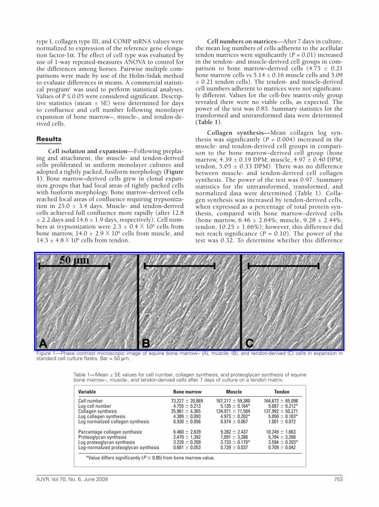
Cell isolation and expansion—Following preplat-ing and attachment, the muscle- and tendon-derived cells proliferated in uniform monolayer cultures and adopted a tightly packed, fusiform morphology (Figure 1). Bone marrow–derived cells grew in clonal expan-sion groups that had focal areas of tightly packed cells with fusiform morphology. Bone marrow–derived cells reached focal areas of confluence requiring trypsoniza-tion in 25.0 ± 3.4 days. Muscle- and tendon-derived cells achieved full confluence more rapidly (after 12.8 ± 2.2 days and 14.6 ± 1.9 days, respectively). Cell num-bers at trypsonization were 2.3 ± 0.4 X 10⁶ cells from bone marrow, 14.0 ± 2.9 X 10⁶ cells from muscle, and 14.3 ± 4.8 X 10⁶ cells from tendon.
Cell numbers on matrices—After 7 days in culture, the mean log numbers of cells adherent to the acellular tendon matrices were significantly (P = 0.01) increased in the tendon- and muscle-derived cell groups in com-parison to bone marrow–derived cells (4.75 ± 0.21 bone marrow cells vs 5.14 ± 0.16 muscle cells and 5.09 ± 0.21 tendon cells). The tendon- and muscle-derived cell numbers adherent to matrices were not significant-ly different. Values for the cell-free matrix-only group revealed there were no viable cells, as expected. The power of the test was 0.85. Summary statistics for the transformed and untransformed data were determined (Table 1).
Collagen synthesis—Mean collagen log syn-thesis was significantly (P = 0.004) increased in the muscle- and tendon-derived cell groups in compari-son to the bone marrow–derived cell group (bone marrow, 4.39 ± 0.19 DPM; muscle, 4.97 ± 0.40 DPM; tendon, 5.05 ± 0.33 DPM). There was no difference between muscle- and tendon-derived cell collagen synthesis. The power of the test was 0.97. Summary statistics for the untransformed, transformed, and normalized data were determined (Table 1). Colla-gen synthesis was increased by tendon-derived cells, when expressed as a percentage of total protein syn-thesis, compared with bone marrow–derived cells (bone marrow, 6.46 ± 2.64%; muscle, 9.28 ± 2.44%; tendon, 10.25 ± 1.66%); however, this difference did not reach significance (P = 0.10). The power of the test was 0.32. To determine whether this difference
Figure1—Phasecontrastmicroscopicimageofequinebonemarrow–(A),muscle-(B),andtendon-derived(C)cellsinexpansioninstandardcellcultureflasks.Bar=50µm.
Variable Bonemarrow Muscle Tendon
Cell number 73,227 20,869 167,217 59,380 164,672 65,098Log cell number 4.755 0.213 5.135 0.164* 5.087 0.212*Collagen synthesis 25,961 4,365 134,971 71,504 137,992 50,271Log collagen synthesis 4.389 0.093 4.973 0.202* 5.050 0.163*Log normalized collagen synthesis 0.930 0.056 0.974 0.067 1.001 0.072
Percentage collagen synthesis 6.460 2.639 9.282 2.437 10.249 1.663Proteoglycan synthesis 2,470 1,392 7,091 3,388 5,784 3,268Log proteoglycan synthesis 3.220 0.209 3.733 0.170* 3.594 0.203*Log-normalized proteoglycan synthesis 0.681 0.053 0.729 0.037 0.709 0.042
*Value differs significantly (P 0.05) from bone marrow value.
Table1—Mean±SEvaluesforcellnumber,collagensynthesis,andproteoglycansynthesisofequinebonemarrow–,muscle-,andtendon-derivedcellsafter7daysofcultureonatendonmatrix.
08-05-0158r.indd 753 5/20/2009 2:10:22 PM
754 AJVR,Vol70,No.6,June2009
was significant, cells from an additional 24 horses would have been required.
When the mean log values of collagen synthesis were normalized to cell number, significant differences between the cell groups were not evident (bone mar-row, 0.93 ± 0.06 DPM/cell; muscle, 0.97 ± 0.07 DPM/cell; tendon, 1.00 ± 0.07 DPM/cell; Table 1). The power for the test was 0.37.
Proteoglycan synthesis—Mean log proteoglycan synthesis was significantly (P = 0.001) increased in the muscle- and tendon-derived cell groups, in com-parison to the bone marrow–derived cell group (bone marrow, 3.22 ± 0.21 CPM; muscle, 3.73 ± 0.17 CPM; tendon, 3.60 ± 0.20 CPM). There was no difference between muscle- and tendon-derived cell proteogly-can synthesis. The power of the test evaluating proteo-glycan synthesis was 1.00. Summary statistics for the
transformed and untransformed data were determined (Table 1). When mean log proteoglycan synthesis was adjusted for cell number, there were no differences (P = 0.19) between cell types (bone marrow, 0.681 ± 0.053 CPM/cell; muscle, 0.729 ± 0.037 CPM/cell; tendon, 0.709 ± 0.042 CPM/cell). The power for this test was 0.17.
Extracellular matrix mRNA expression—As ex-pected, no mRNA was isolated from the acellular control tendon matrices that were subjected to 4 freeze-thaw cycles to kill resident cells. Quantitative PCR analyses of collagen type I mRNA expression in the cell-matrix groups revealed expression that was approximately 10% to 15% of in vivo expression. There were no significant (P = 0.64) differences in collagen type I mRNA expres-sion among the different cell types (bone marrow, 0.14 ± 0.037; muscle, 0.12 ± 0.010; tendon, 0.11 ± 0.18). The power for this test was 0.05.
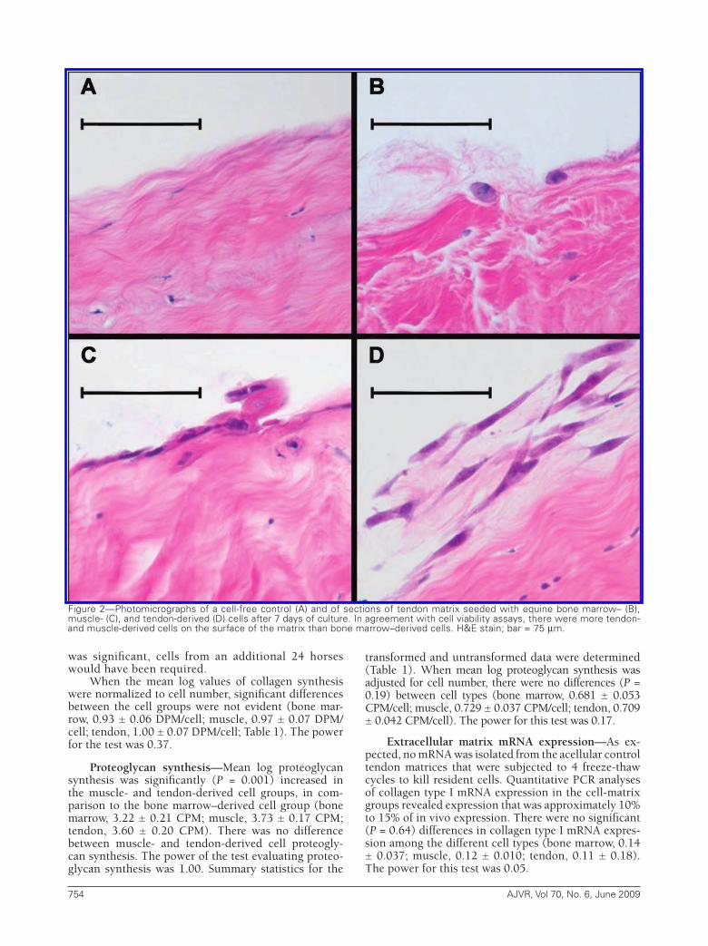
Figure2—Photomicrographsofacell-freecontrol (A)andofsectionsoftendonmatrixseededwithequinebonemarrow–(B),muscle-(C),andtendon-derived(D)cellsafter7daysofculture.Inagreementwithcellviabilityassays,thereweremoretendon-andmuscle-derivedcellsonthesurfaceofthematrixthanbonemarrow–derivedcells.H&Estain;bar=75µm.
08-05-0158r.indd 754 5/20/2009 2:10:25 PM
AJVR,Vol70,No.6,June2009 755
Collagen type III mRNA expression exceeded en-dogenous tenocyte expression in all 3 cell groups. Ex-pression by tendon-derived cells was significantly (P = 0.04) increased in comparison to the bone marrow–de-rived cell group (bone marrow, 3.10 ± 1.05; muscle, 7.30 ± 1.92; tendon, 9.27 ± 2.56). There were no differ-ences between muscle- and tendon-derived cells. The power of this test was 0.56.
The COMP expression in the cell-matrix samples was consistently lower than endogenous tenocyte expression. Tendon-derived cells had more COMP mRNA expression, compared with muscle-derived cells (bone marrow, 0.07 ± 0.04; muscle, 0.02 ± 0.006; tendon, 0.32 ± 0.14), but these differences were not significant (P = 0.10) because of large inter-animal variation in expression. The power of this test was 0.31. To determine whether this difference was significant, cells from an additional 8 horses would have been required.
Histologic examination—Small, condensed nu-clear remnants of endogenous tenocytes were evident throughout the cross-sections of the freeze-thawed ma-trices. All 3 exogenous cell types were predominantly attached to the surface of the autogenous matrices (Fig-ure 2). More tendon- and muscle-derived cells were ad-herent to the matrix surface than bone marrow–derived cells, consistent with the cell count data.
Discussion
The goal of the present study was to determine whether cells isolated from tendon or skeletal muscle constitute an alternative to bone marrow–derived cells for cell-based therapies for tendon injuries. Results in-dicated that cells can be easily isolated from tendon and muscle tissue biopsy samples. These cells can be rapid-ly expanded in monolayer culture to produce clinically useful numbers within 2 weeks. In contrast, there are few proliferative cells in bone marrow aspirates (esti-mated to be approx 1 cell in 10,000).42 Consequently, monolayer expansion of these populations is driven by a small number of the total cell isolates and requires a minimum of 3 to 4 weeks for adequate expansion.18,35 In this respect, cell isolates from skeletal muscle or tendon biopsy samples have a clear advantage over bone marrow aspirates. These findings are supported by those of a recent study43 that revealed that tendon-derived cells proliferated faster than bone marrow–de-rived MSCs from the same individuals. The differences in cell culture techniques also could have contributed to the higher cell numbers and shorter times to conflu-ence seen with the tendon- and muscle-derived cells, compared with bone marrow–derived cells. Apart from the issues of isolation and expansion, tendon- and mus-cle-derived cell attachment and survival on tendon ma-trix were significantly higher than those of bone-mar-row–derived cells after 7 days.
In this study, the muscle- and tendon-derived cell groups synthesized more collagen and proteoglycan than did bone marrow–derived cells. After normalizing matrix synthesis to cell number, the difference was not significant; much of the difference in matrix synthesis is attributable to the differences in cell attachment and
survival over time. These results were similar to those of previous studies44,45 of matrix synthesis by equine bone marrow–derived MSCs, in which increased syn-thesis reflected increases in cell numbers rather than per-cell changes. Nevertheless, this outcome is relevant to therapeutic applications because colonization of the tendon matrix and persistence at the site of administra-tion would likely influence the clinical efficacy of these cells for treatment of horses with tendon injuries.
In the present study, expression of collagen type I and COMP were markedly lower than expression de-tected in normal tendon. There was no significant (P = 0.64) difference in collagen type I expression among the cell groups. Tendon-derived cells had increased COMP expression, compared with the other groups, but the difference was not significant (P = 0.10). Carti-lage oligomeric matrix protein influences collagen type I fibril formation and is important for tendon matrix production and tendon repair.24,26 The pentameric na-ture of COMP brings collagen molecules in close prox-imity to promote further collagen assembly. In 1 study,24 COMP interacted with loose collagen type I to form fi-brils that increased in diameter and in length.
Collagen type III mRNA expression was higher than endogenous expression in all 3 cell groups, and expression by tendon-derived cells was significantly higher than that of bone marrow–derived cells. Colla-gen type III expression is upregulated during embryonic tenogenesis and during tendon healing.25,27 In some re-spects, the acellular matrix model used in this study represents the situation cells might encounter after injection or migration into injured tendon, and interac-tion with the acellular matrix might stimulate a reparative response in these cells. The repeated freeze-thaw protocol was designed to remove confounding biosynthetic contri-butions from endogenous tenocytes. Previous studies46,47 reveal that freeze-thawing tendon does not alter the bio-mechanical properties of the tissue, suggesting that the collagenous matrix of tendon is not adversely affected by this procedure. However, it is possible that endogenous cellular debris might have influenced the activities of the exogenously administered cells.
These data were derived from cells collected from 4 young adult horses. There was considerable interani-mal variation in the data, and as a consequence, many of the outcomes (mean percentage collagen synthesis-to-total protein ratio, mean log collagen synthesis-to-log of cell number ratio, and COMP mRNA expression) were greater in tendon-derived cells, but differences did not reach significance. All the analyses that yielded P values between 0.10 and 0.05 had low statistical pow-ers (≤ 0.37) and should be interpreted with caution. On the basis of these data, a minimum of 12 horses would be necessary to provide sufficient power (0.8) for these analyses. However, in our experience, marked interdo-nor variability is a biological reality of mesenchymal cell isolation and in vitro activity18,44,45 and is likely to be reflected in in vivo therapeutic efficacies.
In the present study, an in vitro model was de-veloped to assess the ability of cells to adhere to, and survive on, tendon matrix. On the basis of results of this study, we propose that muscle- and tendon-derived cells may provide an alternative to bone marrow–de-
08-05-0158r.indd 755 5/20/2009 2:10:25 PM
756 AJVR,Vol70,No.6,June2009
rived cells for cell-based strategies to promote tendon healing. This study evaluated autogenous cell-based strategies, and more research is necessary to determine whether allogenous cell-based strategies would yield similar results. For all autogenous cell-based strate-gies, the donor morbidity site will remain an important consideration, particularly for tendon-derived cells. In recent projects we have harvested tendon-derived cells from the lateral digital extensor tendon, muscle-derived cells from the semitendinosus muscle, and bone mar-row–derived cells from the sternum of horses without any obvious donor site complications. However, donor site morbidity and in-vivo efficacy still need to be deter-mined for these cell types.
The assays used in this study determined cell sur-vival and matrix biosynthesis, rather than characteriz-ing the phenotypes and lineages of these cells. Previous studies43,48,z reveal that MSCs with multilineage poten-tial can be isolated from human and murine tendon as well as bone marrow and muscle. Other studiesaa–bb indicate that cells expanded from equine tendon by use of techniques described in the present study have similar capacities. It remains to be seen whether these cells are effective in vivo, but efforts to optimize the cell isolation, expansion, and purification protocols could improve the clinical efficacy of muscle- and tendon-derived cells for treatment of performance horses with tendon and other musculoskeletal injuries.
a. Jamshidi bone marrow biopsy needle, Cardinal Health, Dublin, Ohio.
b. DMEM, Mediatech Inc, Herndon, Va.c. Gemini Bioproducts, Woodland, Calif.d. l-glutamine, 200 mM, Invitogen, Carlsbad, Calif.e. Penicillin-streptomycin, BioWhittaker, Cambrex Bio Science,
Walkersville, Md.f. Sodium pyruvate, Sigma Chemical Co, St Louis, Mo.g. Collagenase type II, Worthington Biochemical Corp, Lakewood,
NJ.h. Dispase, Roche, Indianapolis, Ind.i. Ascorbic acid, WAKO, Richmond, Va.j. Filters, BD Biosciences, Bedford, Mass.k. Trypan blue, Sigma Chemical Co, St Louis, Mo.l. Cell Titer 96 Aq
ueous One Solution Cell Proliferation Assay, Pro-
mega, Madison, Wis.m. Microplate reader, FLUOstar Optima, BMG Laboratories, Dur-
ham, NC.n. [3H] proline, Sigma Chemical Co, St Louis, Mo.o. Collagenase, purified, Worthington Biochemical Corp, Lake-
wood, NJ.p. LS6500 multipurpose scintillation counter, Beckman Coulter
Inc, Fullerton, Calif.q. 35S- labeled sodium sulphate, MP Biochemicals, Irvine, Calif.r. Papain, Sigma Chemical Co, St Louis, Mo.s. Multiwell punch plates, PDVF plate, Millipore, Bedford, Mass.t. RNeasy Mini Kit, Qiagen, Valencia, Calif.u. Superscript II, Invitrogen, Carlsbad, Calif.v. iQ SYBR Green Supermix, Bio-Rad Laboratories, Hercules, Calif.w. ClustalW, European Molecular Biology Laboratory European
Bioinformatics Institute (EMBL-EMBI), Cambridge, England. Available at: www.ebi.ac.uk. Accessed September 3, 2004.
x. iCycler iQ real-time PCR detection system, Bio-Rad Laborato-ries, Hercules, Calif.
y. Sigma Stat, Systat Software Inc, San Jose, Calif.z. de Mos M, Koevoet WJ, Jahr H, et al. Intrinsic differentiation po-
tential of adolescent human tendon tissue: an in-vitro cell differ-entiation study (abstr). BMC Musculoskelet Disord 2007;8:16.
aa. Barrett JG, Stewart AA, Stewart M, et al. In vitro comparison
of tendon, muscle, and bone marrow-derived progenitor cells cultured on tendon matrix (abstr). Vet Surg 2005;34:E2.
bb. Barrett JG, Stewart AA, Yates AC, et al. Tendon-derived progeni-tor cells can differentiate along multiple lineages (abstr), in Pro-ceedings. 34th Annu Conf Vet Orthop Soc 2007;31.
References
1. Lam KH, Parkin TD, Riggs CM, et al. Descriptive analysis of retirement of Thoroughbred racehorses due to tendon inju-ries at the Hong Kong Jockey Club (1992–2004). Equine Vet J 2007;39:143–148.
2. Goodship AE, Birch HL, Wilson AM. The pathobiology and re-pair of tendon and ligament injury. Vet Clin North Am Equine Pract 1994;10:323–349.
3. Dowling BA, Dart AJ, Hodgson DR, et al. Superficial digital flex-or tendonitis in the horse. Equine Vet J 2000;32:369–378.
4. Alves AG, Nicoletti JM, Thomassian A, et al. Tendon splitting as surgical treatment on experimental equine acute tendinitis. Arch Vet Sci 2002;7:45–51.
5. Dahlgren LA, van der Meulen MC, Bertram JE, et al. Insulin-like growth factor-I improves cellular and molecular aspects of heal-ing in a collagenase-induced model of flexor tendinitis. J Orthop Res 2002;20:910–919.
6. Chen YJ, Wang CJ, Yang KD, et al. Extracorporeal shock waves promote healing of collagenase-induced Achilles tendinitis and increase TGF-beta1 and IGF-I expression. J Orthop Res 2004;22:854–861.
7. Archambault JM, Wiley JP, Bray RC. Exercise loading of tendons and the development of overuse injuries. A review of current literature. Sports Med 1995;20:77–89.
8. Pufe T, Petersen WJ, Mentlein R, et al. The role of vasculature and angiogenesis for the pathogenesis of degenerative tendons disease. Scand J Med Sci Sports 2005;15:211–222.
9. Birch HL, Bailey AJ, Goodship AE. Macroscopic ‘degenera-tion’ of equine superficial digital flexor tendon is accompanied by a change in extracellular matrix composition. Equine Vet J 1998;30:534–539.
10. Abrahamsson SO. Matrix metabolism and healing in the flexor tendon. Experimental studies on rabbit tendon. Scand J Plast Reconstr Surg Hand Surg Suppl 1991;23:1–51.
11. Banes AJ, Tsuzaki M, Hu P, et al. PDGF-BB, IGF-I and mechani-cal load stimulate DNA synthesis in avian tendon fibroblasts in vitro. J Biomech 1995;28:1505–1513.
12. Dowling BA, Dart AJ, Hodgson DR, et al. Recombinant equine growth hormone does not affect the in vitro biomechanical properties of equine superficial digital flexor tendon. Vet Surg 2002;31:325–330.
13. Butler DL, Juncosa-Melvin N, Boivin GP, et al. Functional tissue engineering for tendon repair: a multidisciplinary strategy using mesenchymal stem cells, bioscaffolds, and mechanical stimula-tion. J Orthop Res 2008;26:1–9.
14. Li HY, Zhou XF. Potential conversion of adult clavicle-derived chondrocytes into neural lineage cells in vitro. J Cell Physiol 2008;214:630–644.
15. Bruder SP, Fink DJ, Caplan AI. Mesenchymal stem cells in bone development, bone repair, and skeletal regeneration therapy. J Cell Biochem 1994;56:283–294.
Gene Sequence Amplicon(bp)
Eq Col I (S) GAA AAC ATC CCA GCC AAG AA 231Eq Col I (A) GAT TGC CAG TCT CCT CAT CC Eq Col III (S) AGG GGA CCT GGT TAC TGC TT 215Eq Col III (A) TCT CTG GGT TGG GAC AGT CT Eq COMP (S) TCA TGT GGA AGC AGA TGG AG 223Eq COMP (A) TAG GAA CCA GCG GTA GGA TG Eq EF1-α (S) CCC GGA CAC AGA GAC TTC AT 328Eq EF1-α (A) AGC ATG TTG TCA CCA TTC CA
Appendix
Primersusedforreal-timePCRamplificationofgenesinastudyofequinebonemarrow–,muscle-,andtendon-derivedcells.
08-05-0158r.indd 756 5/20/2009 2:10:25 PM
AJVR,Vol70,No.6,June2009 757
16. Solchaga LA, Welter JF, Lennon DP, et al. Generation of plu-ripotent stem cells and their differentiation to the chondrocytic phenotype. Methods Mol Med 2004;100:53–68.
17. Fraser JK, Schreiber RE, Zuk PA, et al. Adult stem cell therapy for the heart. Int J Biochem Cell Biol 2004;36:658–666.
18. Worster AA, Nixon AJ, Brower-Toland BD, et al. Effect of transforming growth factor β1 on chondrogenic differentia-tion of cultured equine mesenchymal stem cells. Am J Vet Res 2000;59:1003–1010.
19. McDuffee LA, Anderson GI. In vitro comparison of equine can-cellous bone graft donor sites and tibial periosteum as sources of viable osteoprogenitors. Vet Surg 2003;32:455–463.
20. Yoshimura H, Muneta T, Nimura A, et al. Comparison of rat mesenchymal stem cells derived from bone marrow, synovi-um, periosteum, adipose tissue, and muscle. Cell Tissue Res 2007;327:449–462.
21. Izadpanah R, Trygg C, Patel B, et al. Biologic properties of mes-enchymal stem cells derived from bone marrow and adipose tis-sue. J Cell Biochem 2006;99:1285–1297.
22. Kannus P. Structure of the tendon connective tissue. Scand J Med Sci Sports 2000;10:312–320.
23. Liu SH, Yang RS, al-Shaikh R, et al. Collagen in tendon, liga-ment, and bone healing. A current review. Clin Orthop Relat Res 1995;318:265–278.
24. Halasz K, Kassner A, Morgelin M, et al. COMP acts as a catalyst in collagen fibrillogenesis. J Biol Chem 2007;282:31166–31173.
25. Liu X, Wu H, Byrne M, et al. Type III collagen is crucial for col-lagen I fibrillogenesis and for normal cardiovascular develop-ment. Proc Natl Acad Sci U S A 1997;94:1852–1856.
26. Smith RK, Heinegard D. Cartilage oligomeric matrix protein (COMP) levels in digital sheath synovial fluid and serum with tendon injury. Equine Vet J 2000;32:52–58.
27. Dahlgren LA, Brower-Toland BD, Nixon AJ. Cloning and ex-pression of type III collagen in normal and injured tendons of horses. Am J Vet Res 2005;66:266–270.
28. Berglund M, Reno C, Hart DA, et al. Patterns of mRNA expres-sion for matrix molecules and growth factors in flexor tendon injury: differences in the regulation between tendon and tendon sheath. J Hand Surg [Am] 2006;31:1279–1287.
29. Dahlgren LA, Mohammed HO, Nixon AJ. Temporal expression of growth factors and matrix molecules in healing tendon le-sions. J Orthop Res 2005;23:84–92.
30. Awad HA, Boivin GP, Dressler MR, et al. Repair of patellar tendon injuries using a cell-collagen composite. J Orthop Res 2003;21:420–431.
31. Guest DJ, Smith MR, Allen WR. Monitoring the fate of autolo-gous and allogeneic mesenchymal progenitor cells injected into the superficial digital flexor tendon of horses: preliminary study. Equine Vet J 2008;40:178–181.
32. Young RG, Butler DL, Weber W. Use of mesenchymal stem cells
in a collagen matrix for Achilles tendon repair. J Orthop Res 1998;16:406–413.
33. Qu Z, Balkir L, van Deutekom JC, et al. Development of ap-proaches to improve cell survival in myoblast transfer therapy. J Cell Biol 1998;142:1257–1267.
34. Jankowski RJ, Haluszczak C, Trucco M, et al. Flow cytometric characterization of myogenic cell populations obtained via the preplate technique: potential for rapid isolation of muscle-de-rived stem cells. Hum Gene Ther 2001;12:619–628.
35. Fortier LA, Nixon AJ, Williams J, et al. Isolation and chondro-cytic differentiation of equine bone marrow-derived mesenchy-mal stem cells. Am J Vet Res 1998;59:1182–1187.
36. Mather JP, Robert PE. Introduction to cell and tissue culture. New York: Plenum Press, 1998;67–68.
37. Diegelmann RF. Analysis of collagen synthesis. Methods Mol Med 2003;78:349–358.
38. Masuda K, Shirota H, Thonar EJ. Quantification of 35S-labeled proteoglycans complexed to alcian blue by rapid filtration in multiwell plates. Anal Biochem 1994;217:167–175.
39. Stewart MC, Saunders KM, Burton-Wurster N, et al. Phenotypic stability of articular chondrocytes in vitro: the effects of culture models, bone morphogenetic protein 2, and serum supplemen-tation. J Bone Miner Res 2000;15:166–174.
40. Livak KJ, Schmittgen TD. Analysis of relative gene expression data using real-time quantitative PCR and the 2-∆∆CT Method. Methods 2001;25:402–408.
41. Ramsey FL, Schafer DW. A closer look at assumptions. In: Ramsey FL, Schafer DW, eds. The statistical sleuth: a course in methods of data analysis. Belmont, Calif: Duxbury Press, 1997;69.
42. Caplan AI. The mesengenic process. Clin Plast Surg 1994;21:429–435.
43. Bi Y, Ehirchiou D, Kilts TM, et al. Identification of tendon stem/progenitor cells and the role of the extracellular matrix in their niche. Nat Med 2007;13:1219–1227.
44. Stewart AA, Byron CR, Pondenis H, et al. Effect of fibroblast growth factor-2 on equine mesenchymal stem cell monolayer expansion and chondrogenesis. Am J Vet Res 2007;68:941–945.
45. Stewart AA, Byron CR, Pondenis HC, et al. Effect of dexametha-sone supplementation on chondrogenesis of equine mesenchy-mal stem cells. Am J Vet Res 2008;69:1013–1021.
46. Giannini S, Buda R, Di Caprio F. Effects of freezing on the bio-mechnical and structural properties of human posterior tibial tendons. Int Orthop 2008;32:145–151.
47. Clavert P, Kempf JF, Bonnomet F, et al. Effects of freezing/thaw-ing on the biomechanical properties of human tendons. Surg Radiol Anat 2001;23:259–262.
48. Salingcarnboriboon R, Yoshitake H, Tsuji K, et al. Establishment of tendon-derived cell lines exhibiting pluripotent mesenchy-mal stem cell-like property. Exp Cell Res 2003;287:289–300.
08-05-0158r.indd 757 5/20/2009 2:10:25 PM


![[David E. Anderson DVM MS DACVS] Bovine Orthoped(BookZZ.org)](https://img.pdfslide.net/doc/110x75/577c82141a28abe054af5971/david-e-anderson-dvm-ms-dacvs-bovine-orthopedbookzzorg.jpg)