Embed Size (px)
Citation preview
Comparison of panoramic radiography and
computed tomography
for the detection of altered sensation
of inferior alveolar nerve
after implantation
Huijun Kim
Department of Dentistry
The Graduate School, Yonsei University
Comparison of panoramic radiography and
computed tomography
for the detection of altered sensation
of inferior alveolar nerve
after implantation
Directed by Professor Chang-Seo Park, D.M.D., Ph.D.
The Doctoral Dissertation
submitted to the Department of Dentistry,
and the Graduate School of Yonsei University
in partial fulfillment of the requirements for the degree of
Doctor of Philosophy
Huijun Kim
Dec 2012
This certifies that the Doctoral Dissertation of
Huijun Kim is approved.
----------------------------------------------------------------
Thesis Supervisor : Prof. Chang-Seo Park
----------------------------------------------------------------
Thesis Committee Member : Prof. Sung-Taek Kim
----------------------------------------------------------------
Thesis Committee Member : Prof. Jae-Ho Lee
The Graduate School
Yonsei University
Dec 2012

감사의 글
이 논문이 완성되기까지 많은 관심과 격려로 늘 변함없이 따뜻하게 지도해주신 박창서 지도
교수님께 깊은 감사의 마음을 올립니다. 아울러 논문의 완성을 위해서 세심한 조언을 아끼지
않으신 김성택 교수님, 이제호 교수님께도 진심으로 감사드립니다.
항상 저의 일을 늘 돌봐주시고 생활의 본보기를 보여주며 다정하게 대해 주신 박혁, 정호걸
선생님과 옆에서 궂은 일을 묵묵히 도와준 박재규 선생님께도 감사를 드립니다.
힘든 업무 중에도 밝은 표정으로 격려해주고 힘이 되어준 구강악안면방사선과 가족들에게도
감사드립니다.
또한 지금까지 부모님께 받은 사랑에 대한 감사함을 이 글을 빌어 전해드립니다. 아울러
친아들처럼 항상 따뜻하게 대해 주시고 사랑을 베풀어 주신 장인어른, 장모님께도 진심으로
감사드립니다.
마지막으로 항상 내게 용기를 주고 희망을 준 사랑하는 아내 조윤주와 항상 웃는 얼굴로 삶의
기쁨을 주는 사랑스러운 딸 소정이에게 글을 바칩니다.
2012 년 12 월
저 자 씀

i
TABLE OF CONTENTS
Abstract (English)…………………………………………………………………………1
Ⅰ. Introduction…………………………………………………………………………… 2
Ⅱ. Materials and Methods
1. Materials………………………………………………………………………………… 3
2. Methods
A. Clinival evaluation………………………………………………………………………… 3
B. Radiographic evaluation…………………………………………………………………… 4
C. Statistical evaluation ………………………………………………………………………5
Ⅲ. Results
1. Distribution of VAS in clinical examinations………………………………………………5
2. Distribution of scores in radiographic evaluations…………………………………………6
3. The correlation between VAS and radiographic evaluation………………………………6
4. Comparison between estimated panoramic radiography result……………………………8
Ⅳ. DISCUSSION……………………………………………………………………………8
Ⅴ. CONCLUSION…………………………………………………………………………11
REFERENCES……………………………………………………………………………11
Abstract (Korean)…………………………………………………………………………13

ii
LIST OF FIGURES
Fig. 1. Example of subgroup on panoramic radiography……………………4
Fig. 2. Example of subgroup on CT……………………………………………5
Fig. 3. In panoramic radiography, actual distance between implant fixture and
IAN canal could be evaluated variously due to its imaging principle. ……9
iii
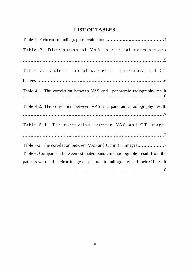
LIST OF TABLES
Table 1. Criteria of radiographic evaluation …………………………………4
T a b l e 2 . D i s t r i b u t i o n o f VA S i n c l i n i c a l e x a m i n a t i o n s
…………………………………………………………………………………5
T a b l e 3 . D i s t r i b u t i o n o f s c o r e s i n p a n o r a m i c a n d C T
images.…………………………………………………………………………6
Table 4-1. The correlation between VAS and panoramic radiography result
…………………………………………………………………………………6
Table 4-2. The correlation between VAS and panoramic radiography result.
…………………………………………………………………………………7
T a b l e 5 - 1 . T h e c o r r e l a t i o n b e t w e e n VA S a n d C T i m a g e s
…………………………………………………………………………………7
Table 5-2. The correlation between VAS and CT in CT images………………7
Table 6. Comparison between estimated panoramic radiography result from the
patients who had unclear image on panoramic radiography and their CT result
…………………………………………………………………………………8
1
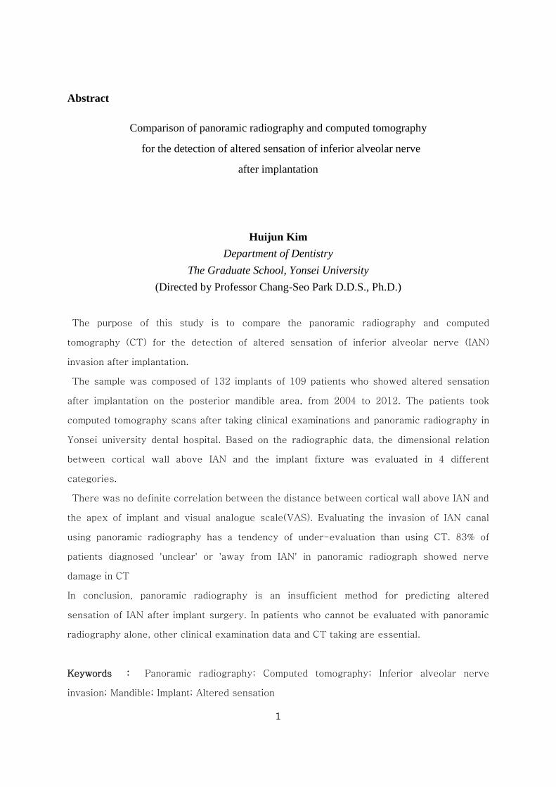
Abstract
Comparison of panoramic radiography and computed tomography
for the detection of altered sensation of inferior alveolar nerve
after implantation
Huijun Kim
Department of Dentistry
The Graduate School, Yonsei University
(Directed by Professor Chang-Seo Park D.D.S., Ph.D.)
The purpose of this study is to compare the panoramic radiography and computed
tomography (CT) for the detection of altered sensation of inferior alveolar nerve (IAN)
invasion after implantation.
The sample was composed of 132 implants of 109 patients who showed altered sensation
after implantation on the posterior mandible area, from 2004 to 2012. The patients took
computed tomography scans after taking clinical examinations and panoramic radiography in
Yonsei university dental hospital. Based on the radiographic data, the dimensional relation
between cortical wall above IAN and the implant fixture was evaluated in 4 different
categories.
There was no definite correlation between the distance between cortical wall above IAN and
the apex of implant and visual analogue scale(VAS). Evaluating the invasion of IAN canal
using panoramic radiography has a tendency of under-evaluation than using CT. 83% of
patients diagnosed 'unclear' or 'away from IAN' in panoramic radiograph showed nerve
damage in CT
In conclusion, panoramic radiography is an insufficient method for predicting altered
sensation of IAN after implant surgery. In patients who cannot be evaluated with panoramic
radiography alone, other clinical examination data and CT taking are essential.
Keywords : Panoramic radiography; Computed tomography; Inferior alveolar nerve
invasion; Mandible; Implant; Altered sensation
2
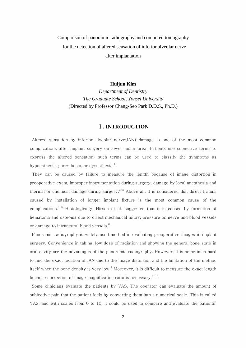
Comparison of panoramic radiography and computed tomography
for the detection of altered sensation of inferior alveolar nerve
after implantation
Huijun Kim
Department of Dentistry
The Graduate School, Yonsei University
(Directed by Professor Chang-Seo Park D.D.S., Ph.D.)
Ⅰ. INTRODUCTION
Altered sensation by inferior alveolar nerve(IAN) damage is one of the most common
complications after implant surgery on lower molar area. Patients use subjective terms to
express the altered sensation; such terms can be used to classify the symptoms as
hypoesthesia, paresthesia, or dysesthesia.1
They can be caused by failure to measure the length because of image distortion in
preoperative exam, improper instrumentation during surgery, damage by local anesthesia and
thermal or chemical damage during surgery.3-5
Above all, it is considered that direct trauma
caused by installation of longer implant fixture is the most common cause of the
complications.4-6
Histologically, Hirsch et al. suggested that it is caused by formation of
hematoma and osteoma due to direct mechanical injury, pressure on nerve and blood vessels
or damage to intraneural blood vessels.6
Panoramic radiography is widely used method in evaluating preoperative images in implant
surgery. Convenience in taking, low dose of radiation and showing the general bone state in
oral cavity are the advantages of the panoramic radiography. However, it is sometimes hard
to find the exact location of IAN due to the image distortion and the limitation of the method
itself when the bone density is very low.7 Moreover, it is difficult to measure the exact length
because correction of image magnification ratio is necessary.8-13
Some clinicians evaluate the patients by VAS. The operator can evaluate the amount of
subjective pain that the patient feels by converting them into a numerical scale. This is called
VAS, and with scales from 0 to 10, it could be used to compare and evaluate the patients’
3
condition before and after treatment.15
The intensity of subjective pain that patients
experienced was surveyed from the records of patients.
Despite some disadvantages of CT such as the high radiation dose and expensive cost, it is
used to get more specific information before and after implant installation since it makes less
image distortion and enables to get three dimensional analysis and more precise
measurement in length.3-5,13
The purpose of this study is to compare the different radiographic methods for the detection
of altered sensation by implant surgery after implantation.
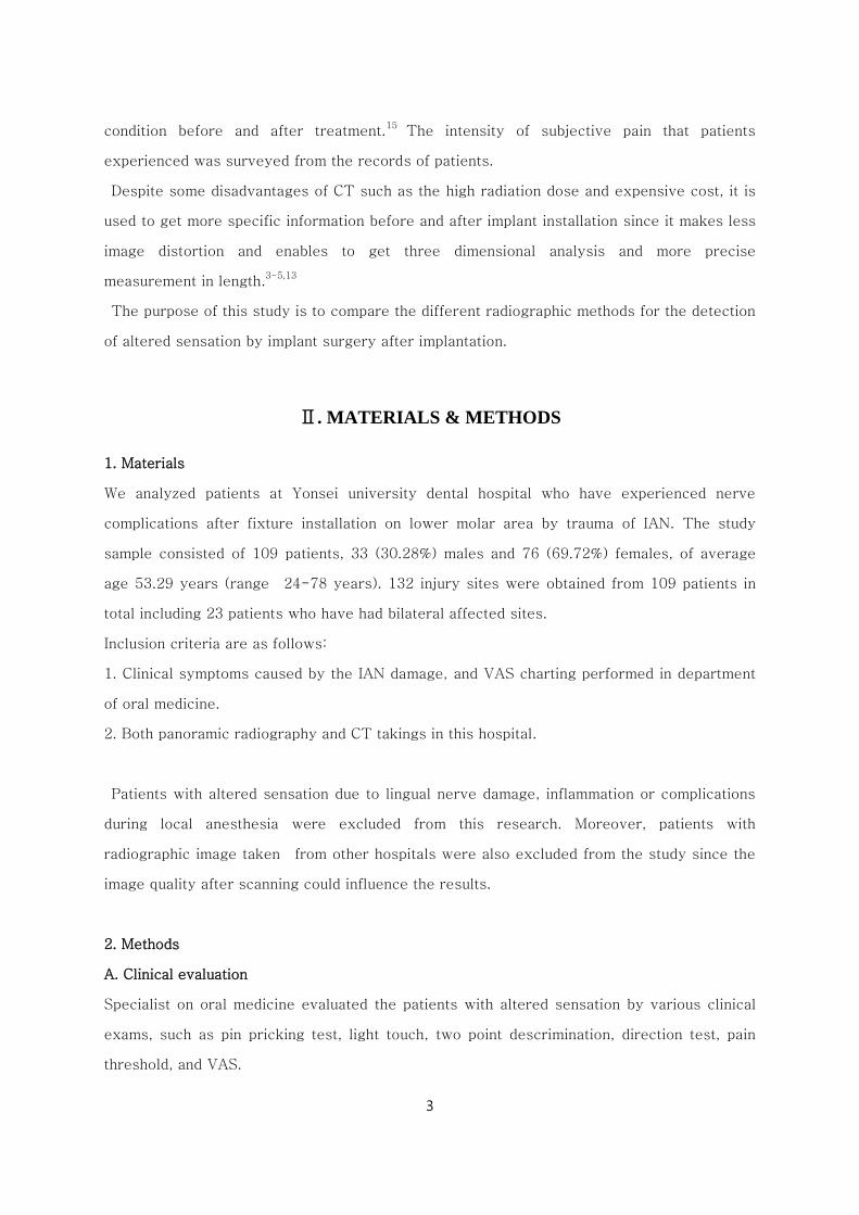
Ⅱ. MATERIALS & METHODS
1. Materials
We analyzed patients at Yonsei university dental hospital who have experienced nerve
complications after fixture installation on lower molar area by trauma of IAN. The study
sample consisted of 109 patients, 33 (30.28%) males and 76 (69.72%) females, of average
age 53.29 years (range 24-78 years). 132 injury sites were obtained from 109 patients in
total including 23 patients who have had bilateral affected sites.
Inclusion criteria are as follows:
1. Clinical symptoms caused by the IAN damage, and VAS charting performed in department
of oral medicine.
2. Both panoramic radiography and CT takings in this hospital.
Patients with altered sensation due to lingual nerve damage, inflammation or complications
during local anesthesia were excluded from this research. Moreover, patients with
radiographic image taken from other hospitals were also excluded from the study since the
image quality after scanning could influence the results.
2. Methods
A. Clinical evaluation
Specialist on oral medicine evaluated the patients with altered sensation by various clinical
exams, such as pin pricking test, light touch, two point descrimination, direction test, pain
threshold, and VAS.
4
B. Radiographic evaluation
Panoramic radiography was performed with CRANEX3+® (SOREDEX, Helsinki, Finland), and
CT was performed with HISPEED advantage® (GE medical, Milwakee, USA). All of the
panoramic radiography and CT images were interpreted two times by two experienced oral
and maxillofacial radiology specialists.
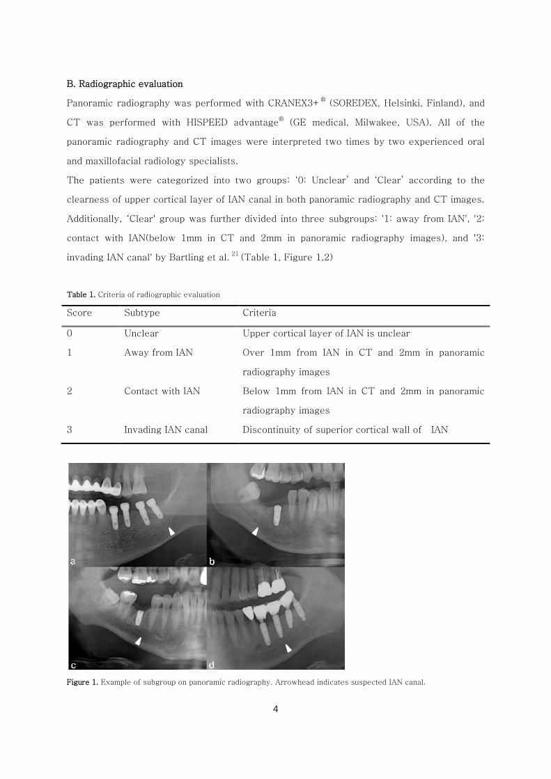
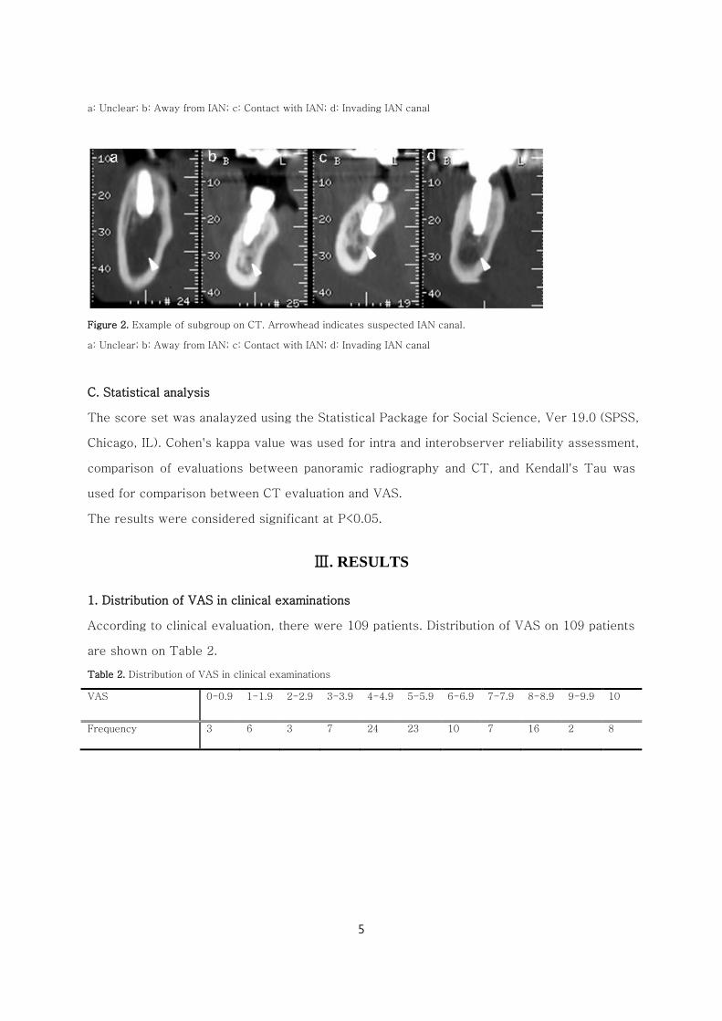
The patients were categorized into two groups: ‘0: Unclear’ and ‘Clear’ according to the
clearness of upper cortical layer of IAN canal in both panoramic radiography and CT images.
Additionally, ‘Clear' group was further divided into three subgroups: '1: away from IAN', '2:
contact with IAN(below 1mm in CT and 2mm in panoramic radiography images), and '3:
invading IAN canal' by Bartling et al. 21
(Table 1, Figure 1,2)
Table 1. Criteria of radiographic evaluation
Score Subtype Criteria
0 Unclear Upper cortical layer of IAN is unclear
1 Away from IAN Over 1mm from IAN in CT and 2mm in panoramic
radiography images
2 Contact with IAN Below 1mm from IAN in CT and 2mm in panoramic
radiography images
3 Invading IAN canal Discontinuity of superior cortical wall of IAN
Figure 1. Example of subgroup on panoramic radiography. Arrowhead indicates suspected IAN canal.
5
a: Unclear; b: Away from IAN; c: Contact with IAN; d: Invading IAN canal
Figure 2. Example of subgroup on CT. Arrowhead indicates suspected IAN canal.
a: Unclear; b: Away from IAN; c: Contact with IAN; d: Invading IAN canal
C. Statistical analysis
The score set was analayzed using the Statistical Package for Social Science, Ver 19.0 (SPSS,
Chicago, IL). Cohen's kappa value was used for intra and interobserver reliability assessment,
comparison of evaluations between panoramic radiography and CT, and Kendall's Tau was
used for comparison between CT evaluation and VAS.
The results were considered significant at P<0.05.
Ⅲ. RESULTS
1. Distribution of VAS in clinical examinations
According to clinical evaluation, there were 109 patients. Distribution of VAS on 109 patients
are shown on Table 2.
Table 2. Distribution of VAS in clinical examinations
VAS 0-0.9 1-1.9 2-2.9 3-3.9 4-4.9 5-5.9 6-6.9 7-7.9 8-8.9 9-9.9 10
Frequency 3 6 3 7 24 23 10 7 16 2 8
6
2. Distribution of scores in radiographic evaluations
132 injury sites were obtained from 109 patients in total including 23 patients who have had
bilateral affected sites. Distribution of scores in panoramic and CT images are showin on
Table 3.
Table 3. Distribution of scores in panoramic and CT images.
Panoramic radiography CT
Number of implant % Number of implant %
0 45 34.1 2 1.5
1 16 12.1 10 7.6
2 26 19.7 14 10.6
3 45 34.1 106 80.3
Total 132 100.0 132 100.0
Evaluating the results of the final exam of the CT scans, 80.3% of the patients showed
disruption in continuity of the IAN canal, 10.6% of the patients showed with close proximity
of the canal and the implant fixture but with continuity, and another 7.6% of the patients
showed IAN canal that were free from contact with the fixtures.
Cohen’s kappa value of both intra-observer and interobserver agreement show almost
perfect agreement in both panoramic radiography and CT.
3. The correlation between VAS and radiographic evaluation.
The correlation between VAS and radiologic evaluation are shown on the table 4-1, 4-2, 5-1,
5-2.
Table 4-1. The correlation between VAS and panoramic radiography result
VAS
Panoramic
radiography
evaluation
0-0.9 1-1.9 2-2.9 3-3.9 4-4.9 5-5.9 6-6.9 7-7.9 8-8.9 9-9.9 10
0 0 2 2 4 9 8 4 4 5 0 1
1 0 0 0 1 3 5 0 1 1 1 1
2 1 2 0 1 6 3 2 1 2 1 3
3 2 2 1 1 6 7 4 1 8 0 3
7
Table 4-2. The correlation between VAS and panoramic radiography result.
VAS
Result of Panoramic
radiography evaluation
N 109
Kendall's Tau -0.075
p-value 0.438
Table 5-1. The correlation between VAS and CT images
VAS
CT evaluation
0-0.9 1-1.9 2-2.9 3-3.9 4-4.9 5-5.9 6-6.9 7-7.9 8-8.9 9-9.9 10
0 0 0 0 0 1 0 1 0 0 0 0
1 0 0 0 1 0 4 0 0 0 1 1
2 0 1 0 1 5 1 3 1 1 1 0
3 3 5 3 5 18 18 6 6 15 0 7
Table 5-2. The correlation between VAS and CT in CT images
VAS
Result of CT evaluation
N 109
Kendall's Tau -0.068
p-value 0.460
Kendall’s Tau statistic method was used to compare the result of VAS with CT evaluation.
The obtained result shows p-value higher than 0.05 in all values, suggesting that Tau value
has no significance. Therefore it could be concluded that there are no correlation between
the results of VAS and radiographic images.
In evaluating the diagnostic capacity of panoramic radiographs, patients who were diagnosed
'unclear' or 'away from IAN' in panoramic radiography were classified then compared with CT
results. Grouping in CT follow the previous division category, and the result is shown in table
6.
8
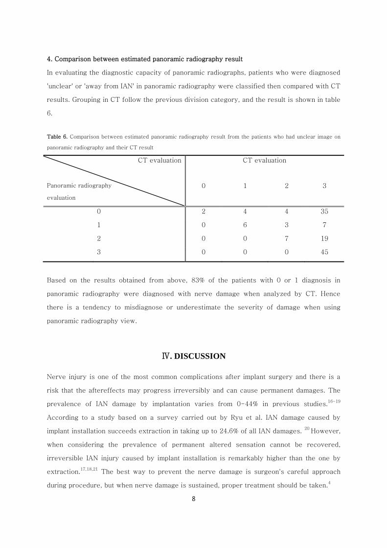
4. Comparison between estimated panoramic radiography result
In evaluating the diagnostic capacity of panoramic radiographs, patients who were diagnosed
'unclear' or 'away from IAN' in panoramic radiography were classified then compared with CT
results. Grouping in CT follow the previous division category, and the result is shown in table
6.
Table 6. Comparison between estimated panoramic radiography result from the patients who had unclear image on
panoramic radiography and their CT result
CT evaluation
Panoramic radiography
evaluation
CT evaluation
0 1 2 3
0 2 4 4 35
1 0 6 3 7
2 0 0 7 19
3 0 0 0 45
Based on the results obtained from above, 83% of the patients with 0 or 1 diagnosis in
panoramic radiography were diagnosed with nerve damage when analyzed by CT. Hence
there is a tendency to misdiagnose or underestimate the severity of damage when using
panoramic radiography view.
Ⅳ. DISCUSSION
Nerve injury is one of the most common complications after implant surgery and there is a
risk that the aftereffects may progress irreversibly and can cause permanent damages. The
prevalence of IAN damage by implantation varies from 0-44% in previous studies.16-19
According to a study based on a survey carried out by Ryu et al. IAN damage caused by
implant installation succeeds extraction in taking up to 24.6% of all IAN damages. 20
However,
when considering the prevalence of permanent altered sensation cannot be recovered,
irreversible IAN injury caused by implant installation is remarkably higher than the one by
extraction.17,18,21
The best way to prevent the nerve damage is surgeon's careful approach
during procedure, but when nerve damage is sustained, proper treatment should be taken.4
9
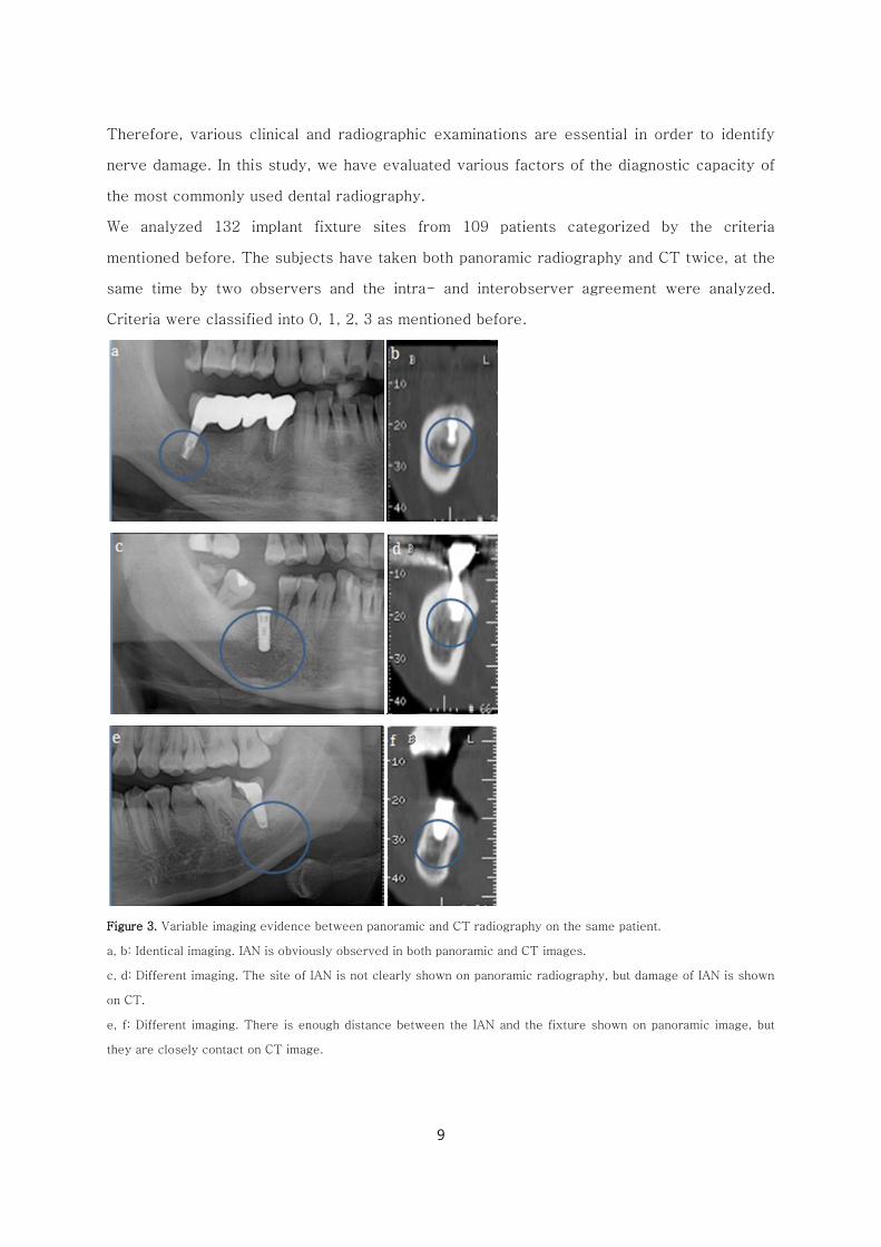
Therefore, various clinical and radiographic examinations are essential in order to identify
nerve damage. In this study, we have evaluated various factors of the diagnostic capacity of
the most commonly used dental radiography.
We analyzed 132 implant fixture sites from 109 patients categorized by the criteria
mentioned before. The subjects have taken both panoramic radiography and CT twice, at the
same time by two observers and the intra- and interobserver agreement were analyzed.
Criteria were classified into 0, 1, 2, 3 as mentioned before.
Figure 3. Variable imaging evidence between panoramic and CT radiography on the same patient.
a, b: Identical imaging. IAN is obviously observed in both panoramic and CT images.
c, d: Different imaging. The site of IAN is not clearly shown on panoramic radiography, but damage of IAN is shown
on CT.
e, f: Different imaging. There is enough distance between the IAN and the fixture shown on panoramic image, but
they are closely contact on CT image.
10
Intraobserver and interobserver agreement were calculated by Cohen's Kappa value and it
showed almost perfect agreement. Therefore both panoramic radiography and CT results are
dependable, and these results may be used in following procedures.
In panoramic radiography, magnification occurs due to its imaging principle, and actual
distance between implant fixture and IAN canal could be overestimated, which implies an
under-evaluation of the IAN damage by implants.7 Figure 3 is the examples chosen by this
study. All CT scans manifestly show fixtures invading the IAN canal. However, in some
panoramic radiographies, either the distance between the fixture and the IAN canal cannot be
accurately evaluated, or the invasion of the nerve canal is unclear due to the uncertain
location of the IAN canal.
Therefore, we performed method pair samples test on the samples excluding the patients
with unreadable panoramic radiography. According to the result on the table, panoramic
radiography images tend to underestimate the degree of the damage compared to that of CT
in all cases which showed significant difference. These results imply that even when a
considerable amount of distance seems to be obtained in panoramic radiographs, CT scans
could show implant fixtures that are invading and thus damaging the IAN canals. Therefore, if
panoramic radiography is the only imaging device used before implant installation, the safety
margin should be set over 2mm.22-24
It could be proven that there is a risk in making a exam
of nerve complication using panoramic radiography alone.
As in results deduced by Choi et al, there were no definite correlation between VAS and
nerve damage in all cases,26
and this suggests that the degree of nerve damage cannot be
assessed by the amount of patient discomfort.
Likewise, due to the limitations of panoramic radiography itself, interpretion result of
panoramic radiograph tends to underestimate the degree of nerve damage. Accordingly, it is
necessary to take CT for the patients suspected to have nerve complications after implant
surgery for precise exam. Since there is no significant correlation between the intensity of
subjective pain and the degree of nerve damage, it is more reasonable to identify nerve
damage by the existence of symptom rather than the intensity of pain and take care of
patients who have symptoms immediately
11
Ⅴ. CONCLUSION
Based on these results, it is difficult to evaluate altered sensation caused by the IAN damage
by VAS score. Moreover, in case where panoramic radiograph is insufficient for accurate
evaluation of the patient discomfort, additional CT taking is needed for the precise diagnosis.
REFERENCES
1. Kim YK, Kim SG, Kim JH. Altered sensation after orthognathic surgery. Journal of Oral and
Maxillofacial Surgery 2011; 69(3): 893-898.
2. Girad Kr. Considerations in the management of damage to the mandibular nerve. Journal of
American Dental Association. 1970; 98: 65-71.
3. Kraut RA, Chahal O. Management of patients with trigeminal nerve injuries after
mandibular implant placement. Journal of American Dental Association 2002; 133: 1351-1354.
4. Juodzbalys G, Wang H, S Gintautas. Injury of the inferior alveolar nerve during implant
placement: a literature review. Journal of Oral and Maxillofacial Research 2011; 2(1): e1
5. Misch CE. Mandibular nerve neurosensory impairment after dental implant surgery:
Management and protocol, Implant dentistry 2010; 19: 378-84.
6. Hirsch JM, Branemark PI. Fixture stability and nerve function after transposition and
lateralization of the inferior alveolar nerve and fixture installation. British Journal of Oral
Maxillofacial Surgery 1995; 33: 276-81.
7. Kim EK, Comparison of different radiographic methods for the detection of the mandibular
canal. Korean Journal of Oral and Maxillofacial Radiology 2003; 33: 199-205.
8. Kim JW, Um YJ, Jung UW, Kim ST, Kim CS, Cho KS, Choi SH. The use of Cone beam
computed tomography and 3D-imaging for preventing mandibular nerve injury. Implantology
2009; 13(3): 162-168.
9. Ji JH, Lee SR, Lee BD. Comparative study on alveolar bone height of pantomography and
multiplanar reformatted computed tomography. Journal of Oral and Maxillofacial Radiology
2003; 34: 159-64.
10. Yu SK, Lee JU, Kim KA, Koh KJ. Positional relationship between mandibular third molar
and mandibular canal in cone beam computed tomographs. Korean Journal of Oral and
Maxillofacial Radiology 2007; 37: 197-203.
11. Szalma J, Lempel E, Jeges S, Szabo G, Olasz L. The prognostic value of panoramic
radiography of inferior alveolar nerve damage after mandibular third molar removal:
12
retrospective study of 400 cases. Oral Surgery, Oral Medicine, Oral Pathology, Oral
Radiology and Endodontics 2010; 109: 294-302.
12. Jung YH, Cho BH. Comparison of panoramic radiography and cone beam computed
tomography for assessing the relationship between the maxillary sinus floor and maxillary
molars. Korean Journal of Oral and Maxillofacial Radiology 2009; 39: 69-73.
13. Bagheri SC, Meyer RA. Management of mandibular nerve injuries from dental implants.
Atlas of the Oral and Maxillofacial Surgery Clinics of North America 2011; 19: 47-61.
14. Ziccardi VD, Steinberg MJ. Timing of trigeminal nerve microsurgery: A review of the
literature. Journal of Oral and Maxillofacial Surgery 2007; 65: 1341-1345.
15. Williamson A, Hoggart B. Pain: a review of three commonly used pain rating scales.
Journals of clinical nursing 2005; 14: 798-804.
16. Gregg JM. Neuropathic complications of mandibular implant surgery: Review and case
presentation. Annals of the Royal Australasian College of Dental Surgeons 2000; 15: 176-80.
17. van Stenberghe D, Lekholm U, Bolender C, et al. Applicability of osseointegrated oral
implants in the rehabilitation of partial edentulism: A prospective multicenter study of 558
fistures. International journal of Oral Maxillofacial implants 1990; 5(3): 272-281.
18. Ellies L, Hawker P. The prevalence of altered sensation associated with implant surgery.
International journal of Oral and Maxillofacial implants. 1993; 8: 674-679.
19. Smith MH, Lung KE. Nerve injuries after dental injection: A review of the literature:
Journal of can. Dent association,2006:72:559-564.
20. Ryu JW, Kwon JS. Dysesthesia after tooth extraction and implant surgery reported by
dentists. Korean Journal of Oral Medicine 2007; 32(3):263-272.
21. Bartling R, Freeman K, Kraut RA. The incidence of altered sensation of the mental nerve
after mandibular implant placement. Journal of Oral Maxillofacial Surgery 1999; 57: 1408-
1412.
22. Misch CE. Root form surgery in the edentulous mandible: Stage Ⅰ implant insertion.
Misch CE, Implant dentistry, 2nd
ed. St. Louis, The CV Mosby company; 1999: 347-370.
23. Greenstein G, Tarnow D. The mental foramen and nerve: Clinical and anatomical factors
related to dental implant placement: a literature review. Journal of Periodontology 2006;
77(12): 1933-43.
24. Choi YC, Kwon JS, Kim ST, Ahn HJ. Analysis of patients with dysesthesia after
mandibular nerve injury. Korean Journal of Oral Medicine 2009; 34(4): 379-385.

13
국문요약
임플란트 식립 후 하치조신경 감각 이상의
식별을 위한 파노라마 방사선사진과
전산화 단층 사진의 비교
김 희 준
연세대학교 대학원 치의학과
(지도교수 박 창 서)
임플란트 식립 후 감각 이상 환자의 평가 방법에서 파노라마와 CT가 사용되고 있으며 해부학적
구조물의 위치관계를 파악함에 있어 CT가 가장 정확한 영상 정보를 제공하는 것으로 알려져
있으며 식립체와 하치조신경관의 위치 관계 평가에 있어서 가장 정확한 정보를 제공해 주고 있다.
이와 더불어 하치조신경 손상의 진단을 위한 임상 검사 결과들이 종합적으로 고려되어
하치조신경 손상 여부를 파악하게 된다. 그러나 이러한 검사들의 정확도가 CT영상에서 얻어지는
해부학적 위치 관계와의 상관성에 대한 연구는 많지 않다. 이에 임플란트 식립 후 감각 이상
환자의 임상 검사 결과와 CT영상 정보를 비교 분석하여 각각의 진단능을 평가하여 임상 검사
결과에서의 CT촬영 필요성에 대한 기준을 제시하고자 한다.
2004년부터 2012년까지 하악 구치부에 임플란트 식립 후 감각 이상이 발생하여 연세대학교
치과eogkr병원에 내원한 109명의 환자, 132개의 식립체 중 임상 검사상에서 이상 소견을 보여
임플란트에 의한 신경 손상이 실제로 나타난 것으로 추정되어 파노라마와 CT를 모두 촬영한
환자들을 선택하였다. 모든 환자들의 영상은 구강악안면 방사선학 전문가들에 의해 4단계로
분류되었으며 이를 바탕으로 관찰자내 일치도, 관찰자간 일치도, 두 영상 진단 간의 비교,
VAS와의 비교, 파노라마 영상에서 명확히 하치조신경관의 위치를 평가할 수 없는 환자의 CT
진단 등과의 평가를 시행하였다.
관찰자내 일치도와 관찰자간 일치도는 0.6-0.8로 거의 완벽에 가까운 일치도를 보였다. 파노라마
영상과 CT영상의 직접 비교시 통계학적으로 유의성있는 차이를 보였으며, 파노라마 영상이
실제의 손상 정도를 과소 평가하는 경향이 있었다. VAS 결과는 전체, 성별, 연령에 따른 차이
없이 신경 손상의 정도와 상관 관계를 보이지 않았다. 파노라마 영상에서 하치조 신경관의
해부학적 위치를 평가할 수 없는 환자의 83%에서 CT영상에서 하치조 신경관의 손상을 보이고
있었다.
결론적으로, 파노라마 영상은 임플란트 식립 후 감각 이상이 나타난 환자의 진단에 있어서
하치조 신경의 손상 여부를 평가하는 데 불충분한 방법이며, 이러한 환자의 진단에 있어서는
CT와 다른 임상 검사를 포함한 부가적인 진단 도구를 이용한 종합적인 평가가 이루어져야

14
한다고 생각된다.
핵심되는 말 : 파노라마 영상; 전산화 단층 촬영; 하치조신경 손상; 하악; 임플란트; 감각 이상