Embed Size (px)
Citation preview
Complement is dispensable for neurodegenerationin Niemann-Pick disease type CLopez et al.
JOURNAL OF NEUROINFLAMMATION
Lopez et al. Journal of Neuroinflammation 2012, 9:216http://www.jneuroinflammation.com/content/9/1/216
SHORT REPORT Open Access
Complement is dispensable for neurodegenerationin Niemann-Pick disease type CManuel E Lopez, Andres D Klein and Matthew P Scott*
Abstract
Background: The immune system has been implicated in neurodegeneration during development and disease. Invarious studies, the absence of complement (that is, C1q deficiency) impeded the elimination of apoptotic neurons,allowing survival. In the genetic lysosomal storage disease Niemann-Pick C (NPC), caused by loss of NPC1 function,the expression of complement system components, C1q especially, is elevated in degenerating brain regions ofNpc1-/- mice. Here we test whether complement is mediating neurodegeneration in NPC disease.
Findings: In normal mature mice, C1q mRNA was found in neurons, particularly cerebellar Purkinje neurons (PNs).In Npc1-/- mice, C1q mRNA was additionally found in activated microglia, which accumulate during diseaseprogression and PN loss. Interestingly, C1q was not enriched on or near degenerating neurons. Instead, C1q wasconcentrated in other brain regions, where it partially co-localized with a potential C1q inhibitor, chondroitin sulfateproteoglycan (CSPG). Genetic deletion of C1q, or of the downstream complement pathway component C3, did notsignificantly alter patterned neuron loss or disease progression. Deletion of other immune response factors, aToll-like receptor, a matrix metalloprotease, or the apoptosis facilitator BIM, also failed to alter neuron loss.
Conclusion: We conclude that complement is not involved in the death and clearance of neurons in NPC disease.This study supports a view of neuroinflammation as a secondary response with non-causal relationship to neuroninjury in the disease. This disease model may prove useful for understanding the conditions in which complementand immunity do contribute to neurodegeneration in other disorders.
Keywords: Complement, Neurodegeneration, Lysosomal storage disease, Niemann-Pick, Purkinje neurons, Microglia,Extracellular matrix
FindingsIntroductionElevated immune and inflammatory factors are suspect incausing or promoting neurodegeneration in several neuro-logical disorders. For the neurodegenerative lysosomal stor-age disease Niemann-Pick C (NPC), multiple independentgene profile studies analyzingNpc1-/-mouse tissues and pa-tient blood samples have identified immune response andinflammation pathway genes as the largest group whose ex-pression is modified during disease progression [1]. Al-though these genes can be used as indicators of diseaseseverity, the relevance of these inflammatory mediators tothe pathology remains unclear. Previously, we observedthat deletion of the inflammatory chemokine Ccl3 gene
did not have the beneficial effect on neurodegenerationin NPC-diseased mice that was evident for anotherlysosomal storage disorder, Sandhoff disease [2]. Inaddition to chemokines, complement components, Toll-like receptors, proteases, and apoptotic facilitators arealso found to be elevated in the brains of Npc1-/- mice.Components in these pathways could play critical rolesin the disease progression. Here, we focus primarily ondefining the role of the innate immune complementcomponent C1q in NPC disease, since in other neuro-degenerative scenarios C1q has been proposed to medi-ate synapse removal and mark apoptotic neurons forlysis and clearance [3-5].
Expression and localization of C1q in mice with NPCdiseaseUsing microarrays to analyze cerebellar mRNAs, inde-pendent studies using the Npc1-/- NPC mouse model
* Correspondence: [email protected] of Developmental Biology, Genetics, and Bioengineering,Howard Hughes Medical Institute, Stanford University School of Medicine,Clark Center W200, 318 Campus Drive, Stanford, CA, USA
JOURNAL OF NEUROINFLAMMATION
© 2012 Lopez et al.; licensee BioMed Central Ltd. This is an Open Access article distributed under the terms of the CreativeCommons Attribution License (http://creativecommons.org/licenses/by/2.0), which permits unrestricted use, distribution, andreproduction in any medium, provided the original work is properly cited.
Lopez et al. Journal of Neuroinflammation 2012, 9:216http://www.jneuroinflammation.com/content/9/1/216
have found increased levels of complement genes, sug-gesting that complement may play an important role inthe disease. The complement pathway involves serialprocessing of several protein complexes [4]. The classicalcomplement cascade begins with C1q (C1qA, C1qB, andC1qC complex). Elevated levels of C1qa, b, and c mRNA
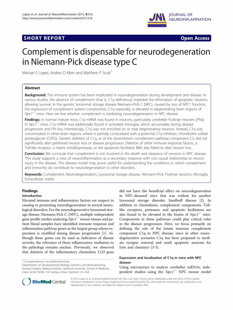
have been detected in the cerebellum of Npc1-/- mice asearly as postnatal day 21 (P21), an age well before onsetof severe neurodegeneration [6,7] (Figure 1A). At aroundP50, an age just prior to major decline in health, C1qa,b, and c mRNA levels are markedly higher [2,7]. Inaddition to the cerebellum, increased C1qa expression
Figure 1 C1q and complement component expression is increasingly elevated with age in NPC disease. (A) Compilation of analyses fromthree independent cerebellar array datasets accessible through the National Center for Biotechnology Information (NCBI) Gene ExpressionOmnibus (GEO) database: a, GSE36119; b, GSE5944; and c, GSE20450. The differential gene expression of components of the complement cascadefor Npc1-/- mice compared to wild-type, Npc1+/- or Npc1+/+, mice are shown at two ages. (B) An outline of the initial components of thecomplement cascade (genes are in bold) showing factors in the pathway that have been identified as elevated in at least one array dataset. (C)Representative in situ signal of C1qa mRNA probe hybridized to thalamic region of P60 wild-type, Npc1-/-, and C1qa-/-; Npc1-/- mice. Scale bars are100μm.
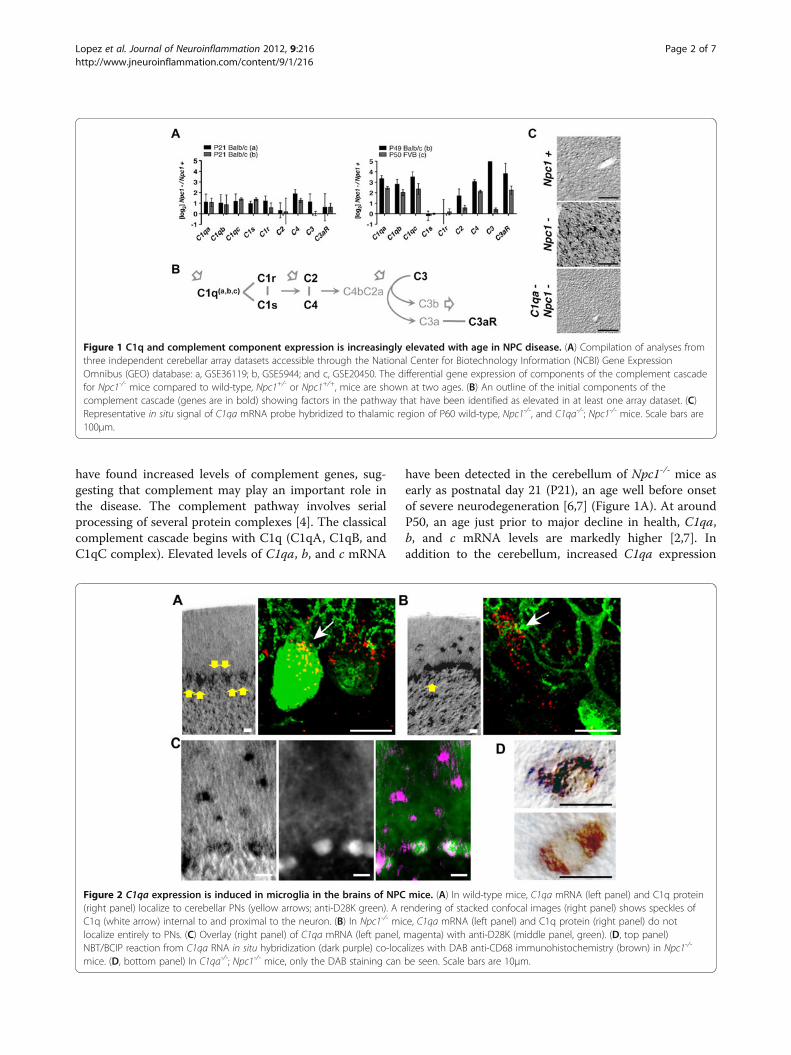
Figure 2 C1qa expression is induced in microglia in the brains of NPC mice. (A) In wild-type mice, C1qa mRNA (left panel) and C1q protein(right panel) localize to cerebellar PNs (yellow arrows; anti-D28K green). A rendering of stacked confocal images (right panel) shows speckles ofC1q (white arrow) internal to and proximal to the neuron. (B) In Npc1-/- mice, C1qa mRNA (left panel) and C1q protein (right panel) do notlocalize entirely to PNs. (C) Overlay (right panel) of C1qa mRNA (left panel, magenta) with anti-D28K (middle panel, green). (D, top panel)NBT/BCIP reaction from C1qa RNA in situ hybridization (dark purple) co-localizes with DAB anti-CD68 immunohistochemistry (brown) in Npc1-/-
mice. (D, bottom panel) In C1qa-/-; Npc1-/- mice, only the DAB staining can be seen. Scale bars are 10μm.
Lopez et al. Journal of Neuroinflammation 2012, 9:216 Page 2 of 7http://www.jneuroinflammation.com/content/9/1/216
was found in the thalamus (Figure 1C), another brain re-gion that is highly vulnerable in the disease. Down-stream gene components of the complement cascadeidentified in cerebellar arrays include the anaphylatoxinand opsonin precursor C3 and the anaphylatoxin recep-tor C3aR. The mRNA of C3, which plays a central rolein complement activation (Figure 1B), is not as robustlydetected as C3aR (Figure 1A). The mRNAs of C1r andC1s, components needed to initiate the classical comple-ment cascade, are also not consistently elevated betweenstudies. However, even without efficient activation of thecomplement cascade, C1q alone may act as a recogni-tion molecule that tags apoptotic cells to facilitate theirclearance by phagocytes [5].We next determined if C1q, a secreted molecule, loca-
lizes around degenerating neurons in the NPC mousemodel. In other neurodegenerative disease models, suchas amyotrophic lateral sclerosis [8] and glaucoma [3], C1qis produced in and localizes around injured neurons. Innormal mice, we detected C1qa mRNA in cerebellar Pur-kinje neurons (PNs) (Figure 2A). Antibody stainingshowed that C1q co-localized with PN soma, marked by
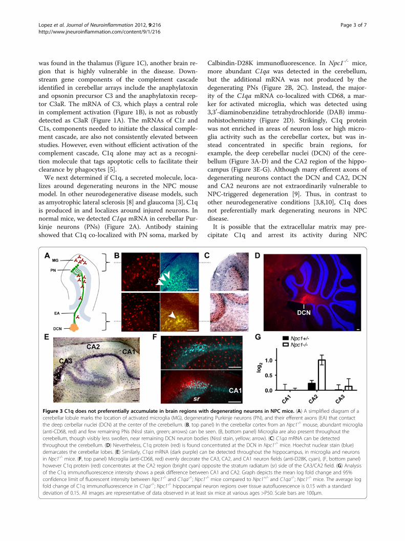
Calbindin-D28K immunofluorescence. In Npc1-/- mice,more abundant C1qa was detected in the cerebellum,but the additional mRNA was not produced by thedegenerating PNs (Figure 2B, 2C). Instead, the major-ity of the C1qa mRNA co-localized with CD68, a mar-ker for activated microglia, which was detected using3,30-diaminobenzidine tetrahydrochloride (DAB) immu-nohistochemistry (Figure 2D). Strikingly, C1q proteinwas not enriched in areas of neuron loss or high micro-glia activity such as the cerebellar cortex, but was in-stead concentrated in specific brain regions, forexample, the deep cerebellar nuclei (DCN) of the cere-bellum (Figure 3A-D) and the CA2 region of the hippo-campus (Figure 3E-G). Although many efferent axons ofdegenerating neurons contact the DCN and CA2, DCNand CA2 neurons are not extraordinarily vulnerable toNPC-triggered degeneration [9]. Thus, in contrast toother neurodegenerative conditions [3,8,10], C1q doesnot preferentially mark degenerating neurons in NPCdisease.It is possible that the extracellular matrix may pre-
cipitate C1q and arrest its activity during NPC
Figure 3 C1q does not preferentially accumulate in brain regions with degenerating neurons in NPC mice. (A) A simplified diagram of acerebellar lobule marks the location of activated microglia (MG), degenerating Purkinje neurons (PN), and their efferent axons (EA) that contactthe deep cerbellar nuclei (DCN) at the center of the cerebellum. (B, top panel) In the cerebellar cortex from an Npc1-/- mouse, abundant microglia(anti-CD68, red) and few remaining PNs (Nissl stain, green; arrows) can be seen. (B, bottom panel) Microglia are also present throughout thecerebellum, though visibly less swollen, near remaining DCN neuron bodies (Nissl stain, yellow; arrow). (C) C1qa mRNA can be detectedthroughout the cerebellum. (D) Nevertheless, C1q protein (red) is found concentrated at the DCN in Npc1-/- mice. Hoechst nuclear stain (blue)demarcates the cerebellar lobes. (E) Similarly, C1qa mRNA (dark purple) can be detected throughout the hippocampus, in microglia and neuronsin Npc1-/- mice. (F, top panel) Microglia (anti-CD68, red) evenly decorate the CA3, CA2, and CA1 neuron fields (anti-D28K, cyan), (F, bottom panel)however C1q protein (red) concentrates at the CA2 region (bright cyan) opposite the stratum radiatum (sr) side of the CA3/CA2 field. (G) Analysisof the C1q immunofluorescence intensity shows a peak difference between CA1 and CA2. Graph depicts the mean log fold change and 95%confidence limit of fluorescent intensity between Npc1-/- and C1qa-/-; Npc1-/- mice compared to Npc1+/- and C1qa-/-; Npc1-/- mice. The average logfold change of C1q immunofluorescence in C1qa-/-; Npc1-/- hippocampal neuron regions over tissue autofluorescence is 0.15 with a standarddeviation of 0.15. All images are representative of data observed in at least six mice at various ages >P50. Scale bars are 100μm.
Lopez et al. Journal of Neuroinflammation 2012, 9:216 Page 3 of 7http://www.jneuroinflammation.com/content/9/1/216
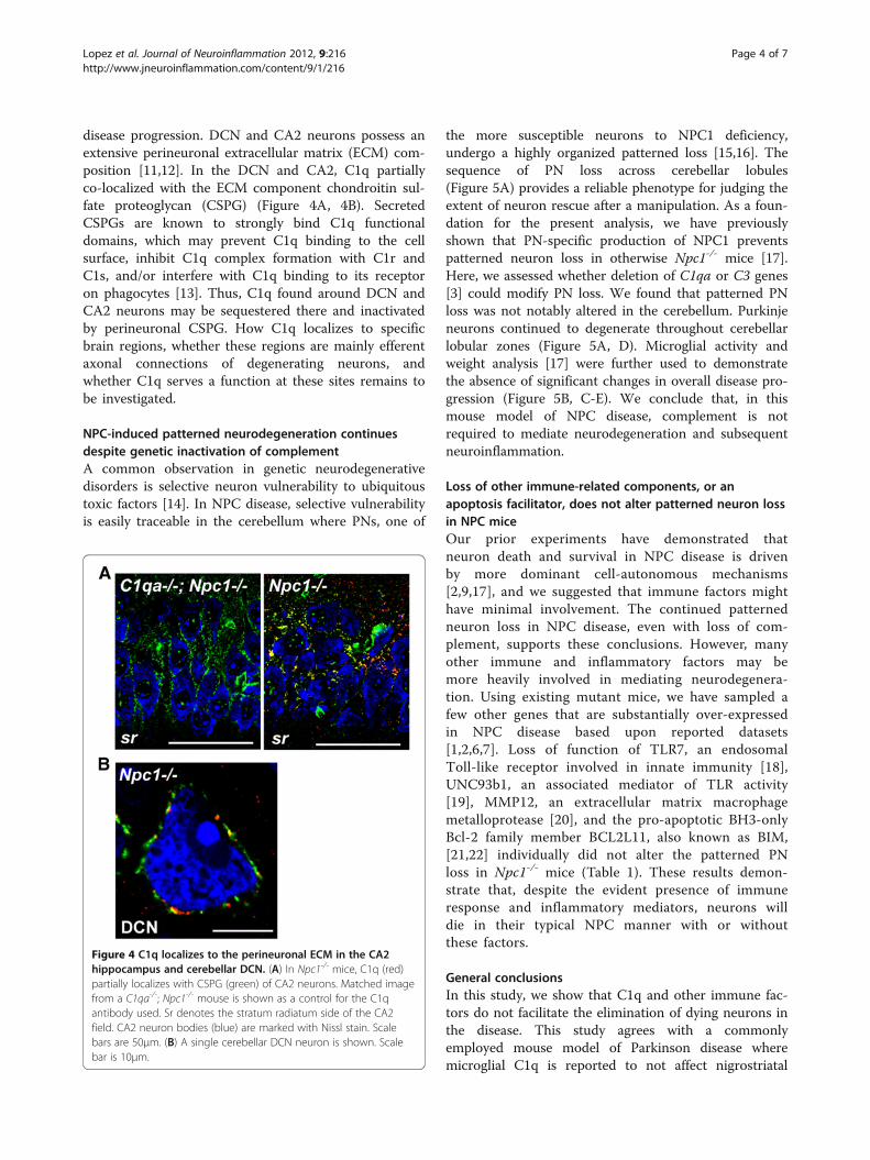
disease progression. DCN and CA2 neurons possess anextensive perineuronal extracellular matrix (ECM) com-position [11,12]. In the DCN and CA2, C1q partiallyco-localized with the ECM component chondroitin sul-fate proteoglycan (CSPG) (Figure 4A, 4B). SecretedCSPGs are known to strongly bind C1q functionaldomains, which may prevent C1q binding to the cellsurface, inhibit C1q complex formation with C1r andC1s, and/or interfere with C1q binding to its receptoron phagocytes [13]. Thus, C1q found around DCN andCA2 neurons may be sequestered there and inactivatedby perineuronal CSPG. How C1q localizes to specificbrain regions, whether these regions are mainly efferentaxonal connections of degenerating neurons, andwhether C1q serves a function at these sites remains tobe investigated.
NPC-induced patterned neurodegeneration continuesdespite genetic inactivation of complementA common observation in genetic neurodegenerativedisorders is selective neuron vulnerability to ubiquitoustoxic factors [14]. In NPC disease, selective vulnerabilityis easily traceable in the cerebellum where PNs, one of
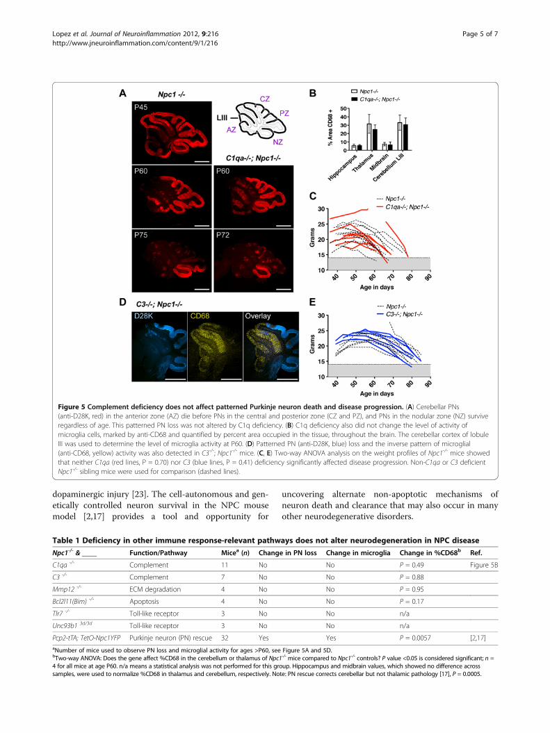
the more susceptible neurons to NPC1 deficiency,undergo a highly organized patterned loss [15,16]. Thesequence of PN loss across cerebellar lobules(Figure 5A) provides a reliable phenotype for judging theextent of neuron rescue after a manipulation. As a foun-dation for the present analysis, we have previouslyshown that PN-specific production of NPC1 preventspatterned neuron loss in otherwise Npc1-/- mice [17].Here, we assessed whether deletion of C1qa or C3 genes[3] could modify PN loss. We found that patterned PNloss was not notably altered in the cerebellum. Purkinjeneurons continued to degenerate throughout cerebellarlobular zones (Figure 5A, D). Microglial activity andweight analysis [17] were further used to demonstratethe absence of significant changes in overall disease pro-gression (Figure 5B, C-E). We conclude that, in thismouse model of NPC disease, complement is notrequired to mediate neurodegeneration and subsequentneuroinflammation.
Loss of other immune-related components, or anapoptosis facilitator, does not alter patterned neuron lossin NPC miceOur prior experiments have demonstrated thatneuron death and survival in NPC disease is drivenby more dominant cell-autonomous mechanisms[2,9,17], and we suggested that immune factors mighthave minimal involvement. The continued patternedneuron loss in NPC disease, even with loss of com-plement, supports these conclusions. However, manyother immune and inflammatory factors may bemore heavily involved in mediating neurodegenera-tion. Using existing mutant mice, we have sampled afew other genes that are substantially over-expressedin NPC disease based upon reported datasets[1,2,6,7]. Loss of function of TLR7, an endosomalToll-like receptor involved in innate immunity [18],UNC93b1, an associated mediator of TLR activity[19], MMP12, an extracellular matrix macrophagemetalloprotease [20], and the pro-apoptotic BH3-onlyBcl-2 family member BCL2L11, also known as BIM,[21,22] individually did not alter the patterned PNloss in Npc1-/- mice (Table 1). These results demon-strate that, despite the evident presence of immuneresponse and inflammatory mediators, neurons willdie in their typical NPC manner with or withoutthese factors.
General conclusionsIn this study, we show that C1q and other immune fac-tors do not facilitate the elimination of dying neurons inthe disease. This study agrees with a commonlyemployed mouse model of Parkinson disease wheremicroglial C1q is reported to not affect nigrostriatal
Figure 4 C1q localizes to the perineuronal ECM in the CA2hippocampus and cerebellar DCN. (A) In Npc1-/- mice, C1q (red)partially localizes with CSPG (green) of CA2 neurons. Matched imagefrom a C1qa-/-; Npc1-/- mouse is shown as a control for the C1qantibody used. Sr denotes the stratum radiatum side of the CA2field. CA2 neuron bodies (blue) are marked with Nissl stain. Scalebars are 50μm. (B) A single cerebellar DCN neuron is shown. Scalebar is 10μm.
Lopez et al. Journal of Neuroinflammation 2012, 9:216 Page 4 of 7http://www.jneuroinflammation.com/content/9/1/216
dopaminergic injury [23]. The cell-autonomous and gen-etically controlled neuron survival in the NPC mousemodel [2,17] provides a tool and opportunity for
uncovering alternate non-apoptotic mechanisms ofneuron death and clearance that may also occur in manyother neurodegenerative disorders.
Figure 5 Complement deficiency does not affect patterned Purkinje neuron death and disease progression. (A) Cerebellar PNs(anti-D28K, red) in the anterior zone (AZ) die before PNs in the central and posterior zone (CZ and PZ), and PNs in the nodular zone (NZ) surviveregardless of age. This patterned PN loss was not altered by C1q deficiency. (B) C1q deficiency also did not change the level of activity ofmicroglia cells, marked by anti-CD68 and quantified by percent area occupied in the tissue, throughout the brain. The cerebellar cortex of lobuleIII was used to determine the level of microglia activity at P60. (D) Patterned PN (anti-D28K, blue) loss and the inverse pattern of microglial(anti-CD68, yellow) activity was also detected in C3-/-; Npc1-/- mice. (C, E) Two-way ANOVA analysis on the weight profiles of Npc1-/- mice showedthat neither C1qa (red lines, P = 0.70) nor C3 (blue lines, P = 0.41) deficiency significantly affected disease progression. Non-C1qa or C3 deficientNpc1-/- sibling mice were used for comparison (dashed lines).
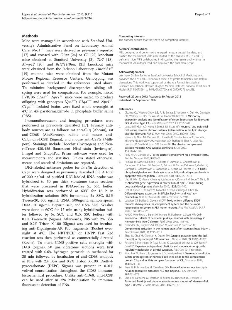
Table 1 Deficiency in other immune response-relevant pathways does not alter neurodegeneration in NPC disease
Npc1-/- & ____ Function/Pathway Micea (n) Change in PN loss Change in microglia Change in %CD68b Ref.
C1qa -/- Complement 11 No No P = 0.49 Figure 5B
C3 -/- Complement 7 No No P = 0.88
Mmp12 -/- ECM degradation 4 No No P = 0.95
Bcl2l11(Bim) -/- Apoptosis 4 No No P = 0.17
Tlr7 -/- Toll-like receptor 3 No No n/a
Unc93b1 3d/3d Toll-like receptor 3 No No n/a
Pcp2-tTA; TetO-Npc1YFP Purkinje neuron (PN) rescue 32 Yes Yes P = 0.0057 [2,17]aNumber of mice used to observe PN loss and microglial activity for ages >P60, see Figure 5A and 5D.bTwo-way ANOVA: Does the gene affect %CD68 in the cerebellum or thalamus of Npc1-/- mice compared to Npc1-/- controls? P value <0.05 is considered significant; n =4 for all mice at age P60. n/a means a statistical analysis was not performed for this group. Hippocampus and midbrain values, which showed no difference acrosssamples, were used to normalize %CD68 in thalamus and cerebellum, respectively. Note: PN rescue corrects cerebellar but not thalamic pathology [17], P = 0.0005.
Lopez et al. Journal of Neuroinflammation 2012, 9:216 Page 5 of 7http://www.jneuroinflammation.com/content/9/1/216
MethodsMice were managed in accordance with Stanford Uni-versity’s Administrative Panel on Laboratory AnimalCare. Npc1+/- mice were derived as previously reported[17] and crossed with C1qa [24] or C3 [25] knockoutmice obtained at Stanford University [3]. Tlr7 [18],Mmp12 [20], and Bcl2l11(Bim) [21] knockout micewere obtained from the Jackson Laboratory. Unc93b13d
[19] mutant mice were obtained from the MutantMouse Regional Resource Centers. Genotyping wasperformed as detailed in the references listed above.To minimize background discrepancies, sibling off-spring were used for comparisons. For example, mixedFVB/B6 C1qa+/-; Npc1+/- mice were mated to produceoffspring with genotypes Npc1-/-; C1qa+/+ and Npc1-/-;C1qa-/-. Isolated brains were fixed whole overnight at4°C in 4% paraformaldehyde in phosphate buffer saline(PBS).Immunofluorescent and imaging procedures were
performed as previously described [17]. Primary anti-body sources are as follows: rat anti-C1q (Abcam), ratanti-CD68 (AbdSerotec), rabbit and mouse anti-Calbindin-D28K (Sigma), and mouse anti-CSPG (Milli-pore). Stainings include Hoechst (Invitrogen) and Neu-roTrace 435/455 fluorescent Nissl stain (Invitrogen).ImageJ and GraphPad Prism software were used formeasurements and statistics. Unless stated otherwise,means and standard deviations are reported.DIG-labeled antisense probes for in situ detection of
C1qa were designed as previously described [3]. A totalof 200 ng/mL of purified DIG-labeled RNA probe washybridized to 50 μm thick vibratome tissue sectionsthat were processed in RNAse-free 5x SSC buffer.Hybridization was performed at 60°C for 16 h inhybridization solution: 50% formamide, 5x SSC, 0.1%Tween-20, 500 ug/mL tRNA, 500ug/mL salmon spermDNA, 50 ug/mL Heparin salt, and 0.5% SDS. Washeswere done at 60°C for 15 min using hybridization buf-fer followed by 5x SCC and 0.2x SSC buffers with0.1% Tween-20 (Sigma). Afterwards, PBS with 2% BSAand 0.2% Triton X-100 (Sigma) was used for incubat-ing anti-Digoxigenin-AP, Fab fragments (Roche) over-night at 4°C. The NBT/BCIP or HNPP Fast Redreaction was then performed as commercially directed(Roche). To mark CD68-positive cells microglia withDAB (Sigma), 50 μm vibratome sections were firsttreated with 0.6% hydrogen peroxide in methanol for30 min followed by incubation of anti-CD68 antibodyin PBS with 2% BSA and 0.2% Triton X-100. Diethyl-pyrocarbonate (DEPC; Sigma) was present in 0.01%vol/vol concentration throughout the CD68 immuno-histochemical procedure. Unlike anti-CD68, anti-D28Kcan be used after in situ hybridization for immuno-fluorescent detection of PNs.
Competing interestsThe authors declare that they have no competing interests.
Authors’ contributionsMEL designed and performed the experiments, analyzed the data, anddrafted the manuscript. ADK contributed to the analysis of C1q and C3deficient mice. MPS collaborated in discussing the results and writing themanuscript. All authors read and approved the final manuscript.
AcknowledgementsWe thank Dr Ben Barres at Stanford University School of Medicine, whoprovided the C1q and C3 knockout mice, C1q probe templates, and helpfuldiscussions. This work was supported by the Ara Parseghian MedicalResearch Foundation; Howard Hughes Medical Institute; National Institutes ofHealth [R01 NS073691 to MPS, GM07790 and GM007276 to MEL.
Received: 29 June 2012 Accepted: 30 August 2012Published: 17 September 2012
References1. Cluzeau CV, Watkins-Chow DE, Fu R, Borate B, Yanjanin N, Dail MK, Davidson
CD, Walkley SU, Ory DS, Wassif CA, Pavan WJ, Porter FD: Microarrayexpression analysis and identification of serum biomarkers for Niemann-Pick disease, type C1. Hum Mol Genet 2012, 21:3632–3646.
2. Lopez ME, Klein AD, Hong J, Dimbil UJ, Scott MP: Neuronal and epithelialcell rescue resolves chronic systemic inflammation in the lipid storagedisorder Niemann-Pick C. Hum Mol Genet 2012, 21:2946–2960.
3. Stevens B, Allen NJ, Vazquez LE, Howell GR, Christopherson KS, Nouri N,Micheva KD, Mehalow AK, Huberman AD, Stafford B, Sher A, Litke AM,Lambris JD, Smith SJ, John SW, Barres BA: The classical complementcascade mediates CNS synapse elimination. Cell 2007,131:1164–1178.
4. Perry VH, O’Connor V: C1q: the perfect complement for a synaptic feast?.Nat Rev Neurosci 2008, 9:807–811.
5. Paidassi H, Tacnet-Delorme P, Garlatti V, Darnault C, Ghebrehiwet B,Gaboriaud C, Arlaud GJ, Frachet P, Paidassi H, Tacnet-Delorme P, Garlatti V,Darnault C, Ghebrehiwet B, Gaboriaud C, Arlaud GJ, Frachet P: C1q bindsphosphatidylserine and likely acts as a multiligand-bridging molecule inapoptotic cell recognition. J Immunol 2008, 180:2329–2338.
6. Liao G, Wen Z, Irizarry K, Huang Y, Mitsouras K, Darmani M, Leon T, Shi L, BiX: Abnormal gene expression in cerebellum of Npc1-/- mice duringpostnatal development. Brain Res 2010, 1325:128–140.
7. Distl R, Kuban R, Komkov V, Kallwellis K, von Deimling A, Ohm TG:Differential gene expression in BALB/c Npc1−/− and Npc1+/+cerebellum. NCBI GEO DataSets 2007, accession GSE5944.
8. Lobsiger CS, Boillee S, Cleveland DW: Toxicity from different SOD1mutants dysregulates the complement system and the neuronalregenerative response in ALS motor neurons. Proc Natl Acad Sci U S A2007, 104:7319–7326.
9. Ko DC, Milenkovic L, Beier SM, Manuel H, Buchanan J, Scott MP: Cell-autonomous death of cerebellar purkinje neurons with autophagy inNiemann-Pick type C disease. PLoS Genet 2005, 1:81–95.
10. Bellander BM, Singhrao SK, Ohlsson M, Mattsson P, Svensson M:Complement activation in the human brain after traumatic head injury. JNeurotrauma 2001, 18:1295–1311.
11. Zhao M, Choi YS, Obrietan K, Dudek SM: Synaptic plasticity (and the lackthereof) in hippocampal CA2 neurons. J Neurosci 2007, 27:12025–12032.
12. Foscarin S, Ponchione D, Pajaj E, Leto K, Gawlak M, Wilczynski GM, Rossi F,Carulli D: Experience-dependent plasticity and modulation of growthregulatory molecules at central synapses. PLoS One 2011, 6:e16666.
13. Kirschfink M, Blase L, Engelmann S, Schwartz-Albiez R: Secreted chondroitinsulfate proteoglycan of human B cell lines binds to the complementprotein C1q and inhibits complex formation of C1. J Immunol 1997,158:1324–1331.
14. Ilieva H, Polymenidou M, Cleveland DW: Non-cell autonomous toxicity inneurodegenerative disorders: ALS and beyond. J Cell Biol 2009,187:761–772.
15. Sarna JR, Larouche M, Marzban H, Sillitoe RV, Rancourt DE, Hawkes R:Patterned Purkinje cell degeneration in mouse models of Niemann-Picktype C disease. J Comp Neurol 2003, 456:279–291.
Lopez et al. Journal of Neuroinflammation 2012, 9:216 Page 6 of 7http://www.jneuroinflammation.com/content/9/1/216
16. Sillitoe RV, Vogel MW, Joyner AL: Engrailed homeobox genes regulateestablishment of the cerebellar afferent circuit map. J Neurosci 2010,30:10015–10024.
17. Lopez ME, Klein AD, Dimbil UJ, Scott MP: Anatomically defined neuron-based rescue of neurodegenerative Niemann-Pick type C disorder.J Neurosci 2011, 31:4367–4378.
18. Lund JM, Alexopoulou L, Sato A, Karow M, Adams NC, Gale NW, Iwasaki A,Flavell RA: Recognition of single-stranded RNA viruses by Toll-likereceptor 7. Proc Natl Acad Sci U S A 2004, 101:5598–5603.
19. Tabeta K, Hoebe K, Janssen EM, Du X, Georgel P, Crozat K, Mudd S, Mann N,Sovath S, Goode J, Shamel L, Herskovits AA, Portnoy DA, Cooke M,Tarantino LM, Wiltshire T, Steinberg BE, Grinstein S, Beutler B: The Unc93b1mutation 3d disrupts exogenous antigen presentation and signaling viaToll-like receptors 3, 7 and 9. Nat Immunol 2006, 7:156–164.
20. Shipley JM, Wesselschmidt RL, Kobayashi DK, Ley TJ, Shapiro SD:Metalloelastase is required for macrophage-mediated proteolysis andmatrix invasion in mice. Proc Natl Acad Sci U S A 1996, 93:3942–3946.
21. Bouillet P, Metcalf D, Huang DC, Tarlinton DM, Kay TW, Kontgen F, AdamsJM, Strasser A: Proapoptotic Bcl-2 relative Bim required for certainapoptotic responses, leukocyte homeostasis, and to precludeautoimmunity. Science 1999, 286:1735–1738.
22. Bouillet P, Robati M, Adams JM, Strasser A: Loss of pro-apoptotic BH3-onlyBcl-2 family member Bim does not protect mutant Lurcher mice fromneurodegeneration. J Neurosci Res 2003, 74:777–781.
23. Depboylu C, Schorlemmer K, Klietz M, Oertel WH, Weihe E, Hoglinger GU,Schafer MK: Upregulation of microglial C1q expression has no effects onnigrostriatal dopaminergic injury in the MPTP mouse model ofParkinson disease. J Neuroimmunol 2011, 236:39–46.
24. Botto M, Dell'Agnola C, Bygrave AE, Thompson EM, Cook HT, Petry F, LoosM, Pandolfi PP, Walport MJ: Homozygous C1q deficiency causesglomerulonephritis associated with multiple apoptotic bodies. Nat Genet1998, 19:56–59.
25. Wessels MR, Butko P, Ma M, Warren HB, Lage AL, Carroll MC: Studies ofgroup B streptococcal infection in mice deficient in complementcomponent C3 or C4 demonstrate an essential role for complement inboth innate and acquired immunity. Proc Natl Acad Sci U S A 1995,92:11490–11494.
doi:10.1186/1742-2094-9-216Cite this article as: Lopez et al.: Complement is dispensable forneurodegeneration in Niemann-Pick disease type C. Journal ofNeuroinflammation 2012 9:216.
Submit your next manuscript to BioMed Centraland take full advantage of:
• Convenient online submission
• Thorough peer review
• No space constraints or color figure charges
• Immediate publication on acceptance
• Inclusion in PubMed, CAS, Scopus and Google Scholar
• Research which is freely available for redistribution
Submit your manuscript at www.biomedcentral.com/submit
Lopez et al. Journal of Neuroinflammation 2012, 9:216 Page 7 of 7http://www.jneuroinflammation.com/content/9/1/216