Embed Size (px)
Citation preview
Comprehensive systematic review: Treatment of cerebellar motor dysfunction and ataxia
Report of the Guideline Development, Dissemination, and Implementation Subcommittee of the
American Academy of Neurology
Theresa A. Zesiewicz, MD1; George Wilmot, MD2; Sheng-Han Kuo, MD3; Susan Perlman,
MD4; Patricia E. Greenstein, MB, BCh5; Sarah H. Ying, MD6; Tetsuo Ashizawa, MD7; S.H.
Subramony, MD8; Jeremy D. Schmahmann, MD9; K.P. Figueroa10; Hidehiro Mizusawa, MD11;
Ludger Schöls, MD12; Jessica D. Shaw, MPH1; Richard M. Dubinsky, MD, MPH13; Melissa J.
Armstrong, MD, MSc8; Gary S. Gronseth, MD13; Kelly L. Sullivan, PhD14
1) Department of Neurology, University of South Florida, Tampa
2) Department of Neurology, Emory University, Atlanta, GA
3) Department of Neurology, Columbia University, New York, NY
4) Department of Neurology, University of California, Los Angeles
5) Department of Neurology, Beth Israel Deaconess Medical Center, Boston, MA
6) Shire, Lexington, MA, and the Johns Hopkins University School of Medicine, Baltimore, MD
7) Department of Neurology, Houston Methodist Research Institute, TX
8) Department of Neurology, University of Florida College of Medicine, Gainesville
9) Department of Neurology, Massachusetts General Hospital, and Department of Neurology,
Harvard Medical School, Boston, MA
10) Department of Neurology, University of Utah, Salt Lake City
11) National Center of Neurology and Psychiatry, Tokyo, Japan
12) Department of Neurology and Hertie-Institute for Clinical Brain Research, Tübingen,
Germany
13) Department of Neurology, University of Kansas Medical Center, Kansas City
14) Jiann-Ping Hsu College of Public Health, Georgia Southern University, Statesboro
Address correspondence and reprint requests to
American Academy of Neurology:
Title character count: 71
Abstract word count: 254
Manuscript word count: 7,891
Approved by the Guideline Development, Dissemination, and Implementation Subcommittee on
October 22, 2016; by the Practice Committee on October 2, 2017; and by the AAN Institute
Board of Directors on December 5, 2017.
This comprehensive systematic review was endorsed by the A-TCP Children’s Project on
July 5, 2017.
AUTHOR CONTRIBUTIONS
Dr. Zesiewicz: study concept and design, acquisition of data, analysis or interpretation of data,
drafting/revising the manuscript, critical revision of the manuscript for important intellectual
content, study supervision.
Dr. Wilmot: acquisition of data, analysis or interpretation of data, drafting/revising the
manuscript, critical revision of the manuscript for important intellectual content.
Dr. Kuo: acquisition of data, analysis or interpretation of data, drafting/revising the manuscript,
critical revision of the manuscript for important intellectual content.
Dr. Perlman: acquisition of data, analysis or interpretation of data, drafting/revising the
manuscript, critical revision of the manuscript for important intellectual content.
Dr. Greenstein: analysis or interpretation of data, drafting/revising the manuscript, critical
revision of the manuscript for important intellectual content.
Dr. Ying: analysis or interpretation of data, drafting/revising the manuscript, critical revision of
the manuscript for important intellectual content.
Dr. Ashizawa: acquisition of data, analysis or interpretation of data, drafting/revising the
manuscript, critical revision of the manuscript for important intellectual content.
Dr. Subramony: acquisition of data, analysis or interpretation of data, drafting/revising the
manuscript, critical revision of the manuscript for important intellectual content.
Dr. Schmahmann: acquisition of data, analysis or interpretation of data, drafting/revising the
manuscript, critical revision of the manuscript for important intellectual content.
Dr. Figueroa: analysis or interpretation of data, drafting/revising the manuscript, critical revision
of the manuscript for important intellectual content.
Dr. Mizusawa: analysis or interpretation of data, drafting/revising the manuscript, critical
revision of the manuscript for important intellectual content.
Dr. Schöls: analysis or interpretation of data, drafting/revising the manuscript, critical revision of
the manuscript for important intellectual content.
Ms. Shaw: analysis or interpretation of data, drafting/revising the manuscript, critical revision of
the manuscript for important intellectual content.
Dr. Dubinsky: acquisition of data, analysis or interpretation of data, drafting/revising the
manuscript, critical revision of the manuscript for important intellectual content.
Dr. Armstrong: study concept and design, acquisition of data, analysis or interpretation of data,
drafting/revising the manuscript, critical revision of the manuscript for important intellectual
content, study supervision.
Dr. Gronseth: acquisition of data, analysis or interpretation of data, drafting/revising the
manuscript, critical revision of the manuscript for important intellectual content, study
supervision.
Dr. Sullivan: study concept and design, acquisition of data, analysis or interpretation of data,
drafting/revising the manuscript, critical revision of the manuscript for important intellectual
content, study supervision.
STUDY FUNDING
This comprehensive systematic review was developed with financial support from the American
Academy of Neurology (AAN). Authors who serve or served as AAN subcommittee members or
methodologists (T.A.Z., R.M.D., M.J.A., G.S.G., K.L.S.) were reimbursed by the AAN for
expenses related to travel to subcommittee meetings where drafts of manuscripts were reviewed.
DISCLOSURE
T. Zesiewicz has served as a clinical advisor for Steminent Biotherapeutics; has received travel
reimbursement from the Department of Neurology at University of Southern Florida; has
received travel reimbursement for a Biohaven Pharmaceuticals meeting; has served on the
editorial board for Neurodegenerative Disease Management and Tremor and other Hyperkinetic
Movements; has a patent for Methods of Treating Disease-Induced Ataxia and Non-Ataxic
Imbalance (US Patent No. 9463190 B2); and has received research support for her division for
approximately 20 clinical trials for Parkinson disease (PD), Friedreich ataxia, and
spinocerebellar ataxias (SCAs).
G. Wilmot has served on scientific advisory panels for Biohaven Pharmaceuticals and Santhera
Pharmaceuticals, and has received financial or material research support or compensation from
Friedreich’s Ataxia Research Alliance, Reata Pharmaceuticals, and Shire.
S. Kuo has no relevant disclosures to report.
S. Perlman has no relevant disclosures to report.
P. Greenstein has received an R21 grant award from the NIH to study the effect of transcranial
magnetic stimulation on SCA (grant awarded in August 2013; the study began in January 2014;
no preliminary data yet available).
S. Ying received a salary from Shire; received a salary during her employment with Pfizer Inc.
and Takeda Pharmaceuticals Inc.; and received grant funding from the NIH.
T. Ashizawa has nonfinancial competing interests with the Marigold Foundation, Myotonic
Dystrophy Foundation, and the Muscular Dystrophy Association (MDA); receives honoraria
from the NIH National Institute of Neurological Disorders and Stroke (NINDS) Neurological
Sciences and Disorders B Study Section; received travel reimbursement from the MDA Medical
Advisory Committee and the National Ataxia Foundation for the Ataxia Investigator Meeting;
serves as an editor for PLoS ONE; has a patent (US Patent No. 6855497) on a DNA test for SCA
type 10 (SCA10); receives funding for an NINDS research grant award R01NSNS083564;
participates in a clinical trial of BHV-4157 (NCT02960893); and has received royalty payments
from Baylor College of Medicine for a DNA test for SCA10 (US Patent No. 6855497).
S. Subramony has received compensation for a lecture from Athena Diagnostics in October
2013; and has received travel reimbursement or honoraria from Reata Pharmaceuticals, ISIS
Pharmaceuticals (now Ionis Pharmaceuticals), the NIH, the National Ataxia Foundation, and the
Friedreich’s Ataxia Research Alliance.
J. Schmahmann serves as a consultant to Ataxion, Biogen, Biohaven, Pfizer, and Takeda, and
receives grant support from the National Ataxia Foundation and the A-T Children’s Project.
K. Figueroa has no relevant disclosures to report.
H. Mizusawa has no relevant disclosures to report.
L. Schöls has no relevant disclosures to report.
J. Shaw has no relevant disclosures to report.
R. Dubinsky serves on the scientific advisory board for Allergan Pharmaceuticals; has received
travel funding from the American Academy of Neurology (AAN), Allergan Pharmaceuticals,
and the Huntington Study Group; serves as Level of Evidence associate editor for the AAN;
receives honoraria from Allergan Pharmaceuticals; serves on the speakers bureau for Allergan
Pharmaceuticals; and is involved with the commercial entity Allergan Pharmaceuticals and the
government entities the NIH and the Agency for Healthcare Research and Quality. His spouse
holds stock in Abbott Laboratories.
M. Armstrong serves on the Level of Evidence editorial board for Neurology (not compensated
financially) and is an AAN evidence-based methodologist.
G. Gronseth serves as an associate editor (level of evidence review) for Neurology, serves on the
editorial advisory board for Neurology Now, and is compensated by the AAN for methodologic
activities.
K. Sullivan has received research support from the Georgia Governor’s Office of Highway
Safety and has a patent for Methods of Treating Disease-Induced Ataxia and Non-Ataxic
Imbalance (US Patient No. 9463190 B2).
ABBREVIATIONS
4-AP: 4-aminopyridine
5-HT1A: 5-hydroxytryptamine subtype 1A
5-HT3: 5-hydroxytryptamine subtype 3
AAN: American Academy of Neurology
AEs: adverse events
CCA: cerebellar cortical atrophy
EA2: Episodic ataxia type 2
FA: Friedreich ataxia
FARS: Friedreich’s Ataxia Rating Scale
FIM: Functional Independence Measure
FXTAS: fragile X tremor ataxia syndrome
GAD: glutamic acid decarboxylase
ICARS: International Cooperative Ataxia Rating Scale
ILOCA: idiopathic late-onset cerebellar ataxia
MS: multiple sclerosis
MSA: multiple-system atrophy
MSA-C: multiple-system atrophy-cerebellar type
NESSCA: Neurological Examination Score for the Assessment of Spinocerebellar Ataxia
OPCA: olivopontocerebellar atrophy
OR: odds ratio
RCT: randomized controlled trial
SARA: Scale for the Assessment and Rating of Ataxia
SCA: spinocerebellar ataxia
SCA1: spinocerebellar ataxia type 1
SCA2: spinocerebellar ataxia type 2
SCA3: spinocerebellar ataxia type 3
SCA28: spinocerebellar ataxia type 28
SCD: spinocerebellar degeneration
tDCS: transcranial direct current stimulation
TMS: transcranial magnetic stimulation
TRH: thyrotropin-releasing hormone
VPA: valproic acid

6
7
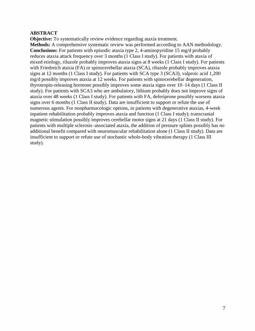
ABSTRACT
Objective: To systematically review evidence regarding ataxia treatment.
Methods: A comprehensive systematic review was performed according to AAN methodology.
Conclusions: For patients with episodic ataxia type 2, 4-aminopyridine 15 mg/d probably
reduces ataxia attack frequency over 3 months (1 Class I study). For patients with ataxia of
mixed etiology, riluzole probably improves ataxia signs at 8 weeks (1 Class I study). For patients
with Friedreich ataxia (FA) or spinocerebellar ataxia (SCA), riluzole probably improves ataxia
signs at 12 months (1 Class I study). For patients with SCA type 3 (SCA3), valproic acid 1,200
mg/d possibly improves ataxia at 12 weeks. For patients with spinocerebellar degeneration,
thyrotropin-releasing hormone possibly improves some ataxia signs over 10–14 days (1 Class II
study). For patients with SCA3 who are ambulatory, lithium probably does not improve signs of
ataxia over 48 weeks (1 Class I study). For patients with FA, deferiprone possibly worsens ataxia
signs over 6 months (1 Class II study). Data are insufficient to support or refute the use of
numerous agents. For nonpharmacologic options, in patients with degenerative ataxias, 4-week
inpatient rehabilitation probably improves ataxia and function (1 Class I study); transcranial
magnetic stimulation possibly improves cerebellar motor signs at 21 days (1 Class II study). For
patients with multiple sclerosis–associated ataxia, the addition of pressure splints possibly has no
additional benefit compared with neuromuscular rehabilitation alone (1 Class II study). Data are
insufficient to support or refute use of stochastic whole-body vibration therapy (1 Class III
study).
8
The cerebellum is composed of the vermis, the hemispheres, and 3 cerebellar peduncles on each
side, and contributes largely to balance and motor coordination. The causes of cerebellar
dysfunction are numerous and include vitamin deficiencies, structural lesions (caused by tumors
or trauma), infection, inflammation, toxins, neurodegeneration, genetics, stroke, multiple
sclerosis (MS), and metabolic disorders. Motor signs resulting from cerebellar dysfunction may
include some or all of the following: imbalance, impaired coordination, limb and body tremor,
dysarthria, and oculomotor abnormalities. Other neurologic symptoms and signs may accompany
cerebellar dysfunction, including dystonia, muscle weakness, oculomotor abnormalities,
neuropathy, parkinsonism, spasticity, impaired visual acuity, and sensory impairment; these
symptoms and signs are beyond the scope of this review. Mood, cognitive disorders, and
autonomic dysfunction may also occur. Ataxia may result from cerebellar or sensory impairment.
There is currently no approved therapy to treat cerebellar motor dysfunction, and no
pharmacologic or surgical treatment is routinely used. Various therapies have been studied in
clinical trials for the past 40 years, although no consensus has been reached on their
effectiveness. This comprehensive systematic review synthesizes the literature on the treatment
of cerebellar motor dysfunction to answer the following questions:
(1) For patients with cerebellar motor dysfunction, do pharmacologic therapies, compared with
no (or alternative) treatment, improve motor symptoms with acceptable safety and
tolerability?
(2) For patients with cerebellar motor dysfunction, do surgical or other interventional therapies
(e.g., physical training), compared with no (or alternative) treatment, improve motor
symptoms with acceptable safety and tolerability?
(3) For patients with cerebellar motor dysfunction, does transcranial magnetic stimulation (TMS)
or transcranial direct current stimulation (tDCS), compared with no (or alternative) treatment,
improve motor symptoms with acceptable safety and tolerability?
This comprehensive systematic review focuses on treatment of cerebellar motor dysfunction
(cerebellar ataxia), which often constitutes symptomatic management. Management of other
elements of the included diseases, such as ataxia resulting from sensory changes, other
neurologic disturbance (e.g., parkinsonism), mood changes, and extraneurologic manifestations,
are not included in this review.
DESCRIPTION OF THE ANALYTIC PROCESS
The American Academy of Neurology (AAN) Guideline Development, Dissemination, and
Implementation Subcommittee (appendices e-1 and e-2) invited neurologists and scientists with
expertise in ataxia and methodology to perform this comprehensive systematic review. Conflicts
of interest were assessed and judged to be balanced when the comprehensive systematic review
was initiated and again at its conclusion. Although new conflicts appeared during the multiyear
process, at least half of the panel was without conflict throughout the entirety of the process. No
panelist of the systematic review was permitted to rate or assess his or her own work; articles
authored by individuals participating in the systematic review were assessed by nonconflicted
panel members.
9
The project used a hybrid systematic review methodology, using the AAN’s 2004 process
manuale1 for the overall approach, but the updated classification of evidence scheme for
therapeutic studies that was already approved and later published as an amendment to the 2011
manual.e2 There was also a public comment period for a near-final draft, a process described in
the 2011 manual.e2 The MEDLINE and EMBASE databases were searched from 1966 to June
2012. An updated pragmatic literature search of MEDLINE was performed on September 22,
2016, to capture studies published after 2012. Appendix e-3 lists the key words and phrases used
in the search. Because tremor is a sign of several disease processes, it was not specifically
included in the search strategy; however, if it was assessed as an outcome in studies of ataxia
treatment, tremor results were reviewed. Central vestibular dysfunction, which may accompany
some cerebellar disorders, was not specifically included in the search strategy.
The searches identified 9,195 articles pertaining to the treatment of motor signs of cerebellar
dysfunction. The titles and abstracts of these articles were reviewed by at least 2 panel members.
Complete articles were reviewed if they were controlled trials, observational studies, cohort
studies, or open-label studies. Studies without an independent control group receiving a different
intervention were not further reviewed once identified, as they are considered Class IV under the
updated classification of evidence scheme. Articles were also excluded if they examined only
basic science, diagnostic methods, or phenotypic descriptions, or if motor signs of cerebellar
dysfunction were not an outcome measure. The panel selected 369 articles for full-text review,
which were then reviewed for relevancy and, if appropriate, rated by at least 2 panelists working
independently of each other using the AAN criteria for therapeutic classification (appendix e-4).
Panel members did not rate their own research. A summary classification table including
formally rated Class IV articles is available at Neurology.org (appendix e-5). Articles that were
rated Class IV were not further considered; 32 studies rated Class III or higher were included in
the final review (appendix e-6). When drawing conclusions, panelists considered not only
whether results were statistically significant, but also whether the 95% CIs included or excluded
potentially clinically important differences. In circumstances where study results were not
statistically significant, only those studies with CIs precluding the possibility of a clinically
important benefit resulted in conclusions of lack of benefit or of harm. Results are presented as
strong, moderate, or weak according to the number and class of available studies, as stipulated by
the 2004 AAN methodology.e1 The draft systematic review was posted for 30-day public
comment ending September 12, 2016; 11 individuals provided feedback. A table of responses to
comments is available upon request.
Because many studies predate the determination of genotypes causing cerebellar motor
dysfunction, the development panel retained the nosology used by the authors of each article. As
the pathophysiology and neurochemistry of the ataxias may vary between types, the different
diagnoses were considered separately wherever possible. The term olivopontocerebellar atrophy
or OPCA was coined by Dejerine and Thomas in 1900 on the basis of neuropathologic
presentation, and referred to patients with sporadic adult-onset progressive cerebellar ataxia.e3
However, the term OPCA defined a cerebellar-plus syndrome encompassing several
neurodegenerative syndromes, including multiple-system atrophy (MSA), autosomal dominant
ataxia, and spastic paraplegia.e4 Because the term OPCA referred to a variety of
10
neurodegenerative diseases that included ataxia and is not thought to represent a single
neurologic entity, the development panel has described the phenomenology of patients whose
diagnoses used this term in the literature review described here.
Clinical rating scales are commonly used to assess ataxia severity and are often used as the
endpoint of clinical trials for ataxia. Frequently used scales include the International Cooperative
Ataxia Rating Scale (ICARS), Scale for the Assessment and Rating of Ataxia (SARA), and
Friedreich’s Ataxia Rating Scale (FARS). These scales assess clinical features of ataxia,
including gait, balance, speech, and limb movement through various tasks. Generally, these
scales provide subscale scores for a specific sign or focus and a total score to quantify overall
ataxia sign severity.
ANALYSIS OF EVIDENCE
Question 1: For patients with cerebellar dysfunction, do pharmacologic therapies,
compared with no (or alternative) treatment, improve motor symptoms with acceptable
safety and tolerability?
Medications with evidence of benefit
Strong evidence
No pharmacologic therapies had strong evidence of benefit in patients with cerebellar motor
dysfunction.
Moderate evidence
4-aminopyridine
Episodic ataxia type 2 (EA2) is an autosomal dominant disorder characterized by distinct
episodes of ataxia, vertigo, dysarthria, and progressive cerebellar atrophy, and is caused by
mutations of the calcium channel gene CACNA1A on chromosome 19p13.10.e5
Aminopyridines have been hypothesized as potential therapeutic agents in patients with ataxia
owing to their antagonistic effect on potassium channels and potential enhancement of axonal
conduction.e6,e7 Ten patients with familial EA2 were administered 4-aminopyridine (4-AP) 15
mg/d in a randomized, double-blind, placebo-controlled, crossover study (1 Class I study).e8
After 3 months of treatment, the median monthly attack frequency was 1.65 (interquartile range
1.00–4.78) compared with a median monthly attack frequency of 6.50 (interquartile range 2.33–
13.75) with placebo (p = 0.03). Adverse events (AEs) included nausea (2 patients given 4-AP, 1
patient given placebo), epigastric discomfort (2 patients given 4-AP), and palpitations (1 patient
given 4-AP); no AEs led to treatment discontinuation.
Conclusion
For patients with EA2, 4-AP 15 mg/d probably reduces the frequency of ataxia attacks over a 3-
month period (1 Class I study).
Riluzole
Although the exact mechanism of action of riluzole is unknown, experts have hypothesized that
this medication increases the uptake of glutamate by cerebellar astrocytes in several ataxia types
in order to ameliorate damage caused by excitotoxicity, which lends support to riluzole use as a
potential therapy for cerebellar ataxia.e9 Other possible mechanisms of action include riluzole’s
11
effect on sodium and potassium channels.e10–e13 Forty patients with ataxia of mixed etiology
(fragile X tremor ataxia syndrome [FXTAS]; Friedreich’s ataxia [FA]; spinocerebellar ataxia
type 1 [SCA1], SCA type 2 [SCA2], and SCA type 28 [SCA28]; MS; MSA-cerebellar type
[MSA-C]; anti-glutamic acid decarboxylase [GAD] and anti-Yo cerebellar ataxia) were
administered riluzole 100 mg/d in a randomized, double-blind, placebo-controlled single-center
Class I study.e14 Among the patients on riluzole, a 5-point drop in ICARS after 4 weeks was seen
in 47.4% (9 of 19 participants) vs 5.3% (1 of 19 participants) in placebo group (odds ratio [OR]
= 16.2, 95% CI 1.8–147.1). After 8 weeks, this difference became greater: 68.4% (13/19) vs
5.3% (1/19) (OR = 39.0, 95% CI 4.2–364.2). Absolute risk difference was 63.2% (95% CI
33.5%–79.9%) after 8 weeks. Treatment with riluzole for 8 weeks resulted in greater mean
decreases in the ICARS total and subscale scores compared with placebo in the combined
population (mean difference in ICARS total change -7.05 [95% CI -9.74 to -4.68]; mean
difference in static function change -2.79 [95% CI -4.30 to -1.28], mean difference in kinetic
function change -4.48 [95% CI -6.09 to -2.87], mean difference in dysarthria change -0.79 [95%
CI -1.20 to -0.38]). Whether these changes reflect clinically meaningful changes is unknown.
The largest clinical improvement (ICARS decrease) among patients taking riluzole was noted in
patients with FXTAS (n = 1, 12-point improvement), anti-GAD antibodies (n = 1, 12-point
improvement), SCA1 (n = 2, 9.5-point improvement), anti-Yo cerebellar ataxia (n = 1, 10-point
improvement), and MSA-C (n = 3, 8-point improvement). Patients with ataxia syndromes of
unknown origin (n = 5) improved 1.6 points, and patients with FA (n = 3) improved 3 points.
Those with MS (n = 2) and SCA28 (n = 2) were only in the placebo group. Because of the small
number of participants with each condition and the varied signs and physiology of each
condition, this study cannot inform treatment of specific diseases. Two patients experienced an
increase in alanine aminotransferase, and one patient experienced transient vertigo during
treatment.
A follow-up randomized, double-blind, placebo-controlled Class I study investigated the benefit
of riluzole 50 mg BID for 12 months in 60 patients with SCA or FA.e15 The primary endpoint
was the proportion of patients with an improved SARA score at 12 months, which was better in
the riluzole group (OR 8.00, 95% CI 1.95–32.83), including after a post hoc logistic regression
analysis adjusting for sex, age, and ataxia type (OR 9.76, 95% CI 2.08–45.80), in the 55 patients
who received treatment. Mean difference in change in SARA score was also better in the riluzole
group (-1.50, 95% CI -2.59 to -0.40, at 3 months; -2.68, 95% CI -3.98 to -1.39, at 12 months).
Two patients in the riluzole group had an increase in liver enzymes (less than 2 times above
normal limits) that did not require treatment withdrawal.
Conclusion
For patients with ataxia of various etiologies, riluzole 100 mg/d is probably effective for short-
term treatment as measured by the ICARS at 8 weeks (1 Class I study). In patients with SCA or
FA, riluzole 100 mg/d is probably effective for improving ataxia as measured by the SARA at 12
months (1 Class I study). Patients receiving riluzole require monitoring of liver enzymes.
Weak evidence
Valproic acid
Valproic acid (VPA) acts via various mechanisms, including by inhibiting certain histone
deacetylase isoforms. Via this mechanism, VPA is hypothesized to have neuroprotective and
12
anti-inflammatory properties. In a Class II randomized, double-blind, placebo-controlled
study,e16 patients with SCA3/Machado–Joseph disease (MJD) were randomized to receive high-
dose VPA (1,200 mg/d), low-dose VPA (800 mg/d), or placebo for 12 weeks. The study included
12 patients who had previously participated in a single-dose VPA tolerance study. Mean change
in SARA total score over 12 weeks was significantly greater in the 1,200-mg/d group (-2.05)
compared with both the 800-mg/d (-1.58) and placebo (-0.75) groups (ANOVA p = 0.021). The
clinical importance of this difference in mean change (1,200 mg/d vs 800 mg/d = -0.47, 1,200
mg/d vs placebo = -1.3, 800 mg/d vs placebo = -0.83) is uncertain. The only subscale for which
the ANOVA analysis showed a statistically significant difference in mean change between
groups was for stance (p = 0.009); the mean change in this subscale was greater in the 1,200-
mg/d group (-0.83) than in the 800-mg/d (-0.17) and placebo (-0.25) groups. The most frequent
AEs in the VPA group were dizziness (36%), loss of appetite (32%), and abdominal distension
(23%). VPA may also cause tremor and parkinsonism (CNS Drugs 2016).e17,e18
Conclusion
For patients with SCA3, VPA 1,200 mg/d is possibly effective for improving SARA total score
at 12 weeks (1 Class II study).
Thyrotropin-releasing hormone
Thyrotropin-releasing hormone (TRH) has been hypothesized as a treatment for ataxia because
of its effects on noradrenaline metabolism in the cerebellum and brainstem. A randomized,
double-blind, placebo-controlled Class II study of 254 patients with “spinocerebellar
degeneration” (SCD) administered 0.5 and 2 mg of TRH, intramuscularly, once daily for 2
weeks.e19 This study predates genetic testing. The primary outcome was a 14-grade visual analog
scale. A higher percentage of patients with late-onset cerebellar cortical atrophy and OPCA—
ataxias thought to be more cerebellar than spinocerebellar—were rated as “markedly improved”
or “moderately improved” at 2 weeks when treated with TRH compared with placebo (p < 0.05,
exact value not reported). In the overall group, more patients treated with TRH had a higher
“improvement ratio” for the signs of dysarthria, standing, and gait disorder (p < 0.05, exact value
not reported). The article focused only on signs that improved. The clinical significance of these
change scores is unknown. AEs were reported in 50% of patients taking TRH 2 mg, 38% of
patients taking TRH 0.5 mg, and 21% of patients taking placebo. The most common AEs fit in
the category of gastrointestinal symptoms (38% of patients taking 2 mg, 42% of patients taking
0.5 mg, and 3% of patients taking placebo), cardiovascular (19% of patients taking 2 mg, 17% of
patients taking 0.5 mg, and 12% of patients taking placebo), and “psychoneurologic” (19% of
patients taking 2 mg, 6% of patients taking 0.5 mg, and 11% of patients taking placebo). One
patient experienced a decrease in the white blood cell count (severity not reported).
Conclusion
For patients with SCD, TRH use possibly improves some signs of ataxia over 10–14 days (1
Class II study). The clinical significance of these changes is uncertain.
Medications with evidence against benefit
Strong evidence
No pharmacologic therapies had strong evidence against benefit in patients with ataxia.
13
Moderate evidence
Lithium carbonate
Lithium carbonate is hypothesized to be a treatment for cerebellar motor dysfunction owing to
reports of inhibition of glycogen synthase kinase-3β in preclinical models of Huntington’s
disease, SCA1 and SCA3.e20 A double-blind, randomized, placebo-controlled Class I study
evaluated lithium carbonate (dosed to serum target levels of 0.5–0.8 mEq/L) in 62 patients with
SCA3 who were ambulatory.e20 After 48 weeks of treatment, no difference was seen in mean
scores on the primary endpoint, the Neurological Examination Score for the Assessment of
Spinocerebellar Ataxia (NESSCA), as assessed by a generalized estimation equation using
baseline measurements as covariates (NESSCA total score -0.38 points in the lithium group vs
placebo, 95% CI -1.7 to 1.0). No difference was observed on the SARA total score (a secondary
outcome measure) at 48 weeks (lithium effect vs placebo -0.96, 95% CI -2.38 to 0.46). Small but
statistically significant changes were noted in certain secondary outcome measures when those
receiving lithium were compared with the placebo group (word speed-PATA rate 0.37, 95% CI
0.14–0.73; Spinocerebellar Ataxia Functional Index 0.32, 95% CI 0.10–0.54; and Composite
Cerebellar Functional Score -0.03, 95% CI -0.05 to -0.003); the clinical relevance of these scales
is not established. In further analysis,e21 the treatment group had less worsening on the cerebellar
NESSCA (range: 0–7 points) at 24 weeks (-0.81, 95% CI -1.18 to -0.44) and 48 weeks (-0.64,
95% CI -1.05 to -0.23). There was no difference in progression on the SARA subscales between
groups. AE type and severity were reportedly similar between groups, but supplementary tables
detailing AEs were not accessible.
Conclusion
For patients with SCA3 who are ambulatory, lithium probably does not improve ataxia over 48
weeks as measured by the NESSCA and SARA total scores (1 Class I study), although minimal
clinically important differences on these scales have not been established and small changes
cannot be excluded.
Weak evidence
Deferiprone
Deferiprone is an orally administered iron chelator, hypothesized to target mitochondrial
dysfunction and altered iron metabolism that occurs in patients with FA.e22 A Class II study
described the administration of deferiprone (20, 40, and 60 mg/kg/d divided in 2 doses) over 6
months to 72 patients with FA who were ambulatory.e22 The 60 mg/kg/d group was discontinued
because of perceived/observed worsening of ataxia. Additional withdrawals among patients
receiving active treatment included serum ferritin decrease (n = 2) and neutropenia (n = 1).
Patients receiving 40 mg/d experienced significant worsening of ataxia compared with the
placebo group, as measured by the FARS total score (difference in mean change 5.4, 95% CI
1.5–9.3) and the ICARS total score (difference in mean change 4.7, 95% CI 0.5–8.9). There were
no significant differences between the group treated with 20 mg/kg/d and the placebo group
(difference in FARS total score mean change -0.3, 95% CI -3.8 to 3.2; difference in ICARS total
score mean change -0.6, 95% CI -4.5 to 3.3). Cardiac outcomes were also evaluated but are
outside the scope of this review.
Conclusion
14
For patients with FA, deferiprone 40 mg/kg/d possibly worsens ataxia signs over 6 months (1
Class II study).
Medications with conflicting results
Idebenone
Idebenone is an antioxidant and has been hypothesized to be a treatment for FA targeting
oxidative stress and impaired cellular energy production that result from reduced frataxin
expression. Two Class I studies evaluated idebenone for the treatment of ataxia in patients with
FA. In the study randomizing 48 patients to 1 of 4 treatment arms (5 mg/kg, 15 mg/kg, 45 mg/kg,
and placebo), there was no difference in ICARS change scores at 6 months by ANCOVA
analysis (p = 0.17), but the intermediate- and high-dose groups had a greater mean change on the
ICARS compared with the placebo group (difference in change vs placebo: low-dose 5 mg/kg -
1.99 [95% CI -7.54 to 3.57], Bonferroni-adjusted p = 1.00; intermediate-dose 15 mg/kg -6.24
[95% CI -10.89 to -1.60], Bonferroni-adjusted p = 0.03; high-dose 45 mg/kg -7.76 [95% CI -
12.56 to -2.96], Bonferroni-adjusted p = 0.010), with the Jonckheere trend test showing dose-
dependent improvement on the ICARS (p = 0.03).e23 The publication describes no difference in
change from baseline on the FARS after 6 months of idebenone treatment (p = 0.47), including
when assessing for dose-dependent trends (p = 0.14). However, when using the figure for the full
cohort to calculate the difference in mean score changes on the FARS between the treatment and
control groups, the CIs for each dose included the possibility of clinically important effects (low
dose 0.8, 95% CI -13.2 to 14.8; intermediate dose -2.2, 95% CI -15.9 to 11.6; high dose -3.5,
95% CI -17.3 to 10.3). A prespecified analysis of patients with ICARS scores of 10–54 (patients
who were not wheelchair bound) showed improvement in ICARS scores (p = 0.01) but not in
FARS scores (p = 0.31) by ANCOVA.e23 The AE frequency was similar in each group. One
pediatric patient receiving the high dose developed neutropenia after 6 months, leading to
treatment discontinuation.
The second study randomized 70 patients with FA and baseline ICARS scores of 10–54 to either
450 or 900 mg/d of idebenone (in those with a body weight ≤ or > 45 kg, respectively,
corresponding to 10–20 mg/kg; n = 22), 1,350 or 2,250 mg/d of idebenone (corresponding to 30-
54 mg/kg; n = 24), or placebo (n = 24).e24 Although this study concluded that there was no
difference in improvement on the ICARS scores between groups, analysis of the figures suggests
that the study did not have sufficient precision to exclude a clinically important effect (mean
difference in score change on the ICARS when comparing intermediate-dose idebenone to
placebo -1.2, 95% CI -7.4 to 5.0; high-dose vs placebo -1.1, 95% CI -6.2 to 4.0). The same was
true in assessment of figures for the FARS, a secondary endpoint measure (mean difference in
change on the FARS when intermediate-dose idebenone was compared with placebo -2.1, 95%
CI -9.2 to 5.0; high-dose idebenone compared with placebo -1.8, 95% CI -7.7 to 4.0). Patients
receiving high-dose treatment were more likely to experience gastrointestinal tract irritations (n =
14) than those receiving low-dose treatment (n = 7) or placebo (n=10).
To address the limited precision with the second study, the guideline panel performed a random-
effects meta-analyses of ICARS change scores between baseline and 6 months, combining
similar doses. When the 15-mg/kg group in the first study was combined with the 10- to 20-
mg/kg group of the second study, the random-effects meta-analysis showed a greater mean
change in the idebenone group, but with CIs that include the possibility of no effect (difference
15
in mean change -4.2, 95% CI -9.0 to 0.7, I2 = 38%). When data for the 45-mg/kg group in the
first study were combined with those for the 30- to 54-mg/kg group in the second study, the
difference in mean score change between idebenone treatment vs placebo was -4.5 (95% CI -
11.0 to 2.0, I2 = 71%). Results were similar when comparisons used only patients with ICARS
scores of 10–54 from the first study (data not shown; mean change scores calculated from
figures).
A third double-blind, placebo-controlled trial investigating idebenone for use in FA was
identified. The MICONOS studye25 studied 3 idebenone doses (low-dose 180 mg or 360 mg,
depending on body weight; mid-dose 450 mg or 900 mg; and high-dose 1,350 or 2,250 mg) over
12 months in 232 patients with FA of all ages and disease severity. The study was completed in
2010; however, no related publication was identified, nor were results reported via
clinicaltrials.gov. The study cannot be classified on the basis of available evidence. According to
a press release,e26 there was no difference in the primary outcome (mean change in ICARS score
from baseline) between the active arms and placebo. The press release also stated that a meta-
analysis of the manufacturer’s 3 studies showed no statistically significant mean change in
ICARS score between high-dose idebenone and placebo groups, or between combined mid- and
high-dose groups and placebo. This meta-analysis could not be repeated for this systematic
review because MICONOS study results were unavailable.
Conclusion
For patients with FA, there is insufficient evidence to support or refute a change in ataxia with
idebenone treatment (1 Class I study showed benefit at intermediate and high doses; 1 Class I
study provided insufficient evidence to support or refute an effect; 1 RCT of unknown AAN
class disclosed unpublished results showing no statistically significant change when treatment
was compared with placebo).
Clinical context
Without publication of the MICONOS trial completed in 2010, it is difficult to fully assess the
impact of idebenone in patients with FA. From the available evidence, the AAN class of the
MICONOS trial cannot be determined; moreover, it is also unknown whether the MICONOS
trial and the associated meta-analysis are sufficient to conclude that idebenone has no benefit, or
whether the 95% CIs from these trials included the possibility of a clinically important effect.
The manufacturer of idebenone is not currently pursuing approval or further study of idebenone
for the treatment of FA and this medication is not routinely used for this indication in clinical
practice. Idebenone is not approved for use within the United States.
Buspirone
The serotonergic system’s role in regulating motor output has motivated the investigation of
buspirone, a serotonin 5-hydroxytryptamine subtype 1A (5-HT1A) receptor agonist, to treat
ataxia. Two Class III studies investigating buspirone use in ataxia were identified. One
randomized placebo-controlled Class III study evaluated buspirone 1 mg/kg for 4 months in
patients with cerebellar cortical atrophy (CCA) and reported a 32% improvement in kinetic score
(p = 0.04) and time standing with feet together (p = 0.006) (methodology and detailed data not
reported).e27,e28 Buspirone, 30 mg twice per day, was evaluated over a 2-week period in a Class
III double-blind, randomized, placebo-controlled, crossover study of 20 patients with ataxia,
16
including SCA1, SCA2, SCA3, SCA type 6 (SCA6), SCA type 17; FA; dentatorubral-
pallidoluysian atrophy, and idiopathic.e29 No difference in ICARS score was observed in a
comparison of mean posttreatment scores between the buspirone and placebo arms (-1.74, 95%
CI -6.24 to 2.76). Dizziness and drowsiness were experienced by approximately 10% of patients.
Conclusion
There is insufficient evidence to support or refute a benefit of buspirone for treatment of
cerebellar motor dysfunction (conflicting Class III studies).
L-Tryptophan
L-tryptophan has been hypothesized to alter cerebellar motor function through its serotonergic
effects. Two Class III studies investigated the use of L-tryptophan for the treatment of cerebellar
motor dysfunction. In the first study, L-tryptophan was evaluated in 30 patients with ataxia due
to a variety of causes (inherited and acquired, including infarctions, MS, FA, and cerebellar
atrophy). The total daily dose was not specified, and treatment was administered in a double-
blind fashion for 4 months. Statistical improvements in timed walk, speech, and writing were
noted in patients receiving L-tryptophan (estimated difference in timed walk with L-tryptophan -
2.6 sec, 95% CI -4.9 to -0.3 sec; estimated difference in time to pronounce an arbitrary phrase
with L-tryptophan 0.4 sec, 95% CI -0.7 to -0.1 sec; and estimated difference time to write name
with L-tryptophan -2.0, 95% CI -3.2 to -0.8). The clinical significance of these differences is
uncertain. No differences were observed between groups on other measures, including rapid
alternating movements and standing tasks. No patients dropped out of the study, and there were
no important AEs observed.e30 The second study evaluated hydroxytryptophan up to 1,000 mg/d
for 10 months in patients with FA, OPCA, and cerebellar atrophy. No effects on cerebellar signs
were noted (data not reported). Eight patients reported minor gastrointestinal AEs.e31
Conclusion
There is insufficient evidence to support or refute a benefit of L-tryptophan for treatment of
cerebellar motor dysfunction (conflicting Class III studies with limited available data).
Choline
A deficiency of brain acetylcholine has been proposed in patients with various ataxia types,
suggesting that choline, the precursor to acetylcholine, might provide symptomatic benefit. Four
Class III placebo-controlled crossover studies evaluated various choline doses in patients with
various types of cerebellar degeneration, predating genetic testing.
The first study enrolled 6 inpatients with “marked chronic cerebellar ataxia” and included 4-day
treatment arms where patients received either 5 g of choline/d (divided QID) or placebo.e32
Outcome measures included the Purdue pegboard score, spiral drawing, handwriting, and
clinician assessments of finger–nose and heel–shin testing and gait, including a turn where each
patient’s performance was ranked from 1 to 12. No statistically significant differences were
identified; insufficient details were provided to calculate CIs.
The second study enrolled 11 patients with cerebellar degeneration and 5 with spinocerebellar
degeneration.e33 Patients were randomized to receive either 3 g of choline/d for 3 weeks followed
by 6 g of choline/d for 3 weeks, or matching placebo, with each treatment arm lasting 6 weeks.
17
The outcome measure was the “mean dot distance” from the 2-mm target. There was no
significant difference between treatment arms (estimated posttreatment difference -0.61 mm
when choline treatment at 6 weeks was compared with placebo, 95% CI -1.67 to 0.46). The
clinical relevance of this outcome measure is uncertain.
In the third study, 14 patients with “predominantly cerebellar disability” from a variety of
sources were randomized to two 6-week treatment arms separated by a 1-week washout, where
patients received either choline (4 g/d for 3 weeks followed by 150 mg/kg/d for 3 weeks) or
placebo.e34 Of the 13 patients receiving active drug, 1 improved on multiple assessments, and 12
reported no functional improvement. Limited outcomes were reported.
In the final study, 20 patients with ataxia (7 with FA, 7 with mixed spinocerebellar ataxia, and 6
with primary cerebellar degeneration) were randomized to 6-week treatment arms consisting
either of placebo or choline, with choline treatment consisting of either 6 g/d (divided QID) or 12
g/d (divided QID) dosing.e35 Fourteen patients alternated between placebo and 1 of the 2 choline
doses, and 6 patients alternated between the 2 choline doses. Ataxia signs were rated on a 0- to 5-
point scale, where 0 indicated normal function and 5 indicated the task was impossible owing to
ataxia. Change scores were recorded as no change, improvement, or worsening. A functional
disability questionnaire was also administered. Four of 6 patients with primary cerebellar
degeneration, 3 of 7 patients with mixed ataxia, and 3 of 7 patients with FA showed “definite
degrees of improvement” while taking choline, although statistical comparisons between groups
were not performed.
AEs in these studies included dose-dependent nausea, abdominal discomfort, diarrhea, and
headaches.
Conclusion
There is insufficient evidence to support or refute a benefit of choline for treatment of ataxia
(conflicting Class III studies with limited available data).
Medications with insufficient evidence
Varenicline
Varenicline is a partial agonist at α4β2 neuronal nicotinic acetylcholine receptors and is indicated
for smoking cessation. This medication has been evaluated in one controlled Class II study of 20
patients with genetically confirmed SCA3.e36 After a 4-week stable dosing period, a mean dose
of 1.67 mg/d had no impact on the SARA total score (effect size 0.40, 95% CI -0.02 to 0.82),
although CIs were broad and included the possibility of important and unimportant effects.
Findings were similar for SARA subscale scores. After correction for multiple comparisons, only
rapid alternating movements (effect size 0.32, 95% CI 0.11–0.53) remained statistically
significant. The other 2 SARA subscale items described as statistically significant (gait effect
size 0.55 [95% CI 0.03–1.08] and stance effect size -0.61 [95% CI -1.16 to -0.06]) were no
longer significant after correction for multiple comparisons. The same was true for outcome
measures other than the SARA scores. AEs were mild and included nausea in 30% of patients,
vivid dreaming in 7% of patients, and leg tingling in 2% of patients.
Conclusion
18
For patients with SCA3, there is insufficient evidence to support or refute whether varenicline
(mean dose of 1.67 mg/d) is effective in treating ataxia over 4 weeks, as measured by the SARA
total score (1 Class II study with insufficient precision for the primary outcome measure).
Ondansetron
One Class II and 2 Class III studies evaluated the use of ondansetron, a serotonin 5-
hydroxytryptamine subtype 3 (5-HT3) receptor antagonist, in patients with ataxia. In a Class II
study of 46 patients with CCA, MSA, FA, familial cerebellar degeneration, and other disorders,
ondansetron 8 mg twice a day for 1 week was compared with placebo.e37 No difference was seen
in the posttreatment ICARS scores between groups (ondansetron: 37.5 ± 19.4, placebo 36.4 ±
14.1; mean difference with ondansetron 1.1, 95% CI -8.8 to 11.0), but the 95% CI included
potentially clinically important benefit and harm.
A Class III crossover study evaluated 4 patients with ataxia after traumatic brain injury. Patients
underwent a 1-week baseline assessment and a 1-week single-blind placebo assessment, and
then, for each of the subsequent 3 weeks were randomized to receive ondansetron 4 mg TID,
ondansetron 8 mg TID, or placebo TID in 1-week blocks. The 5 outcome measures were a self-
assessment rating, measures of upper limb ataxia, measures of lower limb ataxia, measures of
truncal ataxia, and the Functional Independence Measure (FIM). When the 5 areas of testing
were considered, the greatest combined improvement was seen in the area of lower limb ataxia,
where patients improved 10.4% vs baseline during the 4-mg arm, and 10.7% vs baseline during
the 8-mg arm.e38 No statistical analyses were performed because of the small sample size; thus,
this study had limited ability to contribute to conclusions. AEs in these studies were mild and
included constipation, headache, and dystonia.
A second Class III crossover study compared a single 8-mg intravenous dose of ondansetron
with placebo in 20 patients with “moderate to severe cerebellar tremor” from MS (n = 16),
familial cerebellar degeneration (n = 3), and residual ataxia from lithium toxicity (n = 1).e39
Patients received a single injection and were evaluated within 90 minutes of treatment; the
alternate agent was administered similarly approximately 1 week later. The primary outcome
measure was rater-judged change in spiral copying between pretreatment and posttreatment
(approximately 60 minutes later), where the blinded evaluator rated the change as “no difference
apparent,” “mild improvement,” or “moderate/marked improvement.” One patient with
cerebellar degeneration was not tested because of the severity of her symptoms. Spiral copying
was “superior” in 13 of 19 patients after receiving ondansetron, compared with only 1 of 19 with
placebo (p < 0.001 using a 2-tailed McNemar test). The nine-hole peg test was a secondary
outcome measure, but 8 patients could not perform this test because of tremor severity. Of
patients completing the test, the mean time for completion was 79 seconds after treatment with
ondansetron and 86 seconds after treatment with placebo (p = 0.08 using a Wilcoxon rank sum 2-
tailed test for paired data). Of the 20 patients, 12 perceived superior tremor control with
ondansetron and 8 noticed no difference between treatment arms; none identified placebo as
superior (p = 0.01 using a 2-tailed McNemar test). Because only 2 assessable patients had
cerebellar degeneration, however, generalizability cannot be determined.
Conclusion
19
There is insufficient evidence to support or refute a benefit of ondansetron for patients with
ataxia (1 Class II study with insufficient precision, 1 Class III study with no statistics/insufficient
precision, and 1 Class III cerebellar tremor study with only 2 assessable patients with cerebellar
degeneration).
Dolasetron mesylate
One Class III single-dose crossover study of dolasetron mesylate (a 5-HT3 receptor antagonist)
studied 34 patients with a cerebellar syndrome secondary to MS. Patients with MS who had
either a relapsing-remitting (n = 10) or secondary progressive (n = 24) form of the disease and
who presented with a cerebellar syndrome and a Kurtzke score greater than 2 were randomized
to receive a single 100-mg IV dose of dolasetron mesylate or placebo.e40 There was a 1-week
washout period between study arms. After correction for multiple comparisons, no statistically
significant between-group differences were observed on any outcome measure. Data were
insufficient for calculation of CIs. No AEs were reported.
Conclusion
There is insufficient evidence to support or refute a benefit of dolasetron mesylate for patients
with a cerebellar syndrome secondary to MS (1 Class III study).
Trimethoprim-sulfamethoxazole
Trimethoprim-sulfamethoxazole has been hypothesized to improve spasticity and increase
biopterin and homovanillic acid levels, which have been reported to be reduced in patients with
SCA3. A Class III crossover study evaluated trimethoprim-sulfamethoxazole (combination of
trimethoprim 160 mg and sulfamethoxazole 800 mg, twice daily for 2 weeks, followed by a
combination of trimethoprim 80 mg and sulfamethoxazole 400 mg, twice daily for 5.5 months)
in 22 patients with SCA3.e41 The two 6-month treatment arms were separated by a 4-week
washout period. On a modified ataxia rating scale (where higher scores indicate worse
performance), mean 6-month scores were mildly worse in the treatment group (mean difference
0.8, estimated 95% CIs -3.3 to 4.9), but the 95% CI included potentially clinically important
changes favoring both trimethoprim-sulfamethoxazole and placebo. Two patients dropped out:
one because of rash while taking placebo and one because of a suicide attempt while on active
drug. Other AEs included minor gastrointestinal symptoms.
Conclusion
There is insufficient evidence to support or refute a benefit of trimethoprim-sulfamethoxazole for
patients with SCA3 (1 Class III study).
Zinc
In a Class II randomized, double-blind, placebo-controlled trial, 36 Cuban patients with SCA2
were randomized to receive zinc 50 mg/d or placebo for 6 months.e42 Both groups had a
reduction in SARA score over the 6 months of the study, with no difference in mean change in
SARA total scores or subscores (numbers not provided). When end-of-study SARA scores were
estimated from a figure, no difference was seen in SARA scores after 6 months of treatment
(mean difference in zinc group vs placebo -1.25, 95% CI -4.75 to 2.25). The 95% CI included
potentially clinically important changes favoring both zinc and placebo. AEs were considered
mild, and only 2 were considered treatment related (details not reported).
20
Conclusion
There is insufficient evidence to support or refute a benefit of zinc for patients with SCA2 (1
Class II study with limited precision).
L-acetylcarnitine
L-acetylcarnitine acts on oxidative metabolism and has been hypothesized to improve signs of
cerebellar motor dysfunction in patients with degenerative ataxias. One Class III crossover study
evaluated the use of L-acetylcarnitine 2,000 mg/d vs placebo in 6-month treatment arms. Thirty
patients with degenerative cerebellar ataxias were enrolled: 11 with FA, 10 with idiopathic late-
onset cerebellar ataxia (ILOCA), and 3 with an SCA type (1 with SCA1 and 2 with SCA2).e43
Only 24 patients completed the study and were analyzed (dropouts included 1 patient with FA
who died of cardiac rupture, 1 with ILOCA with a fractured hip after a fall, and 4 who dropped
out for nonmedical reasons; treatment arm and purported relation of AEs to treatment are not
stated). Otherwise, no AEs (including changes in heart rate, blood pressure, or laboratory results)
were reported in either treatment group. Patients with FA and those with ILOCA were analyzed
as separate groups, but analyses were primarily within-group analyses rather than comparisons of
treatment and placebo arms.
Conclusion
There is insufficient evidence to support or refute a benefit of L-acetylcarnitine for patients with
degenerative cerebellar ataxia (1 Class III study).
Physostigmine
Two Class III studies examined the effect of the cholinergic alkaloid physostigmine on ataxia. In
the first study,e44 patients with FA, OPCA, progressive peroneal atrophy with ataxia, combined
atrophy of the cerebral and cerebellar cortices, “ragged-red ataxia,” sensory neuropathy with
cerebellar atrophy, ataxia-telangiectasia, adolescent-onset arylsulfatase deficiency, and Ramsay-
Hunt syndrome received either physostigmine salicylate 1 mg orally (starting at 3 mg/d and
increasing to 8 mg/d) or placebo in 3-month blocks. Some patients received treatment in a
random fashion, and others were randomized to a triple crossover with 2 treatment and 2 placebo
phases. The outcomes were physician-graded rating of ataxia, with each task graded on a 0–5
scale and assessed via video. Twenty-eight patients provided informed consent, but 7 were
excluded owing to loss of videotape (n = 1) or use of a random rather than triple-crossover
pattern. Of the remaining 21, the study considered 13 to be responders and 8 to be
nonresponders, with the effect of physostigmine reported to be significantly better than placebo
(p < 0.025), but with little detailed outcome information provided.
In the second study, 8 patients with idiopathic cerebellar ataxia and 11 patients with autosomal
dominant cerebellar ataxia received either physostigmine administered via a patch (6 mg/24
hours) or placebo for 4 weeks.e45 A lack of clinical or statistical change in the overall group was
reported (clinical score improved an average of 0.63 with placebo and 1.84 with treatment, p
value not reported). Eight patients improved more or deteriorated less with placebo, and 7
patients improved more or deteriorated less with physostigmine.
21
AEs of oral treatment included nausea in 1 patient; treatment administered via patch was
associated with mild itching and rash at the patch site, minor headache, and diarrhea.
Conclusion
There is insufficient evidence to support or refute a benefit of physostigmine for patients with
cerebellar ataxia (2 Class III studies over different time periods and with limited descriptions of
results).
Amantadine
Amantadine, an antiviral and antiparkinsonian agent, at doses of 200 mg/d was evaluated for 3 to
5 months in 36 patients with OPCA and 27 patients with phenotypic FA in a randomized,
double-blind, placebo-controlled study (Class III).e46 Blinded assessors indicated that 15/29
patients receiving amantadine had improvement in upper extremity function vs only 1/28 with
placebo (p < 0.001); when OPCA and FA groups were analyzed separately, improvement was
noted in both groups with amantadine vs placebo (p < 0.05 in FA and p < 0.001 in OPCA). Six
participants with OPCA dropped out (3 were noncompliant on placebo, 1 due to gastrointestinal
AEs on placebo, 1 with weight loss on active treatment, and 1 with a severe sleep disorder on
active treatment) as did 1 participant with FA (due to a severe sleep disorder while receiving
active treatment).
Conclusion
There is insufficient evidence to support or refute a benefit of amantadine for patients with
cerebellar ataxia (1 Class III study).
Branched-chain amino acids
Branched-chain amino acids are proposed to stimulate intracellular glutamate metabolism. One
Class III crossover study evaluated the use of branched-chain amino acids (1.5, 3.0, or 6.0 mg
daily) vs placebo in the treatment of 16 patients with ataxia (SCA6 [8], OPCA [5], SCA type 7
[1], and CCA [10]).e47 At 4 weeks, a significant decrease in ICARS scores occurred with
branched-chain amino acid use when the 3.0-mg daily dose was compared with placebo (mean
difference -2.89, 95% CI -5.3 to -0.5), but the differences for the 1.5-mg and 6.0-mg doses were
not significant (data insufficient to calculate CIs). No AEs were reported.
Conclusion
There is insufficient evidence to support or refute a benefit of branched-chain amino acids for
patients with cerebellar ataxia (1 Class III study).
Betamethasone
Steroids have been reported to improve cases of ataxia-telangiectasia in case reports and case
series.e48,e49 A Class III double-blind crossover study evaluated betamethasone 0.1 mg/kg/d for
30 days in 13 children with ataxia-telangiectasia (1 Class III study) and symptomatic ataxia. The
median difference in the change in ICARS score was 13 points better in the treatment arm (-13,
95% CI -19 to -5.5), with betamethasone treatment also resulting in statistically greater
improvements in the posture/gait subscale (median -5, 95% CI -9.5 to -1.5) and kinetic subscale
(median -8, 95% CI -10 to -0.5). One patient receiving active treatment discontinued the study
because of asthenia that occurred during dose tapering. Other AEs included increased body mass
22
index, cholesterol and high-density lipoprotein cholesterol, and decreased blood phosphorus,
none of which led to treatment discontinuation.e50
Conclusion
There is insufficient evidence to support or refute a benefit of betamethasone for patients with
ataxia-telangiectasia (1 Class III study).
Question 2: For patients with cerebellar dysfunction, do surgical or other interventional
therapies (e.g., physical training), compared with no (or alternative) treatments, improve
motor symptoms with acceptable safety and tolerability?
Pressure splints
A Class II study of patients with MS-associated ataxia randomized patients to receive
neuromuscular rehabilitation only (control group, n = 13) or neuromuscular rehabilitation plus
pressure splints (treatment group, n = 13) 3 times weekly for 4 weeks.e51 Although both groups
improved on a number of measures using pre- and posttreatment comparisons, no posttreatment
differences were noted between treatment groups for most gait parameters or equilibrium tests.
Data were insufficient to calculate 95% CIs for between-group change scores. No difference in
posttreatment Expanded Disability Status Scale scores was noted between groups (-0.3 in
treatment vs control group, 95% CI -0.6 to 0.04).
Conclusion
For patients with MS-associated ataxia, the addition of pressure splints to neuromuscular
rehabilitation possibly has no additional benefit over neuromuscular rehabilitation alone (1 Class
II study).
Physical and occupational therapy
Various therapy approaches have been evaluated to improve symptoms of ataxia. In a single
Class I study, daily inpatient physical and occupational therapy for 4 weeks was compared with a
4-week wait list in a randomized controlled clinical trial enrolling 42 patients with isolated
cerebellar ataxia caused by degenerative cerebellar diseases. The SARA and FIM were the
primary outcomes. The study was rated Class I for 4-week outcomes; functional status was also
described at 12 and 24 weeks, but these data are considered Class IV, as there was no control
group (after 4 weeks, the control group also received the intervention such that there was no
control group at 12 and 24 weeks).e52 Patients with SCA6 (n = 20), SCA type 31 (n = 6), and
idiopathic cerebellar ataxia (n = 16) were included. At 4 weeks, patients receiving rehabilitation
had a greater reduction in the SARA total score (mean difference -3.0, 95% CI -4.3 to -1.8) and a
small but significant improvement in the FIM total score (mean difference 1.3, 95% CI 0.4–2.0).
Other outcomes were also significantly improved in the treatment group.
Conclusion
Four-week inpatient rehabilitation with physical and occupational therapy in patients with
isolated degenerative ataxias probably improves ataxia and functional abilities as measured at 4
weeks (1 Class I study).
Stochastic vibration therapy
23
Stochastic whole-body vibration therapy was used in 32 patients with SCA1, SCA2, SCA3 and
SCA6 in 1 Class III study.e53 Vibration treatment consisted of 5 episodes of 60 seconds of
stimulation at 6.5 Hz followed by 60 seconds of rest. A sham group followed the same procedure
but only received 1 Hz of stimulation. After 4 treatments over 8 days, the day 8 SARA total
score improved -1.3 points in the treatment group (95% CI -2.8 to 0.08) and -0.6 in the control
group (95% CI -1.8 to 0.5), but the control group had lower SARA total scores at baseline, and
no between-group differences were calculated. There was no significant difference in
posttreatment scores between the 2 groups (-2.0, 95% -5.6 to 1.7), but CIs could not exclude the
possibility of a meaningful effect.
Conclusion
There is insufficient information to support or refute the use of stochastic whole-body vibration
therapy in patients with SCAs (1 Class III study).
Question 3: For patients with cerebellar dysfunction, does TMS or tDCS, compared with
no (or alternative) treatments, improve motor symptoms with acceptable safety and
tolerability?
A double-blind Class II study compared 21 daily TMS treatments over the cerebellum with sham
treatments in 74 patients with sporadic and hereditary cerebellar degeneration (including SCA6)
and OPCA.e54 The patients treated with TMS had a greater reduction in timed 10-m walk (-1.1
sec, estimated 95% CI -2.3 to -0.005) and 10-m steps (-1.7, estimated 95% CI -3.4 to -0.007), a
greater improvement in the number of tandem steps (1.0, estimated 95% CI 0.3–1.7), and a
greater improvement in standing capacities as assessed on a 0- to 6-point scale with lower scores
indicating better function (-0.32, estimated 95% CI -0.6 to -0.001). The clinical significance of
these differences is uncertain.
A Class III randomized, double-blind, crossover studye55 compared a single session of anodal
cerebellar tDCS with sham stimulation separated by at least 1 week in 19 patients with ataxia
(relating to SCA1, SCA2, SCA type 38, FA, ataxia with oculomotor apraxia type 2, MSA-C,
FXTAS, or an idiopathic process). The SARA score was better after tDCS treatment vs sham
(mean difference 1.40, 95% CI 0.94–1.85), as was the ICARS (mean difference 4.37, 95% CI
3.27–5.47).
Conclusion
TMS over the cerebellum possibly improves cerebellar motor function at 21 days in patients with
SCD and OPCA (1 Class II study). There is insufficient evidence to support or refute use of a
single session of anodal cerebellar tDCS for the treatment of ataxia (1 Class III study).
DISCUSSION AND SUGGESTIONS FOR FUTURE RESEARCH
This comprehensive systematic review identified a paucity of studies investigating the treatment
of cerebellar motor dysfunction despite recent advances in the understanding of pathogenicity
and genetic contributions. Although studies of populations with rare diseases are challenging,
rigorous study design is critical to assess the outcomes associated with new therapeutic options.
This is true for both pharmacologic and nonpharmacologic studies. In addition to the studies
described here, numerous Class IV studies were identified in the literature search. Under the
24
2011 AAN process, as amended, masked pretreatment and posttreatment study designs are
insufficient to achieve Class III status.e2 Only 2 rehabilitation studies were identified with a
classification better than Class IV, and yet in practice, many clinicians find it helpful to refer
patients with ataxia for therapy to help with daily function if not the ataxia itself. This review
focused specifically on treatment of cerebellar motor dysfunction and ataxia; many of these
conditions have associated signs and symptoms both within and outside the neurologic system
that could potentially benefit from therapies not covered in this review. Dietary changes,
including the use of a gluten-free diet to treat ataxia, were outside the scope of this systematic
review. In addition, historical treatment approaches, such as the use of acetazolamide for the
treatment of EA2, can have clinical value even in the absence of clinical trial evidence. No
studies of acetazolamide in EA2 identified for this review achieved higher than a Class IV
evidence rating.
Future research in cerebellar motor dysfunction should analyze and document specific causes
(genotype); define groups of diseases according to their mechanism of action (e.g., gain vs loss
of function, toxicity); and utilize more precise outcome measures, including clinical and
functional rating scales. More specific and potent candidate drugs for both symptomatic and
disease-modifying studies are needed, as well as more sensitive clinical measures and
biomarkers. Moreover, long-term studies to detect disease-modifying potential beyond
symptomatic treatment should be conducted. Finally, the clinical trials must be adequately
powered to detect a meaningful difference for each etiology.
25
DISCLAIMER
Clinical practice guidelines, practice advisories, systematic reviews and other guidance published
by the American Academy of Neurology and its affiliates are assessments of current scientific
and clinical information provided as an educational service. The information: 1) should not be
considered inclusive of all proper treatments, methods of care, or as a statement of the standard
of care; 2) is not continually updated and may not reflect the most recent evidence (new evidence
may emerge between the time information is developed and when it is published or read); 3)
addresses only the question(s) specifically identified; 4) does not mandate any particular course
of medical care; and 5) is not intended to substitute for the independent professional judgment of
the treating provider, as the information does not account for individual variation among
patients. In all cases, the selected course of action should be considered by the treating provider
in the context of treating the individual patient. Use of the information is voluntary. AAN
provides this information on an “as is” basis, and makes no warranty, expressed or implied,
regarding the information. AAN specifically disclaims any warranties of merchantability or
fitness for a particular use or purpose. AAN assumes no responsibility for any injury or damage
to persons or property arising out of or related to any use of this information or for any errors or
omissions.
CONFLICT OF INTEREST STATEMENT
The American Academy of Neurology is committed to producing independent, critical, and
truthful comprehensive systematic reviews (SRs). Significant efforts are made to minimize the
potential for conflicts of interest to influence the conclusions of this SR. To the extent possible,
the AAN keeps separate those who have a financial stake in the success or failure of the products
appraised in the SRs and the developers of the SRs. Conflict of interest forms were obtained
from all authors and reviewed by an oversight committee prior to project initiation. AAN limits
the participation of authors with substantial conflicts of interest. The AAN forbids commercial
participation in, or funding of, systematic review projects. Drafts of the SR have been reviewed
by at least three AAN committees, a network of neurologists, Neurology peer reviewers, and
representatives from related fields. The AAN Guideline Author Conflict of Interest Policy can be
viewed at www.aan.com. For complete information on this process, access the 2004 AAN
process manual.e1

26

27
Appendix e-1. AAN GDDI mission
The mission of the GDDI is to develop, disseminate, and implement evidence-based systematic
reviews and clinical practice guidelines related to the causation, diagnosis, treatment, and
prognosis of neurologic disorders.
The GDDI is committed to using the most rigorous methods available within its budget, in
collaboration with other available AAN resources, to most efficiently accomplish this mission.
28
Appendix e-2. AAN GDDI members 2015–2017
The AAN has structured its subcommittee overseeing guideline development in several ways in
recent years. The GDDI was first formed in 2014; it existed under a previous name and structure
when this guideline project was inaugurated. At the time this guideline was approved to advance
beyond subcommittee development, the subcommittee was constituted as below.
Cynthia Harden, MD (Chair); Steven R. Messé, MD (Co-Vice-Chair); Sonja Potrebic, MD,
PhD; (Co-Vice-Chair); Eric J. Ashman, MD; Stephen Ashwal, MD; Brian Callaghan, MD; Jane
Chan, MD; Gregory S. Day, MD, MSc; Diane Donley, MD; Richard M. Dubinsky, MD, MPH;
Jeffrey Fletcher, MD; Gary S. Gronseth, MD (Senior Evidence-based Medicine Methodology
Expert); Michael Haboubi, DO; John J. Halperin, MD; Yolanda Holler-Managan, MD; Annette
M. Langer-Gould, MD, PhD; Nicole Licking, DO; David Michelson,
MD; Pushpa Narayanaswami, MBBS, DM; Maryam Oskoui, MD; Alejandro A. Rabinstein, MD;
Alexander Rae-Grant, MD; Kevin Sheth, MD; Kelly Sullivan, PhD; Jacqueline French, MD
(Guideline Process Historian)
29
Appendix e-3. Complete search strategy
((((((ataxia*) OR "spinocerebellar ataxia" OR "spinocerebellar ataxias" OR "spinocerebellar
degeneration" OR "spinocerebellar degenerations" OR “spinocerebellar atrophy”) OR SCA) OR
"idiopathic late onset cerebellar ataxia") OR iloka) OR “machado-joseph”) AND
(((((((((((((((((((((((((((((((((((((((((((((((((((((treatment*) OR acetazolamide) OR amantadine) OR
aminopyridine) OR "branched chain amino acid") OR buspirone) OR carnitine) OR choline) OR
cronassial) OR cycloserine) OR dexamphetamine) OR dopamine) OR fluoxetine) OR
gabapentin) OR hydroxytryptophan) OR "insulin like growth factor") OR "intravenous
immunoglobulin") OR lamotrigine) OR levodopa) OR lisuride) OR "local anesthetic") OR
memantine) OR mexiletine) OR ondansetron) OR physostigmine) OR pregabalin) OR
rapamycin) OR sulfamethoxazole) OR "sulfamethoxazole trimethoprim") OR taltirelin) OR
tandospirone) OR tetrahydrobiopterin) OR "thyrotropin-releasing hormone") OR "trimethoprim-
sulphamethoxazole") OR varenicline) OR vigabatrin) OR zinc sulphate) OR zolpidem) OR
"botulinum toxin") OR "deep brain stimulation") OR "fetal tissue graft") OR "umbilical cord")
OR "occupational therapy") OR "physical training") OR "physical therapy") OR "psychological
rehabilitation") OR "thalamic stimulation") OR "transcranial magnetic stimulation") OR
"branched chain amino acids") OR "fetal tissue grafts") OR “voluntary postural control”) OR
“fatty acid”) OR lecithin)
Medline Search strategy October 2015
((((((ataxia*) OR "spinocerebellar ataxia" OR "spinocerebellar ataxias" OR "spinocerebellar
degeneration" OR "spinocerebellar degenerations" OR “spinocerebellar atrophy”) OR SCA) OR
"idiopathic late onset cerebellar ataxia") OR iloka) OR “machado-joseph”) AND
(((((((((((((((((((((((((((((((((((((((((((((((((((((treatment*) OR acetazolamide) OR amantadine) OR
aminopyridine) OR "branched chain amino acid") OR buspirone) OR carnitine) OR choline) OR
cronassial) OR cycloserine) OR dexamphetamine) OR dopamine) OR fluoxetine) OR
gabapentin) OR hydroxytryptophan) OR "insulin like growth factor") OR "intravenous
immunoglobulin") OR lamotrigine) OR levodopa) OR lisuride) OR "local anesthetic") OR
memantine) OR mexiletine) OR ondansetron) OR physostigmine) OR pregabalin) OR
rapamycin) OR sulfamethoxazole) OR "sulfamethoxazole trimethoprim") OR taltirelin) OR
tandospirone) OR tetrahydrobiopterin) OR "thyrotropin-releasing hormone") OR "trimethoprim-
sulphamethoxazole") OR varenicline) OR vigabatrin) OR zinc sulphate) OR zolpidem) OR
"botulinum toxin") OR "deep brain stimulation") OR "fetal tissue graft") OR "umbilical cord")
OR "occupational therapy") OR "physical training") OR "physical therapy") OR "psychological
rehabilitation") OR "thalamic stimulation") OR "transcranial magnetic stimulation") OR
"branched chain amino acids") OR "fetal tissue grafts") OR “voluntary postural control”) OR
“fatty acid”) OR lecithin)
30
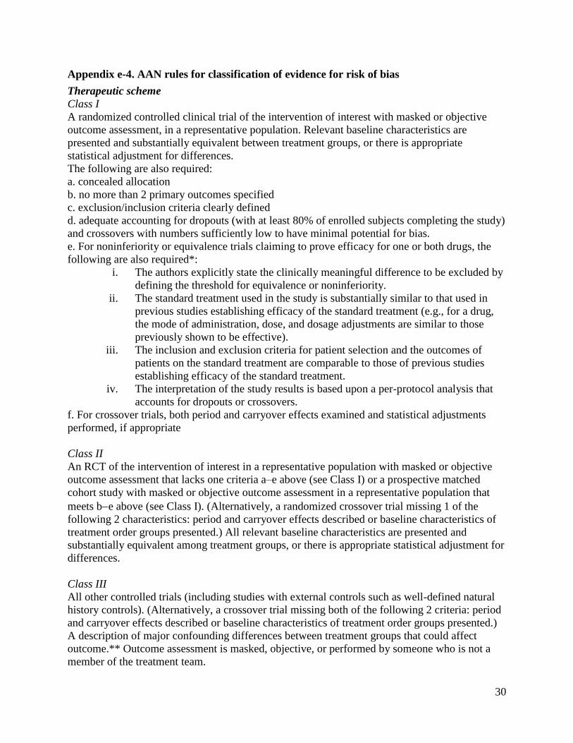
Appendix e-4. AAN rules for classification of evidence for risk of bias
Therapeutic scheme
Class I
A randomized controlled clinical trial of the intervention of interest with masked or objective
outcome assessment, in a representative population. Relevant baseline characteristics are
presented and substantially equivalent between treatment groups, or there is appropriate
statistical adjustment for differences.
The following are also required:
a. concealed allocation
b. no more than 2 primary outcomes specified
c. exclusion/inclusion criteria clearly defined
d. adequate accounting for dropouts (with at least 80% of enrolled subjects completing the study)
and crossovers with numbers sufficiently low to have minimal potential for bias.
e. For noninferiority or equivalence trials claiming to prove efficacy for one or both drugs, the
following are also required*:
i. The authors explicitly state the clinically meaningful difference to be excluded by
defining the threshold for equivalence or noninferiority.
ii. The standard treatment used in the study is substantially similar to that used in
previous studies establishing efficacy of the standard treatment (e.g., for a drug,
the mode of administration, dose, and dosage adjustments are similar to those
previously shown to be effective).
iii. The inclusion and exclusion criteria for patient selection and the outcomes of
patients on the standard treatment are comparable to those of previous studies
establishing efficacy of the standard treatment.
iv. The interpretation of the study results is based upon a per-protocol analysis that
accounts for dropouts or crossovers.
f. For crossover trials, both period and carryover effects examined and statistical adjustments
performed, if appropriate
Class II
An RCT of the intervention of interest in a representative population with masked or objective
outcome assessment that lacks one criteria a–e above (see Class I) or a prospective matched
cohort study with masked or objective outcome assessment in a representative population that
meets be above (see Class I). (Alternatively, a randomized crossover trial missing 1 of the
following 2 characteristics: period and carryover effects described or baseline characteristics of
treatment order groups presented.) All relevant baseline characteristics are presented and
substantially equivalent among treatment groups, or there is appropriate statistical adjustment for
differences.
Class III
All other controlled trials (including studies with external controls such as well-defined natural
history controls). (Alternatively, a crossover trial missing both of the following 2 criteria: period
and carryover effects described or baseline characteristics of treatment order groups presented.)
A description of major confounding differences between treatment groups that could affect
outcome.** Outcome assessment is masked, objective, or performed by someone who is not a
member of the treatment team.
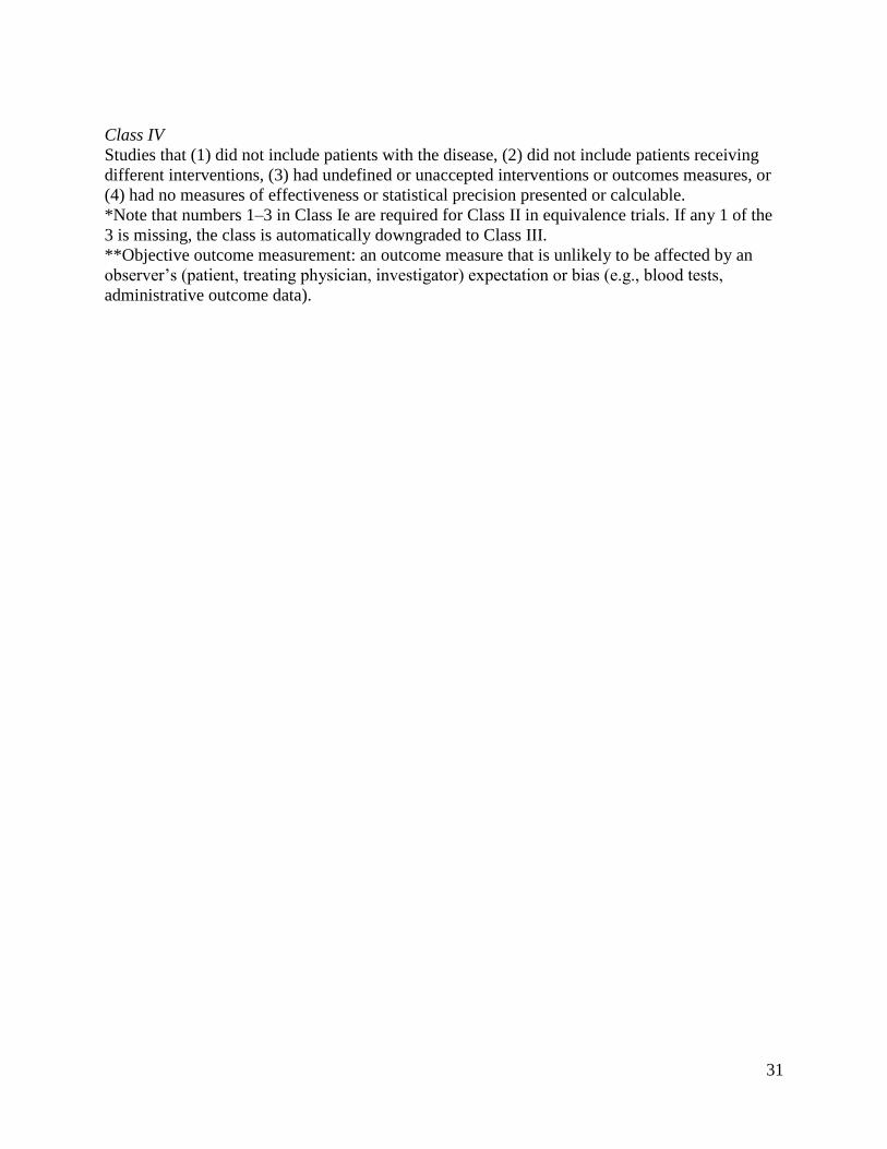
31
Class IV
Studies that (1) did not include patients with the disease, (2) did not include patients receiving
different interventions, (3) had undefined or unaccepted interventions or outcomes measures, or
(4) had no measures of effectiveness or statistical precision presented or calculable.
*Note that numbers 1–3 in Class Ie are required for Class II in equivalence trials. If any 1 of the
3 is missing, the class is automatically downgraded to Class III.
**Objective outcome measurement: an outcome measure that is unlikely to be affected by an
observer’s (patient, treating physician, investigator) expectation or bias (e.g., blood tests,
administrative outcome data).
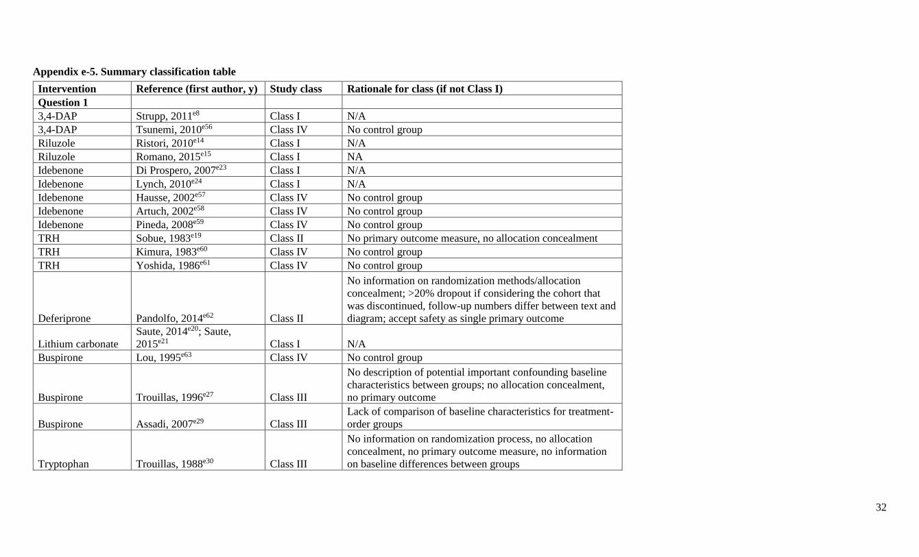
32
Appendix e-5. Summary classification table
Intervention Reference (first author, y) Study class Rationale for class (if not Class I)
Question 1
3,4-DAP Strupp, 2011e8 Class I N/A
3,4-DAP Tsunemi, 2010e56 Class IV No control group
Riluzole Ristori, 2010e14 Class I N/A
Riluzole Romano, 2015e15 Class I NA
Idebenone Di Prospero, 2007e23 Class I N/A
Idebenone Lynch, 2010e24 Class I N/A
Idebenone Hausse, 2002e57 Class IV No control group
Idebenone Artuch, 2002e58 Class IV No control group
Idebenone Pineda, 2008e59 Class IV No control group
TRH Sobue, 1983e19 Class II No primary outcome measure, no allocation concealment
TRH Kimura, 1983e60 Class IV No control group
TRH Yoshida, 1986e61 Class IV No control group
Deferiprone Pandolfo, 2014e62 Class II
No information on randomization methods/allocation
concealment; >20% dropout if considering the cohort that
was discontinued, follow-up numbers differ between text and
diagram; accept safety as single primary outcome
Lithium carbonate
Saute, 2014e20; Saute,
2015e21 Class I N/A
Buspirone Lou, 1995e63 Class IV No control group
Buspirone Trouillas, 1996e27 Class III
No description of potential important confounding baseline
characteristics between groups; no allocation concealment,
no primary outcome
Buspirone Assadi, 2007e29 Class III
Lack of comparison of baseline characteristics for treatment-
order groups
Tryptophan Trouillas, 1988e30 Class III
No information on randomization process, no allocation
concealment, no primary outcome measure, no information
on baseline differences between groups
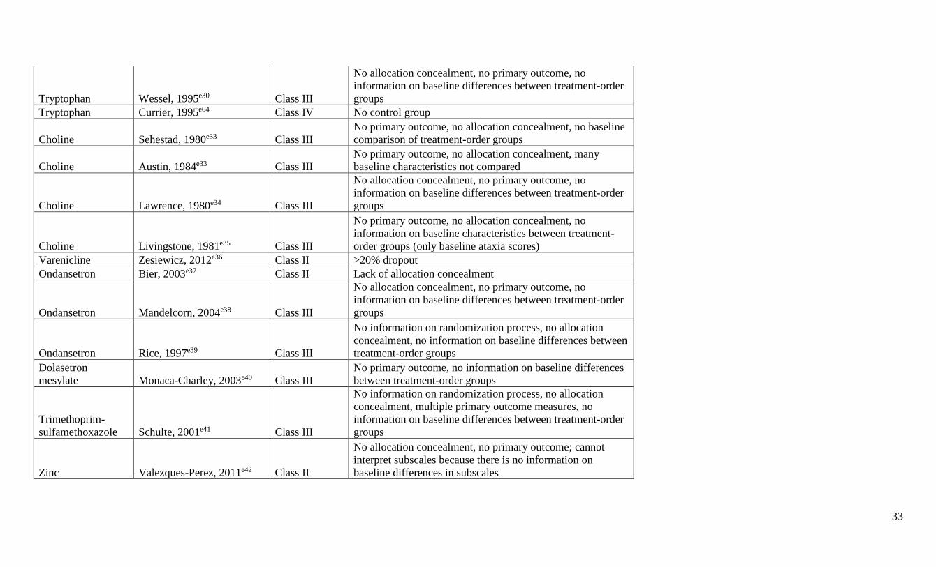
33
Tryptophan Wessel, 1995e30 Class III
No allocation concealment, no primary outcome, no
information on baseline differences between treatment-order
groups
Tryptophan Currier, 1995e64 Class IV No control group
Choline Sehestad, 1980e33 Class III
No primary outcome, no allocation concealment, no baseline
comparison of treatment-order groups
Choline Austin, 1984e33 Class III
No primary outcome, no allocation concealment, many
baseline characteristics not compared
Choline Lawrence, 1980e34 Class III
No allocation concealment, no primary outcome, no
information on baseline differences between treatment-order
groups
Choline Livingstone, 1981e35 Class III
No primary outcome, no allocation concealment, no
information on baseline characteristics between treatment-
order groups (only baseline ataxia scores)
Varenicline Zesiewicz, 2012e36 Class II >20% dropout
Ondansetron Bier, 2003e37 Class II Lack of allocation concealment
Ondansetron Mandelcorn, 2004e38 Class III
No allocation concealment, no primary outcome, no
information on baseline differences between treatment-order
groups
Ondansetron Rice, 1997e39 Class III
No information on randomization process, no allocation
concealment, no information on baseline differences between
treatment-order groups
Dolasetron
mesylate Monaca-Charley, 2003e40 Class III
No primary outcome, no information on baseline differences
between treatment-order groups
Trimethoprim-
sulfamethoxazole Schulte, 2001e41 Class III
No information on randomization process, no allocation
concealment, multiple primary outcome measures, no
information on baseline differences between treatment-order
groups
Zinc Valezques-Perez, 2011e42 Class II
No allocation concealment, no primary outcome; cannot
interpret subscales because there is no information on
baseline differences in subscales
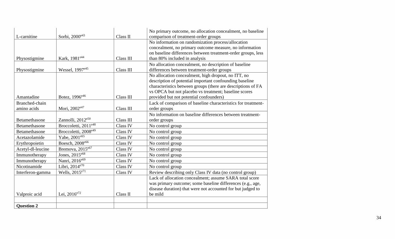
34
L-carnitine Sorbi, 2000e43 Class II
No primary outcome, no allocation concealment, no baseline
comparison of treatment-order groups
Physostigmine Kark, 1981e44 Class III
No information on randomization process/allocation
concealment, no primary outcome measure, no information
on baseline differences between treatment-order groups, less
than 80% included in analysis
Physostigmine Wessel, 1997e45 Class III
No allocation concealment, no description of baseline
differences between treatment-order groups
Amantadine Botez, 1996e46 Class III
No allocation concealment, high dropout, no ITT, no
description of potential important confounding baseline
characteristics between groups (there are descriptions of FA
vs OPCA but not placebo vs treatment; baseline scores
provided but not potential confounders)
Branched-chain
amino acids Mori, 2002e47 Class III
Lack of comparison of baseline characteristics for treatment-
order groups
Betamethasone Zannolli, 2012e50 Class III
No information on baseline differences between treatment-
order groups
Betamethasone Broccoletti, 2011e48 Class IV No control group
Betamethasone Broccoletti, 2008e49 Class IV No control group
Acetazolamide Yabe, 2001e65 Class IV No control group
Erythropoietin Boesch, 2008e66 Class IV No control group
Acetyl-dl-leucine Bremova, 2015e67 Class IV No control group
Immunotherapy Jones, 2015e68 Class IV No control group
Immunotherapy Nanri, 2016e69 Class IV No control group
Nicotinamide Libri, 2014e70 Class IV No control group
Interferon-gamma Wells, 2015e71 Class IV Review describing only Class IV data (no control group)
Valproic acid Lei, 2016e72 Class II
Lack of allocation concealment; assume SARA total score
was primary outcome; some baseline differences (e.g., age,
disease duration) that were not accounted for but judged to
be mild
Question 2
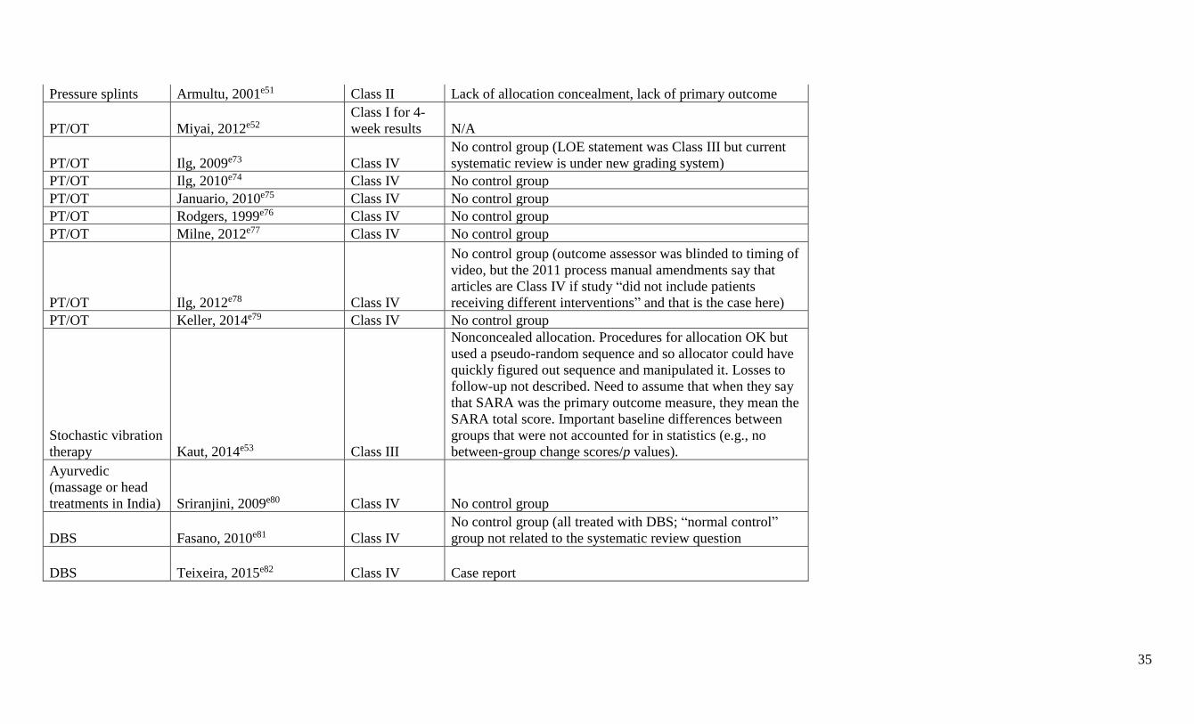
35
Pressure splints Armultu, 2001e51 Class II Lack of allocation concealment, lack of primary outcome
PT/OT Miyai, 2012e52
Class I for 4-
week results N/A
PT/OT Ilg, 2009e73 Class IV
No control group (LOE statement was Class III but current
systematic review is under new grading system)
PT/OT Ilg, 2010e74 Class IV No control group
PT/OT Januario, 2010e75 Class IV No control group
PT/OT Rodgers, 1999e76 Class IV No control group
PT/OT Milne, 2012e77 Class IV No control group
PT/OT Ilg, 2012e78 Class IV
No control group (outcome assessor was blinded to timing of
video, but the 2011 process manual amendments say that
articles are Class IV if study “did not include patients
receiving different interventions” and that is the case here)
PT/OT Keller, 2014e79 Class IV No control group
Stochastic vibration
therapy Kaut, 2014e53 Class III
Nonconcealed allocation. Procedures for allocation OK but
used a pseudo-random sequence and so allocator could have
quickly figured out sequence and manipulated it. Losses to
follow-up not described. Need to assume that when they say
that SARA was the primary outcome measure, they mean the
SARA total score. Important baseline differences between
groups that were not accounted for in statistics (e.g., no
between-group change scores/p values).
Ayurvedic
(massage or head
treatments in India) Sriranjini, 2009e80 Class IV No control group
DBS Fasano, 2010e81 Class IV
No control group (all treated with DBS; “normal control”
group not related to the systematic review question
DBS Teixeira, 2015e82 Class IV Case report
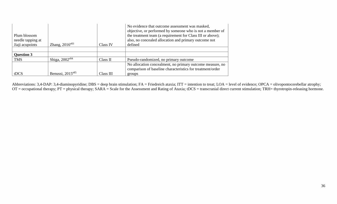
36
Plum blossom
needle tapping at
Jiaji acupoints Zhang, 2016e83 Class IV
No evidence that outcome assessment was masked,
objective, or performed by someone who is not a member of
the treatment team (a requirement for Class III or above);
also, no concealed allocation and primary outcome not
defined
Question 3
TMS Shiga, 2002e84 Class II Pseudo-randomized, no primary outcome
tDCS Benussi, 2015e85 Class III
No allocation concealment, no primary outcome measure, no
comparison of baseline characteristics for treatment/order
groups
Abbreviations: 3,4-DAP: 3,4-diaminopyridine; DBS = deep brain stimulation; FA = Friedreich ataxia; ITT = intention to treat; LOA = level of evidence; OPCA = olivopontocerebellar atrophy;
OT = occupational therapy; PT = physical therapy; SARA = Scale for the Assessment and Rating of Ataxia; tDCS = transcranial direct current stimulation; TRH= thyrotropin-releasing hormone.

37
Appendix e-6. Study review process diagram
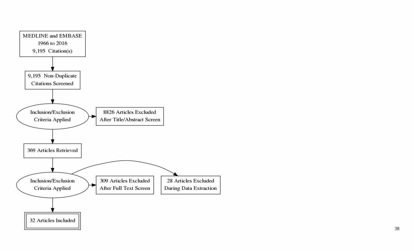
38

39
40
E-REFERENCES
e1. American Academy of Neurology. Clinical Practice Guidelines Process Manual, 2004 ed. St. Paul, MN: The American Academy of Neurology, 2004.
https://www.aan.com/uploadedFiles/Website_Library_Assets/Documents/2.Clinical_Guidelines/4.About_Guidelines/1.How_Guidelines_Are_Developed/2004%20AAN%20Process%20Manual.
pdf.
e2. American Academy of Neurology. Clinical Practice Guideline Process Manual. St. Paul, MN: The American Academy of Neurology, 2011.
https://www.aan.com/Guidelines/Home/Development.
e3. Dejerine JJ, Thomas, A. L’atrophie olivo-ponto-cérébelleuse. Nouvelle iconographie de la Salpêtrière 1900;13:21.
e4. Berciano J, Boesch, S., Pérez-Ramos, J.M., Wenning, G.K. Olivopontocerebellar atrophy: Toward a better nosological definition. Movement disorders: official journal of the Movement
Disorder Society 2006;21:7.
e5. Ophoff RA, Terwindt GM, Vergouwe MN, et al. Familial hemiplegic migraine and episodic ataxia type-2 are caused by mutations in the Ca2 channel gene CACNL1A4. Cell 1996;87:543–
552.
e6. Gao Z, Todorov B, Barrett CF, et al. Cerebellar ataxia by enhanced Ca(V)2.1 currents is alleviated by Ca2+-dependent K+-channel activators in Cacna1a(S218L) mutant mice. J Neurosci
2012; 32: 15533–15546.
e7. Strupp M Zwergal A, Brandt T. Episodic ataxia type 2. Neurotherapeutics 2007;4:267–273.
e8. Strupp M, Kalla R, Claassen J, et al. A randomized trial of 4-aminopyridine in EA2 and related familial episodic ataxias. Neurology 2011;77:268–275.
e9. Cifra A, Mazzone GL, Nistri A. Rilzole: what it does to spinal and brainstem neurons and how it does it. Neuroscientist 2012;19:137–144.
e10. Cao YJ, Dreixler JC, Couey JJ, Houamed KM. Modulation of recombinant and native neuronal SK channels by the neuroprotective drug riluzole. Eur J Pharmacol 2002;449:47–54.
e11. Shakkottai VG, Chou CH, Oddo S, et al. Enhanced neuronal excitability in the absence of neurodegeneration induces cerebellar ataxia. J Clin Invest 2004;113:582–590.
e12. Shakkottai VG, do Carmo Costa M, Dell’Orco JM, Sankaranarayanan A, Wulff H, Paulson HL. Early changes in cerebellar physiology accompany motor dysfunction in the polyglutamine
disease spinocerebellar ataxia type 3. J Neurosci 2011;31:13002–13014.
e13. Gao Z, Todorov B, Barrett CF, et al. Cerebellar ataxia by enhanced Ca(V)2.1 currents is alleviated by Ca2+-dependent K+-channel activators in Cacna1a(S218L) mutant mice. J Neurosci
2012;32:15533–15546.
e14. Ristori G, Romano, S., et al. Riluzole in cerebellar ataxia A randomized, double-blind, placebo-controlled pilot trial. Neurology 2010;74:7.
e15. Romano S, Coarelli G, Marcotulli C, et al. Riluzole in patients with hereditary cerebellar ataxia: a randomised, double-blind, placebo-controlled trial. The Lancet Neurology 2015;14:985-
991.
e16. Lei LF, Yang GP, Wang JL, et al. Safety and efficacy of valproic acid treatment in SCA3/MJD patients. Parkinsonism Relat Disord 2016;26:55-61.
e17. Silver M, Factor SA. Valproic acid-induced parkinsonism: levodopa responsiveness with dyskinesia. Parkinsonism Relat Disord 2013;19:758–760. Epub 2013 Apr 28.
e18. Brugger F, Bhatia KP, Besag FM. Valproate-associated parkinsonism: a critical review of the literature. CNS Drugs 2016;30:527–540.
e19. Sobue I, Takayanagi, T., et al. Controlled Trial of Thyrotropin Releasing Hormone Tartrate in Ataxia of Spinocerebellar Degenerations. Journal of the neurological sciences 1983;61:14.
e20. Saute JA, de Castilhos RM, Monte TL, et al. A randomized, phase 2 clinical trial of lithium carbonate in Machado-Joseph disease. Movement disorders: official journal of the Movement
Disorder Society 2014;29:568-573.
41
e21. Saute JA, Rieder CR, Castilhos RM, et al. Planning future clinical trials in Machado Joseph disease: Lessons from a phase 2 trial. J Neurol Sci 2015;358:72-76.
e22. Pandolfo M, Arpa J, Delatycki MB, et al. Deferiprone in Friedreich ataxia: a 6-month randomized controlled trial. Ann Neurol 2014;76:509-521.
e23. Di Prospero NA, Baker, A., et al. Neurological effects of high-dose idebenone in patients with Friedreich's ataxia: a randomised, placebo-controlled trial. The Lancet Neurology 2007;6:9.
e24. Lynch D, SL Perlman, and T Meier. A phase 3, double-blind, placebo-controlled trial of idebenone in friedreich ataxia. Arch Neurol 2010;67:7.
e25. A Phase III Double-blind, Randomised, Placebo-controlled Study of the Efficacy, Safety and Tolerability of Idebenone in the Treatment of Friedreich's Ataxia Patients [online]. Available at:
https://clinicaltrials.gov/ct2/show/NCT00905268?term=idebenone+ataxia&rank=2. Accessed December 22, 2016.
e26. Santhera’s MICONOS Trial with Catena in Friedreich’s Ataxia Misses Primary Endpoint. Ataxia Canada. http://lacaf.org/en/santhera-trial-with-catena-in-friedreich-ataxia-misses-primary-
endpoint.
e27. Trouillas P. Treatment of cerebellar ataxia with buspirone: a double-blind study. Lancet 1996;348:1.
e28. Trouillas P, Xie J, Adeleine P, et al. Buspirone, a serotonergic 5-HT1A agonist, is active in cerebellar ataxia. A new fact in favor of the serotonergic theory of ataxia. Prog Brain Res
1997;114.
e29. Assadi M, Campellone, J., et al. Treatment of spinocerebellar ataxia with buspirone. Journal of the neurological sciences 2007;260:4.
e30. Trouillas P, Brudon F, Adeleine P. Improvement of cerebellar ataxia with levorotatory form of 5-hydroxytryptophan. Arch Neurol 1988;45:1217-1222.
e31. Wessel K, Hermsdorfer, J., et al. Double-blind crossover study with levorotatory form of hydroxytryptophan in patients with degenerative cerebellar diseases. Arch Neurol 1995;52:5.
e32. Sehested P. Oral Choline in Cerebellar Ataxia. Acta Neurol Scandinavia 1980;62:3.
e33. Austin CA. Low dose choline chloride in cerebellar degeneration. Journal of Neurology, Neurosurgery, and Psychiatry 1984;47:3.
e34. Lawrence CM, Millac, P., et al. The use of choline chloride in ataxic disorders. Journal of Neurology, Neurosurgery, and Psychiatry 1980;43:3.
e35. Livingstone IR, Mastaglia FL, Pennington RJT, Skilbeck C. Choline chloride in the treatment of cerebellar and spinocerebellar ataxia. J Neurol Sci 1981;50:161-174.
e36. Zesiewicz TA, Greenstein, P.E., et al. A randomized trial of varenicline (Chantix) for the treatment of spinocerebellar ataxia type 3. Neurology 2012;78:6.
e37. Bier JC, Dethy, S., et al. Effects of the oral form of ondansetron on cerebellar dysfunction. A multi-center double-blind study. Journal of Neurology 2003;250:5.
e38. Mandelcorn J, N Cullen, et al. A preliminary study of the efficacy of ondansetron in the treatment of ataxia, poor balance and incoordination from brain injury. Brain Injury 2004;18:15.
e39. Rice GPA, Lesaux, J, et al. Ondansetron, a 5-HT3 antagonist, improves cerebellar tremor. Journal of Neurology, Neurosurgery, and Psychiatry 1997;62:3.
e40. Monaca-Charley C, Stojkovic, T., et al. Double-blind crossover study with dolasetron mesilate, a 5-HT3 receptor antagonist in cerebellar syndrome secondary to multiple sclerosis. J Neurol
2003;250:5.
e41. Schulte T, Mattern, R., et al. Double-blind crossover trial of trimethoprim-sulfamethoxazole in spinocerebellar ataxia type 3/machado-joseph disease. Arch Neurol 2001;58:7.
e42. Velazquez-Perez L, Rodriguez-Chanfrau, J., et al. Oral Zinc Sulphate Supplementation for Six Months in SCA2 Patients: A Randomized, Double-Blind, Placebo-Controlled Trial.
Neurochem Res 2011;36:8.
e43. Sorbi S, Forleo, P., et al. Double-blind, crossover, placebo-controlled clinical trial with L-acetylcarnitine in patients with degenerative cerebellar ataxia. Clinical Neuropharmacology
2000;23:5.
e44. Kark RA. Double-blind, triple-crossover trial of low doses of oral physostigmine in inherited ataxias. Neurology 1981;31:5.
e45. Wessel K. Double-blind crossover study with Physostigmine in patients with Degenerative Cerebellar Diseases. Arch Neurol 1997;54:4.
e46. Botez MI, Botez-Marquard, T., et al. Amantadine hydrochloride treatment in heredodegenerative ataxias: a double blind study. Journal of Neurology, Neurosurgery, and Psychiatry
1996;61:15.
42
e47. Mori M, Yoshiki, Adachi, et al. Double-blind crossover study of branched-chain amino acid therapy in patients with spinocerebellar degeneration. Journal of the neurological sciences
2002;195:4.
e48. Broccoletti T, Del Giudice, T., et al. Efficacy of very-low-dose betamethasone on neurological symptoms in ataxia-telangiectasia. European Journal of Neurology 2011;18:7.
e49. Broccoletti T. Steroid-induced improvement of neurological signs in ataxia-telangiectasia patients. European Journal of Neurology 2008;15:6.
e50. Zannolli R, Buoni S, Betti G, et al. A randomized trial of oral betamethasone to reduce ataxia symptoms in ataxia telangiectasia. Movement disorders : official journal of the Movement
Disorder Society 2012;27:1312-1316.
e51. Armultu K, Karabudak R, Niurlu G. Physiotherapy approaches in the treatment of ataxic multiple sclerosis: A pilot study. Neurorehabilitation and Neural Repair 2001;15:9.
e52. Miyai I, Ito, M., et al. Cerebellar Ataxia Rehabilitation Trial in Degenerative Cerebellar Diseases. Neurorehabilitation and Neural Repair 265 (2012): 515-22 2012;26:8.
e53. Kaut O, Jacobi H, Coch C, et al. A randomized pilot study of stochastic vibration therapy in spinocerebellar ataxia. Cerebellum (London, England) 2014;13:237-242.
e54. Shiga Y. Transcranial magnetic stimulation alleviates truncal ataxia in spinocerebellar degeneration. Journal of Neurology, Neurosurgery, and Psychiatry 2002;72:3.
e55. Benussi A, Koch G, Cotelli M, Padovani A, Borroni B. Cerebellar transcranial direct current stimulation in patients with ataxia: A double-blind, randomized, sham-controlled study.
Movement disorders: official journal of the Movement Disorder Society 2015;30:1701-1705.
e56. Tsunemi T, Ishikawa K, Tsukui K, Sumi T, Kitamura K, Mizusawa H. The effect of 3,4-diaminopyridine on the patients with hereditary pure cerebellar ataxia. J Neurol Sci 2010;292:81-84.
Epub 2010 Feb 23.
e57. Hausse AO, Aggoun Y, Bonnet D, et al. Idebenone and reduced cardiac hypertrophy in friedreich's ataxia. Heart 2002;87:346-350.
e58. Artuch R, Aracil A, Mas A, et al. Friedreich’s ataxia: idebenone treatment in early stage patients. Neuropediatrics 2002;33:190-193.
e59. Pineda M, Arpa J. Idebenone treatment in paediatric and adult patients with Friedreich ataxia: long-term follow-up. Eur J Paediatr Neurol 2008;12:470-475.
e60. Kimura I. Motor disabilities and effects of thyrotropin-releasing hormone spinocerebellar degeneration. Thoku J Exp Med 1983;139;331-137.
e61. Yoshida M, Nakanishi T. Acute and chronic effects of thyrotropin-releasing hormone (TRH) on stabilogram in spinocerebellar degenerations. Thoku J Exp Med 1986;148:327-331.
e62. Pandolfo M, Arpa J, Delatycki MB, et al. Deferiprone in Friedreich ataxia: a 6-month randomized controlled trial. Ann Neurol 2014;76:509-521.
e63. Lou JS, Goldfarb L. Use of Buspirone for Treatment of Cerebellar Ataxia. Arch Neurol 1995;52:982-988.
e64. Currier RD. A treatment for ataxia. Arch Neurol 1995;52:449.
e65. Yabe I, Sasaki H, Yamashita I, Takei A, Tashiro K. Clinical trial of acetazolamide in SCA6, with assessment using the Ataxia Rating Scale and body stabilometry. Acta Neurol Scand
2001;104:44-47.
e66. Boesch S, Sturm B, Hering S, et al. Neurological effects of recombinant human erythropoietin in Friedreich's ataxia: a clinical pilot trial. Mov Disord 2008;23:1940-1944.
e67. Bremova T, Malinova V, Amraoui Y, et al. Acetyl-dl-leucine in Niemann-Pick type C: A case series. Neurology 2015;85:1368-1375.
e68. Jones AL, Flanagan EP, Pittock SJ, et al. Responses to and outcomes of treatment of autoimmune cerebellar ataxia in adults. JAMA Neurol 2015;72:1304-1312.
e69. Nanri K, Okuma M, Sato S, et al. Prevalence of Autoantibodies and the Efficacy of Immunotherapy for Autoimmune Cerebellar Ataxia. Intern Med 2016;55:449-454.
e70. Libri V, Yandim C, Athanasopoulos S, et al. Epigenetic and neurological effects and safety of high-dose nicotinamide in patients with Friedreich's ataxia: an exploratory, open-label, dose-
escalation study. Lancet 2014;384:504-513.
e71. Wells M, Seyer L, Schadt K, Lynch DR. IFN-gamma for Friedreich ataxia: present evidence. Neurodegener Dis Manag 2015;2015;5:497-504.
e72. Lei L, Yang G, Wang J, et al. Safety and efficacy of valproic acid treatment in SCA3/MJD patients. Parkinsonism Relat Disord 2016;26:55-61.
43
e73. Ilg W, Synofzik M, Brötz D, Burkard S, Giese MA, Schöls L. Intensive coordinative training improves motor performance in degenerative cerebellar disease. Neurology 2009;73:1923-
1930.
e74. Ilg W, Brötz D, Burkard S, Giese MA, Schölz L, Synofzik M. Long-term effects of coordinative training in degenerative cerebellar disease. Mov Disord 2010;25:2239-2246.
e75. Januário F, Campos I, Amaral C. Rehabilitation of postural stability in ataxic/hemiplegic patients after stroke. Disabil Rehabil 2010;32:1775-1779.
e76. Rodgers MM, Mulcare JA, King DL, Mathews T, Gupta SC, Glaser RM. Gait characteristics of individuals with multiple sclerosis before and after a 6-month aerobic training program. J
Rehabil Res Dev 1999;36:183-188.
e77. Milne SC, Campagna EJ, Corben LA, et al. Retrospective study of the effects of inpatient rehabilitation on improving and maintaining functional independence in people with Friedreich
ataxia. Arch Phys Med Rehabil 2012;93:1860-1863.
e78. Ilg W, Schatton C, Schicks J, Giese MA, Schöls L, Matthis S. Video game–based coordinative training improves ataxia in children with degenerative ataxia. Neurology 2012;79:2056-2060.
e79. Keller JL, Bastian AJ. A Home Balance Exercise Program Improves Walking in People with Cerebellar Ataxia. Neurorehabil Neural Repair 2014;28:770-778.
e80. Sriranjini SJ, Pal PK, Devidas KV, Ganpathy S. Improvement of balance in progressive
degenerative cerebellar ataxias after Ayurvedic therapy: A preliminary report. Neurology India 2009;57:166-171.
e81. Fasano A, Herzog, J, Raethjen J, et al. Gait ataxia in essential tremor is differentially
modulated by thalamic stimulation. Brain 2010;133:3635-3648.
e82. Teixeira MJ, Cury RG, Galhardoni R, et al. Deep brain stimulation of the dentate nucleus improves cerebellar ataxia after cerebellar stroke. Neurology 2015;85:2075-2076.
e83. Zhang L, Wang F, Shi Y, Wang J, Xie Q. Plum-blossom needle tapping at Jiaji(EX-B 2) combined with rehabilitation for ataxia of cerebellar apoplexy: a randomized controlled trial.
Zhongguo Zhen Jiu 2016;36:131-134.
e84. Shiga Y Tsuda T, Itoyama Y, et al. Transcranial magnetic stimulation alleviates truncal ataxia in spinocerebellar degeneration. J Neurol Neurosurg Psychiatry 2002;72:124-126.
e85. Benussi A, Koch G, Cotelli M, Padovani A, Borroni B. Cerebellar transcranial direct
current stimulation in patients with ataxia: a double-blind, randomized, sham-controlled study. Mov Disord 2015;30:1701-1705.