Embed Size (px)
Citation preview

Conductive Keratoplasty for the Correctionof Low to Moderate Hyperopia: U.S.Clinical Trial 1-Year Results on 355 Eyes
Marguerite B. McDonald, MD,1 Peter S. Hersh, MD,2 Edward E. Manche, MD,3 Robert K. Maloney, MD,4
Jonathan Davidorf, MD,5 Moataz Sabry, MD,1 and the Conductive Keratoplasty United States Investigators Group
Objective: To document the 1-year safety, efficacy, and stability results of 355 eyes treated in the multi-center study of conductive keratoplasty (CK) used to correct low to moderate hyperopia.
Design: Nonrandomized comparative (self-controlled) trial.Participants: Twenty surgeons at 13 centers performed CK on the eyes of all patients enrolled in a
multicenter, 2-year, U.S. phase III clinical trial. Treated eyes had �0.75 to �3.00 diopters (D) of hyperopia and�0.75 D of cylinder. Patients were 40 years of age or older.
Intervention: Low-energy, high-frequency current was applied directly into the peripheral corneal stromathrough a delivery tip inserted at 8 to 32 treatment spots. The number of treatment spots was increased forincreasing levels of hyperopia, but the amount of radiofrequency energy remained constant. Emmetropia wasintended. All eyes were treated once (there were no retreatments).
Main Outcome Measures: Data from 355 eyes with 1 year of follow-up were analyzed for safety andstability, and data from 318 eyes were analyzed for efficacy and predictability, as well as stability and safety. Allpatients reported on satisfaction and quality of vision after surgery.
Results: At 1 year, uncorrected visual acuity was �20/20 in 56%, �20/25 in 75%, and �20/40 in 92% ofeyes. The manifest refractive spherical equivalent refraction was within 0.50 D in 63%, within �1.00 D in 89%,and within �2.00 D in 99%. Seven of 355 eyes lost 2 lines of best spectacle-corrected visual acuity at 1 year,but no eye lost �2 lines. One eye of 355 had induced cylinder of �2.00 D. The cycloplegic refractive sphericalequivalent changed a mean of 0.25 � 0.50 D between months 3 and 6, 0.11 � 0.41 D between months 6 and9, and 0.11 � 0.35 D between months 9 and 12. Refractive stability seemed to be attained by 6 months andremained stable through 12 months. Histology and confocal microscopy showed deep penetration of thetreatment into the stroma. Endothelial cell counts were not changed by the treatment.
Conclusions: CK seems to be safe, effective, and stable for correcting low to moderate spherical hyperopiain patients 40 years old or older. Treatment penetration is deep and cylindrical in shape, and it does not damagethe corneal endothelium. Uncorrected visual acuity, predictability, and stability are as good as or better thanthose obtained with other techniques used to correct hyperopia. Ophthalmology 2002;109:1978–1989 © 2002by the American Academy of Ophthalmology, Inc.
Development of thermal techniques to shrink peripheralcorneal collagen and thereby steepen the central cornea haschallenged ophthalmologists for longer than 100 years. Hot-wire thermokeratoplasty, used in the 1980s to produce ther-mal burns that penetrated to 95% of corneal depth in hy-peropic eyes, showed a lack of predictability and stability,and further development was abandoned.1–4 More current
Originally received: November 11, 2001.Accepted: April 11, 2002. Manuscript no. AA 2105831 Southern Vision Institute, New Orleans, Louisiana.2 Cornea and Laser Vision Center, Teaneck, New Jersey.3 Stanford University School of Medicine, Palo Alto, California.4 Maloney Vision Institute, Los Angeles, California.5 Davidorf Eye Group, West Hills, California.
The members of the Conductive Keratoplasty United States InvestigatorsGroup are listed in the appendix.
Presented in part at the Annual Meeting of the American Academy ofOphthalmology, New Orleans, Louisiana, November, 2001.
Drs. Hersh, Manche, Maloney, and Davidorf and the Conductive Kerato-plasty Investigators Group participated as clinical investigators in this Foodand Drug Administration phase III study sponsored by Refractec, Inc.(Irvine, CA). These authors participate on Refractec’s Medical AdvisoryBoard and are paid for their time. They otherwise have no proprietary orfinancial interest in the ViewPoint conductive keratoplasty system from
Refractec, Inc. Dr. Moataz Sabry was not an investigator but provided theconfocal microscopy photographs. Dr. Marguerite McDonald is a paidmedical monitor for the phase III study on conductive keratoplasty byRefractec, Inc.
The nature of the procedure was explained to all participating patients, andthey all signed informed consent forms before undergoing the procedure.
Reprint requests to Marguerite McDonald, MD, Southern Vision Institute,2820 Napoleon Avenue, New Orleans, LA 70115. E-mail: [email protected].
1978 © 2002 by the American Academy of Ophthalmology, Inc. ISSN 0161-6420/02/$–see front matterPublished by Elsevier Science Inc. PII S0161-6420(02)01255-1
techniques using thermal keratoplasty include noncontactholmium:yttrium–aluminum–garnet laser thermal kerato-plasty (Ho:YAG)(LTK; Sunrise Technologies, Fremont,CA),5–9 contact holmium:yttrium–aluminum–garnet LTK(Holmium 25; Technomed, Baesweiler, Germany),9–12 con-tinuous-wave diode LTK (DTK; Rodenstock, ProLaserMedical Systems, Inc., Dusseldorf, Germany),13,14 and con-ductive keratoplasty (CK; ViewPoint; Refractec, Inc., Ir-vine, CA).15 Nonthermal, excimer laser–based techniquesfor correcting hyperopia include photorefractive keratecto-my16–21 and laser in situ keratomileusis.22–28
CK is a laserless radiofrequency-based technique for thecorrection of low to moderate hyperopia. The treatmentproduces a homogenous temperature increase that shrinkscollagen in the treated area and forms a cylindrical footprintdeep in the stroma. After a full circle of treatment spots, theperipheral cornea flattens and the central corneal steepens.This article presents the 1-year, single-treatment results of355 available eyes of the 401 enrolled in a U.S. phase IIIFood and Drug Administration (FDA) clinical trial andcompares them with results obtained with other nonexcimerlaser techniques for the correction of hyperopia.
Materials and Methods
Study Design
This is a report of the 12-month results from the cohort of con-secutive enrolled patients whose eyes were treated in a prospec-tive, consecutive-series, multicenter clinical study (FDA phase III)evaluating the safety, efficacy, and stability of the CK procedurefor the correction of 0.75 to 3.00 diopters (D) of hyperopia and�0.75 D of cylinder. The study follow-up will be 24 months.
One-year data are available from 318 eyes for the variables thatindicate treatment efficacy (uncorrected visual acuity [UCVA],best spectacle-corrected visual acuity [BSCVA], manifest refrac-tive spherical equivalent refraction [MRSE], and cycloplegic re-fractive spherical equivalent). For variables that indicate treatmentsafety and stability, data are available for 355 eyes. The discrep-ancy in number of eyes can be accounted for by the change innomogram after treatment of the first 54 eyes. The original nomo-gram specifying the number of treatment spots to correct a givenlevel of hyperopia was modified after the first 54 eyes were treatedbecause it had a tendency toward undercorrection (Table 1). Datafrom 24 of those 54 eyes could not be analyzed for efficacyvariables but could be analyzed for safety and stability. Theremaining 30 eyes, however, were found to have been treated withthe number of spots indicated by what later became the revised
(current) nomogram and were included in efficacy analyses. Forexample, an eye with 1.50 D of hyperopia would be treated with 16spots under both the original and current nomograms, and theefficacy results could be included in the current nomogram eyes.
Study Device
The ViewPoint CK system (Fig 1) used to perform the CK pro-cedure consists of a radiofrequency energy-generating console; ahandheld, reusable, pen-shaped handpiece attached by a removablecable and connector; a foot pedal that controls the release ofradiofrequency energy; and a speculum that provides a largesurface for an electrical return path. The energy level default is60% of 1 W, and the exposure time default is 0.6 seconds.Attached to the probe is a single-use, disposable, stainless-steelKeratoplast Refractec Inc., Irvine, California tip, 90 �m in diameterand 450 �m long, that delivers the current directly to the cornealstroma (Fig 2). The tip has a proximal bend of 45° and a distal bendof 90° to allow access to the cornea over the patient’s brow and nasalregions. At the very distal portion of the tip is an insulated stainless-steel stop (cuff) that ensures correct depth of penetration.
Table 1. Original and Current Nomograms
Number ofTreatment
Spots Original Nomogram Current Nomogram
8* �0.75 D to � � 1.00 D �0.75 D to �0.875 D16 �1.00 D to � � 2.00 D �1.0 D to �1.625 D24 �2.00 D to � � 3.00 D �1.75 D to �2.25 D32 �3.00 D to � � 4.00 D �2.375 D to �3.00 D
* None of the first 54 eyes were treated with 8 spots.
D � diopters.
Figure 1. The ViewPoint conductive keratoplasty system, including thehandpiece with the Keratoplast tip and a choice of two lid specula that actas the electrical return path. The console weighs 14 pounds.
Figure 2. Conductive keratoplasty handpiece with Keratoplast tip (90 �mwide, 450 �m long) and insulated stop at the distal end. Shown next to a7-0 suture.
McDonald et al � Keratoplasty for the Correction of Low to Moderate Hyperopia
1979
Patients
Institutional review board approval was obtained at each institu-tion. A total of 401 patients at 13 centers in the United States whomet eligibility requirements were enrolled consecutively into thestudy from February 10, 1999, to December 1, 2000, signedinformed consent forms, and had 8 to 32 treatment spots applied tothe cornea during the CK procedure. The intended correction wasemmetropia.
Enrolled patients had no existing ocular or chronic systemicdisease, previous ocular surgery, history of herpes infection, ste-roid-responsive increase in intraocular pressure, intractable kera-toconjunctivitis sicca, history of keloid formation, or unstable,progressive hyperopia. Hard contact lens wearers were to discon-tinue lens use 3 weeks before the final measurements and theprocedure, and soft contact lens wearers were to discontinue lensuse 2 weeks before the final measurements and the procedure.They also had to have clear, undistorted mires on the centralkeratometry examination. Eyes with pachymetry readings of �560
�m at the 6-mm optical zone (OZ) and those with a distanceUCVA of better than 20/32 were excluded from study participation.
Examination MethodsThe preoperative and postoperative examinations for all eyes in-cluded manifest and cycloplegic refractions; uncorrected visualacuity and BSCVA with use of Early Treatment Diabetic Retinop-athy Study visual acuity charts or Bailey–Lovie charts (distance)and Jaeger visual acuity charts (near); slit-lamp and funduscopicexamination; and computerized corneal topography. Cycloplegicrefraction was measured 30 minutes after two applications of oneor two drops of 1% cyclopentolate 5 minutes apart. Intraocularpressure was measured with the surgeon’s choice of standardapplanation instruments, including Goldmann, Perkins, or Draegertonometry.
The preoperative examinations were performed by the surgeonsor their assistants 1 to 60 days before the CK procedure. Postop-erative examinations were performed on days 1 and 7 and months
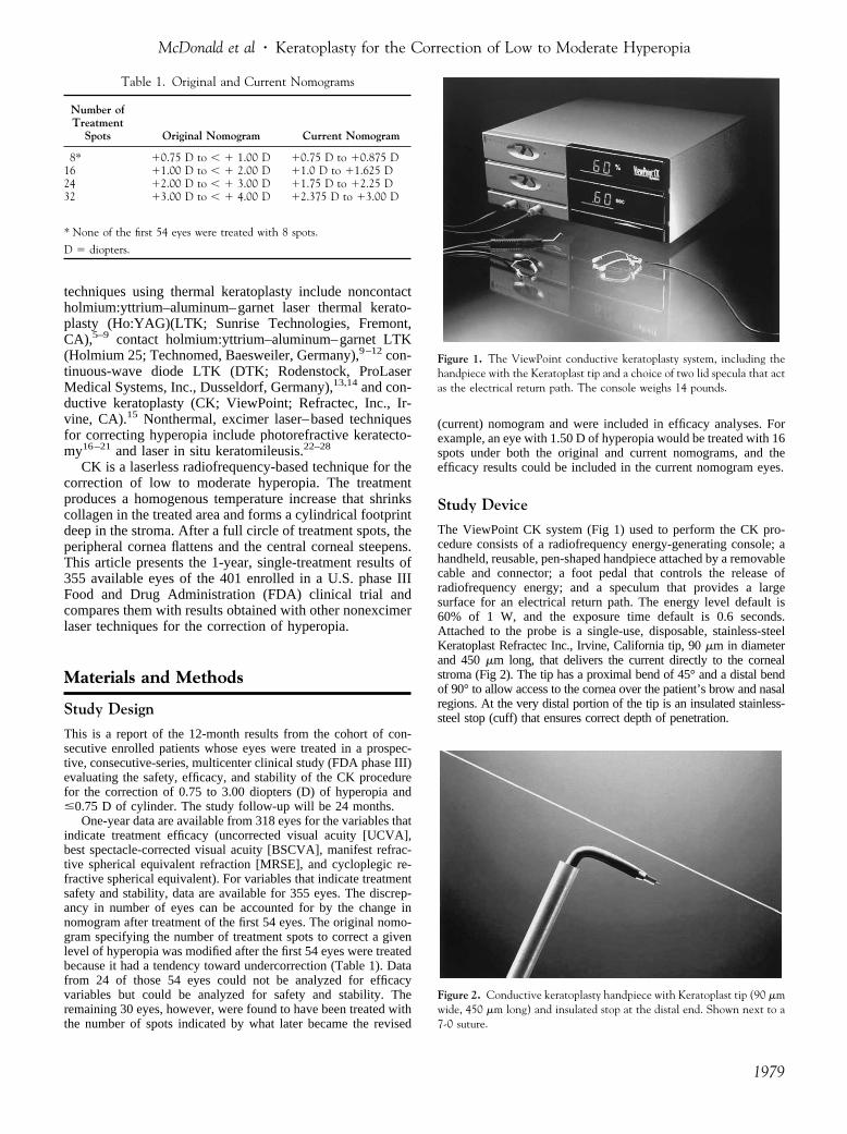
Figure 3. Treatment application. D � diopter; OZ � optical zone.
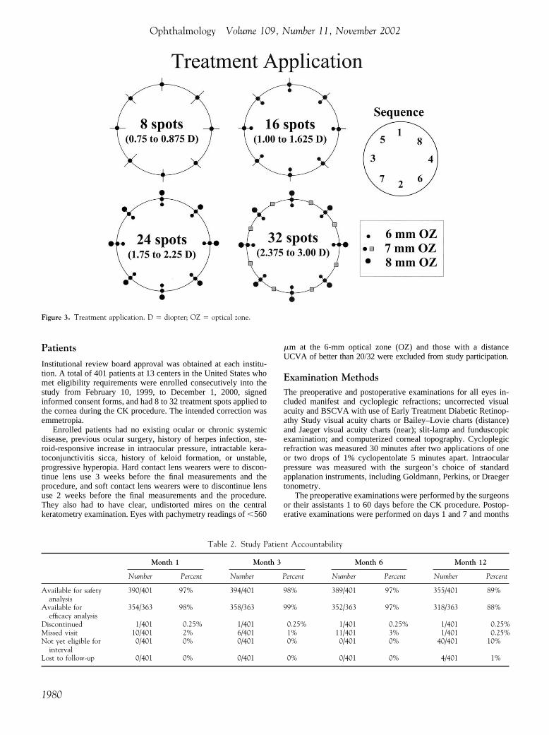
Table 2. Study Patient Accountability
Month 1 Month 3 Month 6 Month 12
Number Percent Number Percent Number Percent Number Percent
Available for safetyanalysis
390/401 97% 394/401 98% 389/401 97% 355/401 89%
Available forefficacy analysis
354/363 98% 358/363 99% 352/363 97% 318/363 88%
Discontinued 1/401 0.25% 1/401 0.25% 1/401 0.25% 1/401 0.25%Missed visit 10/401 2% 6/401 1% 11/401 3% 1/401 0.25%Not yet eligible for
interval0/401 0% 0/401 0% 0/401 0% 40/401 10%
Lost to follow-up 0/401 0% 0/401 0% 0/401 0% 4/401 1%
Ophthalmology Volume 109, Number 11, November 2002
1980
1, 3, 6, 9, and 12, and results were recorded on standardized dataforms. Patients were also asked to subjectively evaluate the qualityof their postoperative vision and indicate their level of satisfactionon standardized forms.
Mesopic contrast sensitivity testing was performed with theOptec 1600X, both with and without a glare source, in a subset of158 study patients before surgery and at 6 and 12 months aftersurgery. Testing without glare was performed with the targetillumination set to 5 cd/m2 and the glare source turned off. A trialframe with the patient’s best correction and test slide 1 wasinserted. The patient was asked to make a forced response to thedirection (left or right) of the point of the line in boxes A1 throughA8. The box number (i.e., A6) of the last correct response wasrecorded. This was repeated with test slide 2. The procedure wasrepeated with lines B1 through B8, C1 through C8, D1 through D8,and E1 through E8. The procedure for testing with glare was thesame except that glare illumination was set to 2 lux.
Specular microscopy studies were performed to study the effectof the treatment on the corneal endothelium on 162 eyes at 5 of the13 investigational sites. Measurements were taken with the Konan
NonCon Robopachy (Konan Medical, Inc., Hyogo, Japan) at thecentral cornea, the 3-mm OZ, and the 6-mm OZ. Changes frompreoperative levels at these sites were calculated by using paired-differences statistical testing.
Corneal haze was evaluated by slit lamp on a five-level scale ofclear, minimal, trace, mild, moderate, and marked. The protocolfailed to specify that haze only in the central cornea was to bereported and that haze at the treatment site was expected.
Confocal microscopy, a noninvasive technique for revealinghistology, was performed 12 months after the procedure by usingthe Nidek Confoscan II (Nidek Ltd., Gamagori, Japan). Thistechnique optically sections living tissues to allow in vivo exam-ination of individual layers.
Surgical ProcedureTopical anesthesia was induced with one drop of 0.5% tetracaine,administered three times at 5-minute intervals. Pilocarpine was notadministered. A lid speculum was placed in the eye to be treatedto obtain maximal exposure and to provide the electrical returnpath; the fellow eye was taped closed. Illumination was providedby the operating microscope. While the patient fixated on themicroscope’s light, the cornea was marked with a gentian violet–dampened, eight-intersection CK marker that marks the 7-mm OZand makes radial marks that extend from the 6-mm to the 8-mmOZs (Fig 3). The surface of the cornea was dried with a fiber-freesponge to avoid dissipation of applied energy through a damp surface.The surgeons placed the Keratoplast tip on the cornea at the treatmentmarkings and attempted to place it perpendicular to the cornealsurface. The cuff around the probe, which settles perpendicular to thecornea, helped to achieve perpendicular placement. Light pressurewas applied until the tip penetrated the stroma to its insulator stop.Energy was applied by depressing the foot pedal. All eyes weretreated at the default setting of 350 kHz at 60% power for 0.6 seconds.
All eyes were treated with the number of spots indicated by thecurrent nomogram (Table 1; Fig 3). For example, for the lowestamount of correction, �0.75 to �0.875 D, eight spots were placed onthe 7-mm OZ. For correcting �1.00 to �1.625 D, 16 spots wereplaced: 8 on the 6-mm OZ and another 8 on the 7-mm OZ. Forcorrecting �1.75 to � 2.25 D, 24 spots were placed: 8 each on the 6-,7-, and 8-mm OZs. For correcting �2.375 to �3.00 D, 24 spots were
Table 3. Demographic and Baseline Information
Number of Patients/Eyes 233/401
Mean age (SD) 55.3 years (6.4)Range 40 to 74Median 55.6
Range of Treatment CRSEMean (SD) �1.86 D (0.63)Range �0.75 D to �4.00 DMedian �1.75 D
Range of Treatment MRSEMean (SD) �1.80 D (0.64)Range �0.38 D* to �3.75 DMedian �1.75 D
CRSE – Cycloplegic refractive spherical equivalent; MRSE – Manifestrefractive spherical equivalent.
* Includes 2 ineligible eyes with minus CRSE values.
SD � standard deviation.
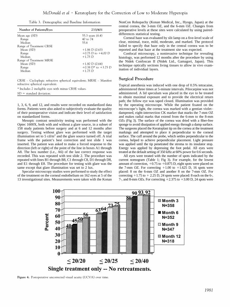
Figure 4. Postoperative uncorrected visual acuity (UCVA) over time.
McDonald et al � Keratoplasty for the Correction of Low to Moderate Hyperopia
1981
placed: 8 each were placed on the 6-, 7-, and 8-mm OZs, as wereplaced for �1.75 to �2.25 D of correction, and then 8 additional spotswere added between the spots previously made on the 7-mm OZ. Thesequence of spot placement is shown in Fig 3. The probe tip wascleaned of tissue debris with a fiber-free sponge after each treatmentspot. All eyes received a single CK treatment; i.e., no retreatmentswere performed.
Postoperative CareAfter treatment, one drop of topical antibiotic solution and onedrop of diclofenac sodium 0.1% (Voltaren; Ciba Vision Ophthal-mics, Duluth, Georgia) were administered and continued for 2days. Unpreserved artificial tear solution was the only ocularmedication permitted in the study. The treated eye was notpatched.
Results
Accountability and Demographics
One eye was enrolled and treated, but no energy was deliveredduring the procedure. Thus the total number of treated eyes was400 of the 401 enrolled (Table 2). Complete follow-up datawere available for 355 eyes for safety variables and for 318 eyesfor efficacy variables. The mean age of enrolled patients was 55� 6.4 years (range, 40 –74 years). There were 136 femalepatients and 97 male patients. Most (81%) of the patients werewhite and the others were Black, Asian, or other, or the racewas not recorded. Demographic information is shown in Table3.
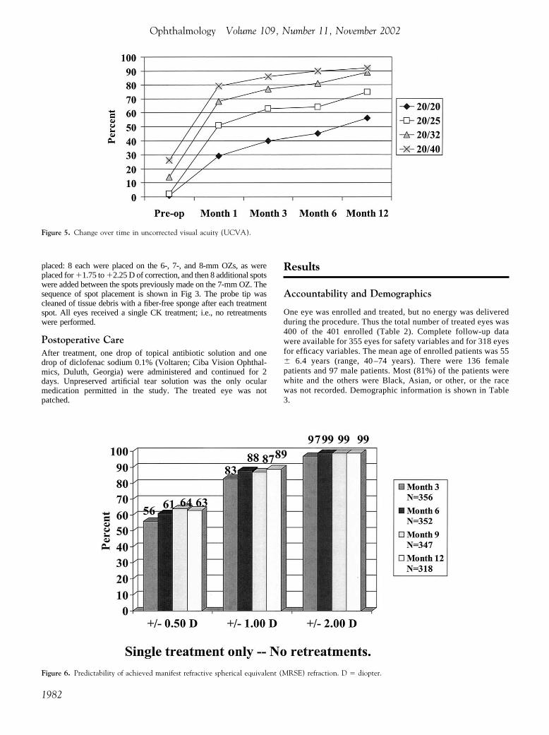
Figure 5. Change over time in uncorrected visual acuity (UCVA).
Figure 6. Predictability of achieved manifest refractive spherical equivalent (MRSE) refraction. D � diopter.
Ophthalmology Volume 109, Number 11, November 2002
1982
Uncorrected Visual Acuity
Before treatment, 5 (1%) of 363 eyes had 20/20 or better UCVA,96 (26%) of 363 had UCVA of 20/40 or better, and 358 (99%) of363 had 20/200 or better. After surgery, at 1 year, 178 (56%) of318 eyes had UCVA of 20/20 or better, 240 (75%) of 318 had20/25 or better, 294 (92%) of 318 had 20/40 or better, 315 (97%)of 318 had 20/80 or better, and 100% had 20/200 or better. Fig 4shows postoperative UCVA over time. Fig 5 shows UCVAchanges over time for each acuity level. UCVA progressivelyimproved with time for acuity levels of 20/20 or better, 20/25 orbetter, and 20/32 or better and showed no leveling off. For loweracuity levels, however, a leveling-off was apparent within the 12months. The 20/40 acuity leveled off at 6 months, and 20/80leveled off at approximately 1 month.
Predictability and StabilityPredictability. At 1 year, 199 (63%) of 318 eyes were within�0.50 D of emmetropia, 282 (89%) of 318 were within �1.00 D,and 316 (99%) of 318 were within �2.00 D (Fig 6).
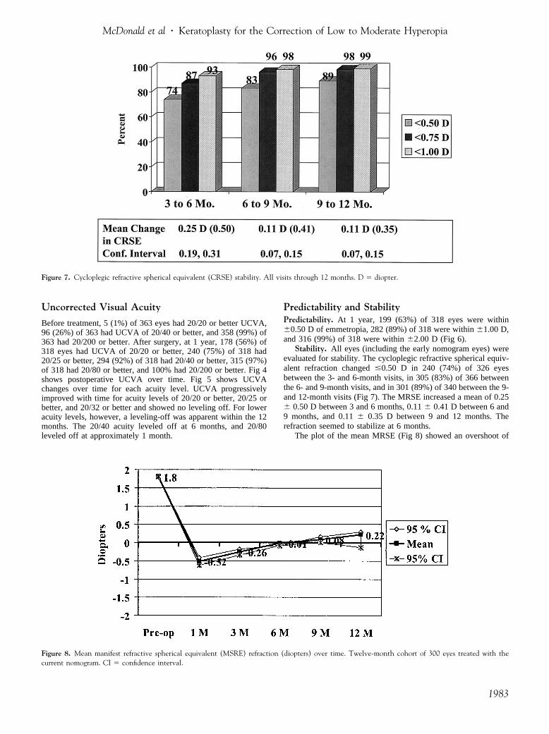
Stability. All eyes (including the early nomogram eyes) wereevaluated for stability. The cycloplegic refractive spherical equiv-alent refraction changed �0.50 D in 240 (74%) of 326 eyesbetween the 3- and 6-month visits, in 305 (83%) of 366 betweenthe 6- and 9-month visits, and in 301 (89%) of 340 between the 9-and 12-month visits (Fig 7). The MRSE increased a mean of 0.25� 0.50 D between 3 and 6 months, 0.11 � 0.41 D between 6 and9 months, and 0.11 � 0.35 D between 9 and 12 months. Therefraction seemed to stabilize at 6 months.
The plot of the mean MRSE (Fig 8) showed an overshoot of
Figure 7. Cycloplegic refractive spherical equivalent (CRSE) stability. All visits through 12 months. D � diopter.
Figure 8. Mean manifest refractive spherical equivalent (MSRE) refraction (diopters) over time. Twelve-month cohort of 300 eyes treated with thecurrent nomogram. CI � confidence interval.
McDonald et al � Keratoplasty for the Correction of Low to Moderate Hyperopia
1983
approximately 0.5 D at month 1, followed by a slight regression.At 12 months, the eyes had an MRSE of 0.22 D, which is veryclose to emmetropia. Confidence intervals are shown surroundingthe mean values (instead of standard deviations). Confidence in-tervals are a better indicator of deviations from mean valuesbecause they take sample size into account. Results at 6 and 12months met all FDA guidelines for UCVA and accuracy ofachieved MSRE (Table 4).
SafetyBest Spectacle-corrected Visual Acuity. Safety variable resultsare summarized in Table 5. Note that n � 355 for safety data (not318, as for efficacy data) because safety data were acquired fromthe first 54 eyes (original nomogram) in addition to the currentnomogram eyes. Twenty-five of the total 390 eyes lost �2 lines ofBSCVA at 1 month, and 7 of 355 lost 2 lines at 12 months. Eighteyes lost more than two lines at 1 month, and none lost more thantwo lines at 12 months. However, the loss of two lines at 12months left all seven eyes with very functional vision. Beforesurgery, all seven of these eyes had 20/10 to 20/16 BSCVA. Aftersurgery, one had 20/16 BSCVA, three had 20/20, and three had20/25. No eye had BSCVA worse than 20/40 at any follow-upvisit. No eye that had 20/20 or better best-corrected vision beforesurgery had worse than 20/25 BSCVA after surgery.
Cylinder. Postoperative absolute cylinder increases are shownin Table 6. No change was defined as �0.75 D. Percentages for alllevels of cylinder were highest at the first month and then declinedwith time. At 12 months, 6% had 1.00 D of induced cylinder, and
4.2% had �1.00 D but �2.00 D of induced cylinder. A total of88% had no change in cylinder at 12 months.
Complications and Adverse Events. No intraoperative com-plications or adverse events occurred during any of the surgeries.There were no treatment-related adverse events, such as peripheralcorneal defect, corneal edema later than 1 week after surgery,recurrent corneal erosion at 1 month or later, double or ghostingimages at any time, foreign body sensation at 1 month or later, andpain at 1 month or later.
Corneal Haze. The investigational sites were expected to mea-sure corneal haze at the central cornea only. However, the evalu-ator at one investigational center measured and reported haze at theCK treatment site, and the results in this section reflect all reports.No haze was seen in 98% (384/390) of the eyes at 1 month aftersurgery, in 96% of eyes at 3 months, in 97% at 6 months, or in100% at 12 months. The highest level of haze was a mild levelseen in 4 (1%) of 390 eyes at month 1 and in 1 (0.25%) of 394 eyesat months 3 and 6.
Intraocular Pressure. Mean intraocular pressure (IOP) in alltreated eyes is shown in Table 7. There were no occurrences of anuncontrolled IOP increase of �5 mmHg above baseline. An IOPreading of �25 mmHg was measured in 2 of 389 eyes at 6 monthsand in 1 eye at 9 and 12 months. However, no patient wasdiagnosed with glaucoma.
Contrast Sensitivity. A comparison of the preoperative and6-month postoperative mesopic contrast sensitivity values showedno differences for patches A, B, C, D, and E, both with (Table 8)and without (Table 9) glare.
Specular Microscopy. Table 10 shows the mean endothelialcell density counts over time, measured at the corneal center, themid periphery, and the periphery. Mean endothelial cell densityshowed values of approximately 2700 cells per square millimeterfor preoperative and postoperative measurements for the central,midperipheral, and peripheral corneal regions. No clinically orstatistically significant changes appeared in the mean cell count
Table 4. Summary Efficacy Variables
3 Months(N � 358)
6 Months(N � 352)
12 Months(N � 344) FDA Target
UCVA �20/20 40% 45% 56% Not stipulatedUCVA �20/25 63% 64% 75% Not stipulatedUCVA �20/40 86% 90% 92% 85%MRSE � 0.50 D 56% 61% 62% 50%MRSE � 1.00 D 83% 88% 89% 75%
MRSE � manifest refractive spherical equivalent; UCVA � uncorrectedvisual acuity.
Table 5. Postoperative Visit
Safety Variable1
Month3
Months6
Months9
Months12
Months
2 line loss BSCVA 25/390 20/392 16/389 13/386 7/381*(6%) (5%) (4%) (3%) (2%)
�2 line loss BSCVA 8/390 4/392 2/389 2/386 0/381(2%) (1%) (0.5%) (0.5%) (0%)
BSCVA worse than 0/390 0/392 0/389 0/386 0/38120/40 (0%) (0%) (0%) (0%) (0%)
Increase �2.00 D 13/390 8/392 3/389 1/386 1/381cylinder (3%) (2%) (0.8%) (0.3%) (0.3%)
BSCVA worse than 14/390 7/392 3/389 2/386 0/38120/25 if better than20/20 preoperatively
(4%) (2%) (0.8%) (0.5%) (0%)
* Preoperatively, these eyes had 20/10 to 20/16 BSCVA. Postoperatively,1/7 eyes had 20/16 BSCVA, 3/7 had 20/20, and 3/7 eyes had 20/25. Thusall eyes with 2-line losses had functional vision.
BSCVA � best spectacle-corrected visual acuity.
Table 6. Cylinder Changes
Cylinder Change Month 1 Month 6 Month 12
Increase �2.00 D 13/390 3/389 1/3553.3% 0.8% 0.3%
Increase 2.00 D 11/390 5/389 2/3552.8% 1.3% 0.6%
Increase 1.25–1.99 D 57/390 47/389 15/35514.6% 12.1% 4.2%
Increase 1.00 D 49/390 40/389 23/35512.6% 10.3% 6.5%
Decrease 1.00 D 2/390 1/389 1/355(0.5%) (0.25%) (0.3%)
No Change 258/390 293/389 311/355(66.1%) (75.3%) (87.6%)
D � diopters.
Table 7. Intraocular Pressure (mm Hg) Over Time
Pre-op Month 1 Month 3 Month 6 Month 12
Mean (SD) 14.9 (2.6) 14.0 (2.6) 14.0 (2.6) 14.1 (2.6) 14.3 (2.5)95% CI 14.6, 15.1 13.7, 14.2 13.8, 14.3 13.9, 14.4 14.0, 14.7Median 15.0 14.0 14.0 14.0 14.0Range 8.0 to 25.0 7.0 to 23.0 6.0 to 24.0 6.0 to 26.0 9.0 to 30.0
CI � confidence interval; SD � standard deviation.
Ophthalmology Volume 109, Number 11, November 2002
1984

over time for any of the corneal regions. Changes in cell density(Table 11) were within error of testing.
Subjective Evaluations. Extreme/marked or moderate im-
provement in postoperative vision was noted by 300 (85%) of 353of patients at 1 month and by 313 (90%) of 347 at 12 months(Table 12). Ten patients (3%) thought they had no improvement at12 months. Patients who were very satisfied or satisfied with theresults of their surgery numbered 77% at 1 month and 81% at 12months (Table 13).
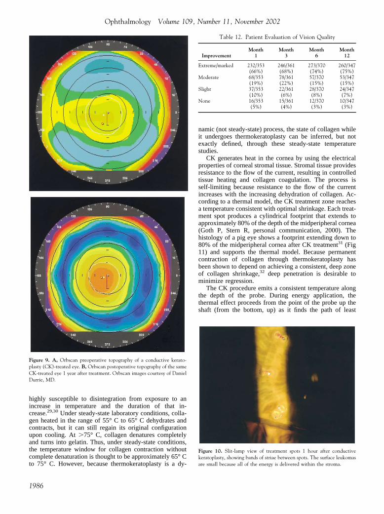
Effects on the CorneaCorneal Topography. Orbscan corneal topography, with a preop-erative view (Fig 9A) and with the same cornea 1 year after CKtreatment (Fig 9B), shows postoperative central corneal steepeningsurrounded by midperipheral flattening.
Slit Lamp. Fig 10 shows a slit-lamp photograph of the cornea1 hour after CK treatment. The paired (6- and 7-mm OZs) treat-ment spots of thermal coagulation are visible as small surfaceleukomas, with lines of tension, or striae, connecting the treatmentspots.
Histology. Histology views of the footprint in the cornealstroma of a pig’s eye 1 week after treatment show the treatmenteffect penetrating to approximately 80% of the depth of the mid-peripheral cornea (Fig 11).
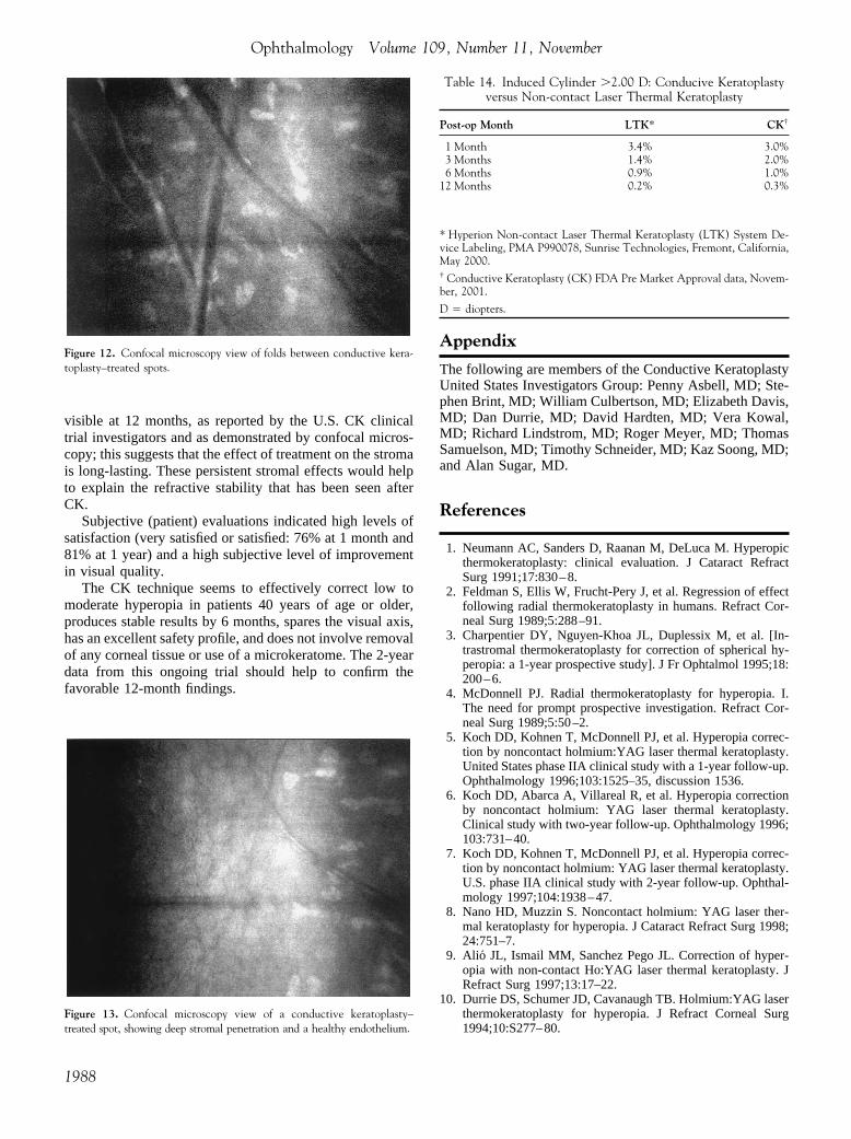
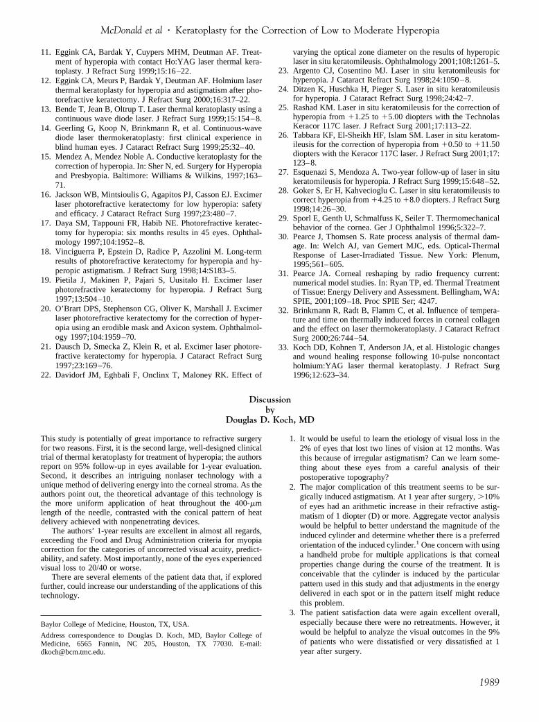
Confocal Microscopy. Confocal microscopy of a patient’s eye12 months after CK treatment (Fig 12) shows a prominent stromalfold in the striae between the two treatment spots. Fig 13 showsdeep stromal penetration without damage to the corneal endothelium.
Discussion
Collagen protein maintains its spiral triple-helix configura-tion through the covalent bonds between its long chains andthe hydrogen bonds between the molecules. These bonds are
Table 8. Mesopic Contrast Sensitivity without Glare
PreoperativeN � 158
Month 6N � 141
Month 12N � 73
Patch AMean (SD) 4.05 (1.51) 4.38 (1.91) 4.64 (1.76)95% CI* 3.81, 4.29 4.07, 4.69 4.23, 5.05Mean change
(95% CI)NA 0.40 (0.07, 0.73) 0.59 (0.16, 1.02)
Patch BMean (SD) 3.74 (1.80) 4.09 (2.16) 4.41 (1.96)95% CI 3.47, 4.01 3.74, 4.44 3.96, 4.86Mean change
(95% CI)NA 0.42 (0.07, 0.77) 0.61 (0.20, 1.02)
Patch CMean (SD) 1.10 (1.29) 0.99 (1.46) 1.11 (2.00)95% CI 0.90, 1.30 0.75, 1.23 0.66, 1.56Mean change
(95% CI)NA �0.16 (�0.45, 0.13) �0.08 (�0.57, 0.41)
Patch DMean (SD) 0.38 (0.95) 0.38 (0.92) 0.60 (1.45)95% CI 0.22, 0.54 0.22, 0.54 0.27, 0.93Mean change
(95% CI)NA �0.22 (�0.22, 0.18) 0.21 (�0.12, 0.54)
Patch EMean (SD) 0.12 (0.44) 0.14 (0.62) 0.44 (1.21)95% CI* 0.04, 0.20 0.04, 0.24 0.17, 0.71Mean change
(95% CI)NA 0.02 (0.12, 0.16) 0.32 (0.05, 0.59)
* CI � confidence interval; NA � not applicable; SD � standard deviation.
Table 9. Mesopic Contrast Sensitivity with Glare
PreoperativeN � 158
Month 6N � 141
Month 12N � 73
Patch AMean (SD) 3.61 (1.57) 3.83 (1.89) 4.14 (1.91)95% CI* 3.36, 3.86 3.52, 4.14 3.71, 4.57Mean change
(95% CI)NA 0.26 (�0.07, 0.59) 0.62 (0.19, 1.05)
Patch BMean (SD) 3.27 (1.67) 3.51 (2.13) 4.19 (2.05)95% CI 3.02, 3.52 3.16, 3.86 3.72, 4.66Mean change
(95% CI)NA 0.24 (�0.11, 0.59) 0.93 (0.46, 1.40)
Patch CMean (SD) 0.83 (1.11) 0.78 (1.30) 0.88 (1.75)95% CI 0.65, 1.01 0.56, 1.00 0.49, 1.27Mean change
(95% CI)NA �0.10 (�0.37, 0.17) �0.07 (�0.48, 0.34)
Patch DMean (SD) 0.26 (0.73) 0.29 (0.95) 0.55 (1.38)95% CI 0.14, 0.38 0.13, 0.45 0.24, 0.86Mean change
(95% CI)NA 0.03 (�0.17, 0.23) 0.31 (0.02, 0.60)
Patch EMean (SD) 0.13 (0.61) 0.11 (0.39) 0.47 (1.35)95% CI* 0.03, 0.23 0.05, 0.17 0.16, 0.78Mean change
(95% CI)NA �0.04 (�0.18, 0.10) 0.31 (0.02, 0.60)
CI � Confidence interval; NA � not applicable; SD � standard deviation.
Table 10. Mean Endothelial Cell Density over Time
Region Pre-Op 3 Months 6 Months 12 Months
Central (n � 162) (n � 127) (n � 123) (n � 42)Mean 2686 2730 2727 2683SD 160.9 163.7 153.6 163.2
Mid-Periphery (n � 162) (n � 111) (n � 108) (n � 31)Mean 2722 2734 2727 2691SD 162 141.3 134.4 158.4
Periphery (n � 159) (n � 107) (n � 104) (n � 28)Mean 2724 2727 2724 2716SD 150.9 140.2 138.4 145.3
SD � standard deviation.
Table 11. Changes in Endothelial Cell Density from Pre-op
Region 3 M 6 M 12 M
Central (N � 127) (N � 123) (N � 42)Mean (SD) �0.31% (4.5) �1.4% (4.2) �1.0% (3.9)CI �0.57, 0.99 0.66, 2.1 �0.19 to �2.2Mid periphery (N � 111) (N � 108) (N � 31)Mean (SD) �0.61% (3.0) �0.23% (3.05) �0.6% (3.6)CI �1.2, �0.08 �0.8, 0.35 �1.87 (0.69)
Periphery (N � 107) (N � 104) (N � 28)Mean (SD) �0.76% (2.9) �0.41% (3.2) 0.2% (3.5)CI �1.3, �0.28 �1.03, 0.21 �1.1, 1.5
CI � confidence interval; SD � standard deviation.
McDonald et al � Keratoplasty for the Correction of Low to Moderate Hyperopia
1985
highly susceptible to disintegration from exposure to anincrease in temperature and the duration of that in-crease.29,30 Under steady-state laboratory conditions, colla-gen heated in the range of 55° C to 65° C dehydrates andcontracts, but it can still regain its original configurationupon cooling. At �75° C, collagen denatures completelyand turns into gelatin. Thus, under steady-state conditions,the temperature window for collagen contraction withoutcomplete denaturation is thought to be approximately 65° Cto 75° C. However, because thermokeratoplasty is a dy-
namic (not steady-state) process, the state of collagen whileit undergoes thermokeratoplasty can be inferred, but notexactly defined, through these steady-state temperaturestudies.
CK generates heat in the cornea by using the electricalproperties of corneal stromal tissue. Stromal tissue providesresistance to the flow of the current, resulting in controlledtissue heating and collagen coagulation. The process isself-limiting because resistance to the flow of the currentincreases with the increasing dehydration of collagen. Ac-cording to a thermal model, the CK treatment zone reachesa temperature consistent with optimal shrinkage. Each treat-ment spot produces a cylindrical footprint that extends toapproximately 80% of the depth of the midperipheral cornea(Goth P, Stern R, personal communication, 2000). Thehistology of a pig eye shows a footprint extending down to80% of the midperipheral cornea after CK treatment31 (Fig11) and supports the thermal model. Because permanentcontraction of collagen through thermokeratoplasty hasbeen shown to depend on achieving a consistent, deep zoneof collagen shrinkage,32 deep penetration is desirable tominimize regression.
The CK procedure emits a consistent temperature alongthe depth of the probe. During energy application, thethermal effect proceeds from the point of the probe up theshaft (from the bottom, up) as it finds the path of least
Table 12. Patient Evaluation of Vision Quality
ImprovementMonth
1Month
3Month
6Month
12
Extreme/marked 232/353 246/361 273/370 260/347(66%) (68%) (74%) (75%)
Moderate 68/353 78/361 57/370 53/347(19%) (22%) (15%) (15%)
Slight 37/353 22/361 28/370 24/347(10%) (6%) (8%) (7%)
None 16/353 15/361 12/370 10/347(5%) (4%) (3%) (3%)
Figure 9. A, Orbscan preoperative topography of a conductive kerato-plasty (CK)-treated eye. B, Orbscan postoperative topography of the sameCK-treated eye 1 year after treatment. Orbscan images courtesy of DanielDurrie, MD.
Figure 10. Slit-lamp view of treatment spots 1 hour after conductivekeratoplasty, showing bands of striae between spots. The surface leukomasare small because all of the energy is delivered within the stroma.
Ophthalmology Volume 109, Number 11, November 2002
1986

resistance. The result is a cylindrical thermal effect that hasalmost no axial component. Thus, a cylindrical footprint ismade in the stroma. The small leukomas seen at CK-treatedspots demonstrate that CK delivers energy deep into thestroma rather than on the surface. In contrast, the noncontactLTK technique generates the greatest amount of heat at thesurface of the cornea because of the high absorption of lightenergy in water. The holmium:yttrium–aluminum–garnetbeam is attenuated as it passes through the cornea so that theheat energy diffuses radially and axially into the tissue. Theresult is corneal denaturation decreasing from top to bottom,forming a cone-shaped zone of collagen shrinkage33 of adepth that seems to be more shallow than that of a CK-treated spot.
In the multicenter clinical trial reported here, the efficacyresults exceeded all FDA guidelines for performance ofrefractive surgery procedures. At 1 year after treatment,56% of the study eyes showed 20/20 or better UCVA, and92% showed 20/40 or better. Eyes were approximately 0.50D myopic at month 1 and were effectively emmetropic at 6months. At 1 year, the mean MRSE was within �0.50 D in63% and within �1.00 D in 89%. These predictabilityvalues match or exceed those of other procedures for similarlevels of hyperopia. During the last two intervals (6 to 9months and 9 to 12 months), the cycloplegic refractivespherical equivalent refraction changed 0.11 and 0.11 D,respectively. Thus, the refraction seemed to stabilize by 6months and to maintain a stable level through 1 year. Thisamount of change is similar to the natural progression ofhyperopia in eyes that have not had refractive surgery.Because we performed no retreatments, our stability resultsreflect actual corneal refractive stability over the 1-yearfollow-up.
BSCVA was generally preserved after the procedure, andno eye had BSCVA worse than 20/40. The incidence ofinduced cylinder of �2.00 D was �1% (the FDA target is5%). At 1 year, 6% showed a 1.00-D increase in cylinder,and 4% showed an increase of �1.00 D but �2.00 D. Thesepercentages are clinically acceptable. This cylinder profileis similar to that of LTK, in which thermal spots are appliedsimultaneously to the cornea (Table 14). CK may owe itssafety profile to the preservation of the visual axis in theprocedure. In comparison, hyperopic photorefractive kera-tectomy16–21 and hyperopic laser in situ keratomileusisstudies22–28 have demonstrated a two-line or greater loss ofBSCVA of up to 6%. Furthermore, CK-treated eyes showedno loss of contrast sensitivity when tested with and withoutglare. This provides additional evidence of safety.
Although the incidence and level of haze seen at 1, 3, and6 months after CK was low, none was expected. The hazethat was observed might be attributed to one investigationalcenter at which haze observed on any part of the cornea(including at the CK treatment site, which is expected to behazy) was recorded instead of at the central cornea only.
Possible damage to the corneal endothelium after deepthermal penetration in the stroma by CK treatment wasassessed from data collected on 162 eyes at 3 investigationalsites. Cell counts taken before surgery and at 3, 6, and 12months after surgery at the central cornea, the 3-mm OZ,and the 6-mm OZ showed no clinically or statisticallysignificant changes from preoperative levels. Confocal mi-croscopy views (Figs 12 and 13) also showed deep pene-tration without damage to the endothelium. Thus, the in-terim data seem to indicate that the CK procedure is safe forthe corneal endothelium. Furthermore, no spikes were seenin postoperative IOP data, suggesting that flattening of thecornea by the procedure did not cause shallowing of theperipheral chamber angle.
Leucomas visible by slit lamp after surgery were smallbecause CK delivers energy deep into the stroma rather thanon the surface. The striae between treatment zones remained
Table 13. Patient Satisfaction
Patient Response Month 1 Month 6 Month 12
Very satisfied/satisfied 273/356 306/371 282/347(77%) (82%) (81%)
Neutral 57/356 34/371 33/347(16%) (9%) (10%)
Dissatisfied 16/356 20/371 25/347(4%) (5%) (7%)
Very dissatisfied 10/356 11/371 7/347(3%) (3%) (2%)
Figure 11. Histology view of a pig eye 1 week after conductive kerato-plasty treatment, showing a cylindrical footprint and a treatment effectextending to 80% of the corneal depth.
McDonald et al � Keratoplasty for the Correction of Low to Moderate Hyperopia
1987
visible at 12 months, as reported by the U.S. CK clinicaltrial investigators and as demonstrated by confocal micros-copy; this suggests that the effect of treatment on the stromais long-lasting. These persistent stromal effects would helpto explain the refractive stability that has been seen afterCK.
Subjective (patient) evaluations indicated high levels ofsatisfaction (very satisfied or satisfied: 76% at 1 month and81% at 1 year) and a high subjective level of improvementin visual quality.
The CK technique seems to effectively correct low tomoderate hyperopia in patients 40 years of age or older,produces stable results by 6 months, spares the visual axis,has an excellent safety profile, and does not involve removalof any corneal tissue or use of a microkeratome. The 2-yeardata from this ongoing trial should help to confirm thefavorable 12-month findings.
Appendix
The following are members of the Conductive KeratoplastyUnited States Investigators Group: Penny Asbell, MD; Ste-phen Brint, MD; William Culbertson, MD; Elizabeth Davis,MD; Dan Durrie, MD; David Hardten, MD; Vera Kowal,MD; Richard Lindstrom, MD; Roger Meyer, MD; ThomasSamuelson, MD; Timothy Schneider, MD; Kaz Soong, MD;and Alan Sugar, MD.
References
1. Neumann AC, Sanders D, Raanan M, DeLuca M. Hyperopicthermokeratoplasty: clinical evaluation. J Cataract RefractSurg 1991;17:830–8.
2. Feldman S, Ellis W, Frucht-Pery J, et al. Regression of effectfollowing radial thermokeratoplasty in humans. Refract Cor-neal Surg 1989;5:288–91.
3. Charpentier DY, Nguyen-Khoa JL, Duplessix M, et al. [In-trastromal thermokeratoplasty for correction of spherical hy-peropia: a 1-year prospective study]. J Fr Ophtalmol 1995;18:200–6.
4. McDonnell PJ. Radial thermokeratoplasty for hyperopia. I.The need for prompt prospective investigation. Refract Cor-neal Surg 1989;5:50–2.
5. Koch DD, Kohnen T, McDonnell PJ, et al. Hyperopia correc-tion by noncontact holmium:YAG laser thermal keratoplasty.United States phase IIA clinical study with a 1-year follow-up.Ophthalmology 1996;103:1525–35, discussion 1536.
6. Koch DD, Abarca A, Villareal R, et al. Hyperopia correctionby noncontact holmium: YAG laser thermal keratoplasty.Clinical study with two-year follow-up. Ophthalmology 1996;103:731–40.
7. Koch DD, Kohnen T, McDonnell PJ, et al. Hyperopia correc-tion by noncontact holmium: YAG laser thermal keratoplasty.U.S. phase IIA clinical study with 2-year follow-up. Ophthal-mology 1997;104:1938–47.
8. Nano HD, Muzzin S. Noncontact holmium: YAG laser ther-mal keratoplasty for hyperopia. J Cataract Refract Surg 1998;24:751–7.
9. Alio JL, Ismail MM, Sanchez Pego JL. Correction of hyper-opia with non-contact Ho:YAG laser thermal keratoplasty. JRefract Surg 1997;13:17–22.
10. Durrie DS, Schumer JD, Cavanaugh TB. Holmium:YAG laserthermokeratoplasty for hyperopia. J Refract Corneal Surg1994;10:S277–80.
Figure 12. Confocal microscopy view of folds between conductive kera-toplasty–treated spots.
Figure 13. Confocal microscopy view of a conductive keratoplasty–treated spot, showing deep stromal penetration and a healthy endothelium.
Table 14. Induced Cylinder �2.00 D: Conducive Keratoplastyversus Non-contact Laser Thermal Keratoplasty
Post-op Month LTK* CK†
1 Month 3.4% 3.0%3 Months 1.4% 2.0%6 Months 0.9% 1.0%
12 Months 0.2% 0.3%
* Hyperion Non-contact Laser Thermal Keratoplasty (LTK) System De-vice Labeling, PMA P990078, Sunrise Technologies, Fremont, California,May 2000.† Conductive Keratoplasty (CK) FDA Pre Market Approval data, Novem-ber, 2001.
D � diopters.
Ophthalmology Volume 109, Number 11, November
1988
11. Eggink CA, Bardak Y, Cuypers MHM, Deutman AF. Treat-ment of hyperopia with contact Ho:YAG laser thermal kera-toplasty. J Refract Surg 1999;15:16–22.
12. Eggink CA, Meurs P, Bardak Y, Deutman AF. Holmium laserthermal keratoplasty for hyperopia and astigmatism after pho-torefractive keratectomy. J Refract Surg 2000;16:317–22.
13. Bende T, Jean B, Oltrup T. Laser thermal keratoplasty using acontinuous wave diode laser. J Refract Surg 1999;15:154–8.
14. Geerling G, Koop N, Brinkmann R, et al. Continuous-wavediode laser thermokeratoplasty: first clinical experience inblind human eyes. J Cataract Refract Surg 1999;25:32–40.
15. Mendez A, Mendez Noble A. Conductive keratoplasty for thecorrection of hyperopia. In: Sher N, ed. Surgery for Hyperopiaand Presbyopia. Baltimore: Williams & Wilkins, 1997;163–71.
16. Jackson WB, Mintsioulis G, Agapitos PJ, Casson EJ. Excimerlaser photorefractive keratectomy for low hyperopia: safetyand efficacy. J Cataract Refract Surg 1997;23:480–7.
17. Daya SM, Tappouni FR, Habib NE. Photorefractive keratec-tomy for hyperopia: six months results in 45 eyes. Ophthal-mology 1997;104:1952–8.
18. Vinciguerra P, Epstein D, Radice P, Azzolini M. Long-termresults of photorefractive keratectomy for hyperopia and hy-peropic astigmatism. J Refract Surg 1998;14:S183–5.
19. Pietila J, Makinen P, Pajari S, Uusitalo H. Excimer laserphotorefractive keratectomy for hyperopia. J Refract Surg1997;13:504–10.
20. O’Brart DPS, Stephenson CG, Oliver K, Marshall J. Excimerlaser photorefractive keratectomy for the correction of hyper-opia using an erodible mask and Axicon system. Ophthalmol-ogy 1997;104:1959–70.
21. Dausch D, Smecka Z, Klein R, et al. Excimer laser photore-fractive keratectomy for hyperopia. J Cataract Refract Surg1997;23:169–76.
22. Davidorf JM, Eghbali F, Onclinx T, Maloney RK. Effect of
varying the optical zone diameter on the results of hyperopiclaser in situ keratomileusis. Ophthalmology 2001;108:1261–5.
23. Argento CJ, Cosentino MJ. Laser in situ keratomileusis forhyperopia. J Cataract Refract Surg 1998;24:1050–8.
24. Ditzen K, Huschka H, Pieger S. Laser in situ keratomileusisfor hyperopia. J Cataract Refract Surg 1998;24:42–7.
25. Rashad KM. Laser in situ keratomileusis for the correction ofhyperopia from �1.25 to �5.00 diopters with the TechnolasKeracor 117C laser. J Refract Surg 2001;17:113–22.
26. Tabbara KF, El-Sheikh HF, Islam SM. Laser in situ keratom-ileusis for the correction of hyperopia from �0.50 to �11.50diopters with the Keracor 117C laser. J Refract Surg 2001;17:123–8.
27. Esquenazi S, Mendoza A. Two-year follow-up of laser in situkeratomileusis for hyperopia. J Refract Surg 1999;15:648–52.
28. Goker S, Er H, Kahvecioglu C. Laser in situ keratomileusis tocorrect hyperopia from �4.25 to �8.0 diopters. J Refract Surg1998;14:26–30.
29. Sporl E, Genth U, Schmalfuss K, Seiler T. Thermomechanicalbehavior of the cornea. Ger J Ophthalmol 1996;5:322–7.
30. Pearce J, Thomsen S. Rate process analysis of thermal dam-age. In: Welch AJ, van Gemert MJC, eds. Optical-ThermalResponse of Laser-Irradiated Tissue. New York: Plenum,1995;561–605.
31. Pearce JA. Corneal reshaping by radio frequency current:numerical model studies. In: Ryan TP, ed. Thermal Treatmentof Tissue: Energy Delivery and Assessment. Bellingham, WA:SPIE, 2001;109–18. Proc SPIE Ser; 4247.
32. Brinkmann R, Radt B, Flamm C, et al. Influence of tempera-ture and time on thermally induced forces in corneal collagenand the effect on laser thermokeratoplasty. J Cataract RefractSurg 2000;26:744–54.
33. Koch DD, Kohnen T, Anderson JA, et al. Histologic changesand wound healing response following 10-pulse noncontactholmium:YAG laser thermal keratoplasty. J Refract Surg1996;12:623–34.
Discussionby
Douglas D. Koch, MD
This study is potentially of great importance to refractive surgeryfor two reasons. First, it is the second large, well-designed clinicaltrial of thermal keratoplasty for treatment of hyperopia; the authorsreport on 95% follow-up in eyes available for 1-year evaluation.Second, it describes an intriguing nonlaser technology with aunique method of delivering energy into the corneal stroma. As theauthors point out, the theoretical advantage of this technology isthe more uniform application of heat throughout the 400-�mlength of the needle, contrasted with the conical pattern of heatdelivery achieved with nonpenetrating devices.
The authors’ 1-year results are excellent in almost all regards,exceeding the Food and Drug Administration criteria for myopiacorrection for the categories of uncorrected visual acuity, predict-ability, and safety. Most importantly, none of the eyes experiencedvisual loss to 20/40 or worse.
There are several elements of the patient data that, if exploredfurther, could increase our understanding of the applications of thistechnology.
1. It would be useful to learn the etiology of visual loss in the2% of eyes that lost two lines of vision at 12 months. Wasthis because of irregular astigmatism? Can we learn some-thing about these eyes from a careful analysis of theirpostoperative topography?
2. The major complication of this treatment seems to be sur-gically induced astigmatism. At 1 year after surgery, �10%of eyes had an arithmetic increase in their refractive astig-matism of 1 diopter (D) or more. Aggregate vector analysiswould be helpful to better understand the magnitude of theinduced cylinder and determine whether there is a preferredorientation of the induced cylinder.1 One concern with usinga handheld probe for multiple applications is that cornealproperties change during the course of the treatment. It isconceivable that the cylinder is induced by the particularpattern used in this study and that adjustments in the energydelivered in each spot or in the pattern itself might reducethis problem.
3. The patient satisfaction data were again excellent overall,especially because there were no retreatments. However, itwould be helpful to analyze the visual outcomes in the 9%of patients who were dissatisfied or very dissatisfied at 1year after surgery.
Baylor College of Medicine, Houston, TX, USA.
Address correspondence to Douglas D. Koch, MD, Baylor College ofMedicine, 6565 Fannin, NC 205, Houston, TX 77030. E-mail:[email protected].
McDonald et al � Keratoplasty for the Correction of Low to Moderate Hyperopia
1989