Embed Size (px)
Citation preview
Conformation of ATP and ADP Molecules in AqueousSolutions Determined by High-Energy X-ray Diffraction
Takuya Miyazaki • Yasuo Kameda • Yasuhiro Umebayashi •
Hiroyuki Doi • Yuko Amo • Takeshi Usuki
Received: 27 November 2013 / Accepted: 28 December 2013� Springer Science+Business Media New York 2014
Abstract High-energy X-ray diffraction measurements were carried out at 26 �C for
aqueous 1.0, 2.0 and 2.05 mol% disodium adenosine 50-triphosphate (ATP) and 2.0 and
2.05 mol% disodium adenosine 50-diphosphate (ADP) solutions in order to obtain direct
experimental information on the intramolecular conformations of ATP and ADP molecules
in aqueous solutions. Observed interference terms were analyzed in terms of the
intramolecular geometry of the ATP and ADP molecules. Dihedral angles between adenine
and the ribose group (t1), ribose-ring and methylene group of ribose (t2), and the methylene
group of ribose and triphosphate (or diphosphate) group (t3), were determined through the
least-squares fitting procedure of the observed interference term.
Keywords ATP � ADP � Intramolecular structure � Conformation � X-ray
diffraction
1 Introduction
Adenosine 50-triphosphate (ATP) is one of the most important biomolecules in the energy
metabolism in the living cell. The energetics of ATP is promoted by the hydrolysis of the
terminal P–O bond in the triphosphate group to form the adenosine 50-diphosphate (ADP)
molecule. The conformation of the ATP molecule plays an important role in determining
the amount energy stored. In crystalline ATP disodium salt, the triphosphate group of ATP
is folded back towards the adenine base [1–3]. This bent conformation is stabilized by the
sodium ions that form a bridge between the phosphate chain and nitrogen (N7) atom in the
T. Miyazaki � Y. Kameda (&) � Y. Amo � T. UsukiDepartment of Material and Biological Chemistry, Faculty of Science, Yamagata University,Yamagata, Yamagata 990-8560, Japane-mail: [email protected]
Y. Umebayashi � H. DoiGraduate School of Science and Technology, Niigata University, Niigata 950-2181, Japan
123
J Solution ChemDOI 10.1007/s10953-014-0153-8
adenosine residue. The conformation of the ATP molecule in the isolated state obtained
from ab initio calculations is characterized by intramolecular hydrogen bonds within the
triphosphate group [4, 5]. In aqueous solution, the triphosphate, ribose and adenine groups
of the ATP molecule should form intermolecular hydrogen bonds with neighboring water
molecules. According to a 1H NMR study, the phosphate groups of ATP and ADP mol-
ecules in aqueous solutions have an unfolded conformation [6].
In principle, information concerning the intramolecular structure (conformation) of ATP
can be determined by diffraction experiments; however, direct determination of the
intramolecular conformation of the ATP molecule has not yet been reported. It is in general
difficult to separate the intra- and intermolecular contributions in the interference term
observed from a single diffraction experiment because of considerable overlap of intra- and
intermolecular distances for relatively large molecule such as ATP. Even if the
intramolecular interference term of ATP could successfully be extracted, it may not be
possible to evaluate the large number of independent intramolecular parameters to be
determined from the least-squares fitting analysis. Since the local structural parameters for
adenine and the ribose ring within the ATP molecule are considered to remain almost
constant for any conformation of the molecule in aqueous solution, the intramolecular
interference term can be evaluated approximately by using the small number of dihedral
angles between local atomic groups within the ATP molecule. The intermolecular con-
tributions such as intermolecular hydrogen bonds between the ATP and neighboring water
molecules should be involved in the observed total interference term. If we assume fixed
values of the dihedral angles between the functional groups of the ATP molecule, then it is
possible to evaluate short-range intermolecular ATP–water, Na?–water, and water–water
interactions and the long-range random contribution through the least-squares fitting
procedure. The optimized set of values for dihedral angles within the ATP molecule can be
determined by searching the minimum of the residual sum-of-squares determined from the
least-squares fit for observed total interference term. In order to carry out the above
structural analysis, scattering data of wide Q-range with high statistical accuracy are
required.
In the present paper, we describe results of high-energy X-ray diffraction measurements
on aqueous disodium ATP and disodium ADP solutions. Observed X-ray interference
terms were analyzed by a least-squares fitting procedure to obtain the dihedral angles
between functional groups within the ATP and ADP molecules.
2 Experimental
2.1 Materials
Weighed amounts of adenosine 50-triphosphate disodium salt (ATP-Na2, 99.8 % in
chemical purity, Wako Pure Chemical Industries, Ltd.) and adenosine 50-diphosphate
disodium salt (ADP-Na2, 96.7 % in chemical purity, Wako Pure Chemical Industries, Ltd.)
were dissolved into distilled water to prepare aqueous 1.0, 2.0 and 2.05 mol% ATP-Na2
and 2.0 and 2.05 mol% ADP-Na2 solutions, respectively. The pH value measured for the
1.0, 2.0 and 2.05 mol% ATP solutions is 2.7. The value pH = 5.1 was obtained for the 2.0
and 2.05 mol% ADP solutions. The sample solution was sealed in a flat plate cell with
thickness of 2 mm, which had X-ray transmission windows made of Kapton� film with
thickness of 25 lm.
J Solution Chem
123
2.2 High-Energy X-ray Diffraction Measurements
Synchrotron X-ray diffraction measurements in the transmission geometry were carried out
at 26 ± 2 �C on the horizontal two-axis diffractometer [7, 8] installed at the beamline
BL04B2 [9] of the SPring-8 synchrotron facility, Hyogo, Japan. An incident X-ray
wavelength k = 0.2009 A was employed. Scattered X-ray photons were collected by a
liquid N2 cooled Ge solid-state detector over an angular range of 0.3 B 2h B 42.08(0.16 B Q B 22.4 A-1, Q = 4p sinh/k) for aqueous 2.05 mol% ATP and ADP solutions,
which was carried out in 2011. Measurements for 1.0 and 2.0 mol% ATP and 2.0 mol%
ADP solutions were made in 2012 in the angular range of 0.3 B 2h B 48.08(0.16 B Q B 25.4 A-1). The total exposure time was ca. 6.1 h for aqueous 2.05 mol%
ATP and ADP solutions and ca. 4.3 h for 2.0 and 1.0 mol% ATP and 2.0 mol% ADP
solutions, respectively.
2.3 Data Reduction
Observed scattering intensities from the sample were normalized by monitor counts from
an incident ionization chamber. Normalized intensities were corrected for instrumental
background, polarization and absorption within the sample and cell [10]. Absorption
coefficients for constituent atoms were taken from those tabulated by Sasaki [11]. Ana-
lytical expressions of the atomic scattering factor and incoherent scattering intensities were
used from the paper by Hajdu [12] and from the International Tables for Crystallography
[13]. The data normalization procedure was carried out by using a least-squares fit by the
high-angle method modified by Habenschuss and Spedding [14].
The observed total interference term, i(Q), is given by
i Qð Þ ¼ Ieu Qð Þ�\f 2 [� �
=\f [ 2; ð1Þ
where
\f 2 [ ¼X
cif2i Qð Þ;
and
\f 2 [ ¼X
cifi Qð Þh i2
;
Ieu(Q) denotes the normalized coherent scattering intensity in electron units, ci corre-
sponds to the number of i-th atom in the stoichiometric unit, (Z)x(H2O)1-x (Z = ATP or
ADP), and fi(Q) is the atomic scattering factor of the i-th atom.
The observed total interference term can be divided into intra- and intermolecular
contributions,
i Qð Þ ¼ iintra Qð Þ þ iinter Qð Þ; ð2ÞThe intramolecular interference term is written as the sum of contributions from ATP
(or ADP) and water molecules.
iintra Qð Þ ¼ xiintraZ Qð Þ þ 1� xð Þiintra
H2O Qð Þ; Z ¼ ATP or ADPð Þ ð3Þ
J Solution Chem
123
where
iintraZ Qð Þ ¼
XX
i 6¼j
fi Qð Þfj Qð Þexp �l2ijQ
2=2� �
sin Qrij
� �= Qrij
� �; Z ¼ ATP or ADPð Þ ð4Þ
iintraH2O Qð Þ ¼ 4fO Qð ÞfH Qð Þexp �l2
OHQ2=2� �
sin QrOHð Þ= QrOHð Þ ð5Þ
þ2f 2H Qð Þexp �l2
HHQ2=2� �
sin QrHHð Þ= QrHHð Þ:
The intermolecular interference term, iinter(Q), can be expressed as the sum of contri-
butions from short-range intermolecular interactions and long-range random distribution of
atoms as follows [15–17]:
iinter Qð Þ ¼2cinij
Xfi Qð Þfj Qð Þexp �l2
ijQ2=2
� �sin Qrij
� �= Qrij
� �
þ 4pqexp �l20Q2=2
� �Qr0cos Qr0ð Þ�sin Qr0ð Þ½ �Q�3:
ð6Þ
where q is the number density of the stoichiometric unit. The long-range parameter, r0,
denotes the distance beyond which a continuous distribution of atoms can be assumed. The
parameter, l0, describes the sharpness of the boundary at r0.
Since the amplitude of the intermolecular interference term is known to diminish much
faster than that for the intramolecular one with increasing Q-value, interference features
observed in the high-Q region are, in general, regarded as the intramolecular interference
term [18]. However, in the present analysis, it is difficult to divide the intra- and inter-
molecular interference contributions because of their considerable overlap. Then, the
dihedral angles that determine the conformation of ATP and ADP molecules were deter-
mined by the following procedure.
(a) The intramolecular interference term for the water molecule, iintraH2O Qð Þ; was evaluated
using parameters rOH = 0.98 A, rHH = 1.55 A, lOH = 0.06 A, and lHH = 0.12 A
[19, 20] and then subtracted from the observed total interference term for the sample
solution to obtain the observed interference term, iobs Qð Þ ¼ i Qð Þ � 1� xð ÞiintraH2O Qð Þ.
This procedure is effectively removes the low-frequency systematic error involved in
the observed interference term.
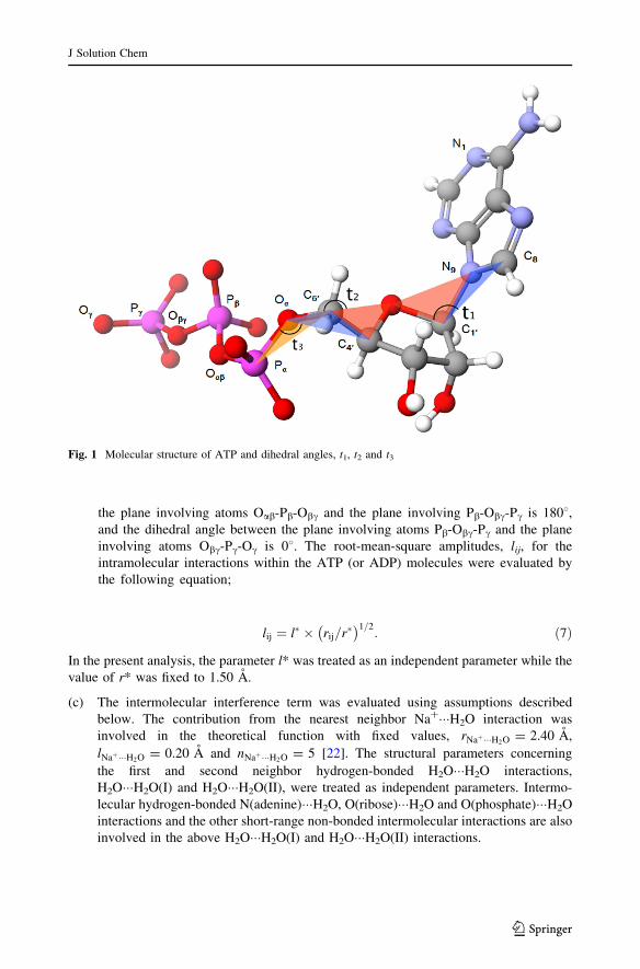
(b) The intramolecular interference term for an ATP (or ADP) molecule, iATPintra(Q) (or
iADPintra (Q)), was estimated for fixed dihedral angles, t1 (between the plane involving
C8(adenine)-N9(adenine)-C10(ribose) atoms and the plane involving atoms N9(ade-
nine)-C10(ribose)-O(ribose)), t2 (between the plane involving O(ribose)-C40(ribose)-
C50(ribose) atoms and the plane involving atoms C40(ribose)-C50(ribose)-O(phos-
phate)), and t3 (between the plane involving C40(ribose)-C50(ribose)-O(phosphate)
and the plane involving atoms C50(ribose)-O(phosphate)-Pa(phosphate)) as indicated
in Fig. 1. Intramolecular bond distances, rij, within the adenine, ribose and
triphosphate groups were fixed at values determined for the crystalline ATP salt [1].
The conformation of the phosphate group within ATP (or ADP) was assumed to
take a linear form in the present analysis which was suggested from previous NMR
[6] and DFT calculation [21] studies; dihedral angle between the plane involving
C50(ribose)-O(phosphate)-Pa(phosphate) and the plane involving Oa-Pa-Oab atoms is
1808 (where Oij denotes an oxygen atom binding with both Pi and Pj atoms), the
dihedral angle between the plane involving Oa-Pa-Oab atoms and the plane
involving Pa-Oab-Pb atoms is 08, dihedral angle between the plane involving atoms
Pa-Oab-Pb and the plane involving Oab-Pb-Obc is 1808, the dihedral angle between
J Solution Chem
123
the plane involving atoms Oab-Pb-Obc and the plane involving Pb-Obc-Pc is 1808,and the dihedral angle between the plane involving atoms Pb-Obc-Pc and the plane
involving atoms Obc-Pc-Oc is 08. The root-mean-square amplitudes, lij, for the
intramolecular interactions within the ATP (or ADP) molecules were evaluated by
the following equation;
lij ¼ l� � rij=r�� �1=2
: ð7Þ
In the present analysis, the parameter l* was treated as an independent parameter while the
value of r* was fixed to 1.50 A.
(c) The intermolecular interference term was evaluated using assumptions described
below. The contribution from the nearest neighbor Na?���H2O interaction was
involved in the theoretical function with fixed values, rNaþ���H2O = 2.40 A,
lNaþ���H2O = 0.20 A and nNaþ���H2O = 5 [22]. The structural parameters concerning
the first and second neighbor hydrogen-bonded H2O���H2O interactions,
H2O���H2O(I) and H2O���H2O(II), were treated as independent parameters. Intermo-
lecular hydrogen-bonded N(adenine)���H2O, O(ribose)���H2O and O(phosphate)���H2O
interactions and the other short-range non-bonded intermolecular interactions are also
involved in the above H2O���H2O(I) and H2O���H2O(II) interactions.
Fig. 1 Molecular structure of ATP and dihedral angles, t1, t2 and t3
J Solution Chem
123
(d) Parameters for the long-range random distribution of atoms, r0 and l0, in Eq. 6 were
allowed to vary independently.
(e) The least-squares fitting analysis was carried out for the observed interference term,
iobs(Q), in the range of 1.0 B Q B 22.0 A-1 for 2.05 mol% ATP and ADP solutions,
and 1.0 B Q B 25.0 A-1 for 2.0 and 1.0 mol% ATP and 2.0 mol% ADP solutions
using the SALS program [23]. The model function, imodel(Q), was evaluated by the
following equation,
imodel Qð Þ ¼ iintraZ Qð Þ þ iinter Qð Þ; Z ¼ ATP or ADPð Þ: ð8Þ
The R-factor defined below, was then determined for the best-fit result with fixed dihedral
angles of t1, t2 and t3:
Rt1;t2;t3 ¼X
iobs Qð Þ�imodel Qð Þ� �2
=X
iobs Qð Þ� �2�100 ð9Þ
(f) Rt1;t2;t3 was obtained for the fixed angles t1, t2 and t3 in the range of 0�–3608 with
angular interval of 308. Least-squares refinements were carried out for
12 9 12 9 12 = 1,728 times to obtain the values of Rt1;t2;t3 : The set of angles t1, t2and t3 which gives the minimum value of Rt1;t2;t3 corresponds to the possible
conformer of the ATP (or ADP) molecule.
The total distribution function, g(r), was evaluated by the Fourier transform of the
observed iobs(Q),
g rð Þ ¼ 1þ ð2p2qrÞ�1
ZQmax
0
Qobsi Qð Þsin Qrð Þ dQ; ð10Þ
The upper limit of the integral, Qmax = 20 A-1, was employed in the present study.
3 Results and Discussion
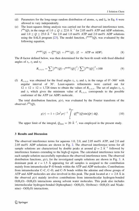
The observed interference terms for aqueous 1.0, 2.0, and 2.05 mol% ATP, and 2.0 and
2.05 mol% ADP solutions are shown in Fig. 2. The observed interference terms for all
sample solutions are characterized by double peaks at around Q = 2 A-1 followed by
interference features extending to the high-Q region. The calculated interference term for
each sample solution successfully reproduces the observed interference term. The observed
distribution functions, g(r), for the investigated sample solutions are shown in Fig. 3. A
dominant peak at r = 1.5 A appearing for all samples is assigned to the contribution
mainly from intramolecular P–O bonds within the ATP and ADP molecules. Contributions
from intramolecular C–C, C–O, and C–N bonds within the adenine and ribose groups of
ATP and ADP molecules are also involved in this peak. The peak located at r = 2.8 A in
the observed g(r) mainly involves contributions from intermolecular hydrogen-bonded
O(H2O)���O(H2O) interactions among solvent water molecules. This peak also includes
intermolecular hydrogen-bonded O(phosphate)���O(H2O), O(ribose)���O(H2O) and N(ade-
nine)���O(H2O) interactions.
J Solution Chem
123
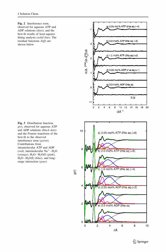
Fig. 2 Interference term,observed for aqueous ATP andADP solutions (dots), and thebest-fit results of least-squaresfitting analysis (solid line). Theresidual functions d(Q) areshown below
Fig. 3 Distribution function,g(r), observed for aqueous ATPand ADP solutions (black dots)and the Fourier transform of thebest-fit to the observedinterference term (green).Contributions fromintramolecular ATP and ADP(red), intermolecular Na?���H2O(orange), H2O���H2O(I) (pink),H2O���H2O(I) (blue), and long-range interaction (gray)
J Solution Chem
123
Structural features observed in the experimental g(r) in each solution are well repro-
duced by the calculated distribution function derived from the least-squares fitting pro-
cedure. In order to obtain information on the conformation of the ATP and ADP molecules,
the least-squares fitting procedure was carried out with fixed dihedral angles t1, t2 and t3 to
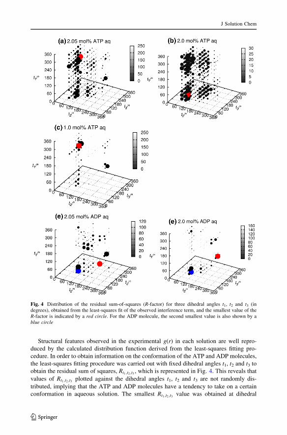
obtain the residual sum of squares, Rt1;t2;t3 , which is represented in Fig. 4. This reveals that
values of Rt1;t2;t3 plotted against the dihedral angles t1, t2 and t3 are not randomly dis-
tributed, implying that the ATP and ADP molecules have a tendency to take on a certain
conformation in aqueous solution. The smallest Rt1;t2;t3 value was obtained at dihedral
Fig. 4 Distribution of the residual sum-of-squares (R-factor) for three dihedral angles t1, t2 and t3 (indegrees), obtained from the least-squares fit of the observed interference term, and the smallest value of theR-factor is indicated by a red circle. For the ADP molecule, the second smallest value is also shown by ablue circle
J Solution Chem
123
angles of t1 = 240�–3008, t2 = 60�–908, and t3 = 120�–2408 for the ATP molecule. These
dihedral angles correspond to an unfolded conformation of the ATP molecule. All inde-
pendent parameters determined from the least-squares fit in the condition of fixed dihedral
angles t1, t2 and t3, which gives the smallest value of the R-factor, are summarized in
Table 1. The value of residual sum-of-squares (R-factor) for each solution ranges from
0.54 to 1.51 %, indicating that the least-squares fitting procedure was adequately per-
formed and assumptions applied in evaluating the theoretical interference term are valid.
The plot of Rt1;t2;t3 for the 2.05 and 2.0 mol% ATP solutions (Fig. 4a, b) indicates that
possible conformation are distributed around t1 = 240�–300� and 0�–908, which implies
that a broadened distribution of the t1 value occurs in these concentrated solutions. On the
other hand, the minimum Rt1;t2;t3 distribution seems more localized in the case of the
1.0 mol% ATP solution. The solute–solute (ATP���ATP and/or ATP���Na?) interactions
become more significant in concentrated solutions. This may cause the difference in the
minimum Rt1;t2;t3 distribution between the 2 and 1 mol% ATP solutions. The conformation
of the ATP molecules for dihedral angles t1 = 2408, t2 = 608, and t3 = 2408 show an
L-shape for the ATP molecule as seen in Fig. 5.
The distribution of the smallest Rt1;t2;t3 value for the ADP molecule in aqueous 2.05 and
2.0 mol% solutions was obtained at the dihedral angles of t1 = 120�–1508, t2 = 2708, and
t3 = 120�–2108 (see Table 1). The conformation of the ADP molecule for dihedral angles
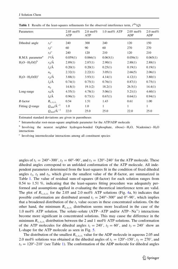
Table 1 Results of the least-squares refinements for the observed interference term, iobs(Q)
Parameters 2.05 mol%ATP
2.0 mol%ATP
1.0 mol% ATP 2.05 mol%ADP
2.0 mol%ADP
Dihedral angle t1/8 240 300 240 120 150
t2/8 60 90 60 270 270
t3/8 240 120 210 120 210
R.M.S. parametera l*/A 0.059(1) 0.066(1) 0.063(1) 0.056(1) 0.065(1)
H2O���H2O(I)b rij/A 2.89(1) 2.87(1) 2.90(1) 2.88(1) 2.88(1)
lij/A 0.20(1) 0.20(1) 0.25(1) 0.19(1) 0.19(1)
nij 2.32(1) 2.22(1) 3.05(1) 2.66(5) 2.06(1)
H2O���H2O(II)c rij/A 3.88(1) 3.93(1) 4.14(1) 4.12(1) 3.80(1)
lij/A 0.74(1) 0.75(1) 0.76(1) 0.87(1) 0.75(1)
nij 14.8(1) 19.1(2) 18.2(1) 26.5(1) 14.4(1)
Long-range r0/A 4.55(1) 4.78(1) 5.06(1) 5.21(1) 4.60(1)
l0/A 0.96(1) 0.73(1) 0.67(1) 0.64(1) 0.94(1)
R-factor Rt1 ;t2 ;t3 0.54 1.51 1.43 0.61 1.00
Fitting Q-range Qmin/A-1 1.0 1.0 1 1 1
Qmax/A-1 22.0 25.0 25.0 22.0 25.0
Estimated standard deviations are given in parenthesesa Intramolecular root-mean-square amplitude parameter for the ATP/ADP moleculeb Involving the nearest neighbor hydrogen-bonded O(phosphate, ribose)���H2O, N(adenine)���H2Ointeractionsc Involving intermolecular interactions among all constituent species
J Solution Chem
123
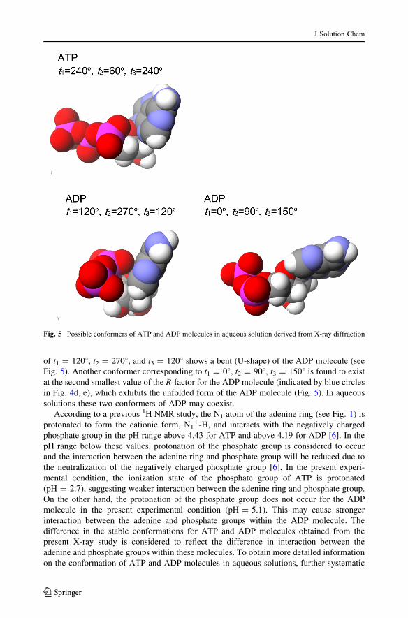
of t1 = 1208, t2 = 2708, and t3 = 1208 shows a bent (U-shape) of the ADP molecule (see
Fig. 5). Another conformer corresponding to t1 = 08, t2 = 908, t3 = 1508 is found to exist
at the second smallest value of the R-factor for the ADP molecule (indicated by blue circles
in Fig. 4d, e), which exhibits the unfolded form of the ADP molecule (Fig. 5). In aqueous
solutions these two conformers of ADP may coexist.
According to a previous 1H NMR study, the N1 atom of the adenine ring (see Fig. 1) is
protonated to form the cationic form, N1?-H, and interacts with the negatively charged
phosphate group in the pH range above 4.43 for ATP and above 4.19 for ADP [6]. In the
pH range below these values, protonation of the phosphate group is considered to occur
and the interaction between the adenine ring and phosphate group will be reduced due to
the neutralization of the negatively charged phosphate group [6]. In the present experi-
mental condition, the ionization state of the phosphate group of ATP is protonated
(pH = 2.7), suggesting weaker interaction between the adenine ring and phosphate group.
On the other hand, the protonation of the phosphate group does not occur for the ADP
molecule in the present experimental condition (pH = 5.1). This may cause stronger
interaction between the adenine and phosphate groups within the ADP molecule. The
difference in the stable conformations for ATP and ADP molecules obtained from the
present X-ray study is considered to reflect the difference in interaction between the
adenine and phosphate groups within these molecules. To obtain more detailed information
on the conformation of ATP and ADP molecules in aqueous solutions, further systematic
Fig. 5 Possible conformers of ATP and ADP molecules in aqueous solution derived from X-ray diffraction
J Solution Chem
123
studies on the pH and concentration dependences of the intramolecular structures of these
molecules will be necessary.
In order to obtain more detailed information on the molecular geometry of ATP and
ADP molecules and hydration structure of these molecules in aqueous solutions, the use of
neutron diffraction with 14N/15N and H/D isotopic substitution techniques [24–26] can be
an effective experimental method to distinguish intramolecular interactions relating to N
and H atoms within the adenine and ribose groups. This will be a future research project.
Acknowledgments The authors would like to thank Dr. Shinji Kohara (Japan Synchrotron ResearchInstitute) for his help during X-ray diffraction measurements. The synchrotron radiation experiments wereperformed with the approval of JASRI (Proposal Nos. 2011A1368 and 2012B1509).
References
1. Kennard, O., Isaacs, N.W., Motherwell, W.D.S., Coppora, J.C., Wamplar, D.L., Larson, A.C., Watson,D.G.: The crystal and molecular structure of adenosine triphosphate. Proc. R. Soc. Lond. A 325,401–436 (1971)
2. Larson, A.C.: Restrained refinement of disodium adenosine 50-triphosphate trihydrate. Acta Cryst. B34,3601–3604 (1978)
3. Sugawara, Y., Kamiya, N., Iwasaki, H., Ito, T., Satow, Y.: Humidity-controlled reversible structuretransition of disodium adenosine 50-triphosphate between dehydrate and trihydrate in a single crystallinestate. J. Am. Chem. Soc. 113, 5440–5445 (1991)
4. Burke, R.M., Pearce, J.K., Boxford, P.W.E., Bruckmann, A., Dessent, C.E.: Stabilization of excesscharge in isolated adenosine 50-triphosphate and adenosine 50-diphosphate multiply and singly chargedanions. J. Phys. Chem. A 109, 9775–9785 (2005)
5. Burke, R.M., Dessent, C.E.H.: Effect of cation complexation on the structure of a conformationallyflexible multiply charged anion: stability of excess charge in the Na?�adenosine 50-triphosphate dianionion-pair complex. J. Phys. Chem. B 113, 2683–2692 (2009)
6. Wang, P., Izatt, R.M., Oscarson, J.L., Gillespie, S.E.: 1H NMR study of protonation and Mg(II)coordination of AM, ADP, and ATP at 25, 50 and 70 �C. J. Phys. Chem. 100, 9556–9560 (1996)
7. Kohara, S., Itou, M., Suzuya, K., Inamura, Y., Sakurai, Y., Ohishi, Y., Takata, M.: Structural studies ofdisordered materials using high-energy X-ray diffraction from ambient to extreme conditions. J. Phys.Condens. Matter 19, 1–15 (2007)
8. Kohara, S., Suzuya, K., Kashihara, Y., Matsumoto, N., Umesaki, N., Sakai, I.: A horizontal two-axisdiffractometer for high-energy X-ray diffraction using synchrotron radiation on bending magnetbeamline BL04B2 at SPring-8. Nucl. Instrum. Method Phys. Res. Sect. A 467, 1030–1033 (2001)
9. Issiki, M., Ohishi, Y., Goto, S., Takeshita, K., Ishikawa, T.: High-energy X-ray diffraction beamlineBL04B2 at SPring-8. Nucl. Instrum. Method Phys. Res. Sect. A 467, 663–666 (2001)
10. Waseda, Y.: The Structure of Non-Crystalline Materials. McGraw–Hill, New York (1980)11. Sasaki, S.: X-ray absorption coefficients of the elements (Li to Bi, U), pp. 16–90. KEK Report, Koide
(1990)12. Hajdu, F.: Revised parameters of the analytic fits for coherent and incoherent scattered X-ray intensities
of the first 36 atoms. Acta Crystallogr. Sect. A 28, 250–252 (1972)13. Kluwer, C.: International Tables for Crystallography. Academic Press, London (1999)14. Habenshuss, A., Spedding, F.H.: The coordination (hydration) of rare earth ion in aqueous chloride
solutions from X-ray diffraction. I. TbCl3, DyCl3, ErCl3, TmCl3, and LuCl3. J. Chem. Phys. 70,2797–2806 (1979)
15. Narten, A.H., Danford, M.D., Levy, H.A.: X-ray diffraction study of liquid water in the temperaturerange 4–200 �C. Discuss. Faraday Soc. 43, 97–107 (1967)
16. Caminiti, R., Cucca, P., Monduzzi, M., Saba, G.: Divalent metal-acetate complexes in concentratedaqueous solutions. An X-ray diffraction and NMR spectroscopic study. J. Chem. Phys. 81, 543–551(1984)
17. Ohtaki, H., Fukushima, N.: A structural study of saturated aqueous solutions of some alkali halides byX-ray diffraction. J. Solution Chem. 21, 23–38 (1992)
18. Clarke, H., Granada, J.R., Dore, J.C.: Structural studies of tetrachloride liquids I. Pulsed neutronscattering by carbon tetrachloride—Molecular structure. Mol. Phys. 37, 1263–1279 (1979)
J Solution Chem
123
19. Powles, J.G.: The structure of the water molecule in liquid water. Mol. Phys. 42, 757–765 (1981)20. Kameda, Y., Uemura, O.: The intramolecular structure of oxonium ion in concentrated aqueous deu-
terochloric acid solutions. Bull. Chem. Soc. Jpn. 65, 2021–2028 (1992)21. Akola, J., Jones, R.O.: ATP hydrolysis in water—A density functional study. J. Phys. Chem. B 107,
11774–11783 (2003)22. Kameda, Y., Sugawara, K., Usuki, T., Uemura, O.: Hydration structure of Na? in concentrated aqueous
solutions. Bull. Chem. Soc. Jpn. 71, 2769–2776 (1998)23. Nakagawa, T., Oyanagi, Y.: Recent Development in Statistical Inference and Data Analysis, p. 221.
North-Holland, Amsterdam (1980)24. Enderby, J.E., Neilson, G.W.: X-ray and neutron scattering by aqueous solutions of electrolytes. In:
Franks, F. (ed.) Water A Comprehensive Treatise, pp. 1–6. Plenum Press, New York (1979)25. Kameda, Y., Maki, A., Amo, Y., Usuki, T.: Partial pair correlation functions of highly concentrated
aqueous urea solutions determined by neutron diffraction with 14N/15N and H/D isotopic substitutionmethods. Bull. Chem. Soc. Jpn. 83, 131–144 (2013)
26. Mason, P., Neilson, G.W., Saboungi, M.-L., Brady, J.W.: The conformation of a ribose derivatives inaqueous solution: a neutron-scattering and molecular dynamics study. Biopolymers 99, 739–745 (2013)
J Solution Chem
123