Embed Size (px)
Citation preview
Biophysical Chemistry 17 (1983) 1-I 1 Eisevier Biomedical Press
CONFORMATIONAL CHARACTERISTICS OF THE DINUCLEOSIDE TRIPHOSPHATE pCpGp FROM ENERGY-MINIMIZATION STUDIES
A. ANUKANTH and P-K. PONNUSWAMY Departmerzr of Physics. Bhararhidaran Unioersir_v. TiruchirapaIIi - 620 020. Tumilnadu, India
Received 28th January 1982 Rewsed manuscript received 13th July 1982 Accepted 6th September 1982
Key words: pCpGp; Triphosphare inreracrion; Conformorion; RN.4 buckbwe
The influence of the 3’- and 5’- terminal phosphares on the conformational characteristics of the dinucleoside monophos- phate CpG is described in this paper. The computed potential energy of the system is minimized with respecr to the relevant 10 dihedral angles permitting the two sugar rings to adopt the alternative puckering states. 2, and 3,. Of the 84 conformations considered, 22 become energetically accessible. The familiar A-. B-. Z- and Watson-Crick-type backbone states of DNA subunits become low-energy forms for this RNA unit pCpGp also. The Watson-Crick-type backbone is invariably preferred in all the four sugar pucker sequences, indicating its importance in the dynamics of sugar pucker fluctuations and in the DNA-RNA association. The interphosphate geometries and the possible hydrogen-bonded states are discussed in relation to the varied folded/extended polynucleotide structures.
1. Introduction
Suggestions on the possibility of alternative structures of DNA [l-6] have given a new thrust to the research activity on the conformational characteristics of small and specific nucleotides. The proposed new structures incorporate novel local conformations of the subunits, hitherto ne- glected or underestimated. The existence of differ- ent kinds of conformational (A, B, C and Z) states in the polynucleotide strands, their interconver- sion, the types of helical twist and interstrand alignments, and finally, the overall folding have all become problems of new investigations, both in theoretical and experimental fields. In a few recent articles [7- 111, we have reported our systematic theoretical studies on a number of monomeric and dimeric subunits of DNA and RNA, anti a tri- nucleoside diphosphate unit, d(ApApA). A di- nucleoside triphosphate, pCpGp, is studied in the present report. The special feature of this unit is the existence of two terminal phosphate groups
0301_4622/83/OOOO-OOOO/SO3.00 8 1983 Elsevier Biomedical Press
enclosing a dimeric segment; the dimeric uni: has the bases cytosine and guanine and this pair of bases is under extensive study under different con- ditions [ 12- 141. The conformational characteristics of the monophosphate form CpG were studied previously by us [15]. The present study on the triphosphate pCpGp brings out many salient fea- tures, specifically, the iniluence of the interactions of the terminal phosphates separated by a di- nucleoside monosphosphate segment.
2. Methods
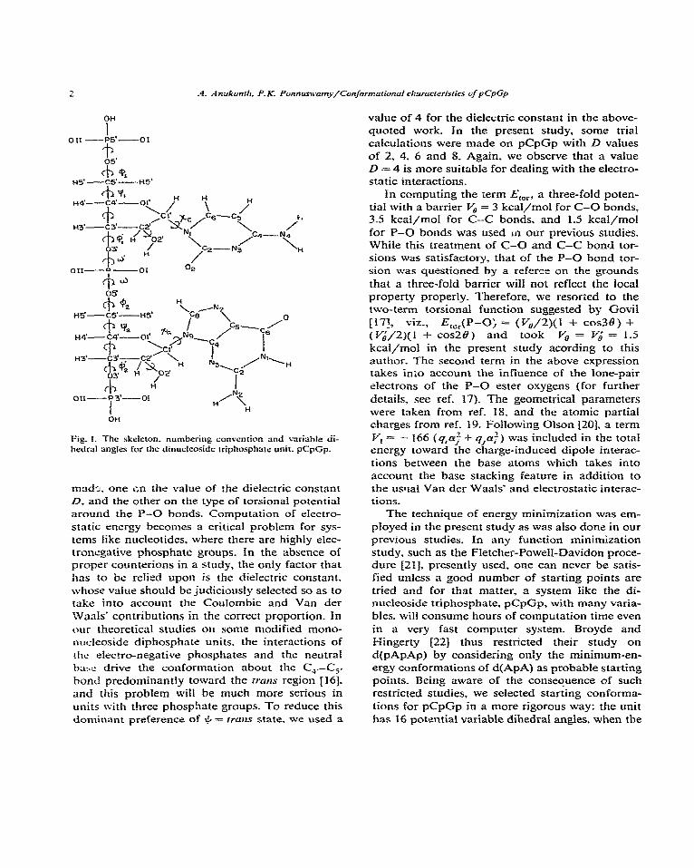
Fig. 1 depicts the skeleton of pCpGp with its variable dihedral angles. The potential energy of the system was considered as E,o,31 = E,, + E, +
Em + Em,- where the subscripts nb, es. hb and tar represent, respectively, nonbonded, electrostatic, hydrogen bonding and torsional; these terms of the total energy were computed using expressions and parameters reported in our previous studies [7,8]. In the present study two additionai tests were
OH
I ou -P5’-ot
on
Fig. I. The skekton. numbering convcnrion and varinbie di- hedral angles for the dinuclcnsidc rriphosphate unit. pCpGp.
mad--_. one on the value of the dielectric constant D. and the other on the type of torsional potential around the P-O bonds. Computation of electro- static energy becomes a critical probIem for sys- tems like nucleotides, where there are highly elec- tronegative phosphate groups. in the absence of proper counterions in a study, the only factor that has to be refied upon is the dielectric constant. whose value should be judiciously selected so as to take into account the Coulombic and Van der WaaIs’ contributions in the correct proportion. In our theoretical studies on some modified mono- nucleoside diphosphate units. the interactions of the electro-negaticte phosphates and the neutral ba:.~ drive the conformation about the C,.-C,. bond predominantly toward the rrcz~ts region 1161. and this problem wiil be much more serious in units with three phosphate groups. To reduce this dominant preference of I& = rtuns state. wc used a
value of 4 for the dielectric constant in the above- quoted work. fn the present study, some trial calculations were made on pCpCp with D values of 2, 4. 6 and 8. Again, we observe that a value D = 4 is more suitable for dealing with the electro- static interactions.
In computing the term E,,,, a three-foId poten- tial with a barrier V, = 3 kcal/mol for C-O bonds, 3.5 kcal/mol for C--C bonds, and 1.5 kcal/mol for P-O bonds was used ta our previous studies. Whiie this treatment of C-O and C-C bond tor- sions was satisfactory, that of the P-O bond tor- sion was questioned by a referee on the grounds that a three-fold barrier will not reflect the local property properly. Therefore, we resorted to the two-term torsional function suggested by GoviI 1171, viz., E,,,(P-0; = (V&2)(1 + cos38) + ( 5/2)(1 + ~0~20) and took kcal/moi in the present study acording to this author. The second term in the above expression takes into account the infiuence of the lone-pair electrons of the P-O ester oxygens (for further details, see ref. 17). The geometrical parameters were taken from ref. 18, and the atomic partial charges from ref. 19. Foliowing Olson 1201, a term Vi = - 166 (q,$ -t ~,cK:) was included in the total energy toward the charge-induced dipole interac- tions between the base atoms which takes into account the base stacking feature in addition to the usral Van der Waais’ and electrostatic interac- tions.
The technique of energy minimization was em- pIoyed in the present study as was also done in our previous studies. In any function minimization study. such as the Fletcher-Powell-Davidon proce- dure [21]. presently used, one can never be satis- fied unless a good number of starting points are tried and for that matter, a system like the di- nucleoside triphosphate, pCpGp, with many varia- bles. will consume hours of computation time even in a very fast computer system. Broyde and I-iingerty 1221 thus restricted their study on d(pApAp) by considering only the minimum-en- ergy conformations of d(ApA) as probable starting points_ Being aware of the conseouence of such restricted studies, we selected starting conforma- tions for pCpCp in a more rigorous way: the unit has 16 potrntial variable dihedral angles, when the
3
**orrm* oOPc#PvrQ ---me...
I I 1
two terminal phosphate hydroxyl oxygens are trea:ed as united-atom entities (fig. 1). The end phosphate groups were kept in staggered states around the O-P bonds such that the P-OH bond is ITUIZS to the O-C bond, thus eliminating two variabtes. Similarty. in :he sugar rings the 2‘-hy- droxyl bond was kept in the gauche- position (in accordance with crystal-state observation) to the C,.-C,. bond, again eliminating two variables. The sugar ring was treated as a fixed system in the 3, or 2,: state. thus eliminating the two ring $’ angles from the variables’ set. Thus. the remaining ten variables 9,. +,. xc* +;. w;, w2, e2, &. Xo and +; alone vvere considered. taking the following values as the best starting locations for them: xc, xc = JOO: a;. +,; = - 1300; (?,. &= 180”; qt. w. w’= 60°. 180°, - 60*: and a$2 = 60”. The possible com- binations of these values yield 27 (1 x 1 x 1 x I x 3 X 3 X 3 X I X 1 X 1) conformations for a specific sugar pucker sequence_ For the four sugar pucker sequences. 3,-3,. 3,2,. 2.-3, and 2,2,. 108 conformations are thus possible, of which we ex- cluded the 24 conformations with (w’, w) = (g+ _ .g- ) as these are not observed in any of the experi- menta! studies reported hitherto. The remaining 84 conformations were considered as starting points for the minimization study. For each of the start- ing points. the computed total energy was mini- mized by allowing the ten dihedral angles to vary simultaneously so as to meet the condition that the difference in energies of two consecutive intera- tions is less than or equa1 to 0.05 kcal/moI. and the gradient for each variable is less than 0.5. To standardise the energy values of the two different sugar pucker states. the internal energies of sugar rings were computed and added to the final mini- mized energy values as detailed in the article of Ponnuswnmy and Thiyagarajan f23J. All the hy- drogens. except the terminal phosphate hydroxyl ones. were consi&red expV1cit\y.
3. Results
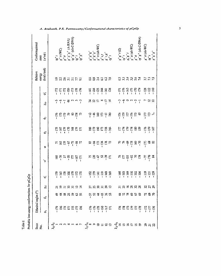
Out of the 84 starting conformational states considered. only 22 states moved toward an energy difference of LIE = 10 kcal/mol with respect to the lowest energy located. These 22 probable confor-
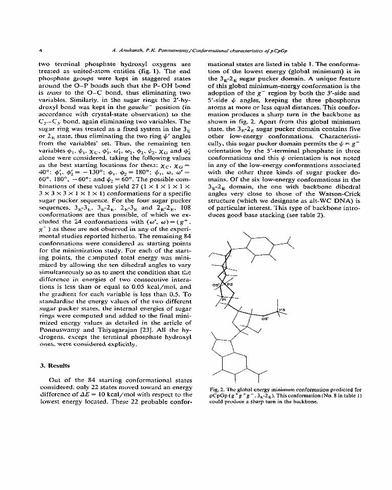
mational states are listed in table 1. The conforma- tion of the lowest energy (global minimum) is in the 3,-2, sugar pucker domain. A unique feature of this global minimum-energy conformation is the adoption of the g- region by both the 3’-side and S-side I& angies, keeping the three phosphorus atoms at more or less equal distances. This confor- mation produces a sharp turn in the backbone as shown in fig. 2, Apart from this global minimum state, the 3,-2, sugar pucker domain contains five other low-energy conformations_ Characteristi- cally, this sugar pucker domain permits the + = g- orientation by the 5’-terminal phosphate in three conformations and this + orientation is not noted in any of the low-energy conformations associated with the other three kinds of sugar pucker do- mains_ Of the six low-energy conformations in the 3,2, domain, the one with backbone dihedral angles very close to those of the Watson-Crick structure (which we designate as ah-WC DNA) is of particular interest. This type of backbone intro- duces good base stacking (see table 2).
Fig. 2. The global energy minimum conformation predicted for pCpGp(g+g+g-. 3,2,). Thisconformation(No. 8 in table 1) could produce a sharp turn in the backbone.
A. Anukanrh. 9 K. Ponn~amy/Confonnafionol chnracreristics of pCpGp
Table 2
Base stacking geometry predicted for the RNA subunit pCpGp
sugar pucker
Conforma- tional state
Base Mean distance stacking between
energy base planes (kcal/mol) (A)
Angle betwsen base planes
(“)
Overlapping area
(I)”
3,-3, A-RNA - 1.913 3.9 35 35 WC - 2.960 3.3 10 94
3,-2, ah-WC - 1.766 4.0 31 65 2E-2E rg+r -0.8-m 4.8 32 12
n Overlapping is 100% when the base with smaller area is completely overlapped by the base of larger area.
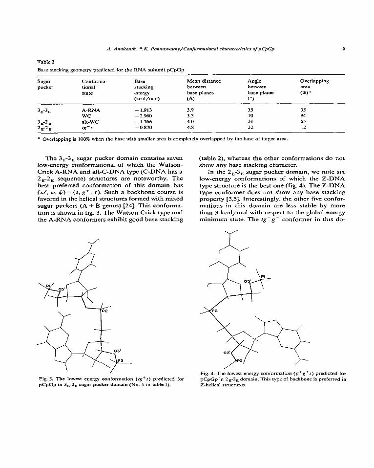
The 3,-3, sugar pucker domain contains seven low-energy conformations, of which the Waison- Crick A-RNA and ah-C-DNA type (C-DNA has a 2,-2, sequence) structures are noteworthy. The best preferred conformation of this domain has (W’,W,+)=(t,g+. t)_ Such a backbone course is favored in the helical structures formed with mixed sugar puckers (A + B genus) [24]. This conforma- tion is shown in fig. 3. The Watson-Crick type and the A-RNA conformers exhibit good base stacking
Fig. 3. The lowest energy conformation (rg+t) predicted for pCpGp in 3,-2, sugar pucker domain (No. 1 in table 1).
(table 2), whereas the other conformations do not show any base stacking character.
in the 2,3, sugar pucker domain, we note six low-energy conformations of which the Z-DNA type structure is the best one (fig. 4). The Z-DNA type conformer does not show any base stacking property [3,5]. Interestingly, the other five confor- mations in this domain are less stable by more than 3 kcal/mol with respect to the global energy minimum state. The fg-g’ conformer in thts do-
Fig.4. The lowest energy coniormatian (g+g+r) predicted for pCpGp in 2,-3, domain. This type of backbone is preferred in Z-helical structures.
6 A. Anukanzh_ P. K. Ponn~anl~/Confornlorionol characreristics of pCpGp
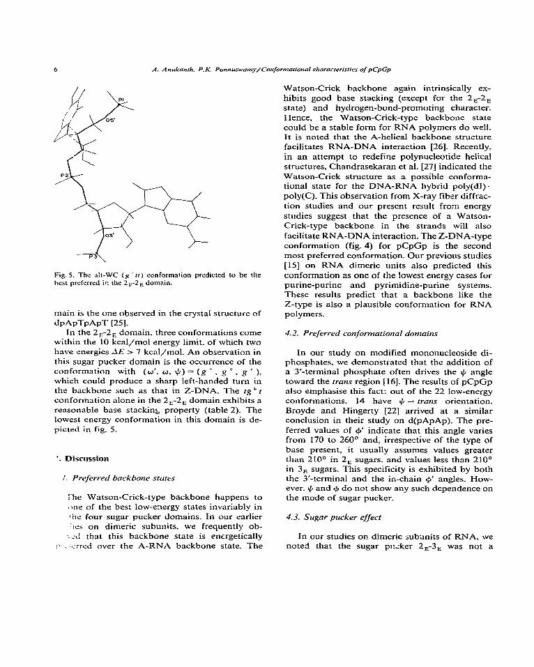
Fig. 5. The St-WC (g-n) conformation predicted to be the best preferred ir. the 2,-2, domain.
main is the one observed in the crystal structure of dpApTpApT [25].
In the 2,-2, domain. three conformations come within the 10 kcal/mol energy limit. of which two have energies dE > 7 kcal/mol. An observation in this sugar pucker domain is the occurrence of the conformation with (w’. o, 4) = (g + , g + , g I ), which could produce a sharp left-handed turn in the backbone such as thai in Z-DNA. The fg+t conformation alone in the 2a-2, domain exhibits a reasonable base stacking property (table 2). The lowest energy conformation in this domain is de-
picted in fig_ 5.
‘. Discussion
1. Preferred backbone states
I-he Watson-Crick-type backbone happens to *ne of the best low-energy states invariably in :he four sugar pucker domains. In our earlier :!a on dimeric subunits. we frequently ob-
: <d that this backbone state is energetically ;‘. . :c:rrcd over the A-RNA backbone state. The
Watson-Crick backbone again intrinsically ex- hibits good base stacking (except for the 2a-2, state) and hydrogen-bond-promoting character. Hence, the Watson-Crick-type backbone state could be a stable form for RNA polymers do well. It is noted that the A-helical backbone structure facilitates RNA-DNA interaction [26]. Recently, in an attempt to redefine polynucleotide helical structures, Chandrasekaran et al. [27] indicated the Watson-Crick structure as a possible conforma- tional state for the DNA-RNA hybrid poly(dI)- poly(C). This observation from X-ray fiber diffrac- tion studies and our present result from energy studies suggest that the presence of a Watson- Crick-type backbone in the strands will also facilitate RNA-DNA interaction. The Z-DNA-type conformation (fig. 4) for pCpGp is the second most preferred conformation. Our previous studies [IS] on RNA dimeric units also predicted this conformation as one of the lowest energy cases for purine-purine and pyrimidine-purine systems. These results predict that a backbone like the Z-type is also a plausible conformation for RNA polymers_
4.2. Preferred conformational domains
In our study on modified mononucleoside di- phosphates, we demonstrated that the addition of a 3’-terminal phosphate often drives the I$ angle toward the tram region [ 161. The results of pCpGp also emphasise this fact: out of the 22 low-energy conformations, 14 have + = trans orientation_
Broyde and Hingerty [22] arrived at a similar conclusion in their study on d(pApAp). The pre- ferred values of cp’ indicate that this angle varies from 170 to 260° and, irrespective of the type of base present, it usually assumes values greater than 21CP in 2, sugars, and values less than 210° in 3, sugars. This specificity is exhibited by both the 3’-terminal and the in-chain 9 angles. How- ever. I$ and + do not show any such dependence on the mode of sugar pucker_
4.3. Sugar pucker effect
In our studies on dimeric subunits of RNA, we noted that the sugar pi:;ker 2,-3. was not a
7
favorable state and no conformation was predicted the 3,-2, segments of the crystal structures of in this domain within an energy limit of 5 kcal/mol tRNA-Phe molecules [28-311. For a similar back- over the lowest energy state located. Interestingly, bone course. the 3,-2, sugar state sometimes brings the present results on pCpGp indicate that the the phosphates closer when compared to the 3,-3, addition of the 5’-terminal phosphate brings in the state and consequently forced + to move toward 2, pucker at 3’-ribose as a favorable case. The 22 the g- region. Another interesting point to be low-energy conformations for pCpGp are distrib- noted is the base-base gap in the alt-WC con- uted in the four sugar pucker domains, respec- former. In this conformation a wider vertical gap tively, seven in 3,-3,, six each in 3,-2, and 2,-3,, is introduced between the bases due to the flipping and three in 2,-2,. The energies of the conforma- of the 3’-sugar from the 3, to the 2, state (normal tions indicate that 2,-2, is the least preferred WC to ah-WC) without large alterations in cp’ and domain. In the 3,2, domain, three out of the six + orientations: this gap provides enough room for low-energy conformations have the # = g- orien- heavy drugs of complex shapes to intercalate be- tation and such a preference of I& is observed in tween the bases.
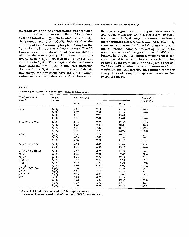
Table 3
Interphosphate geometries of the low-ene.gv conformations
Conformational state =
rg+r
g- tr (WC-DNA)
g+rt
‘g-g- (C-DNA)
g-g-g+ (A-RNA) tg-2 x-g+ s+s+??-
Sugar pucker
3,-3, 3,-2, 2.F3, 2,-2,
3,3, 3,-2, 2,-3, 2&E
3E3E 3,2, 2,3,
3,3, 2,-3,
3,-3, 3E-3E
3,-3, 3,-2,
Distance (A)
P,-P,
6.21 6.07 6.95 7.01
6.04 5.23 6.97 7.05
6.44 4.73 6.90
6.35 6.94
6.32 6.33 6.25 5.13
P_-P? PI-P3
7.17 12.18 7.3 1 12.19
7.20 13.49 7.41 13.47
7.22 12.30 7.33 10.80 7.20 13.18 7.42 12.90
7.18 IO.72 7.47 7.37 7.21 11.20
6.45 11.42 6.54 13.20
6.73 12.78 7.21 9.46
7.24 12.14 6.20 6.6 1
Angle (“)
(P,-P,-P,)
139.2 139.0 157.8 149.4
145.9 120.3 147.3 132.0
104.1 69.2
105.7
132.4 170.4
170.1 88.4
135.1 69.7
s+s-s- 3,2, 6.05 5.77 8.35 89.8
s-r&- 3E-2E 4.89 6.24 8.96 107.2 g+g+ I (Z-DNA) 2,-3, 7.00 7.15 11.43 108.7
s-s-r 2,-3, 7.2 I 7.13 11.76 111.5
s+s*s+ 2E-2, 7.13 6.75 8.65 76.8
11, h 3,-3, 7.24 6.25 12.14 135.1 3,2, 7.72 6.25 12.53 133.8 2,-3, 7.24 6.98 14.10 176.1
2E-2E 7.20 6.98 14.17 176.8
il See table 1 for the dihedral angles of the respective states. b Reference states computed (with a’= o = + = 180°) for comparison.
4.4. Phosphate effect am? interphosphate geometries
Theoretical studies on mononucleosides and di- nucleoside monophosphates have correctly predic- ted the importance of the # = g* o_<entaiion, but fliled to bring to light the importance of the + = I~UU)ZS orientation: the $J = rrans state emerges as an tzp~ally probable case when a phosphate is added at the 3’-terminus. The + = trans conforma- tion is often suggested to be the case in many of the fiber diffraction studies on poIyr.s;!?otides_ In a dinucleoside triphosphate, such as pCpGp, each base has one 3’- and one S-phosphate. and hence it is a good model system to be investigated for purpose of extrapolating the results of the subunit system to polynucieotides. Ths presence of the 5’-phosphate in a few specific situations forces the
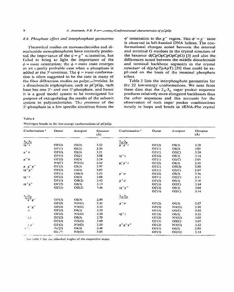
Table 4
Hydrogen bonds in the low-ener&y conformations of pCpGp
6’ orientation to the g- region_ This @’ = g- state is observed in left-handed DNA helices. The con- formational changes noted between the internal and terminal G residues in the crystal structure of the hexamer d(CpGpCpGpCpG) [3] and also the differences noted between the middle dinucleoside and termindi backbone segments in the crystaf structure of d(pApTpApT) [25] thus could be ex- pIEmed on the basis of the terminal phosphate ePfect.
Table 3 Iists the interphosphate geometries for the 22 low-energy conformations. We note from these data that the ZE-3n sugar pucker sequence produces reIativeIy more elongated backbones than the other sequences and this accounts for the observation of such sugar pucker combinations mostIy in loops and bends in tRNA-Phe crystal
Conformation a DOnOr Acceptor Distance Conformation” DOnOr Acceptor Distlrnce
(A) (A)
3,-3, i.9ti cw(2)
02,(l) g*Ir 02y2)
02’( 1)
g-rr 02‘(2)
N4(C)
R-R-R- OT(2)
1.4 - s + OZ’(2) or(l)
rg-I ozq 2) 02’( 1)
rg‘g’ OT(2) OT(l)
a(3) 3.23
Of(Z) 3.30
OI(3) 3.21
ON2) 3.08
ON31 3.24
N7(G) 3.42
W3) 2.97
W3) 3.07 0Iff.Q 3.22
OI(3) 3.0s OH(Z) 3.43
OR3) 3.13 011(Z) 3.46
oq2, ozq 2)
02’(2) i.r 02x2)
027 2-j i_lf - 02’(Z)
: : .7i’( 2) 02 ;?)
.._ ~
OIf3) N3(Gt X3(G)
OK3) N3(G) cfI(3)
2.80 3.35 3.33 3.29 3.20 2.70 3.00
2.59 3.48 3.05
ON3 OI(2)
02(C) OI(3) 02(C) O&3) 011(Z)
O2(Ct 0X(3) 02(C) OK3) 02(C) W3) 02(C)
3.28 3.01 3.24 3.14 3.01 3.35 2.88 2.87 3.16 3.11 3.16 3.24 3.04 3.14
2,-2,
g-rr 02’(2) OK3) 2.87 01’( 2) NJ(G) 2.98 02’( 1) 02(C) 3.02
tg”r 02’(2) w3) 3.22 Ot’(Z) N3fG) 3.05 Oz’(I) 02(C) 3.07
s+.?*s+ 02’(2) N3(Gl 3.33 02’(l) Oi(Z) 1.95 021(l) OXG) 3.14
- ’ Scu r.xh!o 1 for &c dihedral angles of the respective states.
A. Anukanth, P.K. Ponnur~amy/Con~~rmit;onal choracrerisrics of pCpGp
”
\
IO A. And-anth. P.K. Ponntls,~am~/Confor~?tationaI characteristics 01 pCpGp
models. A perusal of the interphosphate angles suggests that the 3,-2, sugar pucker sequence, in
comparison with the other sequences, could pro- duce sharper turns in the backbones, whereas the 2t1-3. pucker sequence could produce extended
backbones. These observations emphasise the fact that the flipping of the sugar pucker between the
3, and 2, modes of 3’-ribose alters the backbone
course appreciably_
Our earlier theoretical study [ 1 I] and a recent NMR study by Bolton and Kearns [32] described
the association features of a water molecule with the 2’-OH group in RNA subunits and also the
influence of solvent on backbone folding. In the present study we have analysed the intrastrand hydrogen bonds in the resultant 22 low-energy
conformations. Table 4 lists the noted hydrogen
bonds. In sorting out the hydrogen-bonded states, only the X - - - Y distance WCS considered and the
linearity condition of X-H - - -Y was not im-
posed. since it could be achieved with minor alter- ations of the angles_ In conformity with solution studies. we note in many low-energy conforma- tions that the ribose oxygen atoms 02’(l) and 02’(2) form hydrogen bonds with one of the 3’-
phosphate oxygens. These hydrogen bonds are. however. broken when the backbone angle o’ takes
up the g- orientation as this condition keeps the central phosphate group away from the 2’-OH of the first rihose unit. Further. these specific hydro-
gen bonds are also not feasible when +’ assumes
values greater than 200”. The Watson-Crick-type conformation alone permits interbase hydrogen
bonding. Intranucleotide hydrogen bonds are
roted between one of the acceptor atoms (N3 for ; and 0, for C) of the bases and the 2’-OH group
:r all the conformations associated with the 2,
ugar state. The 2, sugar pucker. usually. brings ic‘ L’-OH group nearer to the base unit.
Fkvibili~v in the backbone structures
\ comparison of the bzckbone states of the lr’,ws conformations listed in table 1 provides
i!t!ch as to the flesibility inherent in a few specific
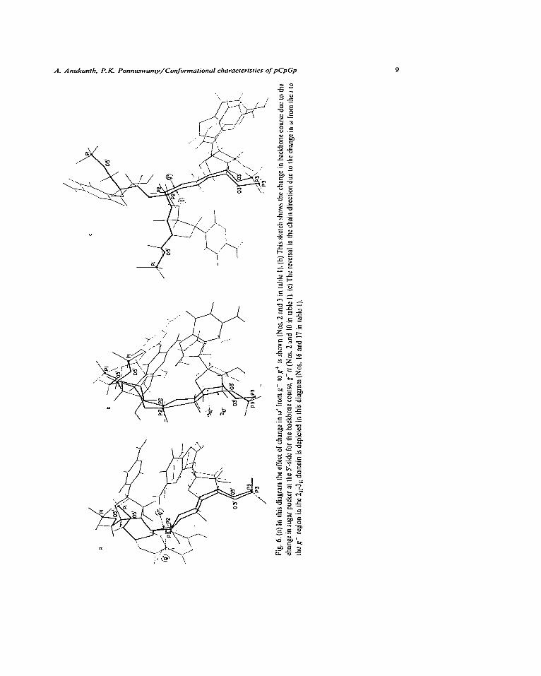
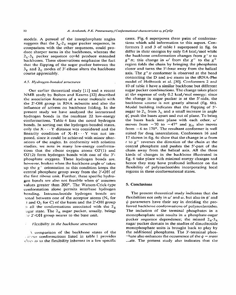
cases. Fig. 6 superposes three pairs of conforma- tions which add information to this aspect. Con-
formers 2 and 3 of table 1 superposed in fig. 6a differ in their energies by only 0.4 kcal/mol while the backbone conformation changes from g- tt to
g+ttt; this change in w’ from the g- to the g+ region folds the chain by bringing the phosphates
closer and turns the S-base away from the helical
axis. The gc tt conformer is observed at the bend
connecting the D and a-c stems in the tRNA-Phe model of Holbrook et al. [30]. Conformers 2 and
10 of tabie 1 have a similar backbone but different sugar pucker combinations_ The change takes place at the expense of only 0.2 kcal/mol energy: since the change in sugar pucker is at the 5’-side, the backbone course is not greatly altered (fig. 6b).
Model building indicates that the flipping of S- sugar to 2, from 3, and a small increase in angle
+; push the bases apart and out of plane. To bring
the bases back into plane with each other, w‘
moves from - 70 to -47” and ho also moves from -6 to 170°. The resultant conformer is well
suited for drug intercalation_ Conformers 16 and
17 shown in fig. 6c show that the change in w from t to g- reverses the direction of the chain at the central phosphate and pushes the 5’-part of the chain away from the helical axis. All the three
kinds of changes in the backbone illustrated in fig. 6 take place with minimal energy changes and hence they may have profound influence on the
flexibility of polynucleotides incorporating local regions in these conformational states.
5. Conclusions
The present theoretical study indicates that the
flexibilities not only in w’ and w, but also in 9’ and
+ parameters have their say in deciding the pre-
ferred backbone conformations of polynucleotides.
The inclusion of the terminal phosphates in a monophosphate unit results in a phosphate-sugar
pucker sequence dependence; the missed 2,-3,
sugar pucker domain in the studies of dinucleoside monophosphate units is brought back to play by
the additional phosphates. The 3’-terminal phos- -hate also enhances the occurrence of the $J = tram
_&e. The present study also indicates that the
A. Anukanrh, P.K. Ponnur~a~l~/Conformorionol characteristics of pCpGp II
Z-type backbone state originally predicted for DNA is possible for RNA molecules also. The Watson-Crick-type backbone state seems to be one of the favorable conformations for DNA-RNA interaction.
References
I G.A. Rodley. R.S. Scobie. R.H.T. Bates and R.W. Lewitt.
Proc. Natl. Acad. Sci. U.S.A. 73 (1976) 29.59.
2 V. Sasisekharan, N. Patta’%raman and G. Gupta. Proc.
Natl. Acad. Sci. U.S.A. 75 (1978) 4092.
3 A.HJ. Wang. G.J. Quigley. F.J. Kolpak. J.L. Crawford.
J.H. van Boom, G. van der Mare1 and A. Rich, Nature 282
( 1979) 680.
4 S. Amott. R. Chandrasekaran. D.L. Birdsall, A.G. Leslie
and R.L. Ratcliff. Nature 283 (1980) 743.
5 H. Drew. T. Takano, S. Tznaka, K. Itakura and R.E. Dickerson, Nature 286 (1980) 557.
6 J.L. Crawford, F.J. Kolpak, A.H.J. Wang. G.J. Quigley.
J.H. van Boom, G. van der Mare1 and A. Rich. Proc. NatI.
Acad. Sci. U.S.A. 77 (1980) 4016.
7 P. Thiyagarajan and P.K. Ponnuswamy. Biopolymers 17
(1978) 533.
8 P. Thiyagarajan and P.K. Ponnuswamy. Biopolymers 17
(1978) 2143.
9 P.K. Ponnu;wnmy and P. Thiyagarajan. Biochem. Biophys.
Res. Cammun. 89 (1979) 3 :4.
10 P. Thiyagarajan and P.K. Ponnuswamy. Biopolymers 18
(1979) 789.
11 P.K. Ponnuswamy and P. Thiyagarajan. Biopolymers 17 (1978) 2503.
12 J.T. Finch, L.C. Lutter, D. Rhodes. R.S. Brown, B. Rush-
ton. M. Levitt and A. Klug. Nature 269 (1977) 29. 13 J.C. Wang. Proc. Natl. Arad. Sci. U.S.A. 76 (1979) 200. 14 S.B. Zimmerman and B.H. Pheiffer. Proc. Natl. Acad. Sci.
U.S.A. 76 (1979) 2703.
15 P. Thiyagarajan and P.K. Ponnuswamy. Biophys. J. 35 (1981) 753.
16 P.K. Ponnuswamy and A. Anukanth. J. Theor. Biol. 96 (1982) 233.
17 G. Govil. Biopolymers 15 (1976) 2303. 18 V. Sasisekharan an3 A.V. Lakshminarayanan. Biochim.
Biophys. Acta 204 (1970) 49. 19 V. Renugopalakrishnan. A.V. Lakshiminarayanan and V.
Sasisekharan, Biopolymers 10 (1971) 1159. 20 W.K. Olson, Biopolymers 17 (1978) 1015.
21 R. Fletcher and M.J.D. Powell, Comput. J. 6 (1963) 163. 22 S. Broyde and B. Hingerty. Nucleic Acids Res. 6 (1979)
2165.
23 P.K. Ponnuswamy and P. Thiyagarajan. Biophys. J. 35 (1981) 731.
24 S. Arnott. R. Chandrasekaran. P.J. Bond. D.L. Birdsall. A.G.W. Leslie and L.C. Puigjaner. Tine 7th Aharon Katzir- Katchalsky conference on str ,tural aspects of recognition and assembly in biological macromolecules. Israel. 2 (1980) 24.
25 M.A. Viswamitra. 0. Kennard. Z. Shakked. P.G. Jones. G.M. Sheldrick, S. Saiisbury and L. Falvello. Curr. Sci. 47 (1978) 289.
28 J.L. Sussman and S.H. Kim. B&hem. Biophys. Rrs. Com- mu”. 68 (1976) 89.
26 G. Millman. R. Langdridge and M.J. Chamberlain. Proc. Natl. Acad. Sci. U.S.A. 57 (1967) 1801.
27 R. Chandrasekaran. S. Arnott. A. Banerjee. S. Campbell- Smith. A.G.W. Leslie and L. Puigjnner. Am. Chem. Sot. Symp. Proc. (1979).
29 ST. Rao. M. Mizuno. P. Swaminathan. CD. Stout and M. Sundaralingam, in: Biomolecular struczure. conformation, function and evolution. vol. i. eds. R. Srinivasan. N. Yathindra and E. Subramanian (Pergamon Press. Oxford. 1981) p. 285.
30 S.R. Holbrook. J.L. Sussman. R.W. Warrant and S.H. Kim. J. Mol. Biol. 123 (1578) 631.
31 B. Hingerty. R.S. Brown and A. Jack. J. Mol. Biol. 124 ( 1978) 523.
32 P.H. Bolton and D.R. Kearns. J. Am. Chem. Sot. 101 (1979) 479.